Article

Osmotic Stress Responses, Cell Wall Integrity, and Conidiation Are Regulated by a Histidine Kinase Sensor in Trichoderma atroviride

by
Gabriela Calcáneo-Hernández
1,2,
Fidel Landeros-Jaime
1,
José Antonio Cervantes-Chávez
1,
Artemio Mendoza-Mendoza
3 and
Edgardo Ulises Esquivel-Naranjo
1,3,*
1
Unit for Basic and Applied Microbiology, Faculty of Natural Sciences, Autonomous University of Queretaro, Queretaro 76230, Mexico
2
Departamento de Genética Molecular, Instituto de Fisiología Celular, Universidad Nacional Autónoma de México, Ciudad de México 04510, Mexico
3
Faculty of Agriculture and Life Sciences, Lincoln University, Lincoln 7647, New Zealand
*
Author to whom correspondence should be addressed.
J. Fungi 2023, 9(9), 939; https://doi.org/10.3390/jof9090939
Submission received: 4 August 2023 / Revised: 12 September 2023 / Accepted: 14 September 2023 / Published: 16 September 2023
(This article belongs to the Section Fungal Cell Biology, Metabolism and Physiology)

Abstract

:
Trichoderma atroviride responds to various environmental stressors through the mitogen-activated protein kinase (MAPK) Tmk3 and MAPK-kinase Pbs2 signaling pathways. In fungi, orthologues to Tmk3 are regulated by a histidine kinase (HK) sensor. However, the role of T. atroviride HKs remains unknown. In this regard, the function of the T. atroviride HK Nik1 was analyzed in response to stressors regulated by Tmk3. The growth of the Δnik1 mutant strains was compromised under hyperosmotic stress; mycelia were less resistant to lysing enzymes than the WT strain, while conidia of Δnik1 were more sensitive to Congo red; however, ∆pbs2 and ∆tmk3 strains showed a more drastic defect in cell wall stability. Light-regulated blu1 and grg2 gene expression was induced upon an osmotic shock through Pbs2-Tmk3 but was independent of Nik1. The encoding chitin synthases chs1 and chs2 genes were downregulated after an osmotic shock in the WT, but chs1 and chs3 expression were enhanced in ∆nik1, ∆pbs2, and ∆tmk3. The vegetative growth and conidiation by light decreased in ∆nik1, although Nik1 was unrequired to activate the light-responsive genes by Tmk3. Altogether, Nik1 regulates responses related to the Pbs2-Tmk3 pathway and suggests the participation of additional HKs to respond to stress.

1. Introduction

Trichoderma species are common inhabitants of the soil and rhizosphere, which sense harmful environmental changes such as osmotic stress, temperature fluctuations, pH changes, nutrient limitations, oxidative stress, light, and wound [1,2]. These stressors act as cues to initiate protective responses driving Trichoderma to conidiation, and the activation of mitogen-activated protein kinase (MAPK) signaling cascades has been related to Trichoderma’s morphological and physiological changes [3].
In yeast, the MAPK Hog1 participates in osmotolerance [4], oxidative stress [5], heat shock resistance [6], cell wall integrity [7], and cadmium tolerance [8], and it is conserved across fungal and animal species [9]. In Trichoderma, the ortholog to yeast Hog1, called Tmk3/ThHog1, plays a role in conidiation, resistance to high osmotic pressure, cell wall integrity maintenance, heavy metal toxicity, and oxidative stress [10,11,12]. In T. harzianum, ThHog1 is phosphorylated and localized in nuclei under osmotic stress, and it is required for antagonistic activity against plant pathogens [12]. In T. reesei, Tmk3 is involved in cellulase production and regulates the synthesis of chitin and β-1,3-glucan [11], while in T. atroviride, Tmk3 and the MAPKK (mitogen-activated protein kinase kinase) Pbs2 regulate cellular stresses, conidiation, and the expression of genes regulated by light [10]. Although Pbs2 has been proposed as the direct activator of Tmk3 in T. atroviride, upstream components of the Pbs2–Tmk3 pathway have not been characterized.
The upstream elements of the yeast Hog1 signaling pathway include the MAPKK Pbs2, which activates the MAPK Hog1 by phosphorylation of Thr174 and Tyr176 residues [4] and three MAPKKKs: Ste11, Ssk2, and Ssk22 [13]. The osmosensors Sho1, Msb2, Hkr1, and Opy2 activate the Ste11-Pbs2-Hog1 branch through interaction with the MAPKKKK Ste20 [14]. At the same time, the redundant MAPKKK Ssk2 and Ssk22 are activated via the Sln1-Ypd1-Ssk1 two-component signal transduction system (TCS) [13].
In filamentous fungi, Hog1 orthologs are specific targets of the TCS. In fungi, TCS consists of a histidine kinase (HK) sensor, a histidine-containing phosphotransferase (HPt), and a response regulator (RR) protein [9,15,16]. In the genome of T. atroviride, twelve HKs were identified, and one of them was assigned as class III [17], a homolog to Nik-1/Os-1, which contains HAMP domain repeats in its amino terminus and is conserved in filamentous fungi and pathogenic yeasts [18]. Nik-1 was identified as a member of the osmotic response signal transduction cascade in Neurospora crassa and was designated Os-1 [19]. In N. crassa, HK Os-1/Nik-1 is essential for hyphal development, cell wall integrity, conidiation, and fludioxonil sensitivity [20,21]. Although Os-1 is considered a possible element in the osmotic stress response pathway, it is dispensable for the phosphorylation of the Os-2 MAPK in response to osmotic and heat shock stress [21]. In Aspergillus fumigatus, HK NikA contributes to osmotic adaptation, conidiation, hyphal morphology, cell wall structure, and fungicide stress responses, but it appears to have roles that are independent of the MAPK SakA [22].
To identify players upstream of the Pbs2-Tmk3 signaling pathway, the main aim was to analyze the role of the HK Nik1 and its potential relationship with the MAPKK Pbs2 and MAPK Tmk3 of T. atroviride. We found that Nik1 regulates osmotic stress responses, cell wall integrity, and asexual reproduction. Altogether, our results suggest that the activation of the MAPK Tmk3 may require additional HKs in response to external stimuli.

2. Materials and Methods

2.1. Strains and Culture Conditions

T. atroviride IMI 206040 was used as the wild type strain (WT). ∆pbs2-7 and ∆tmk3-13 mutants were previously reported [1,10]. All strains were propagated on a potato dextrose agar (PDA; DIFCO) at 27 °C in light or dark conditions. PDA plates supplemented with 100 μg mL−1 Hygromycin B (Invitrogen, Carlsbad, CA, USA) were used as a selection medium or supplemented with different chemicals for cellular stress analysis.

2.2. Construction of ∆nik1 Mutants

The nik1 sequence of T. atroviride (EHK40885.1) was used to design primers (Table 1) to replace the open reading frame (ORF) with the hygromycin phosphotransferase gene (hph) selectable marker. Following the double-joint PCR methodology [23], the nik1 5′ and 3′ flanking regions were amplified using primers Pnik1-F–PQnik1-R and TQnik1-F–Tnik1-R, respectively. The hph gene was amplified using primers Hyg-F–Hyg-R from plasmid pCB1004 as a template [24]. The three PCR fragments were joined in a second PCR reaction through chimeric sequences (primers PQnik1-R and TQnik1-F). The product of the second PCR was used as a template to amplify the selection cassette using the nested primers N5nik1-F–N3nik1-R, and the product was directly used for protoplast transformation. Protoplast isolation and transformation were carried out as described before [25]. The PCR reactions were run using Platinum™ Taq DNA Polymerase High Fidelity (Invitrogen, Carlsbad, CA, USA) with the following conditions: first step at 94 °C/2 min, 35 cycles at 94 °C/15 s, 60 °C/15 s, 68 °C/1 min per kb, and a final extension at 68 °C/5 min. All T. atroviride nik1 transformants were subjected to six rounds of single spore isolation. The gene replacement by double homologous recombination was verified by PCR and RT-PCR. Genomic DNA of the WT and mutant strains were prepared according to Raeder and Broda [26]. Complementary DNA (cDNA) was synthesized as described before [25]. The DreamTaq DNA Polymerase (Thermo Scientific, Waltham, MA, USA) was used for PCR reactions, and the program was as follows: first step at 95 °C/3 min, 35 cycles at 95 °C/30 s, 60 °C/30 s, 72 °C/1 min per kb, and a final extension at 72 °C/5 min.

2.3. Cellular Stress Assays

A conidial suspension (1 × 10⁵ conidia/mL) of the WT, ∆nik1, ∆pbs2, and ∆tmk3 strains was prepared to evaluate stress tolerance in conidia. Then, 5 µL of the conidial suspension was inoculated on PDA plates supplemented with 0.5% Triton X-100, supplemented with or without the different stressors: sorbitol or NaCl to test osmotolerance; CdCl2 for cadmium toxicity; Congo red to challenge cell wall integrity; and H2O2 or menadione for oxidative stress tolerance. Then, the cultures were incubated at 27 °C for six days.
To evaluate stress tolerance in mycelia, precultures were generated on PDA plates with 2 µL of a conidial suspension (1 × 10⁸ conidia mL−1) of the WT and mutant strains and incubated for 48 h at 27 °C in darkness. Mycelial plugs from the precultures (0.5 cm) were inoculated on PDA supplemented with or without the stressors indicated above for conidia. The plates were incubated at 27 °C for four days. All stress assays were performed in triplicate.

2.4. Sensitivity to the Cell Wall Lytic Enzyme Assay

To analyze the impact of cell wall integrity in the WT, ∆nik1, ∆pbs2, and ∆tmk3 strains, a conidial suspension (1 × 10⁶ conidia mL−1) of the respective strains was inoculated in 100 mL of a GYEC liquid medium (1.5% glucose, 0.3% yeast extract, 0.5% casein, and pH adjusted to 5.5 with KOH) in a 500 mL Erlenmeyer flask. The culture was incubated in a constant orbital agitation (160 rpm) at 27 °C for 18 h. The mycelium for each strain was filtered, and 0.1 g of the mycelium was transferred into a 50 mL conical tube containing 7 mL of osmotic solution (50 mM CaCl2, 0.5 M mannitol, 50 mM MES, pH 5.5) and 6 mg mL−1 of lysing enzymes from T. harzianum (Sigma-Aldrich, St. Louis, MO, USA). The mycelium was then incubated in orbital agitation (120 rpm) at room temperature for 2 h to form protoplasts. Protoplasts were filtered through sterile Miracloth, washed with 2 mL of osmotic solution, and collected by centrifugation (8000× g rpm). Then, the supernatant was discarded, the protoplast pellet was resuspended in an osmotic solution, and protoplasts were counted in a Neubauer chamber. The assay was carried out in triplicate.

2.5. Gene Expression Induced by Osmotic Stress

A conidial suspension (1 × 10⁶ conidia mL−1) of the WT and mutant strains was inoculated in 45 mL of PDB medium (potato dextrose broth) in a 250 mL Erlenmeyer flask covered with aluminum foil and incubated in constant orbital agitation (160 rpm) at 27 °C for 48 h. To test the effect of osmotic stress in gene expression of chitin synthase encoding genes (chs1-chs8), the β-1,3-glucan synthase (fks1) and blue light-induced genes blu1, grg2, and env1, 5 mL of 5 M NaCl were added to a liquid medium under red safelight, which were incubated for an additional 5, 15, 30 or 60 min. Then, mycelia were filtered, frozen in liquid nitrogen, and macerated. Mycelia grown in PDB without stress were used as a control. Total RNA was extracted with TRIzol® Reagent (Invitrogen, Carlsbad, CA, USA), following the specifications for RNA isolation. The cDNA was synthesized using 1 µg total RNA using RevertAid Reverse Transcriptase (Thermo Scientific, Waltham, MA, USA). PCR reactions were carried out to analyze the gene expression using the primers listed in Table 2, and they followed the next PCR program: first step at 95 °C/3 min, 35 cycles at 95 °C/30 s, 60 °C/30 s, 72 °C/1 min per kb, and a final extension at 72 °C/5 min.
Primers were designed to produce amplicons around 300 bp. For gene expression, cycle numbers listed in Table 2 were determined experimentally by choosing a cycle where the PCR product exponentially increased before reaching the maximum signal for each transcript. For this approach, we made an end-point PCR that increased for five cycles for each reaction, from 20 to 45 cycles (20, 25, 30, 35, 40, and 45). Then, the signals were analyzed by electrophoresis to determine the cycle when the signal was saturated. PCR was repeated, decreasing one cycle in each reaction from the sutured signal cycle; e.g., if the signal was saturated at 35 cycles, PCR was repeated from 30 to 35 (30, 31, 32, 33, 34, and 35 cycles). Once we determined the exponentially growing cycles, we chose the middle point or two cycles before saturation started. It was essential to make this approach using the treatment with the highest transcript levels to detect differences between the two treatments better. This analysis was carried out with transcripts extracted 15 min after an osmotic shock or 30 min after a blue light pulse, as previously described [10], to easily detect differences among transcript levels.

2.6. Radial Growth Measurement

Mycelial plugs (0.5 cm) from precultures were transferred under a red safelight onto PDA plates. Three plates were incubated under constant white light (0.586 µmol m−2 s−1), whereas three additional plates were kept in darkness. The plates were incubated at 27 °C for 48 h, and a photograph registered the growth. The radial growth was measured using ImageJ software version 1.52a (https://imagej.net/ij/index.html, accessed on 15 September 2023). The assay was carried out in triplicate.

2.7. Conidial Production Induced by Light

Mycelial plugs from precultures (0.5 cm) were inoculated on PDA plates in triplicate and incubated at 27 °C for seven days under constant white light conditions (0.586 µmol m−2 s−1). To analyze conidiation induced by a blue light pulse, colonies grown at 27 °C for 36 h were exposed to a blue light pulse (152.4 μmol m−2) and incubated for 48 h in darkness. Then, conidia were harvested with 16 mL of sterilized water and counted in a Neubauer chamber. The assay was performed in triplicate.

2.8. Expression Assays of Light-Regulated Genes

The WT and mutant strains were photoinduced, as described before [25]. Total RNA extraction and cDNA synthesis were performed, as described, for expression analysis of cell wall-related genes. The primers used are listed in Table 2.

2.9. Statistical Analysis

The graphs and statistical tests were made with GraphPad Prism version 5 (GraphPad Software, San Diego, CA, USA). The graph shows the average of the experiments plus the standard deviation. Data were analyzed by ANOVA followed by the Tukey–Kramer post-test.

3. Results

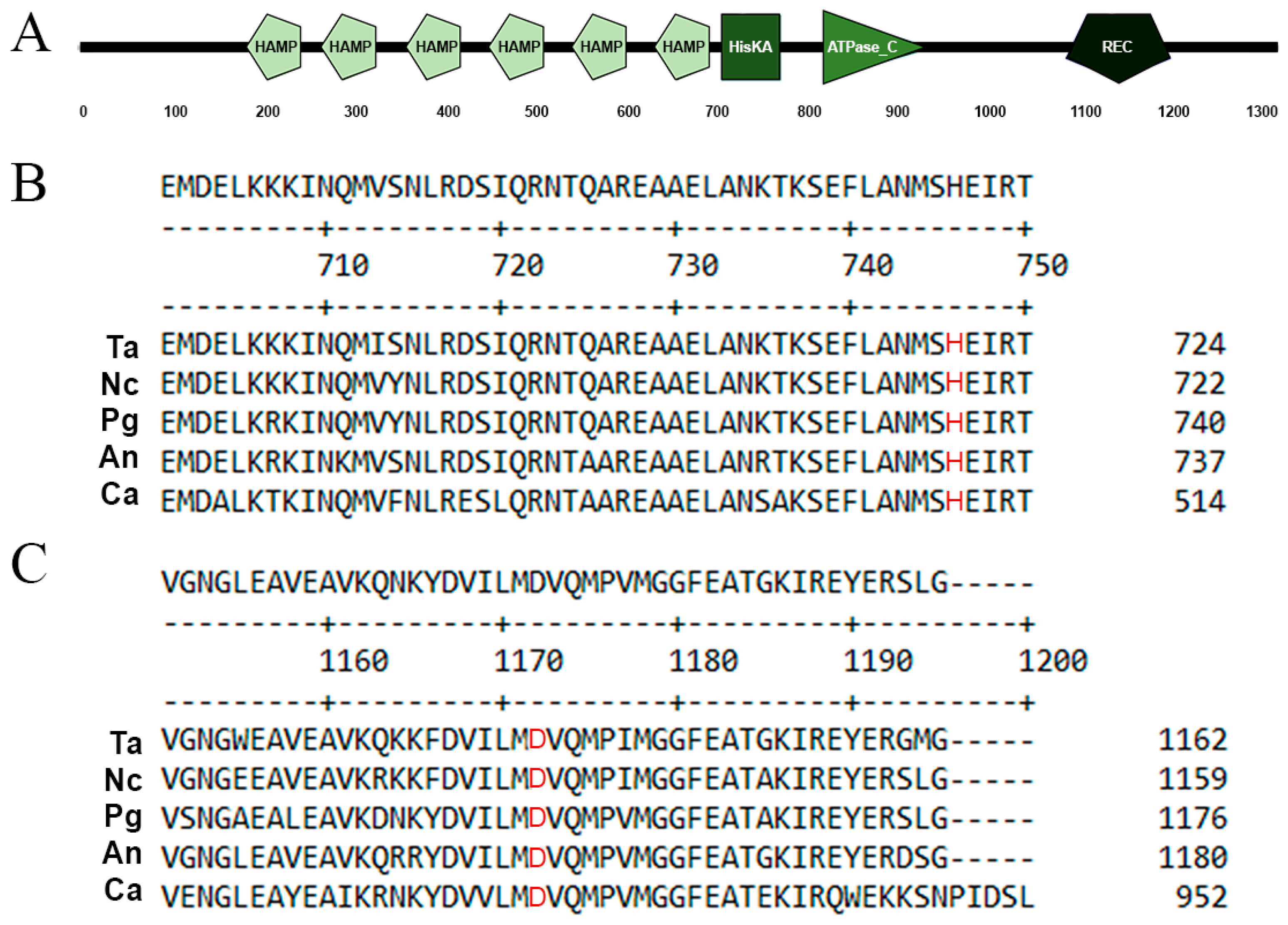
3.1. Identification and Deletion of the T. atroviride nik1 Gene

The T. atroviride Nik1 has an identity of 63.5% to the HK Os-1 of N. crassa (AAB01979.1), 61.1% to the NikA of Aspergillus nidulans (EAA60822.1), 63.5% to the HIK1 of Pyricularia grisea (BAB40947.1), 62.2% to the Bos1 of Botrytis cinerea (ATZ45935.1), and 57.7% to the Nik1 of Candida albicans (AOW30628.1). The nik1 gene encodes a protein of 1324 aa (Figure 1A), containing six repeats of HAMP domains in its amino terminus (191–246, 275–327, 367–419, 459–511, 551–603, 643–695 aa) and the typical domains encoded by fungal HK genes: a phosphoacceptor (710–775 aa) with a conserved His720 residue (Figure 1B), an ATP-binding domain (822–942 aa), and a RR receiver domain (1089–1205 aa) with a conserved Asp1139 residue (Figure 1C). These data suggest that T. atroviride Nik1 has all the conserved motifs for a functional class III HK.
To investigate the role of HK Nik1 in T. atroviride IMI206040, the nik1 gene was replaced by the hph selectable marker (Figure S1A). Six stable mutants were obtained after six rounds of single-spore isolation on PDA supplemented with 0.5% Triton X-100 and 100 μg mL−1 Hygromycin B. The nik1 gene replacement was confirmed by PCR, which detected the integration of the drug-resistance marker gene at the corresponding locus and the absence of the nik1 ORF. Pnik1-F–Hyg-R primers were used to amplify a 2.7 kb fragment comprising the nik1 5′ region and the hph gene only in the ∆nik1 strains (Figure S1B). A 2.8 kb fragment comprising the hph gene and nik1 3′ region was amplified in ∆nik1 strains using Hyg-F–Tnik1-R primers (Figure S1C). To corroborate the lack of the nik1 coding region, 1nik1-F–3nik1-R primers only amplified a 0.6 kb fragment of the nik1 ORF in the WT strain (Figure S1D). All results confirmed that the nik1 ORF was successfully knocked out in the six strains analyzed.

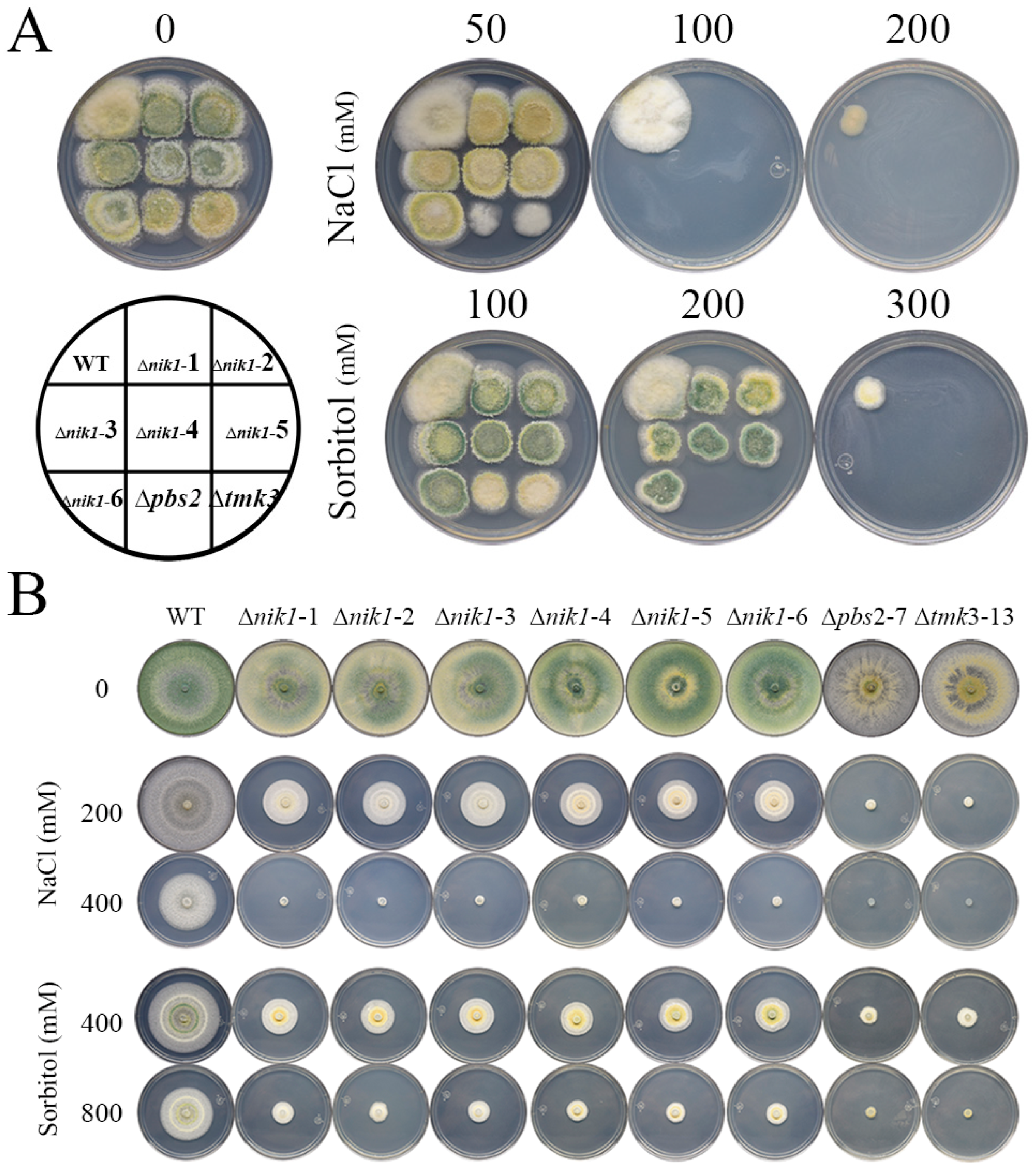
3.2. Nik1 Is Involved in Osmotic Stress Resistance

In ascomycetes, Nik1 orthologs generally participate in hyperosmotic stress responses [18]. To examine the role of the T. atroviride Nik1 in tolerance against high osmotic pressure, the conidia and mycelia of the WT, ∆nik1, ∆pbs2, and ∆tmk3 strains were subjected to high concentrations of NaCl and sorbitol. In the conidia stage, 100 mM NaCl inhibited the growth of ∆nik1, ∆pbs2, and ∆tmk3 strains, and 200 mM sorbitol reduced the growth of ∆nik1 strains. At the same time, that concentration was lethal for ∆pbs2 and ∆tmk3 strains (Figure 2A). In mycelia, 200 mM NaCl and 400 mM sorbitol slightly impaired the radial growth of ∆nik1, and NaCl was more noxious than sorbitol. In contrast, the ∆pbs2 and ∆tmk3 strains did not grow at those osmolyte concentrations (Figure 2B). These results suggest that Nik1 has a role in hyperosmotic stress tolerance in T. atroviride, and because the ∆nik1 strains were not as hypersensitive as ∆psb2 and ∆tmk3 strains, these findings suggest that additional HKs may be participating in osmotic stress responses along with Nik1.

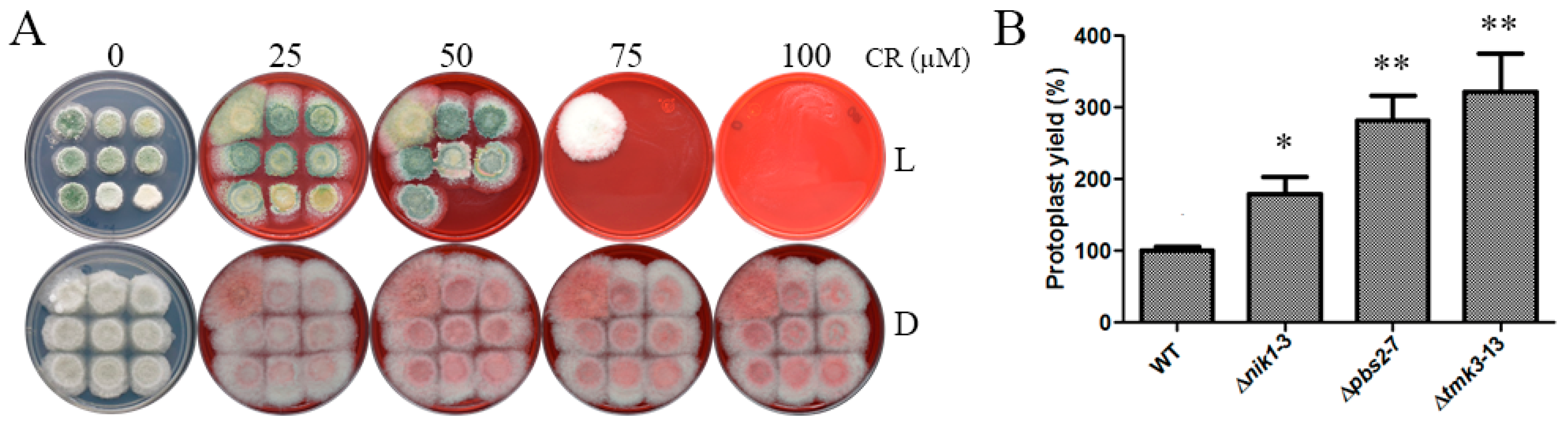
3.3. Nik1 Regulates the Cell Wall Integrity Maintenance of T. atroviride

An alteration in the architecture and elasticity of fungal cell walls provokes sensitivity to osmotic stress [28]. To assess if Nik1 has a role in cell wall integrity, Congo red was used as a cell wall disruptor. Compared to the WT, ∆nik1 strains could not grow at 75 µM of Congo red. In contrast, ∆pbs2 and ∆tmk3 strains were unable to grow at lower Congo red concentrations (Figure 3A). However, when using mycelia plugs, ∆nik1 mycelia displayed a phenotype similar to the WT strain (Figure S2A). In contrast, as reported previously, mycelial growth of ∆psb2 and ∆tmk3 strains was compromised in Congo red media under constant illumination [10]. To validate if ∆nik1 strains have a defect in cell wall integrity, we tested the sensitivity to cell wall lysing enzymes in the WT and mutant strains (Figure 3B). The deletion of the nik1 gene increased the sensitivity to cell wall lysing enzymes in contrast to the WT, producing a double number of protoplasts, while the lack of pbs2 and tmk3 genes affected cell wall composition, producing three times more protoplasts than the WT. These results suggest that Nik1 is involved in cell wall integrity maintenance via the MAPK Tmk3 signaling pathway.
Furthermore, we assessed if additional stressors regulated by Tmk3 are affected in Δnik1 strains. Thus, tolerance assays in oxidative stress, cadmium stress, heat shock, and UV light irradiation were performed. The ∆nik1 strains showed no sensitivity in response to these stressors (Figures S2 and S3).

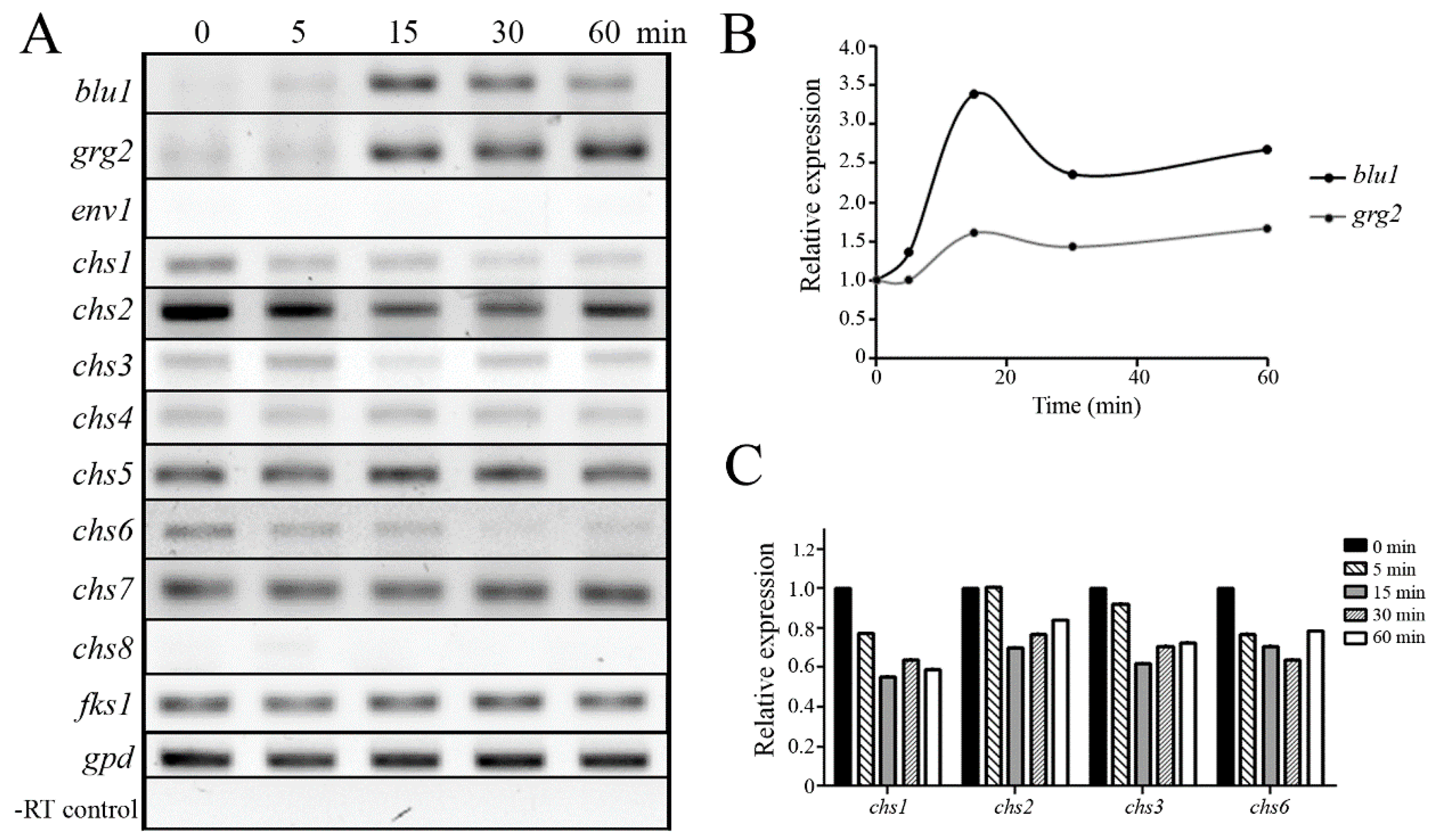
3.4. Osmotic Stress Provokes Changes in Gene Expression through the Tmk3 MAPK Pathway

In T. harzianum, two homologs to Grg-1 of N. crassa, encoding a glucose-repressible protein, were induced in response to high osmotic stress [12]. In contrast, T. atroviride demonstrated that the blu1 and grg2 genes, homologs to N. crassa Grg-1, are induced through the Pbs2–Tmk3 pathway after blue light stimulus [10,29]. To determine if the gene expression of blu1 and grg2 is induced after high osmotic stress in T. atroviride, the mycelia of the WT strain were grown in PDB under dark conditions and subjected to high osmotic stress. After 500 mM NaCl treatment, the blu1 and grg2 genes reached their maximum transcript levels at 15 min. Then, the blu1 expression decreased, and the grg2 transcript levels remained high after 60 min of treatment (Figure 4A,B). The env1 gene, which encodes a PAS/LOV domain protein, is activated by blue light stimulus independent of Tmk3 [10,30]. Transcripts of the env1 gene were not detected during the experiment, indicating that the blu1 and grg2 genes were stimulated explicitly in response to an osmotic shock in T. atroviride.
Considering that a cell wall protects from a hyper-osmotic shock in fungi [31], T. atroviride genes encoding chitin synthases (chs1-8) and β-1,3-glucan synthase (fks1) were identified (Table 2), and their expressions were evaluated in response to high osmotic stress. Transcript levels of chs1, chs2, chs3, and chs6 genes decreased after 15 min of treatment with 500 mM NaCl (Figure 4C), while chs4, chs5, chs7, and fks1 expression remained without apparent change in the time analyzed. chs8 gene expression was not detected under our experimental conditions.
In order to test if the Tmk3 pathway regulates the expression of blu1, grg2, and cell wall-related genes via Nik1, the WT and ∆nik1, ∆pbs2, and ∆tmk3 strains were subjected to high osmotic stress. After 15 min of treatment, the blu1 and grg2 genes were induced in response to osmotic stress only in the WT and ∆nik1, but not induced in the ∆pbs2 and ∆tmk3 strains, indicating that the Tmk3–Pbs2 pathway regulates their expression independent of Nik1 (Figure 5A). Furthermore, env1 transcripts were undetected, indicating that activation was specifically stimulated by osmotic shock. Further, the expression of nik1 and tmk3 was undetected in ∆nik1 and ∆tmk3 strains, respectively, corroborating that the mutant strains lack the corresponding genes. Consistently, transcript levels of chs1 and chs2 genes decreased in the WT, whereas chs1 and chs3 were higher in ∆nik1, ∆pbs2, and ∆tmk3 strains in comparison to the WT strain (Figure 5B). However, their expression was still responsive to a hyperosmotic shock. These data suggest that the Nik1 could regulate chs1 and chs3 genes through the Tmk3 MAPK pathway by acting as a repressor.

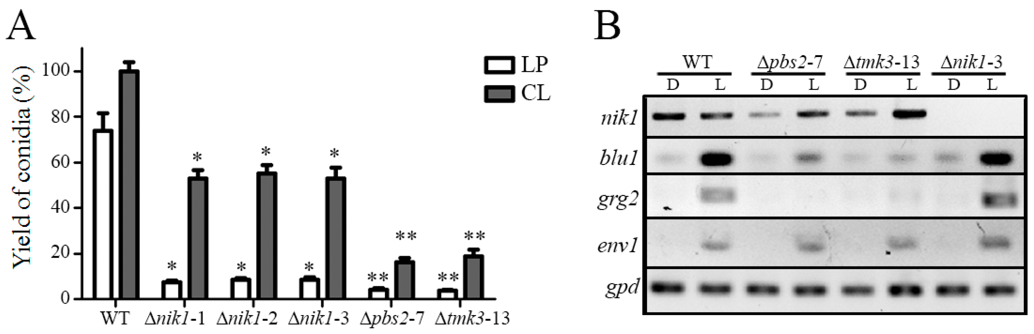
3.5. The HK Nik1 Is Involved in Mycelial Growth and Asexual Reproduction

In T. atroviride, the MAPK Tmk3 regulates mycelial growth and conidiation triggered by light. The vegetative growth of the ∆nik1 strains was analyzed (Figure S4A), and the radial growth was reduced by 10% in darkness and 20% under constant white light in contrast to the WT, whereas the growth of the ∆pbs2 and ∆tmk3 strains was reduced by 30% and 40%, respectively, compared to the WT strain (Figure S4B). Furthermore, when compared to the WT, the conidial production stimulated by a blue light pulse in the ∆nik1 strains was 90% less. In contrast, in constant white light, the ∆nik1 strains produced 50% less conidia than the WT (Figure 6A). The Δtmk3 and Δpbs2 strains produced 80% less conidia in constant white light in comparison to the WT strain, as reported previously [10]. These results suggest that Nik1 is required for vegetative growth and conidiation induced by light in T. atroviride, possibly by a mechanism regulated through the Tmk3 pathway. To evaluate if Nik1 is a specific regulator of the photoconidiation process, the conidial production of the ∆nik1 strains in response to the wound was analyzed, and it was observed that conidiation was also reduced in ∆nik1, similar to ∆pbs2 and ∆tmk3 strains (Figure S5), suggesting that the Tmk3 pathway plays a general role in T. atroviride for asexual reproduction. Based on these results, we examined the expression of light-responsive genes regulated by the Tmk3 MAPK pathway after 30 min of a blue light pulse (Figure 6B). Nik1 is dispensable for blu1, grg2, and env1 expression regulated by light. Taken together, our results indicate that Nik1 regulates conidiation, but the activation of light-responsive genes through the Pbs2–Tmk3 pathway occurs by a mechanism independent of Nik1.

4. Discussion

Prokaryotic organisms use the TCS to sense and respond to a wide range of environmental signals, and the signal transduction is mediated by the phosphorylation of histidine and aspartic acid residues [18]. In eukaryotic cells, an environmental cue is generally transduced by modifications of serine/threonine and tyrosine residues of signaling proteins. However, plants and fungi possess genes that code for proteins of the TCS. One of the most characterized HK in fungi is Nik-1/Os-1, which was first reported in N. crassa and acts upstream of the MAPK Os-2 pathway and regulates osmotic stress responses [21]. In this research, the role of the HK Nik1 in T. atroviride was analyzed in response to diverse cellular stressors regulated by the MAPK Tmk3. Our results suggest that Nik1 participates as a receptor in response to osmotic stress, and another HKs could cooperate as osmosensors to transduce the signal through Tmk3. The first HK identified in fungi was Sln1 in yeast [32], which is an osmotic stress sensor that possesses transmembrane domains and is categorized as class VI and is conserved among filamentous fungi [18]. However, a sensor homolog to yeast Sln1 in T. reesei is nonessential to cope with osmostress [33]. It would be interesting to explore the role of Sln1 (EHK50841.1) in T. atrorivide to elucidate if it is also a player in response to osmotic stress along with Nik1 or another HK that has gained this new role.
A highly saline environment not only imposes osmotic pressure on cells but also intracellular ion toxicity [34,35]. From this perspective, filamentous fungi have a more complex osmoregulatory system than yeast and are capable of discerning stress caused by high levels of salt or sugar. We observed that in T. atroviride, higher levels of NaCl were more toxic than sorbitol, suggesting that HK Nik1 is also required to survive intracellular toxicity by NaCl. On the contrary, a Pyricularia oryzae strain lacking the HIK1 gene, which codes for an HK class III, is more sensitive to high levels of sorbitol than NaCl [36], suggesting that P. oryzae can also distinguish if the osmostress is caused by high concentrations of sugar or salt through Hik1. In Magnaporthe oryzae, the ∆Mosln1 strain is more susceptible to salt stress, whereas ∆Mohik1 is affected by high doses of sorbitol. Interestingly, a phosphorylation signal of MoHog1p was detected in a ∆Mohik1/∆sln1 strain after an osmotic shock, suggesting that more osmosensors participate in the MoHog1p MAPK cascade [37]. A Beauveria bassiana strain lacking a class VIII HK was affected by high NaCl and sorbitol concentrations, and a movement of HK8:GFP to the inside of hyphal cells from the cell periphery after 30 min exposure to NaCl was observed [38]. The class VIII HKs have in common a GAF and a phytochrome domain [18]. According to the reports mentioned above, exploring the role of T. atroviride Sln1 and Phy1 (EHK44075.1) in osmoadaptation would be interesting.
The mechanisms for osmotic stress tolerance in fungi are based on producing osmolytes in the cytoplasm to balance osmotic pressures and cell wall reinforcement [39]. Our results on Congo red and lytic enzyme resistance assays suggested that Nik1 maintains the cell wall integrity in T. atroviride, most likely with other signaling pathways, including other HKs, as observed in other fungal systems. In response to other stressors, such as oxidative, cadmium, thermal shock, and UV irradiation, Nik1 is dispensable. Other fungal HKs are involved in cell wall formation. The PmHHK1 class X of Penicillium marneffei regulates sporulation, polarized growth, and cell wall composition [40]. C. albicans chk1∆ strains are sensitive to high doses of Congo red and Calcofluor white [41]. M. oryzae, ∆Mosln1, ∆Mohik5 (class V), and ∆Mohik9 (class XI) strains released more protoplasts than the wild type strain after an application of lysing enzymes, but the ∆Mohik6 (class X) strain was more resistant of the effect of lysing enzymes than the parental strain [37]. In B. bassiana, the growth of a ∆hk5 strain, which lacks a class V HK, was affected by Congo red and a strain lacking the homolog to Nik1 [38]; these data suggest a complex role among HKs in fungi. Our results open the possibility that the Sln1 and the HK class V (EHK42346.1), X (EHK40776.1), and XI (EHK40433.1) cooperatively could participate in the cell wall homeostasis of T. atroviride.
Chitin is an important structural element of the cell wall in fungi, and chitin synthases are essential enzymes for their construction [42]. Strains lacking chs genes encoding chitin synthases were analyzed in T. atroviride, and the growth and conidiation of the Δchs1, Δchs2, Δchs5, and Δchs7 strains were severally compromised [43]. Consistently, the transcript levels of chs1 and chs2 decreased after 15 min of an osmotic shock, and the fks1 encoding β-1,3-glucan synthase was constitutively expressed, suggesting that chitin is the leading targeted polysaccharide to modulate the cell wall architecture in responses to osmostress. In Ustilago maydis, β-1,3-glucan synthase was expressed constitutively, while the expression of four of the eight genes that code for chitin synthases decreased after treatment with 1 M NaCl [44]. In addition, the expression of five chs genes from Penicillium digitatum evaluated under osmotic stress conditions was downregulated [45]. The studies mentioned above suggest that in response to osmotic shock, chs genes have differences in transcription levels, as suggested by our results.
Furthermore, the C. albicans Hog MAPK pathway mediates the regulation of the CHS gene expression. The chitin synthase activity in a ∆hog1 strain is elevated compared to WT cells [46]. Accordingly, we analyzed the expression of chs1, chs2, chs3, and chs6 genes in the ∆nik1, ∆pbs2, and ∆tmk3 strains of T. atroviride. Our results revealed that chs1 and chs3 showed higher transcriptional levels in the mutant strains in an osmostress-independent manner, and after a hyperosmotic shock, chs2 and chs3 genes remained upregulated. The results suggest that Nik1, Pbs2, and Tmk3 act as negative regulators of these genes to maintain homeostasis in cell wall synthesis; consequently, the absence of nik1, pbs2, and tmk3 genes affects the cell wall integrity and generates cells more sensitive to osmotic stress.
The T. atroviride blu1 and grg2 genes, which encode glucose-repressible proteins, are induced in response to an osmotic shock and blue light pulse through the MAPK Tmk3 and MAPKK Pbs2 pathways. However, HK Nik1 is dispensable for their expression in response to both stressors. MAPK Tmk3 is phosphorylated in response to blue light; once activated, it regulates the expression of blu1 and grg2 genes [10]. Yeast Hog1 is also activated by phosphorylation and is translocated into the nucleus to regulate gene expression, although the nuclear import of Hog1 is not essential for osmoadaptation [47]. The results suggest that Nik1 uses two different mechanisms; it acts as a negative modulator of gene expression and as an osmostress-response activator; this is probably carried out by nuclear and cytosolic functions reported for Hog1. In addition, our results suggest that other signaling pathways, including HKs, collaborate with Nik1 in response to environmental stresses, revealing a functional redundancy. However, Nik1 is required for blue light conidiation but not gene expression induced by blue light. Consistent with this proposal, in A. nidulans, it was demonstrated that an HK, FphA, is stimulated by red and blue light to activate SakA, a homolog to Tmk3 [48]. Also, in M. oryzae, the HPt gene YPD1 was expressed in response to light [49], suggesting that alternative histidine kinases could regulate light responses and support the possible participation of additional proteins upstream of Tmk3 for asexual reproduction.
In conclusion, our results suggest that the T. atroviride Nik1 regulates tolerance to osmotic stress, cell wall integrity, and asexual reproduction. The phenotypes and gene expression indicate a collaborative regulation among histidine kinases to regulate the Tmk3 signaling pathway. Finally, our results suggest that Nik1 modulates stress-independent gene expression through the Tmk3 MAPK signaling pathway.

Supplementary Materials

The following are available online at https://www.mdpi.com/article/10.3390/jof9090939/s1: Figure S1. Deletion of the nik1 gene, Figure S2. Response to oxidative stress, cadmium, and UV light in ∆nik1 strains, Figure S3. Thermal shock resistance in ∆nik1 strains, Figure S4. Radial growth of ∆nik1 strains, Figure S5. Wound response assay in Δnik1 strains.

Author Contributions

Conceptualization and methodology, E.U.E.-N. and G.C.-H.; validation, E.U.E.-N., F.L.-J., J.A.C.-C. and A.M.-M.; formal analysis, E.U.E.-N. and G.C.-H.; investigation, G.C.-H.; resources, E.U.E.-N. and A.M.-M., data curation, E.U.E.-N. and G.C.-H.; writing—original draft preparation, G.C.-H. and E.U.E.-N.; writing—review and editing, G.C.-H., E.U.E.-N., A.M.-M., G.C.-H., J.A.C.-C. and F.L.-J.; supervision, project administration, and funding acquisition, E.U.E.-N. and A.M.-M. All authors have read and agreed to the published version of the manuscript.

Funding

This research was funded by CONACYT (Consejo Nacional de Ciencia y Tecnología), grant numbers CB-2011-169045 and FONDEC-UAQ-2022 (Fondo para el desarrollo del Conocimiento—Universidad Autónoma de Querétaro, FNB-2022-04), The New Zealand Tertiary Education Commission through the Bio-Protection Research Centre, and MBIE LINX2201. Fungal volatile organic compounds for sustainable agriculture in a changing environment supported A.M.-M.’s research.

Institutional Review Board Statement

Not applicable.

Informed Consent Statement

Not applicable.

Data Availability Statement

Not applicable.

Acknowledgments

G.C.-H. is indebted to CONACYT for a postdoctoral fellowship.

Conflicts of Interest

The authors declare no conflict of interest. The funders had no role in the design of the study; in the collection, analyses, or interpretation of data; in the writing of the manuscript, or in the decision to publish the results.

References

  1. Medina-Castellanos, E.; Esquivel-Naranjo, E.U.; Heil, M.; Herrera-Estrella, A. Extracellular ATP activates MAPK and ROS signaling during injury response in the fungus Trichoderma atroviride. Front. Plant Sci. 2004, 5, 659. [Google Scholar] [CrossRef]
  2. Mukherjee, P.K.; Horwitz, B.A.; Herrera-Estrella, A.; Schmoll, M.; Kenerley, C.M. Trichoderma Research in the Genome Era. Annu. Rev. Phytopathol. 2013, 51, 105–129. [Google Scholar] [CrossRef] [PubMed]
  3. Steyaert, J.M.; Weld, R.J.; Mendoza-Mendoza, A.; Stewart, A. Reproduction without sex: Conidiation in the filamentous fungus Trichoderma. Microbiology 2010, 156, 2887–2900. [Google Scholar] [CrossRef]
  4. Brewster, J.; de Valoir, T.; Dwyer, N.; Winter, E.; Gustin, M. An osmosensing signal transduction pathway in yeast. Science 1993, 259, 1760–1763. [Google Scholar] [CrossRef]
  5. Singh, K.K. The Saccharomyces cerevisiae sln1p-ssk1p two-component system mediates response to oxidative stress and in an oxidant-specific fashion. Free Radic. Biol. Med. 2000, 29, 1043–1050. [Google Scholar] [CrossRef] [PubMed]
  6. Winkler, A.; Arkind, C.; Mattison, C.P.; Burkholder, A.; Knoche, K.; Ota, K. Heat Stress Activates the Yeast High-Osmolarity Glycerol Mitogen-Activated Protein Kinase Pathway, and Protein Tyrosine Phosphatases Are Essential under Heat Stress. Eukaryote Cell 2002, 1, 163–173. [Google Scholar] [CrossRef] [PubMed]
  7. García-Rodriguez, L.J.; Durán, A.; Roncero, C. Calcofluor Antifungal Action Depends on Chitin and a Functional High-Osmolarity Glycerol Response (HOG) Pathway: Evidence for a Physiological Role of the Saccharomyces cerevisiae HOG Pathway under Noninducing Conditions. J. Bacteriol. 2000, 182, 2428–2437. [Google Scholar] [CrossRef]
  8. Jiang, L.; Cao, C.; Zhang, L.; Lin, W.; Xia, J.; Xu, H.; Zhang, Y. Cadmium-induced activation of high osmolarity glycerol pathway through its Sln1 branch is dependent on the MAP kinase kinase kinase Ssk2, but not its paralog Ssk22, in budding yeast. FEMS Yeast Res. 2014, 14, 1263–1272. [Google Scholar] [CrossRef]
  9. Saito, H.; Posas, F. Response to Hyperosmotic Stress. Genetics 2012, 192, 289–318. [Google Scholar] [CrossRef]
  10. Esquivel-Naranjo, E.U.; García-Esquivel, M.; Medina-Castellanos, E.; Correa-Pérez, V.A.; Parra-Arriaga, J.L.; Landeros-Jaime, F.; Cervantes-Chávez, J.A.; Herrera-Estrella, A. A Trichoderma atroviride stress-activated MAPK pathway integrates stress and light signals. Mol. Microbiol. 2016, 100, 860–876. [Google Scholar] [CrossRef] [PubMed]
  11. Wang, M.; Zhao, Q.; Yang, J.; Jiang, B.; Wang, F.; Liu, K.; Fang, X. A Mitogen-Activated Protein Kinase Tmk3 Participates in High Osmolarity Resistance, Cell Wall Integrity Maintenance and Cellulase Production Regulation in Trichoderma reesei. PLoS ONE 2013, 8, e72189. [Google Scholar] [CrossRef]
  12. Delgado-Jarana, J.; Sousa, S.; González, F.; Rey, M.; Llobell, A. ThHog1 controls the hyperosmotic stress response in Trichoderma harzianum. Microbiology 2006, 152, 1687–1700. [Google Scholar] [CrossRef]
  13. Horie, T.; Tatebayashi, K.; Yamada, R.; Saito, H. Phosphorylated Ssk1 Prevents Unphosphorylated Ssk1 from Activating the Ssk2 Mitogen-Activated Protein Kinase Kinase Kinase in the Yeast High-Osmolarity Glycerol Osmoregulatory Pathway. Mol. Cell. Biol. 2008, 28, 5172–5183. [Google Scholar] [CrossRef] [PubMed]
  14. Tanaka, K.; Tatebayashi, K.; Nishimura, A.; Yamamoto, K.; Yang, H.Y.; Saito, H. Yeast Osmosensors Hkr1 and Msb2 Activate the Hog1 MAPK Cascade by Different Mechanisms. Sci. Signal. 2014, 7, ra21. [Google Scholar] [CrossRef] [PubMed]
  15. Chang, C.; Kwok, S.; Bleecker, A.; Meyerowitz, E. Arabidopsis ethylene-response gene ETR1: Similarity of product to two-component regulators. Science 1993, 262, 539–544. [Google Scholar] [CrossRef]
  16. Ota, I.; Varshavsky, A. A yeast protein similar to bacterial two-component regulators. Science 1993, 262, 566–569. [Google Scholar] [CrossRef]
  17. Schmoll, M.; Dattenböck, C.; Carreras-Villaseñor, N.; Mendoza-Mendoza, A.; Tisch, D.; Alemán, M.I.; Baker, S.E.; Brown, C.; Cervantes-Badillo, M.G.; Cetz-Chel, J.; et al. The Genomes of Three Uneven Siblings: Footprints of the Lifestyles of Three Trichoderma Species. Microbiol. Mol. Biol. Rev. 2016, 80, 205–327. [Google Scholar] [CrossRef]
  18. Catlett, N.L.; Yoder, O.C.; Turgeon, B.G. Whole-Genome Analysis of Two-Component Signal Transduction Genes in Fungal Pathogens. Eukaryote Cell 2003, 2, 1151–1161. [Google Scholar] [CrossRef]
  19. Schumacher, M.M.; Enderlin, C.S.; Selitrennikoff, C.P. The Osmotic-1 Locus of Neurospora crassa Encodes a Putative Histidine Kinase Similar to Osmosensors of Bacteria and Yeast. Curr. Microbiol. 1997, 34, 340–347. [Google Scholar] [CrossRef]
  20. Alex, L.A.; Borkovich, K.A.; Simon, M.I. Hyphal development in Neurospora crassa: Involvement of a two-component histidine kinase. Proc. Natl. Acad. Sci. USA 1996, 93, 3416–3421. [Google Scholar] [CrossRef] [PubMed]
  21. Noguchi, R.; Banno, S.; Ichikawa, R.; Fukumori, F.; Ichiishi, A.; Kimura, M.; Yamaguchi, I.; Fujimura, M. Identification of OS-2 MAP kinase-dependent genes induced in response to osmotic stress, antifungal agent fludioxonil, and heat shock in Neurospora crassa. Fungal Genet. Biol. 2007, 44, 208–218. [Google Scholar] [CrossRef] [PubMed]
  22. Hagiwara, D.; Takahashi-Nakaguchi, A.; Toyotome, T.; Yoshimi, A.; Abe, K.; Kamei, K.; Gonoi, T.; Kawamoto, S. NikA/TcsC Histidine Kinase Is Involved in Conidiation, Hyphal Morphology, and Responses to Osmotic Stress and Antifungal Chemicals in Aspergillus fumigatus. PLoS ONE 2013, 8, e80881. [Google Scholar] [CrossRef] [PubMed]
  23. Yu, J.H.; Hamari, Z.; Han, K.H.; Seo, J.A.; Reyes-Domínguez, Y.; Scazzocchio, C. Double-joint PCR: A PCR-based molecular tool for gene manipulations in filamentous fungi. Fungal Genet. Biol. 2004, 41, 973–981. [Google Scholar] [CrossRef]
  24. Carroll, A.M.; Sweigard, J.A.; Valent, B. Improved Vectors for Selecting Resistance to Hygromycin. Fungal Genet. Rep. 1994, 41, 22. [Google Scholar] [CrossRef]
  25. Calcáneo-Hernández, G.; Rojas-Espinosa, E.; Landeros-Jaime, F.; Cervantes-Chávez, J.A.; Esquivel-Naranjo, E.U. An efficient transformation system for Trichoderma atroviride using the pyr4 gene as a selectable marker. Braz. J. Microbiol. 2020, 51, 1631–1643. [Google Scholar] [CrossRef] [PubMed]
  26. Raeder, U.; Broda, P. Rapid preparation of DNA from filamentous fungi. Lett. Appl. Microbiol. 1985, 1, 17–20. [Google Scholar] [CrossRef]
  27. Schultz, J.; Copley, R.R.; Doerks, T.; Ponting, C.P.; Bork, P. SMART: A web-based tool for the study of genetically mobile domains. Nucleic Acids Res. 2000, 28, 231–234. [Google Scholar] [CrossRef]
  28. Ene, I.; Walker, L.A.; Schiavone, M.; Lee, K.K.; Martin-Yken, H.; Dague, E.; Gow, N.A.R.; Munro, C.A.; Brown, A.J.P. Cell Wall Remodeling Enzymes Modulate Fungal Cell Wall Elasticity and Osmotic Stress Resistance. mBio 2015, 6, e00986-15. [Google Scholar] [CrossRef]
  29. Rosales-Saavedra, T.; Esquivel-Naranjo, E.U.; Casas-Flores, S.; Martínez-Hernández, P.; Ibarra-Laclette, E.; Cortes-Penagos, C.; Herrera-Estrella, A. Novel light-regulated genes in Trichoderma atroviride: A dissection by cDNA microarrays. Microbiology 2006, 152, 3305–3317. [Google Scholar] [CrossRef]
  30. Schmoll, M.; Franchi, L.; Kubicek, C.P. Envoy, a PAS/LOV Domain Protein of Hypocrea jecorina (Anamorph Trichoderma reesei), Modulates Cellulase Gene Transcription in Response to Light. Eukaryote Cell 2005, 4, 1998–2007. [Google Scholar] [CrossRef]
  31. Levin, D.E. Regulation of Cell Wall Biogenesis in Saccharomyces cerevisiae: The Cell Wall Integrity Signaling Pathway. Genetics 2011, 189, 1145–1175. [Google Scholar] [CrossRef]
  32. Maeda, T.; Wurgler-Murphy, S.M.; Saito, H. A two-component system that regulates an osmosensing MAP kinase cascade in yeast. Nature 1994, 369, 242–245. [Google Scholar] [CrossRef]
  33. Wang, Z.; An, N.; Xu, W.; Zhang, W.; Meng, X.; Chen, G.; Liu, W. Functional characterization of the upstream components of the Hog1-like kinase cascade in hyperosmotic and carbon sensing in Trichoderma reesei. Biotechnol. Biofuels 2018, 11, 97. [Google Scholar] [CrossRef]
  34. Ke, R.; Ingram, P.J.; Haynes, K. An Integrative Model of Ion Regulation in Yeast. PLoS Comput. Biol. 2013, 9, e1002879. [Google Scholar] [CrossRef]
  35. Garcia, M.J.; Rios, G.; Ali, R.; Belles, J.M.; Serrano, R. Comparative physiology of salt tolerance in Candida tropicalis and Saccharomyces cerevisiae. Microbiology 1997, 143, 1125–1131. [Google Scholar] [CrossRef]
  36. Motoyama, T.; Kadokura, K.; Ohira, T.; Ichiishi, A.; Fujimura, M.; Yamaguchi, I.; Kudo, T. A two-component histidine kinase of the rice blast fungus is involved in osmotic stress response and fungicide action. Fungal Genet. Biol. 2005, 42, 200–212. [Google Scholar] [CrossRef]
  37. Jacob, S.; Foster, A.J.; Yemelin, A.; Thines, E. Histidine kinases mediate differentiation, stress response, and pathogenicity in Magnaporthe oryzae. MicrobiologyOpen 2014, 3, 668–687. [Google Scholar] [CrossRef]
  38. Liu, J.; Tong, S.M.; Qiu, L.; Ying, S.H.; Feng, M.G. Two histidine kinases can sense different stress cues for activation of the MAPK Hog1 in a fungal insect pathogen: Two sensors of fungal Hog1 cascade. Environ. Microbiol. 2017, 19, 4091–4102. [Google Scholar] [CrossRef] [PubMed]
  39. Brewster, J.L.; Gustin, M.C. Hog1: 20 years of discovery and impact. Sci. Signal. 2014, 7, re7. [Google Scholar] [CrossRef] [PubMed]
  40. Wang, F.; Tao, J.; Qian, Z.; You, S.; Dong, H.; Shen, H.; Chen, X.; Tang, S.; Ren, S. A histidine kinase PmHHK1 regulates polar growth, sporulation and cell wall composition in the dimorphic fungus Penicillium marneffei. Mycol. Res. 2009, 113, 915–923. [Google Scholar] [CrossRef] [PubMed]
  41. Li, D.; Williams, D.; Lowman, D.; Monteiro, M.A.; Tan, X.; Kruppa, M.; Fonzi, W.; Roman, E.; Pla, J.; Calderone, R. The Candida albicans histidine kinase Chk1p: Signaling and cell wall mannan. Fungal Genet. Biol. 2009, 46, 731–741. [Google Scholar] [CrossRef]
  42. Li, M.; Jiang, C.; Wang, Q.; Zhao, Z.; Jin, Q.; Xu, J.R.; Liu, H. Evolution and Functional Insights of Different Ancestral Orthologous Clades of Chitin Synthase Genes in the Fungal Tree of Life. Front. Plant Sci. 2016, 7, 37. [Google Scholar] [CrossRef] [PubMed]
  43. Kappel, L.; Münsterkötter, M.; Sipos, G.; Escobar-Rodriguez, C.; Gruber, S. Chitin and chitosan remodeling defines vegetative development and Trichoderma biocontrol. PLOS Pathog. 2020, 16, e1008320. [Google Scholar] [CrossRef] [PubMed]
  44. Robledo-Briones, M.; Ruiz-Herrera, J. Transcriptional Regulation of the Genes Encoding Chitin and β-1,3-Glucan Synthases from Ustilago maydis. Curr. Microbiol. 2012, 65, 85–90. [Google Scholar] [CrossRef] [PubMed]
  45. Gandía, M.; Harries, E.; Marcos, J.F. Identification and characterization of chitin synthase genes in the postharvest citrus fruit pathogen Penicillium digitatum. Fungal Biol. 2012, 116, 654–664. [Google Scholar] [CrossRef]
  46. Munro, C.A.; Selvaggini, S.; Bruijn, I.; Walker, L.; Lenardon, M.D.; Gerssen, B.; Milne, S.; Brown, A.J.P.; Gow, N.A.R. The PKC, HOG and Ca 2+ signalling pathways co-ordinately regulate chitin synthesis in Candida albicans. Mol. Microbiol. 2007, 63, 1399. [Google Scholar] [CrossRef]
  47. García-Marqués, S.; Randez-Gil, F.; Prieto, J.A. Nuclear versus cytosolic activity of the yeast Hog1 MAP kinase in response to osmotic and tunicamycin-induced ER stress. FEBS Lett. 2015, 589, 2163–2168. [Google Scholar] [CrossRef]
  48. Yu, Z.; Armant, O.; Fischer, R. Fungi use the SakA (HogA) pathway for phytochrome-dependent light signalling. Nat. Microbiol. 2016, 1, 16019. [Google Scholar] [CrossRef]
  49. Mohanan, V.C.; Chandarana, P.M.; Chattoo, B.B.; Patkar, R.N.; Manjrekar, J. Fungal Histidine Phosphotransferase Plays a Crucial Role in Photomorphogenesis and Pathogenesis in Magnaporthe oryzae. Front. Chem. 2017, 5, 31. [Google Scholar] [CrossRef]
Figure 1. Nik1 amino acid sequence analysis. (A) Domains of the T. atroviride Nik1 protein predicted by SMART [27]. HAMP (histidine kinases, adenylyl cyclases, methyl-accepting chemotaxis proteins, and phosphatases) domain; HisKA, phosphoacceptor domain; HATPase_c, ATP-binding domain; REC, receiver domain. (B,C) Amino acid sequence alignment to identify the conserved phosphorylable His720 residue in the HisKA motif (B) and the Asp1139 residue in the REC domain (C). Alignment was carried out using MegAlign software version 7.1.0 (DNAStar) by the Clustal W Method. Ta, T. atroviride IMI206040; Nc, N. crassa; Pg, P. grisea; An, A. nidulans; Ca, C. albicans.
Jof 09 00939 g001
Figure 2. The HK Nik1 regulates tolerance to high osmolarity challenges. (A) Tolerance to osmotic stress in the conidia of WT, ∆nik1, ∆pbs2, and ∆tmk3 strains. Drops of 500 conidia of the WT and mutant strains were inoculated on PDA plates plus different concentrations of the indicated stressors. Plates were incubated at 27 °C for four days. (B) Tolerance to osmotic stress in the mycelia of WT, ∆nik1, ∆pbs2, and ∆tmk3 strains. NaCl and sorbitol were added to PDA media at the concentrations indicated. Strains were incubated at 27 °C for four days. All experiments were carried out in triplicate.
Jof 09 00939 g002
Figure 3. The HK Nik1 plays a role in cell wall integrity. (A) Tolerance to Congo red in the conidia of WT, ∆nik1, ∆pbs2, and ∆tmk3 strains. Drops of 500 conidia of the WT and mutant strains were inoculated on PDA plates with Congo red at the indicated concentrations, incubated at 27 °C for four days in constant white light and darkness, and pictures were taken. The strain order is indicated in Figure 2. (B) Sensitivity to cell wall lysing enzymes of WT, ∆nik1, ∆pbs2, and ∆tmk3 strains. The total production of protoplasts was determined using a Neubauer chamber. The mean value is represented in bars ± SEM of three independent experiments analyzed with the Tukey–Kramer method (α = 0.001). * and ** indicate mean values that are statistically different from the control.
Jof 09 00939 g003
Figure 4. Expression analysis in response to osmotic stress. (A). The mycelia of the WT strain were challenged by adding 0.5 M NaCl. Then, samples were collected at the indicated time. Total RNA was extracted from samples and used to synthesize cDNA. As a loading control, the gpd gene was amplified. In the same reaction for cDNA synthesis, the RNA of samples without transcriptase was used as a template to amplify gpd as a negative control. (B,C) Semiquantitative transcript levels were determined according to signal intensity from two biological replicates and plotted. The expression level for each gene was normalized by dividing the control gpd signal. Relative expression was adjusted to the unit in the control (0 min) without an osmotic shock and compared with the treatment at different time points.
Jof 09 00939 g004
Figure 5. Role of Nik1, Pbs2, and Tmk3 on gene expression regulated by osmotic stress. (A) Total RNA was extracted from the mycelia of WT and mutant strains that were non-stressed (−) or 15 min after an osmotic shock by 0.5 M NaCl (+) and used to synthesize cDNA. In the same reaction for cDNA synthesis, (1) RNA of samples without transcriptase was used as a negative control and (2) gpd was used as a loading control (cDNA template). (B) Signal intensity was quantified from two biological replicates and plotted as indicated in Figure 4. The expression level for each gene was normalized and divided by the control gpd signal. Relative expression was adjusted to the unit in the WT without osmotic stress (−).
Jof 09 00939 g005
Figure 6. Asexual reproduction and gene expression regulated by light in ∆nik1 mutants. (A) Conidial production of the WT strain and the ∆nik1, ∆pbs2, and ∆tmk3 mutant strains. Mycelial plugs of the indicated strains were inoculated on PDA plates, incubated at 27 °C in darkness for 36 h, and then exposed to a blue light pulse (152.4 μmol m−2) (LP), or grown for 7 days in constant white light (0.586 μmol m−2 s−1) (CL). The mean value of conidia yield is represented in bars ± SEM of three independent experiments analyzed with the Tukey–Kramer method (α = 0.05). * and ** indicate mean values that are statistically different from the control. (B) Expression analysis by RT-PCR of light-induced genes in the WT, ∆pbs2, ∆tmk3, and ∆nik1 strains. D, darkness; L, light.
Jof 09 00939 g006
Table 1. Primers used for gene deletion.
GenePrimerSequence (5′ → 3′)Target Region
nik1
(EHK40885.1)
Pnik1-FCTT GCA GGC ACA TCC TTG ACG5′ flanking region
PQnik1-RTGC TCC TTC AAT ATC AGT TAA CGT CGA TCA CGC TCG GCT CGG GTA AGC
TQnik1-FCCC AGC ACT CGT CCG AGG GCA AAG GAA TAG ACA GAA CCA GCT CAT CCA GAC C3′ flanking region
Tnik1-RCTC TTA TCC ACC TTC CGT CCG
N5nik1-FTCG CCT GAG ACT TCC AAG ACGNested nik1 primers
N3nik1-RTCA AGC CTG CAG CTC TCT CTC
hph
(AEJ60084.1)
Hyg-FGAT CGA CGT TAA CTG ATA TTG AAG GAG CAhph marker
Hyg-RCTA TTC CTT TGC CCT CGG ACG AGT GCT GGG
Table 2. Primers used for the identification of mutants and analysis of gene expression.
GenePrimerSequence (5′ → 3′)Number of PCR Cycles
nik1
(EHK40885.1)
1nik1-FCGA ATG TCG AGG GCA AGT GG35
3nik1-RGTG AAG TCG CCG TCT GTA GC
tmk3
(EHK43400.1)
tmk3-FGTT TGG TCT TGT CTG CTC TGC G35
tmk3-RGCA GGT CGG TTC CGA GAA GC
gpd
(EHK49005.1)
gpd-FGCC GAT GGT GAG CTC AAG GG26
gpd-RGGT CGA GGA CAC GGC GGG A
blu1
(EHK44319.1)
qblu1-FCGT TGG CTC TCG CCT GAC C27
blu1-RGAA CGC CAT TGA AGG CCT CG
grg2
(EHK50625.1)
grg2-FGAT TCC ATC AAG CAG GGT GCC27
grg2-RGTT TAG ATA GCC TGC TTG TGG G
env1
(EHK44161.1)
env1-FGCC AAA ATG GTT CCT TCA GGG TC27
env1-RGTT TGG TCG AGA CAC AAG TCG G
chs1
(EHK39721.1)
chs1-FCTG ACG TTC CCG ACA CTG TTC C40
chs1-RTGC CAG TCC ACC AGC GAC G
chs2
(EHK46279.1)
chs2-FGAT TCG CGC CAA CCA TGT CG32
chs3-RGTA GGA TAA AGC ATC AAC CGA GG
chs3
(EHK39554.1)
chs3-FCCT CAG GCA GTA GCT ACC AC40
chs3-RCGT GGA CAG TGG AGG CAG G
chs4
(EHK40657.1)
chs4-FGTC CGC GAT CTC TGT GGC AC32
chs4-RACC AAG AGT GTG CGG TGA CG
chs5
(EHK48324.1)
chs5-FCCG TGC ATG GCT AAG ACT TGG35
chs5-RGAG TCG GGT GTG TAG ATG CAG
chs6
(EHK48325.1)
chs6-FGTT CGA CTG GGT CAG AAT GGC40
chs6-RCCA TTG GAG AAC TGA GAC GAC G
chs7
(EHK48360.1)
chs7-FTCA TCA CAG CCG CAC CAG C35
chs7-RGAG TCG ATT GAT GCA GAG AAC C
chs8
(EHK41601.1)
chs8-FTCT TCG GAA ATG TCT CGC ACC40
chs8-RCGG AGC CTT GCC TCT TCC
fks1
(EHK39881.1)
fks1-FCTC TTC TGG TTA TTG CCC AGT C35
fks1-RGTT GCT TGG TTG TAA CAG TCG G
Disclaimer/Publisher’s Note: The statements, opinions and data contained in all publications are solely those of the individual author(s) and contributor(s) and not of MDPI and/or the editor(s). MDPI and/or the editor(s) disclaim responsibility for any injury to people or property resulting from any ideas, methods, instructions or products referred to in the content.

