Article

Expanding the Toolbox for Genetic Manipulation in Pseudogymnoascus: RNAi-Mediated Silencing and CRISPR/Cas9-Mediated Disruption of a Polyketide Synthase Gene Involved in Red Pigment Production in P. verrucosus

1
Departamento de Química, Facultad de Ciencias, Universidad de Chile, Santiago 7800003, Chile
2
Departamento de Biología, Facultad de Química y Biología, Universidad de Santiago de Chile, USACH, Santiago 9170022, Chile
3
Núcleo de Investigación en Data Science, Facultad de Ingeniería y Negocios, Universidad de Las Américas, Santiago 7500975, Chile
*
Authors to whom correspondence should be addressed.
These authors contributed equally to this work.
J. Fungi 2024, 10(2), 157; https://doi.org/10.3390/jof10020157
Submission received: 3 November 2023 / Revised: 25 January 2024 / Accepted: 13 February 2024 / Published: 16 February 2024
(This article belongs to the Special Issue Recent Advances in Fungal Secondary Metabolism, 2nd Edition)

Abstract

:
Fungi belonging to the genus Pseudogymnoascus have garnered increasing attention in recent years. One of the members of the genus, P. destructans, has been identified as the causal agent of a severe bat disease. Simultaneously, the knowledge of Pseudogymnoascus species has expanded, in parallel with the increased availability of genome sequences. Moreover, Pseudogymnoascus exhibits great potential as a producer of specialized metabolites, displaying a diverse array of biological activities. Despite these significant advancements, the genetic landscape of Pseudogymnoascus remains largely unexplored due to the scarcity of suitable molecular tools for genetic manipulation. In this study, we successfully implemented RNAi-mediated gene silencing and CRISPR/Cas9-mediated disruption in Pseudogymnoascus, using an Antarctic strain of Pseudogymnoascus verrucosus as a model. Both methods were applied to target azpA, a gene involved in red pigment biosynthesis. Silencing of the azpA gene to levels of 90% or higher eliminated red pigment production, resulting in transformants exhibiting a white phenotype. On the other hand, the CRISPR/Cas9 system led to a high percentage (73%) of transformants with a one-nucleotide insertion, thereby inactivating azpA and abolishing red pigment production, resulting in a white phenotype. The successful application of RNAi-mediated gene silencing and CRISPR/Cas9-mediated disruption represents a significant advancement in Pseudogymnoascus research, opening avenues for comprehensive functional genetic investigations within this underexplored fungal genus.

1. Introduction

The genus Pseudogymnoascus encompasses a diverse array of fungi that have aroused considerable interest in recent years. For instance, the species Pseudogymnoascus destructans is the etiological agent of white-nose syndrome (WNS), a pathogenic condition linked to substantial mortality rates observed in bat populations [1]. The emergence of WNS in 2009, coupled with advancements in the understanding of Pseudogymnoascus phylogeny [2], has stimulated global efforts to search for new members of the genus, particularly in soil environments, caves, and cold habitats. These efforts have yielded notable results. Among the total accepted Pseudogymnoascus species to date (amounting to 31 species according to MycoBank, Supplementary Table S1), 20 have been taxonomically characterized within the last five years [3,4,5,6,7,8].
From a biotechnological perspective, numerous studies have demonstrated that extracts derived from diverse strains of Pseudogymnoascus harbor bioactive metabolites with antibacterial, antifungal, trypanocidal, herbicidal, and antitumoral properties [9,10,11,12,13,14]. Furthermore, chemical analyses of Pseudogymnoascus strains have revealed that members of this fungal genus produce different classes of compounds, such as sesquiterpenes [15], macrolides [16], polyketides [17,18], tetrapeptides [19], and various nitrogenous compounds [20,21]. This underscores the potential of Pseudogymnoascus as a prolific source of specialized metabolites. It is worth noting that none of the specialized metabolites identified in Pseudogymnoascus have been linked to specific biosynthetic gene clusters (BGCs), leaving the molecular basis governing their biosynthesis in the fungus unknown.
To date, there are 26 publicly available genome sequences of Pseudogymnoascus strains in the GenBank database, encompassing known species such as P. destructans, P. pannorum, and P. verrucosus. This constitutes a valuable genetic reservoir for conducting functional and physiological studies in Pseudogymnoascus, particularly in characterizing BGCs responsible for producing specialized metabolites of interest. However, despite the availability of these genomic sequences, a significant hindrance to the molecular investigation of Pseudogymnoascus has been the lack of suitable tools for genetic manipulation. In recent years, important progress has been achieved in transforming different strains of Pseudogymnoascus, facilitated by the availability of suitable plasmids and the development of methodologies for protoplasts transformation, electroporation, and Agrobacterium-mediated transformation [22,23]. While these advancements have laid an important basis for the initial genetic manipulation of the fungus, the implementation of additional methods is necessary for the comprehensive functional study of its genes.
In fungi, two highly valuable and complementary techniques for functional genetic studies are RNAi-mediated gene silencing and CRISPR/Cas9-mediated disruption. RNAi-mediated gene silencing is grounded in the natural phenomenon known as RNA interference (RNAi), which is widely distributed across eukaryotes, including filamentous fungi [24]. In brief, RNAi is a post-transcriptional process where the presence of a double-stranded RNA molecule (dsRNA) is detected by the cell and subsequently processed into smaller RNA duplex molecules by a Dicer enzyme. One strand of the resultant RNA duplex integrates into an RNA-induced silencing complex (RISC), which is then guided to complementary sequences on the target mRNA. This leads to the degradation of the target mRNA through the endonuclease activity of RISC, resulting in the downregulation of gene expression [25]. RNAi-mediated gene silencing is straightforward as it only requires the plasmid-mediated expression of a dsRNA molecule from a small DNA sequence [24,25]. However, this technique does have some drawbacks, the main one being that the target gene remains intact within the genome. Consequently, even though fungi can achieve high silencing efficiencies [26], the technique does not completely suppress the expression of the target gene, resulting in a residual leaky expression.
In recent years, there have been significant advances in adapting CRISPR/Cas9 systems for application in filamentous fungi [27]. Like other organisms, the utilization of fungal CRISPR/Cas9 systems requires the expression of a Cas9 enzyme and a small chimeric protospacer adjacent motif (PAM)-dependent guide RNA (sgRNA). The sgRNA encompasses 20 nucleotides that are complementary to the target DNA region. When base pairing occurs, the Cas9 protein cleaves the sequence, creating a double-stranded break at this specific site [28,29]. This break can subsequently undergo repair through either homologous recombination or non-homologous end joining (NHEJ). In filamentous fungi, the prevailing repair mechanism is NHEJ, resulting in the introduction of small insertions or deletions (indels) in the target sequence. Consequently, this achieves functional disruption of the gene of interest.
The production of red pigments is a shared trait among various strains of Pseudogymnoascus [30,31]. Thus far, the precise chemical nature of these red pigments in Pseudogymnoascus has remained elusive. One interesting characteristic of this pigmentation is its conspicuous presence on agar plates, where the medium is typically stained with an intense reddish coloration, visible to the naked eye (see Results). We postulate that this pigmentation could serve as a visual indicator for evaluating targeting strategies directed toward the putative gene responsible for its biosynthesis. In this study, we successfully applied this proof-of-concept approach. Specifically, we sequenced the genome of a strain of P. verrucosus and identified a BGC that we hypothesize is responsible for the reddish coloration produced by the fungus. Through the implementation of RNAi-mediated silencing and CRISPR/Cas9-mediated disruption, we demonstrated that the azpA gene, which encodes a polyketide synthase within this BGC, is indeed responsible for producing the reddish pigment, thus validating our hypothesis. Additionally, at the molecular level, the success of the genetic manipulation was confirmed through qRT-PCR and sequencing. To the best of our knowledge, this is the first report describing the application of RNAi-mediated gene silencing and CRISPR/Cas9-mediated disruption in a fungus from the genus Pseudogymnoascus.

2. Materials and Methods

2.1. Strains, General Culture Conditions, and DNA Isolation

The fungal strain used in this study, Pseudogymnoascus verrucosus FAE27, was previously isolated from a sponge collected in Fildes Bay, King George Island, South Shetland Islands, Antarctica [10]. This strain and the transformants generated in this work were routinely cultivated at 15 °C in darkness on potato dextrose agar (PDA, BD Difco, Sparks, MD, USA) for a period of 15 days. For the inoculation of liquid cultures, conidia were harvested from these plates and subsequently transferred to 500 mL Erlenmeyer flasks containing 100 mL of CM medium (glucose 5 g/L, yeast extract 5 g/L, and malt extract 5 g/L). For the selection of transformants, PDA or solid CM media supplemented with hygromycin at a concentration of 60 μg/mL were used.
The routine cultivation of Saccharomyces cerevisiae S288 was carried out in YPDA medium (glucose 20 g/L, peptone 20 g/L, yeast extract 10 g/L, adenine 0.2 g/L, and agar 20 g/L).
DNA from P. verrucosus for PCR experiments was isolated from mycelium grown for one week in CM at 15 °C and 150 rpm. The mycelium was collected and washed with a 0.9% NaCl solution, and DNA was extracted following the procedure described by Mahuku [32].

2.2. Genome Sequencing and Identification of azp BGC

Pseudogymnoascus verrucosus FAE27 was cultured in CM medium at 15 °C and 150 rpm. After five days, the mycelium was harvested and washed with a 0.9% NaCl solution, and genomic DNA extraction was conducted using the QIAamp DNA Kit (Qiagen, Hilden, Germany) following the manufacturer’s instructions. This DNA was employed for PacBio sequencing, which was carried out by ADM Lifesequencing (Valencia, Spain). The genome has been assembled and is currently undergoing annotation (unpublished data).
Subsequently, the genome sequence of P. verrucosus FAE27 was submitted for analysis using the antiSMASH web server (fungal version 6.1.0) with default settings [33]. The deduced proteins from the genes constituting the BGC putatively responsible for reddish pigmentation, hereafter referred to as azp BGC, underwent BLASTP analyses to predict the putative function of each gene.
The nucleotide sequence of the azp BGC from P. verrucosus FAE27 described in this study has been deposited in the GenBank database under the accession number OR660048.

2.3. Construction of Plasmid pJLH-RNAi-azpA for RNA-Mediated Silencing of azpA Gene

For RNA-mediated silencing of the azpA gene in P. verrucosus FAE27, a suitable plasmid named pJLH-RNAi-azpA was constructed. A schematic diagram detailing the step-by-step construction process of this plasmid is depicted in Supplementary Figure S1. The process was initiated with the assembly of a hygromycin resistance cassette. For this purpose, two DNA fragments were amplified via PCR. The first fragment, encompassing the Pgdh promoter from Aspergillus awamori, was amplified using primers P1-KpnI and P2-pgdh-hph (Supplementary Table S2) with plasmid pJL43-RNAi [34] as the template. The second fragment, containing the hph gene conferring hygromycin resistance, along with the TtrpC terminator from Aspergillus nidulans, was amplified using primers P3-pgdh-hph and P4-HindIII (Supplementary Table S2), and plasmid pAN7-1 [35] as the template. Both PCR fragments were subjected to in vivo recombination in Saccharomyces cerevisiae following the protocol outlined by Gietz et al. [36], with some modifications. In brief, 200 µL of competent yeast cells were transformed with a mixture containing 5 µL of each PCR product, 2 µg of linearized plasmid pRS426, 240 µL of 50% (w/v) PEG 3350, 36 µL of 1 M lithium acetate, and 50 µL of single-stranded salmon DNA (2 µg/mL) pre-heated. Following transformation, yeast cells were plated onto SC-Ura medium (glucose 20 g/L, Yeast Synthetic Drop-out Medium Supplements without uracil 0.2 g/L, Yeast Nitrogen Base without Amino Acids 6.7 g/L, and agar 20 g/L). Plasmids from the transformed S. cerevisiae colonies were extracted, propagated in E. coli, and subsequently subjected to sequencing to confirm the assembly. Once confirmed, the assembled hygromycin cassette was released by KpnI and HindIII digestion and subsequently ligated into plasmid pJL43-RNAi [34], previously digested with the same enzymes, thus resulting in the obtainment of plasmid pJLH-RNAi.
Finally, a 413 bp fragment from the azpA gene from P. verrucosus FAE27 was amplified by PCR from genomic DNA using primers azpA-RNAi-Fw and azpA-RNAi-Rv (Supplementary Table S2). The fragment was digested with XbaI and subsequently ligated into the plasmid pJLH-RNAi, previously digested with the same enzyme, thus giving rise to the final plasmid pJLH-RNAi-azpA (Supplementary Figure S1).

2.4. Construction of Plasmid pFC332-azpA for azpA Disruption by CRISPR-Cas9

The azpA sequence from P. verrucosus FAE27 was employed to select a target site for CRISPR-Cas9 disruption. To accomplish this, the software package sgRNACas9 v3.0.5 was utilized. This bioinformatic tool is designed to specifically identify Cas9 cleavage sites and predict potential off-target cleavage sites throughout a whole user-provided genome [37]. Following analysis with this software, a target sequence (5′CGTATTACACGGCCAGTAAC 3′) was selected based on its proximity to the 5′end of the gene and the absence of off-target sites. To verify the specificity of this target sequence, a comprehensive alignment was conducted between the chosen target sequence and the complete genome of P. verrucosus FAE27 using BLASTN. Subsequently, a 373 bp sgRNA expression cassette was custom-designed and synthesized by Integrated DNA Technologies (IDT, Coralville, IA, USA). This cassette was based on a previous design successfully used in Penicillium roqueforti and Penicillium rubens [38,39]. The cassette was delivered by IDT cloned into plasmid pUCIDT, and it encompasses the promoter and gene sequence of the proline-tRNA (tRNAPro1) from A. niger, the sgRNA (which includes the target sequence), the terminator of tRNAPro1 and a hammerhead ribozyme sequence positioned between the tRNAPro1 gene sequence and the sgRNA. The cassette also included PacI restriction sites at both ends for subsequent cloning (Supplementary Figure S2).
The cassette was released from pUCIDT by PacI and cloned into the vector pFC332 [40] previously digested with the same enzyme, resulting in the formation of plasmid pFC332-azpA. In addition to the sgRNA expression cassette, pFC332-azpA harbors a segment for Cas9 expression, the AMA region for autonomous replication of the plasmid, and the hygromycin resistance gene (Supplementary Figure S1).

2.5. Transformation of P. verrucosus FAE27 and Transformants Selection

Pseudogymnoascus verrucosus FAE27 was transformed by polyethylene glycol (PEG)-mediated transformation of protoplasts, following the procedure detailed by Diaz et al. [23]. In each transformation experiment, 1 × 10⁷ protoplasts and 10 µg of plasmid were utilized in accordance with the recommendations outlined in that prior study [23]. Transformants resulting from the introduction of both pJLH-RNAi-azpA and pJLH-RNAi were subsequently transferred onto CM agar plates supplemented with 60 μg/mL hygromycin. Monosporic cultures of these transformants were obtained through the collection, dilution, and plating of conidia on selective media once again. To confirm the presence of the silencing plasmid within the transformants, a region of 1619 bp encompassing the fragment of the azpA gene and its flanking promoters was amplified by PCR using genomic DNA of the transformants, and primers RNAi-conf-fw and RNAi-conf-rv (Supplementary Table S2).
Homokaryotic strains derived from transformants resulting from the introduction of pFC332-azpA and pFC332 were obtained by seeding dilutions of transformant conidia onto PDA plates supplemented with 60 μg/mL hygromycin B. Subsequently, the strains were cultivated on PDA under non-selective conditions for seven to ten rounds to induce the loss of hygromycin resistance. Finally, to verify the disruption of the azpA gene in the transformants, sequencing analysis was conducted. Genomic DNA from both wild-type and transformant strains served as templates for amplifying the specific target region of azpA using primers Seq-Cas9-azpA-fw and Seq-Cas9-azpA-rv (Supplementary Table S2). The amplified regions were sequenced by Macrogen Inc. (Seoul, Republic of Korea).

2.6. RNA Extraction and qRT-PCR Experiments

For RNA extraction, P. verrucosus strains were grown on PDA plates at 15 °C for 14 days. Subsequently, mycelia were harvested using a sterile scalpel, frozen in liquid nitrogen, and then pulverized using a mortar. Fifty mg of pulverized mycelia were suspended in 1 mL of TriSure (Bioline, Memphis, TN, USA) and further disrupted using Tissueruptor (Qiagen, Hilden, Germany) to facilitate tissue breakdown. Following this step, total RNA extraction was carried out following established protocols [41]. Extracted RNA underwent treatment with RNase-free DNase I (New England Biolabs, Ipswich, MA, USA) to eliminate any potential DNA contamination. The total RNA concentration was determined using a NanoDrop ND-1000 spectrophotometer (Thermo Fischer Scientific, Braunschweig, Germany). Subsequently, one µg of total RNA was utilized for cDNA synthesis, employing the 5xAll-In-One 5X RT MasterMix (Applied Biological Materials, Richmond, BC, Canada) in accordance with the manufacturer’s instructions.
For gene expression analysis, qRT-PCR was performed with specific primers pairs: qRT-btub-Fw and qRT-btub-Rv for the β-tubulin gene, and azpA-Q-Fw and azpA-Q-Rv for azpA (refer to Supplementary Table S2). Reaction mixtures were prepared in 20 μL volumes, consisting of 10 μL of KAPA SYBR Fast qRT-PCR Master Mix 2x (Kapa Biosystems, Wilmington, MA, USA), 0.4 μL of each primer (at a concentration of 10 μM each), 0.4 μL de 50x ROX High/Low, 6.8 μL of water, and 2 μL of previously synthesized cDNA. qRT-PCR reactions were conducted using the StepOne Real-Time PCR System (Applied Biosystems, Waltham, MA, USA). The amplification conditions included an initial stage of 20 s at 95 °C, followed by 40 cycles of 3 s at 95 °C and 30 s at 50 °C. Three replicates were performed for the analysis. Data were analyzed using the comparative Ct (2−ΔΔCt) method and were normalized to β-tubulin gene expression in each sample.

2.7. Extraction of Red-Pigmented Metabolites and HPLC Analysis

For the extraction of red pigments, fungal strains were cultivated on PDA for 15 days at 15 °C. Subsequently, the mycelium was scraped off with a scalpel, and the remaining agar was cut into 1 cm pieces. These pieces underwent an overnight extraction with a mixture of methanol: ethyl acetate (1:1). Following extraction, the agar was removed, and the solvent was filtered through a 0.45 µm filter. The resulting filtrate was then evaporated to dryness using a rotary evaporator, followed by reconstitution in 500 µL of a methanol: water mixture (1:1) before being subjected to HPLC analysis. The HPLC system employed was a Waters HPLC system (Waters, Wexford, Ireland), consisting of a Waters 1525 Binary HPLC pump, a Waters 2996 Photodiode Array Detector, a Waters 2707 Autosampler, and a 4.6 × 250 mm (5 μm) SunFire C18 column. For the analysis, 50 µL of samples were injected into the HPLC system using a mobile phase comprising water (solvent A) and methanol (solvent B), both acidified with 0.01% trifluoroacetic acid. The elution gradient proceeded as follows: a linear increase from 10% solvent B to 100% solvent B over 20 min, followed by an isocratic phase for 8 min, and finally, a linear decrease from 100% solvent B to 10% solvent B over 2 min. The flow rate was set at 1 mL/min, and the column temperature was maintained at 35 °C.

3. Results

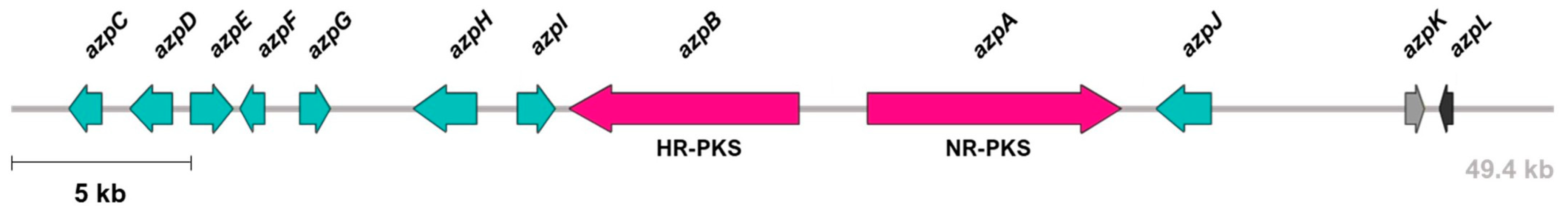
3.1. Identification and Characterization of azp BGC

The antiSMASH analysis of the P. verrucosus FAE27 genome revealed the presence of 29 BGCs. Among these, only one displayed significant similarity to known fungal BGCs associated with the biosynthesis of red-pigmented compounds. This specific BGC, denoted as azp BGC (Figure 1), shares structural resemblances with the ankaflavin and azanigerone BGCs found in Monascus pilosus and Aspergillus niger, respectively. Notably, both ankaflavin and azanigerone are classified as azaphilone-type pigments, suggesting that the azp BGC in P. verrucosus FAE27 may be responsible for the biosynthesis of compounds within this pigment family. It is worth noting that azaphilones can exhibit a color range from yellow to red based on their structural features. Therefore, the biosynthesis of azaphilone-type compounds by the azp BGC could explain the observed red pigmentation in the culture medium of P. verrucosus FAE27. Considering these findings, the genes within this BGC emerge as potential targets for the development of gene deletion and/or attenuation methodologies in Pseudogymnoascus. In order to identify the most suitable target gene, we conducted a bioinformatic analysis of the azp BGC.
The azp BGC spans a length of 49,400 bp and comprises 12 genes (Figure 1). To elucidate the potential functions of these genes, their deduced proteins were subjected to bioinformatic analyses using BLASTP. As depicted in Table 1, five genes encode proteins bearing similarity to those found in the ankaflavin BGC from M. pilosus [42], while two genes encode proteins with similarity to those in the azanigerone BGC from A. niger [43]. These findings provide further support that the azp BGC is responsible for the biosynthesis of an azaphilone compound. Noteworthy, two genes within the azp BGC encode enzymes of the polyketide synthase type (Table 1): azpA (Non-reducing PKS (NR-PKS); 8151 bp; 2698 aa) and azpB (Highly reducing PKS (HR-PKS); 7373 bp; 2315 aa). Currently, there are five known pathways for azaphilone biosynthesis, and all identified azaphilones derive directly from orcinaldehyde precursors synthesized by NR-PKS enzymes [44]. Consequently, we have identified the azpA gene as a potential target for knock-down and knock-out strategies.

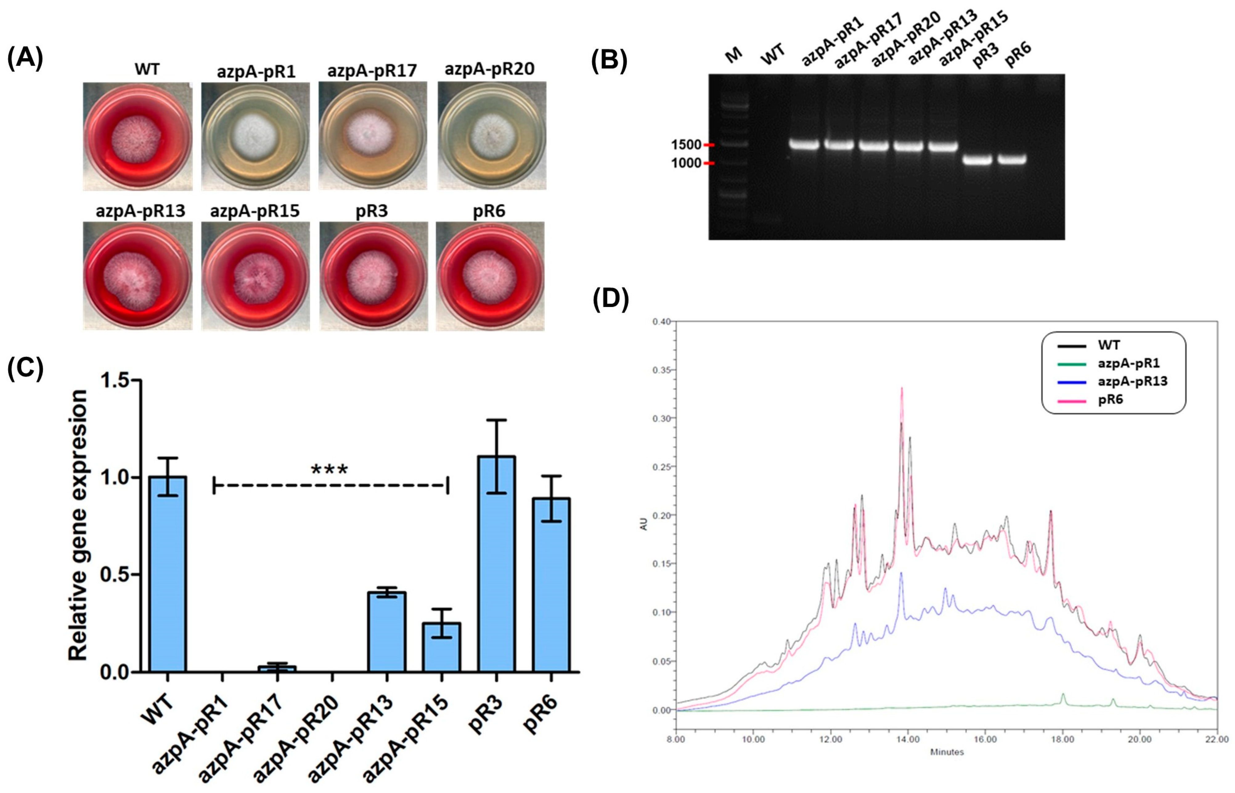
3.2. RNAi-Mediated Silencing of azpA in P. verrucosus FAE27

Following transformation with plasmid pJLH-RNAi-azpA, 60 transformants were obtained, resulting in a transformation efficiency of 6 transformants/µg of plasmid. Among the 60 transformants, 8 (13%) displayed a completely white phenotype, characterized by the absence of pigmentation in both the colony and the agar. For subsequent analyses, we randomly selected three transformant strains with the white phenotype (azpA-pR1, azpA-pR17, and azpA-pR20), along with two transformant strains exhibiting wild-type phenotype (azpA-pR13 and azpA-pR15) (Figure 2A). All of them harbor the full silencing cassette in the genome (Figure 2B).
Quantitative analysis of azpA transcript levels demonstrated a remarkable 90% to 100% reduction in transformants displaying the white phenotype, in contrast to the wild-type strain (Figure 2C). Conversely, transformants exhibiting the red phenotype showed a more moderate decrease of 50–60% in transcript levels. Control strains that underwent transformation with plasmid pJLH-RNAi displayed no significant changes in gene transcript levels when compared to the wild-type strain (Figure 2C). This suggests that the plasmid pJLH-RNAi itself does not influence the observed phenotypic changes in the white transformants.
In order to confirm that azpA would be responsible for the red pigmentation in P. verrucosus FAE27, we conducted a comparative analysis of metabolite profiles between white and red-pigmented transformants. Initially, we examined the overall metabolic profiles of the strains. Extracts from various transformants obtained by RNAi-mediated silencing of azpA were subjected to HPLC analysis at 254 nm, revealing a general resemblance in metabolite composition, irrespective of coloration (Supplementary Figure S3). However, when examined at 530 nm (the optimal wavelength for maximum red pigment absorbance), significant disparities became evident (Figure 2D). Under these conditions, extracts from red transformant strains displayed many peaks with maximal absorption at 530 nm within the retention time range of 10 to 18 min. In contrast, extracts obtained from the white transformants did not show any evidence of compounds with maximal absorption at 530 nm (Figure 2D). These findings strongly support the involvement of the azpA gene in the biosynthesis of metabolites responsible for the red staining of the culture medium.

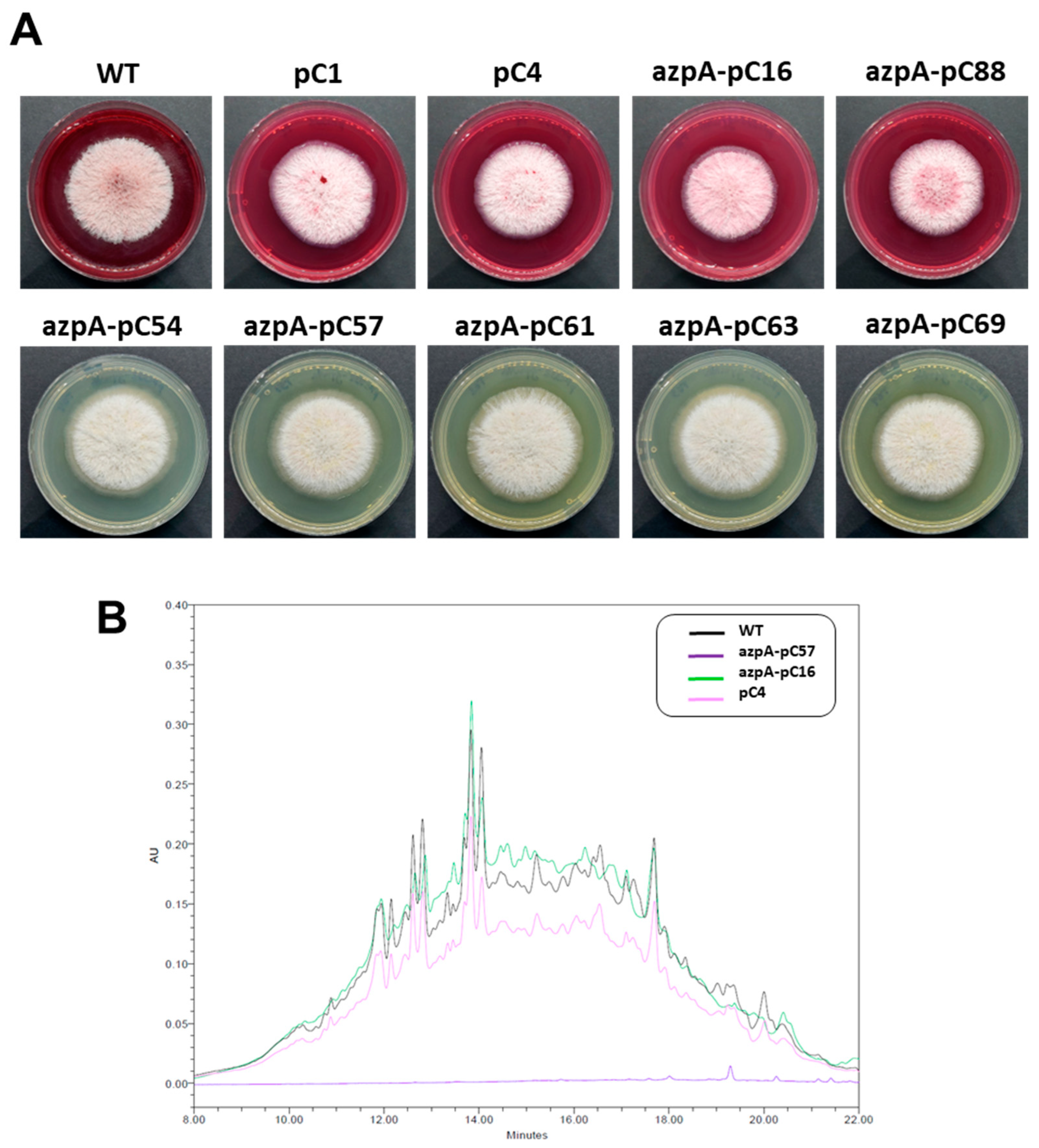
3.3. Disruption of azpA Gene in P. verrucosus FAE27 by CRISPR-Cas9

To specifically target the azpA gene by CRISPR-Cas9, plasmid pFC332-azpA was generated, following the procedures outlined in the Materials and Methods section. Subsequently, we introduced this plasmid into P. verrucosus FAE27 through transformation. As a result, 21 transformants were obtained, yielding a transformation efficiency of 2.1 transformants/μg of plasmid. This efficiency is lower than the achieved with pJLH-RNAi-azpA, which is expected, considering that the size of pFC332-azpA exceeds more than twice that of pJLH-RNAi-azpA (15.6 kb vs. 7.3 kb).
Among the 21 transformants obtained, 16 (76%) displayed a completely white phenotype and showed no pigmentation in the agar. The remaining five transformants exhibited red pigmentation in the culture medium, resembling the wild-type strain. After obtaining monosporic cultures of all transformants, we inoculated them onto PDA plates without hygromycin, resulting in nine transformants (43%) losing hygromycin resistance. From this group, and for subsequent analysis, we selected all transformants with white phenotype (five transformants, namely azpA-pC54, azpA-pC57, azpA-pC61, azpA-pC63, and azpA-pC69) (Figure 3), and two transformants that retained a red pigmentation similar to the wild-type (azpA-pC16 and azpA-pC88, Figure 3). Additionally, we generated two control strains (pC1 and pC4, Figure 3) through transformation with pFC332, which were also included in the subsequent experiments.
As with transformants obtained by RNAi-mediated gene silencing, in transformants obtained through CRISPR-Cas9 methodology, we conducted a comparative HPLC analysis of metabolite profiles between white and red-pigmented strains. At 254 nm, the overall metabolic profiles displayed a general resemblance in composition regardless of coloration (Supplementary Figure S3). However, when examined at 530 nm, extracts from red transformants exhibited multiple peaks within the retention time range of 10 to 18 min. In contrast, extracts obtained from the white transformants showed no evidence of compounds with maximal absorption at 530 nm (Figure 3). These results further support the involvement of the azpA gene in the biosynthesis of metabolites responsible for the red staining in P. verrucosus FAE27.
In order to conclusively establish that the azpA is responsible for the reddish coloration observed in P. verrucosus, we conducted sequencing of the target region of azpA in both the wild-type strain and selected transformants. As depicted in Figure 4, the nucleotide sequences of the red transformant strains, as well as control strains, remained identical to that of the wild-type strain. In contrast, all white transformant strains displayed an insertion in the target sequence, leading to a protein frameshift and the generation of a premature stop codon that effectively disrupts the azpA gene (Figure 4).

4. Discussion

Fungi from the genus Pseudogymnoascus exhibit interesting capabilities in their specialized metabolism [15,16,17,18,19,20,21]. To gain a deeper understanding of Pseudogymnoascus’ metabolic potential, molecular tools enabling its genetic manipulation are essential. While several transformation systems for Pseudogymnoascus have been optimized [22,23], the options for gene manipulation in this important fungal genus have been limited due to the lack of suitable genetic tools. In this study, we contribute to filling this gap by implementing RNAi-mediated gene silencing and CRISPR-Cas9 techniques in the Antarctic strain Pseudogymnoascus verrucosus FAE27. To establish both methodologies, we take advantage of the ability of P. verrucosus FAE27 to secrete a red pigment into the culture medium. The red-pigmented phenotype is associated with the presence of a functional azpA gene. Consequently, the targeting of the azpA gene resulted in a white phenotype that can be easily distinguished from the wild-type phenotype by the naked eye. The availability of this visual reporter greatly facilitated the implementation of both techniques.
The analysis of the genome of P. verrucosus FAE27 revealed the presence of 29 BGCs. One of them, named azp BGC, exhibited similarity to BGCs associated with the production of two metabolites, ankaflavin and azanigerone, both belonging to the azaphilone family. Azaphilones constitute a structurally diverse family of fungal polyketides exhibiting absorbance spectra spanning from 330 to 530 nm. Consequently, they possess the capability to manifest yellow, orange, or red pigmentation depending on their specific chemical structure [45,46].
Azaphilones are synthesized by fungi spanning 61 genera, including Aspergillus, Penicillium, Chaetomium, Talaromyces, Pestalotiopsis, Phomopsis, Emericella, Epicoccum, Monascus and Hypoxylon [44,47]. Over time, chemical analyses of fungal azaphilones and the exploration of their metabolic pathways have supported the idea that these compounds are predominantly genus- or even species-specific [42,46,48,49], to the extent that azaphilones have been proposed as markers for taxonomic purposes in certain fungi [50,51,52]. The findings presented in this work suggest that Pseudogymnoascus is yet another fungal genus capable of azaphilone production. Further efforts, including the isolation of pure compounds and their spectroscopic characterization, will be necessary to elucidate the specific chemical structure of azaphilones originating from the genus Pseudogymnoascus. These endeavors, though, are beyond the scope of the present manuscript.
The core structure of azaphilones consists of a highly oxygenated pyrano-quinone bicyclic [53]. In the biosynthesis of this distinctive functional group, NR-PKS is involved [44]. Consequently, we selected the azpA gene as our target, as it was the sole gene encoding NR-PKS within the azp BGC. Disrupting the azpA gene through CRISPR-Cas9 or achieving substantial RNAi-mediated silencing (at least 90% reduction) led to the generation of white transformants that exhibited no staining in the culture medium. This strongly supports the notion that the azpA gene is essential to produce red compounds in P. verrucosus. In relation to the other genes within the azp BGC, our bioinformatic analysis provides further support indicating the involvement of this BGC in the biosynthesis of compounds belonging to the azaphilone family.
To date, five main biosynthetic pathways for azaphilones have been elucidated [44,47]. Functional analysis of genes in these pathways has been conducted employing various molecular techniques such as homologous recombination-based deletions, overexpression, heterologous expression, and in vitro reconstitution of selected reactions [44,47,54]. It is noteworthy that, as of now, RNA-mediated gene silencing has not been applied to investigate azaphilones BGCs. In the case of CRISPR-Cas9, this methodology was employed in M. ruber to inactivate two negative regulatory genes within the azaphilone BGC, resulting in an enhanced pigment production capacity [55]. To the best of our knowledge, no other examples of CRISPR/Cas9 application for studying azaphilone BGCs have been described.
RNAi-mediated gene silencing is a valuable technique for studying the functionality of fungal genes. Consequently, there has been considerable interest in implementing this technique across various fungal species [26,56]. In several studies using this technique, genes within melanin biosynthesis clusters have been targeted [57,58,59,60,61], following the same rationale as applied in this investigation. Strains deficient in melanin exhibit an albino phenotype in contrast to the corresponding wild-type fungus. Thus, the successful silencing of genes within melanin BGCs results in an easily distinguishable change in phenotype. It is noteworthy that in these studies, RNAi-silenced transformants with phenotypes closely resembling the wild-type were obtained [57,58,59,60,61]. Our findings concerning RNAi-mediated silencing of the azpA gene in P. verrucosus are analogous to these results. In our case, following the RNAi-mediated silencing experiment, we observed that 13% of the transformants displayed visibly distinct phenotypes compared to the wild-type strain, as evidenced by a colorless culture medium. This correlated with a reduction of over 90% in azpA transcripts. Regarding the transformants exhibiting a reddish phenotype identical to the P. verrucosus wild type (constituting 83% of transformants), they displayed only around 60% reduction in azpA transcripts. Therefore, a significant decrease in azpA transcripts is necessary to achieve distinctive phenotypes between these transformants and the wild-type strain of P. verrucosus.
The disruption of the azpA gene via CRISPR-Cas9 yielded more consistent results compared to RNAi-mediated gene silencing. The disruption of azpA effectively resulted in the gene´s inactivation, leading to transformants exhibiting phenotypes characterized by pronounced and unequivocal alterations, in contrast to those observed with RNAi-mediated gene silencing. Furthermore, the efficiency of azpA gene disruption through CRISPR-Cas9 was notably higher, with 76% of the transformants displaying the white phenotype, in contrast to the 13% observed with RNAi-mediated gene silencing. These results indicate that CRISPR-Cas9 is more effective in functionally disrupting azpA than RNAi-mediated silencing. This is expected, considering the distinct mechanisms of these methods. As mentioned, CRISPR-Cas9 induces a double-strand break, repairable by cellular mechanisms, leading to azpA inactivation. In contrast, RNAi-mediated silencing degrades mRNA but keeps the azpA gene intact in the genome. Consequently, with CRISPR-Cas9, azpA is completely inactivated, whereas with RNAi-mediated silencing, despite achieving high silencing efficiencies, complete suppression of azpA expression is not always attained. This partial suppression results in a residual leaky expression, which, in some cases, is sufficient to manifest the red phenotype produced by azaphilone pigments, known for their intense coloration [62,63]. Thus, in mutants that did not achieve substantial RNAi-mediated silencing of azpA (at least a 90% reduction in transcripts), the leaky expression induced fungal coloration, as shown in Figure 2. This contrasts with CRISPR-Cas9 methodology, where azpA was entirely inactivated, preventing any manifestation of the red phenotype.
The examination of CRISPR-Cas9-disrupted transformants revealed a consistent type of mutation (a T insertion) in the target sequence among those displaying the white phenotype. This represents a highly reproducible event. Previous investigations have identified two dominant categories of mutations occurring in the context of DNA double-strand break repair: NHEJ-dependent single-base insertion (entailing duplication of a nucleotide in position -4 from PAM) and MH-mediated deletion [64]. In our case, the single-base insertion corresponds to a thymine at the -4 position relative to the PAM site in the designed sgRNA, thus corresponding to the NHEJ-dependent single-base insertion mechanism.
In this investigation, we implemented RNAi-mediated gene silencing and CRISPR-Cas9 gene disruption methodologies for the analysis of the azpA gene in P. verrucosus. Beyond establishing this proof-of-concept, the methodologies outlined herein expand the repertoire for conducting functional analyses of diverse genes within the wide range of Pseudogymnoascus species. The choice between these two techniques will depend on the specific research objectives of the investigators. As suggestions, we envisaged several scenarios where one technique might be more convenient or suitable than the other. CRISPR-Cas9 is well-suited when precise and permanent genetic modifications are required; however, its application necessitates access to the sequenced genome of the specific Pseudogymnoascus species under investigation. Consequently, the methodology described in this study could be adapted for functional gene analyses in Pseudogymnoascus strains with available genomes, including species such as P. destructans, P. pannorum, and P. verrucosus [65,66]. Of particular interest would be the functional study of genes suspected to encode virulence factors in P. destructans [67], the etiological agent of WNS in bats. In contrast, RNAi-mediated gene silencing is advantageous in scenarios where only a partial coding region of the target gene is known or in cases where comprehensive genome sequences are lacking, a current circumstance for the majority of Pseudogymnoascus species. Moreover, given that RNAi-mediated gene silencing induces gene knockdown without gene elimination, it emerges as a valuable strategy for elucidating the function of genes essential or lethal in Pseudogymnoascus, as demonstrated in other filamentous fungi [68,69]. Furthermore, the straightforward application of RNAi-mediated gene silencing positions it as a good choice for high-throughput functional genomics screens, enabling the systematic evaluation of gene silencing effects on diverse cellular processes. In this context, the genomic analysis of Pseudogymnoascus underscores the presence of numerous biosynthetic gene clusters [16], thereby rendering RNAi-mediated gene silencing useful for the simultaneous experimental analysis of these genes, aiming to establish associations with the production of their respective metabolites.
In summary, this study represents the first implementation of RNAi-mediated gene silencing and CRISPR-Cas9 gene disruption methodologies in Pseudogymnoascus. The availability of these two distinct gene manipulation techniques expands the toolkit for conducting functional analyses of genes in this understudied fungal genus.

Supplementary Materials

The following supporting information can be downloaded at https://www.mdpi.com/article/10.3390/jof10020157/s1, Table S1: Currently accepted Pseudogymnoascus species and their MycoBank identifiers; Table S2: Primers used in this work; Figure S1: Schematic representation depicting the construction process of plasmid pJLH-RNAi-azpA for RNA-mediated silencing of azpA gene; Figure S2: Schematic representation of the sgRNA expression cassette and construction of plasmid pFC332-azpA for azpA disruption by CRISPR-Cas9; Figure S3: Comparative analysis of specialized metabolite profiles of P. verrucosus FAE27 azpA transformants at 254 nm [70].

Author Contributions

Conceptualization, I.V. and R.C.; methodology, D.P., V.O., M.M., R.C., D.T. and I.V.; investigation, D.P, V.O., M.M. and C.G.-D.; resources, I.V. and R.C.; data curation, D.P., V.O., D.T. and I.V.; writing—original draft preparation, I.V. and R.C.; writing—review and editing, R.C. and I.V.; visualization, I.V., R.C., D.P. and V.O.; supervision, I.V. and R.C.; project administration, I.V. and R.C.; funding acquisition, I.V. and R.C. All authors have read and agreed to the published version of the manuscript.

Funding

This research was funded by ANID-FONDECYT grant N° 1211830. V.O. and M.M. have received doctoral fellowships “Beca ANID Doctorado Nacional Folio 21181056” and “Beca ANID Doctorado Nacional Folio 21180477”, respectively.

Institutional Review Board Statement

Not applicable.

Informed Consent Statement

Not applicable.

Data Availability Statement

Data are contained within the article and supplementary materials.

Acknowledgments

We thank Martin E. Kogle and Uffe H. Mortensen from the Technical University of Denmark for providing plasmid pFC332. R.C. acknowledges the support of DICYT-USACH. Finally, we would like to express our gratitude for the invaluable technical expertise provided by Maria Ines Polanco, who recently retired. She will be sorely missed by the team.

Conflicts of Interest

The authors declare no conflict of interest.

References

  1. Cheng, T.L.; Reichard, J.D.; Coleman, J.T.H.; Weller, T.J.; Thogmartin, W.E.; Reichert, B.E.; Bennett, A.B.; Broders, H.G.; Campbell, J.; Etchison, K.; et al. The scope and severity of white-nose syndrome on hibernating bats in North America. Conserv. Biol. 2021, 35, 1586–1597. [Google Scholar] [CrossRef] [PubMed]
  2. Minnis, A.M.; Lindner, D.L. Phylogenetic evaluation of Geomyces and allies reveals no close relatives of Pseudogymnoascus destructans, comb. nov., in bat hibernacula of eastern North America. Fungal Biol. 2013, 117, 638–649. [Google Scholar] [CrossRef] [PubMed]
  3. Villanueva, P.; Vásquez, G.; Gil-Durán, C.; Oliva, V.; Díaz, A.; Henríquez, M.; Álvarez, E.; Laich, F.; Chávez, R.; Vaca, I. Description of the first four species of the genus Pseudogymnoascus from Antarctica. Front. Microbiol. 2021, 12, 713189. [Google Scholar] [CrossRef] [PubMed]
  4. Zhang, Z.; Dong, C.; Chen, W.; Mou, Q.; Lu, X.; Han, Y.; Huang, J.; Liang, Z. The enigmatic Thelebolaceae (Thelebolales, Leotiomycetes): One new genus Solomyces and five new species. Front. Microbiol. 2020, 11, 572596. [Google Scholar] [CrossRef] [PubMed]
  5. Zhang, Z.Y.; Shao, Q.Y.; Li, X.; Chen, W.H.; Liang, J.D.; Han, Y.F.; Huang, J.Z.; Liang, Z.Q. Culturable fungi from urban soils in China I: Description of 10 new taxa. Microbiol. Spectr. 2021, 9, e0086721. [Google Scholar] [CrossRef] [PubMed]
  6. Becker, P.; van den Eynde, C.; Baert, F.; D’hooge, E.; De Pauw, R.; Normand, A.C.; Piarroux, R.; Stubbe, D. Remarkable fungal biodiversity on northern Belgium bats and hibernacula. Mycologia 2023, 115, 484–498. [Google Scholar] [CrossRef] [PubMed]
  7. Zhang, Z.Y.; Han, Y.F.; Chen, W.H.; Tao, G. Additions to Thelebolales (Leotiomycetes, Ascomycota): Pseudogeomyces lindneri gen. et sp. nov. and Pseudogymnoascus campensis sp. nov. MycoKeys 2023, 95, 47–60. [Google Scholar] [CrossRef] [PubMed]
  8. Zhang, Z.Y.; Li, X.; Chen, W.H.; Liang, J.D.; Han, Y.F. Culturable fungi from urban soils in China II, with the description of 18 novel species in Ascomycota (Dothideomycetes, Eurotiomycetes, Leotiomycetes and Sordariomycetes). MycoKeys 2023, 98, 167–220. [Google Scholar] [CrossRef]
  9. Furbino, L.E.; Godinho, V.M.; Santiago, I.F.; Pellizari, F.M.; Alves, T.M.; Zani, C.L.; Junior, P.A.; Romanha, A.J.; Carvalho, A.G.; Gil, L.H.; et al. Diversity patterns, ecology and biological activities of fungal communities associated with the endemic macroalgae across the Antarctic peninsula. Microb. Ecol. 2014, 67, 775–787. [Google Scholar] [CrossRef]
  10. Henríquez, M.; Vergara, K.; Norambuena, J.; Beiza, A.; Maza, F.; Ubilla, P.; Araya, I.; Chávez, R.; San-Martín, A.; Darias, J.; et al. Diversity of cultivable fungi associated with Antarctic marine sponges and screening for their antimicrobial, antitumoral and antioxidant potential. World J. Microbiol. Biotechnol. 2014, 30, 65–76. [Google Scholar] [CrossRef]
  11. Gonçalves, V.N.; Carvalho, C.R.; Johann, S.; Mendes, G.; Alves, T.M.; Zani, C.L.; Junior, P.A.S.; Murta, S.M.F.; Romanha, A.J.; Cantrell, C.L.; et al. Antibacterial, antifungal and antiprotozoal activities of fungal communities present in different substrates from Antarctica. Polar Biol. 2015, 38, 1143–1152. [Google Scholar] [CrossRef]
  12. Gomes, E.C.Q.; Godinho, V.M.; Silva, D.A.S.; de Paula, M.T.R.; Vitoreli, G.A.; Zani, C.L.; Alves, T.M.A.; Junior, P.A.S.; Murta, S.M.F.; Barbosa, E.C.; et al. Cultivable fungi present in Antarctic soils: Taxonomy, phylogeny, diversity, and bioprospecting of antiparasitic and herbicidal metabolites. Extremophiles 2018, 22, 381–393. [Google Scholar] [CrossRef]
  13. Purić, J.; Vieira, G.; Cavalca, L.B.; Sette, L.D.; Ferreira, H.; Vieira, M.L.C.; Sass, D.C. Activity of Antarctic fungi extracts against phytopathogenic bacteria. Lett. Appl. Microbiol. 2018, 66, 530–536. [Google Scholar] [CrossRef]
  14. Vieira, G.; Purić, J.; Morão, L.G.; Dos Santos, J.A.; Inforsato, F.J.; Sette, L.D.; Ferreira, H.; Sass, D.C. Terrestrial and marine Antarctic fungi extracts active against Xanthomonas citri subsp. citri. Lett. Appl. Microbiol. 2018, 67, 64–71. [Google Scholar] [CrossRef]
  15. Shi, T.; Li, X.Q.; Zheng, L.; Zhang, Y.H.; Dai, J.J.; Shang, E.L.; Yu, Y.Y.; Zhang, Y.T.; Hu, W.P.; Shi, D.Y. Sesquiterpenoids from the Antarctic fungus Pseudogymnoascus sp. HSX2#-11. Front. Microbiol. 2021, 12, 688202. [Google Scholar] [CrossRef]
  16. Antipova, T.V.; Zaitsev, K.V.; Zhelifonova, V.P.; Tarlachkov, S.V.; Grishin, Y.K.; Kochkina, G.A.; Vainshtein, M.B. The potential of arctic Pseudogymnoascus fungi in the biosynthesis of natural products. Fermentation 2023, 9, 702. [Google Scholar] [CrossRef]
  17. Shi, T.; Yu, Y.-Y.; Dai, J.-J.; Zhang, Y.-T.; Hu, W.-P.; Zheng, L.; Shi, D.-Y. New polyketides from the Antarctic fungus Pseudogymnoascus sp. HSX2#-11. Mar. Drugs 2021, 19, 168. [Google Scholar]
  18. Han, X.; Gao, H.; Lai, H.; Zhu, W.; Wang, Y. Anti-Aβ42 Aggregative polyketides from the Antarctic psychrophilic fungus Pseudogymnoascus sp. OUCMDZ-3578. J. Nat. Prod. 2023, 86, 882–890. [Google Scholar] [CrossRef]
  19. Hou, X.; Li, C.; Zhang, R.; Li, Y.; Li, H.; Zhang, Y.; Tae, H.S.; Yu, R.; Che, Q.; Zhu, T.; et al. Unusual tetrahydropyridoindole-containing tetrapeptides with human nicotinic acetylcholine receptors targeting activity discovered from Antarctica-derived psychrophilic Pseudogymnoascus sp. HDN17-933. Mar. Drugs 2022, 20, 593. [Google Scholar] [CrossRef]
  20. Shi, T.; Zheng, L.; Li, X.-Q.; Dai, J.-J.; Zhang, Y.-T.; Yu, Y.-Y.; Hu, W.-P.; Shi, D.-Y. Nitrogenous compounds from the Antarctic fungus Pseudogymnoascus sp. HSX2#-11. Molecules 2021, 26, 2636. [Google Scholar]
  21. Figueroa, L.; Jiménez, C.; Rodríguez, J.; Areche, C.; Chávez, R.; Henríquez, M.; de la Cruz, M.; Díaz, C.; Segade, Y.; Vaca, I. 3-Nitroasterric acid derivatives from an Antarctic sponge-derived Pseudogymnoascus sp. fungus. J. Nat. Prod. 2015, 78, 919–923. [Google Scholar] [CrossRef]
  22. Zhang, T.; Ren, P.; Chaturvedi, V.; Chaturvedi, S. Development of an agrobacterium-mediated transformation system for the cold-adapted fungi Pseudogymnoascus destructans and P. pannorum. Fungal Genet. Biol. 2015, 81, 73–81. [Google Scholar] [CrossRef]
  23. Díaz, A.; Villanueva, P.; Oliva, V.; Gil-Durán, C.; Fierro, F.; Chávez, R.; Vaca, I. Genetic transformation of the filamentous fungus Pseudogymnoascus verrucosus of Antarctic origin. Front. Microbiol. 2019, 10, 2675. [Google Scholar] [CrossRef]
  24. Lax, C.; Tahiri, G.; Patiño-Medina, J.A.; Cánovas-Márquez, J.T.; Pérez-Ruiz, J.A.; Osorio-Concepción, M.; Navarro, E.; Calo, S. The evolutionary significance of RNAi in the Fungal Kingdom. Int. J. Mol. Sci. 2020, 21, 9348. [Google Scholar] [CrossRef] [PubMed]
  25. Chang, S.S.; Zhang, Z.; Liu, Y. RNA interference pathways in fungi: Mechanisms and functions. Annu. Rev. Microbiol. 2012, 66, 305–323. [Google Scholar] [CrossRef]
  26. Nakayashiki, H.; Nguyen, Q.B. RNA interference: Roles in fungal biology. Curr. Opin. Microbiol. 2008, 11, 494–502. [Google Scholar] [CrossRef]
  27. Van Leeuwe, T.M.; Arentshorst, M.; Ernst, T.; Alazi, E.; Punt, P.J.; Ram, A.F. Efficient marker free CRISPR/Cas9 genome editing for functional analysis of gene families in filamentous fungi. Fungal Biol. Biotechnol. 2009, 6, 13. [Google Scholar] [CrossRef]
  28. Cong, L.; Ran, F.A.; Cox, D.; Lin, S.; Barretto, R.; Habib, N. Multiplex genome engineering using CRISPR-Cas systems. Science 2013, 339, 197–217. [Google Scholar] [CrossRef]
  29. O’Connell, M.R.; Oakes, B.L.; Sternberg, S.H.; Eastseletsky, A.; Kaplan, M.; Doudna, J.A. Programmable RNA recognition and cleavage by CRISPR/Cas9. Nature 2014, 516, 263–266. [Google Scholar] [CrossRef] [PubMed]
  30. Rice, A.V.; Currah, R.S. Two new species of Pseudogymnoascus with Geomyces anamorphs and their phylogenetic relationship with Gymnostellatospora. Mycologia 2006, 98, 307–318. [Google Scholar] [CrossRef] [PubMed]
  31. Duarte, A.; Menezes, G.; Silva, T.; Bicas, J.; Oliveira, V.; Rosa, L. Antarctic fungi as producers of pigments. In Fungi of Antarctica; Rosa, L., Ed.; Springer: Cham, Switzerland, 2019; pp. 305–318. [Google Scholar]
  32. Mahuku, G. A simple extraction method suitable for PCR-based analysis of plant, fungal, and bacterial DNA. Plant Mol. Biol. Rep. 2004, 22, 71–81. [Google Scholar] [CrossRef]
  33. Blin, K.; Shaw, S.; Kloosterman, A.M.; Charlop-Powers, Z.; van Wezel, G.P.; Medema, M.H.; Weber, T. AntiSMASH 6.0: Improving cluster detection and comparison capabilities. Nucleic Acids Res. 2021, 49, W29–W35. [Google Scholar] [CrossRef] [PubMed]
  34. Ullán, R.V.; Godio, R.P.; Teijeira, F.; Vaca, I.; García-Estrada, C.; Feltrer, R.; Kosalková, K.; Martín, J.F. RNA-silencing in Penicillium chrysogenum and Acremonium chrysogenum: Validation studies using β-lactam genes expression. J. Microbiol. Methods 2008, 75, 209–218. [Google Scholar] [CrossRef] [PubMed]
  35. Punt, P.J.; Oliver, R.P.; Dingemanse, M.A.; Pouwels, P.H.; van den Hondel, C.A. Transformation of Aspergillus based on the hygromycin B resistance marker from Escherichia coli. Gene 1987, 56, 117–124. [Google Scholar] [CrossRef]
  36. Gietz, R.D.; Woods, R.A. Transformation of yeast by lithium acetate/single-stranded carrier DNA/polyethylene glycol method. In Methods in Enzymology; Guthrie, C., Fink, G.R., Eds.; Academic Press: Cambridge, MA, USA, 2002; Volume 350, pp. 87–96. [Google Scholar]
  37. Xie, S.; Shen, B.; Zhang, C.; Huang, X.; Zhang, Y. sgRNAcas9: A software package for designing CRISPR sgRNA and evaluating potential off-target cleavage sites. PLoS ONE 2014, 9, e100448. [Google Scholar] [CrossRef]
  38. Marcano, Y.; Montanares, M.; Gil-Durán, C.; González, K.; Levicán, G.; Vaca, I.; Chávez, R. PrlaeA affects the production of roquefortine C, mycophenolic acid, and andrastin A in Penicillium roqueforti, but it has little impact on asexual development. J. Fungi 2023, 9, 954. [Google Scholar] [CrossRef]
  39. Gil-Durán, C.; Palma, D.; Marcano, Y.; Palacios, J.-L.; Martínez, C.; Rojas-Aedo, J.F.; Levicán, G.; Vaca, I.; Chávez, R. CRISPR/Cas9-mediated disruption of the pcz1 gene and its impact on growth, development, and penicillin production in Penicillium rubens. J. Fungi 2023, 9, 1010. [Google Scholar] [CrossRef]
  40. Nødvig, C.S.; Nielsen, J.B.; Kogle, M.E.; Mortensen, U.H. A CRISPR-Cas9 system for genetic engineering of filamentous fungi. PLoS ONE 2015, 10, e0133085. [Google Scholar] [CrossRef]
  41. Schumann, U.; Smith, N.A.; Wang, M.B. A fast and efficient method for preparation of high-quality RNA from fungal mycelia. BMC Res. Notes 2013, 6, 71. [Google Scholar] [CrossRef] [PubMed]
  42. Balakrishnan, B.; Karki, S.; Chiu, S.H.; Kim, H.-J.; Suh, J.-W.; Nam, B.; Yoon, Y.-M.; Chen, C.-C.; Kwon, H.-J. Genetic localization and in vivo characterization of a Monascus azaphilone pigment biosynthetic gene cluster. Appl. Microbiol. Biotechnol. 2013, 97, 6337–6345. [Google Scholar] [CrossRef] [PubMed]
  43. Zabala, A.O.; Xu, W.; Chooi, Y.H.; Tang, Y. Characterization of a silent azaphilone gene cluster from Aspergillus niger ATCC 1015 reveals a hydroxylation-mediated pyran-ring formation. Chem. Biol. 2012, 19, 1049–1059. [Google Scholar] [CrossRef] [PubMed]
  44. Pavesi, C.; Flon, V.; Mann, S.; Leleu, S.; Prado, S.; Franck, X. Biosynthesis of azaphilones: A review. Nat. Prod. Rep. 2021, 38, 1058–1071. [Google Scholar] [CrossRef] [PubMed]
  45. Feng, Y.; Shao, Y.; Chen, F. Monascus pigments. Appl. Microbiol. Biotechnol. 2012, 96, 1421–1440. [Google Scholar] [CrossRef] [PubMed]
  46. Chen, W.; Chen, R.; Liu, Q.; He, Y.; He, K.; Ding, X.; Kang, L.; Guo, X.; Xie, N.; Zhou, Y.; et al. Orange, red, yellow: Biosynthesis of azaphilone pigments in Monascus fungi. Chem. Sci. 2017, 8, 4917–4925. [Google Scholar] [CrossRef]
  47. Chen, C.; Tao, H.; Chen, W.; Yang, B.; Zhou, X.; Luo, X.; Liu, Y. Recent advances in the chemistry and biology of azaphilones. RSC Adv. 2020, 10, 10197–10220. [Google Scholar] [CrossRef] [PubMed]
  48. Yang, Y.; Liu, B.; Du, X.; Li, P.; Liang, B.; Cheng, X.; Du, L.; Huang, D.; Wang, L.; Wang, S. Complete genome sequence and transcriptomics analyses reveal pigment biosynthesis and regulatory mechanisms in an industrial strain, Monascus purpureus YY-1. Sci. Rep. 2015, 5, 8331. [Google Scholar] [CrossRef] [PubMed]
  49. Chen, W.; Feng, Y.; Molnár, I.; Chen, F. Nature and nurture: Confluence of pathway determinism with metabolic and chemical serendipity diversifies Monascus azaphilone pigments. Nat. Prod. Rep. 2019, 36, 561–572. [Google Scholar] [CrossRef]
  50. Stadler, M.; Fournier, J. Pigment chemistry, taxonomy and phylogeny of the Hypoxyloideae (Xylariaceae). Rev. Iberoam. Micol. 2006, 23, 160–170. [Google Scholar] [CrossRef]
  51. Kuhnert, E.; Surup, F.; Sir, E.B.; Lambert, C.; Hyde, K.D.; Hladki, A.I.; Romero, A.I.; Stadler, M. Lenormandins A—G, new azaphilones from Hypoxylon lenormandii and Hypoxylon jaklitschii sp. nov., recognised by chemotaxonomic data. Fungal Div. 2015, 71, 165–184. [Google Scholar] [CrossRef]
  52. Yang, M.Y.; Wang, Y.X.; Chang, Q.H.; Li, L.F.; Liu, Y.F.; Cao, F. Cytochalasans and azaphilones: Suitable chemotaxonomic markers for the Chaetomium species. Appl. Microbiol. Biotechnol. 2021, 105, 8139–8155. [Google Scholar] [CrossRef]
  53. Osmanova, N.; Schultze, W.; Ayoub, N. Azaphilones: A class of fungal metabolites with diverse biological activities. Phytochem. Rev. 2010, 9, 315–342. [Google Scholar] [CrossRef]
  54. Sousa, T.F.; de Araújo Júnior, M.B.; Peres, E.G.; Souza, M.P.; da Silva, F.M.A.; de Medeiros, L.S.; de Souza, A.D.L.; de Souza, A.Q.L.; Yamagishi, M.E.B.; da Silva, G.F.; et al. Discovery of dual PKS involved in sclerotiorin biosynthesis in Penicillium meliponae using genome mining and gene knockout. Arch. Microbiol. 2023, 205, 75. [Google Scholar] [CrossRef] [PubMed]
  55. Yoon, H.R.; Han, S.; Shin, S.C.; Yeom, S.C.; Kim, H.J. Improved natural food colorant production in the filamentous fungus Monascus ruber using CRISPR-based engineering. Food Res. Int. 2023, 167, 112651. [Google Scholar] [CrossRef] [PubMed]
  56. Dang, Y.; Yang, Q.; Xue, Z.; Liu, Y. RNA interference in fungi: Pathways, functions, and applications. Eukaryot. Cell 2011, 10, 1148–1155. [Google Scholar] [CrossRef]
  57. Moriwaki, A.; Ueno, M.; Arase, S.; Kihara, J. RNA-mediated gene silencing in the phytopathogenic fungus Bipolaris oryzae. FEMS Microbiol. Lett. 2007, 269, 85–89. [Google Scholar] [CrossRef]
  58. Hu, Y.; Hao, X.; Lou, J.; Zhang, P.; Pan, J.; Zhu, X. A PKS gene, pks-1, is involved in chaetoglobosin biosynthesis, pigmentation and sporulation in Chaetomium globosum. Sci. China Life Sci. 2012, 55, 1100–1108. [Google Scholar] [CrossRef]
  59. Hu, Y.; Hao, X.; Chen, L.; Akhberdi, O.; Yu, X.; Liu, Y.; Zhu, X. Gα-cAMP/PKA pathway positively regulates pigmentation, chaetoglobosin A biosynthesis and sexual development in Chaetomium globosum. PLoS ONE 2018, 13, e0195553. [Google Scholar] [CrossRef]
  60. Alhawatema, M.S.; Gebril, S.; Cook, D.; Creamer, R. RNAi-mediated down-regulation of a melanin polyketide synthase (pks1) gene in the fungus Slafractonia leguminicola. World J. Microbiol. Biotechnol. 2017, 33, 179. [Google Scholar] [CrossRef]
  61. Voigt, O.; Knabe, N.; Nitsche, S.; Erdmann, E.A.; Schumacher, J.; Gorbushina, A.A. An advanced genetic toolkit for exploring the biology of the rock-inhabiting black fungus Knufia petricola. Sci Rep. 2020, 10, 12021. [Google Scholar] [CrossRef]
  62. Frisvad, J.C.; Yilmaz, N.; Thrane, U.; Rasmussen, K.B.; Houbraken, J.; Samson, R.A. Talaromyces atroroseus, a new species efficiently producing industrially relevant red pigments. PLoS ONE 2013, 8, e84102. [Google Scholar] [CrossRef]
  63. Fernández, P.; Fañanás, F.J.; Rodríguez, F. Nitrogenated azaphilone derivatives through a silver-catalysed reaction of imines from ortho-alkynylbenzaldehydes. Chemistry 2017, 23, 3002–3006. [Google Scholar] [CrossRef] [PubMed]
  64. Sledzinski, P.; Nowaczyk, M.; Olejniczak, M. Computational tools and supporting CRISPR-Cas experiments. Cells 2020, 9, 1288. [Google Scholar] [CrossRef] [PubMed]
  65. Chibucos, M.C.; Crabtree, J.; Nagaraj, S.; Chaturvedi, S.; Chaturvedi, V. Draft genome sequences of human pathogenic fungus Geomyces pannorum sensu lato and bat white nose syndrome pathogen Geomyces (Pseudogymnoascus) destructans. Genome Announc. 2013, 1, e01045-13. [Google Scholar] [CrossRef] [PubMed]
  66. Palmer, J.M.; Drees, K.P.; Foster, J.T.; Lindner, D.L. Extreme sensitivity to ultraviolet light in the fungal pathogen causing white-nose syndrome of bats. Nat. Commun. 2018, 9, 35. [Google Scholar] [CrossRef] [PubMed]
  67. Davy, C.M.; Donaldson, M.E.; Bandouchova, H.; Breit, A.M.; Dorville, N.A.S.; Dzal, Y.A.; Kovacova, V.; Kunkel, E.L.; Martínková, N.; Norquay, K.J.O.; et al. Transcriptional host-pathogen responses of Pseudogymnoascus destructans and three species of bats with white-nose syndrome. Virulence 2020, 11, 781–794. [Google Scholar] [CrossRef]
  68. Kim, S.; Lee, R.; Jeon, H.; Lee, N.; Park, J.; Moon, H.; Shin, J.; Min, K.; Kim, J.E.; Yang, J.W.; et al. Identification of essential genes for the establishment of spray-induced gene silencing-based disease control in Fusarium graminearum. J. Agric. Food Chem. 2023, 71, 19302–19311. [Google Scholar] [CrossRef]
  69. Zhang, J.; Xiao, K.; Li, M.; Hu, H.; Zhang, X.; Liu, J.; Pan, H.; Zhang, Y. SsAGM1-mediated uridine diphosphate-N-acetylglucosamine synthesis is essential for development, stress response, and pathogenicity of Sclerotinia sclerotiorum. Front. Microbiol. 2022, 13, 938784. [Google Scholar] [CrossRef]
  70. Del-Cid, A.; Gil-Durán, C.; Vaca, I.; Rojas-Aedo, J.F.; García-Rico, R.O.; Levicán, G.; Chávez, R. Identification and functional analysis of the mycophenolic acid gene cluster of Penicillium roqueforti. PLoS ONE 2016, 11, e0147047. [Google Scholar] [CrossRef]
Figure 1. Schematic representation of the putative azaphilone biosynthesis gene cluster (azp BGC) in Pseudogymnoascus verrucosus FAE27. Arrows indicate the genes and their respective transcriptional orientation. Refer to Table 1 for the predicted functions of each gene.
Jof 10 00157 g001
Figure 2. RNA-mediated silencing of azpA in P. verrucosus FAE27. (A) Phenotypic variations observed in selected azpA transformants following the introduction of pJLH-RNAi-azpA. P. verrucosus FAE27 (WT) exhibits its characteristic red pigmentation, while transformants azpA-pR1, azpA-pR17, and azpA-pR20 display a white phenotype. Transformants azpA-pR13 and azpA-pR15 retain the wild-type appearance. Additionally, two control transformants (pR3 and pR6), carrying the empty pJLH-RNAi plasmid lacking the RNAi silencing cassette, were included. These control transformants display the same phenotype as the wild-type fungus. (B) PCR assay confirming the integration of the full silencing cassette in the transformants. The presence of a 1619 bp amplicon confirms the insertion of the cassette in all the transformants. In contrast, control strains pR3 and pR6 show the presence of a 1200 bp amplicon, confirming the absence of azpA insert in the cassette. P. verrucosus FAE27 (WT) was included as negative control. Lane M corresponds to the standard O’GeneRuler 1 kb DNA Ladder (Thermo Fischer Scientific, Braunschweig, Germany), with relevant sizes indicated on the left. (C) qRT-PCR analysis of the expression of azpA in the strains. Wild-type P. verrucosus FAE27 (WT) and transformants pR3 and pR6 containing the empty pJLH-RNAi vector serve as controls. Error bars represent the standard deviation of three replicates in three different experiments. Statistically significant differences were denoted at p < 0.05 (***) using one-way ANOVA and Tukey’s test. (D) Metabolic profiles at 530 nm of P. verrucosus FAE27 (WT) and transformants obtained through RNAi-mediated gene silencing. Alongside WT, strains azpA-pR1 (representative of white transformants), azpA-pR13 (representative of red transformants), and pR6 (control transformed with empty pJLH-RNAi) are included. Note the absence of absorbance at 530 nm in the white transformant azpA-pR1 in comparison to WT and red transformants.
Jof 10 00157 g002
Figure 3. Phenotypic variations in Pseudogymnoascus verrucosus FAE27 and representative strains obtained from pFC332-azpA and pFC332 transformations. (A) P. verrucosus FAE27 (WT) exhibits its characteristic red pigmentation. Following transformation with pFC332-azpA, the phenotypes of the obtained transformants vary. Most transformants (azpA-pC54, azpA-pC57, azpA-pC61, azpA-pC63, and azpA-pC69) display a white phenotype, while some maintain the red coloration, resembling the wild-type appearance (azpA-pC16 and azpA-pC88). Additionally, two control transformants (pC1 and pC4), harboring the empty pFC332 plasmid without the CRISPR-Cas9 expression cassette, were included. As expected, P. verrucosus FAE27 and transformants pC1 and pC4 have the same phenotype. (B) Metabolic profiles at 530 nm of P. verrucosus FAE27 (WT) and transformants obtained by CRISPR/Cas9 methodology: azpA-pC57 (representative of white transformants) and azpA-pC16 (representative of red transformants). Additionally, the control strain pC4, transformed with the empty pFC332 plasmid, is included. Note the absence of absorbance at 530 nm in the white transformant azpA-pC57 in comparison to WT and red transformants.
Jof 10 00157 g003
Figure 4. Comparison of the azpA gene target region in P. verrucosus FAE27 and selected transformants. (A) Sequence alignment of the target region of the azpA gene (highlighted in blue) in P. verrucosus FAE27 (labeled as P. verrucosus WT), white transformants (azpA-pC54, azpA-pC57, azpA-pC61, azpA-pC63, and azpA-pC69), and red transformants (azpA-pC16 and azpA-pC88). Control transformants pC1 and pC4 are also included. The protospacer adjacent motif (PAM) sequences are highlighted in red. All white transformants exhibit a consistent single nucleotide insertion at the same position (indicated by arrows). (B) Analysis of the amino acid sequence of AzpA protein from P. verrucosus FAE27 and the representative transformant azpA-pC54. The amino acid sequence deduced from the azpA target region is highlighted in blue. In strain azpA-pC54, this region displays frameshifts and a premature stop codon (highlighted with a pink asterisk). The same alterations were observed in all other white transformants. Conversely, all red transformants exhibit the same intact sequence as P. verrucosus WT.
Jof 10 00157 g004
Table 1. Analysis of the deduced proteins encoded by the azp genes from Pseudogymnoascus verrucosus FAE27 and their putative functions.
Closest Characterized Homologues
Protein NameSize (Aminoacids)Putative FunctionProtein Name (Organism)GenBank Accession NumberIdentity (%)
AzpA2698Non-reducing polyketide synthaseConidial yellow pigment biosynthesis polyketide synthase (Monascus pilosus)AGN7160461
AzpB2315Highly reducing polyketide synthasePolyketide synthase (Aspergillus niger)EHA2824445
AzpC374KetoreductaseAldehyde reductase (Monascus pilosus)AGN7160850
AzpD455O-acetyltransferaseAcetyltransferase (Monascus pilosus)AGN7160743
AzpE445FAD monooxygenaseMonooxygenase (Phoma sp.)QCO9310953
AzpF268Serine hydrolaseAmino oxidase/esterase (Monascus pilosus)AGN7160958
AzpG364Enoyl reductasePutative quinone-oxidoreductase-like protein (Monascus pilosus)AGN7161050
AzpH644FAD oxidaseIsoamyl alcohol oxidase (Penicillium expansum)AIG6214236
AzpI368Cytochrome P450BuaG cytochrome P450 (Aspergillus burnettii)QBE8564749
AzpJ482FAD oxidaseFAD oxidase (Aspergillus niger)EHA2824347
AzpK216TransporterAflT transporter (Aspergillus flavus)AAS9006945
AzpL119Transcription factorPutative citrinin biosynthesis transcriptional activator CtnR (Monascus pilosus)AGN7160551
Disclaimer/Publisher’s Note: The statements, opinions and data contained in all publications are solely those of the individual author(s) and contributor(s) and not of MDPI and/or the editor(s). MDPI and/or the editor(s) disclaim responsibility for any injury to people or property resulting from any ideas, methods, instructions or products referred to in the content.

