
Compositional Variation and Bioactivity of the Leaf Essential Oil of Montanoa guatemalensis from Monteverde, Costa Rica: A Preliminary Investigation


Abstract
:
1. Introduction
2. Experimental Section
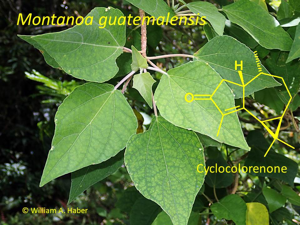
2.1. Plant Material

| Sample | Mass Fresh Leaves | Mass Essential Oil | Yield |
|---|---|---|---|
| 2008, tree A | 46.3 g | 188 mg | 0.41% |
| 2008, tree B | 34.0 g | 160 mg | 0.47% |
| 2009, tree C | 122.1 g | 525 mg | 0.43% |
| 2009, tree D | 192.1 g | 791 mg | 0.41% |
2.2. Gas Chromatographic—Mass Spectral Analysis
| RI a | Compound | 2008 | 2009 | ||
|---|---|---|---|---|---|
| Tree A | Tree B | Tree C | Tree D | ||
| 923 | Artemisia triene | 0.69 | 0.13 | - | - |
| 939 | α-Pinene | 3.18 | 3.78 | 6.91 | 8.56 |
| 958 | Camphene | 0.76 | 0.68 | - | 2.18 |
| 980 | Sabinene | 0.32 | 0.07 | - | - |
| 982 | β-Pinene | 0.74 | 0.93 | 1.81 | 3.71 |
| 990 | Myrcene | 0.12 | 0.12 | 0.10 | 0.27 |
| 1007 | iso-Sylvestrene | 1.65 | 0.48 | - | 1.84 |
| 1030 | Limonene | 8.25 | 8.05 | 21.31 | 12.15 |
| 1040 | (Z)-β-Ocimene | tr b | tr | - | - |
| 1051 | (E)-β-Ocimene | 1.42 | 0.96 | 2.42 | 1.72 |
| 1063 | Artemisia ketone | - | - | 1.77 | - |
| 1075 | cis-Linalool oxide (furanoid) | 0.52 | tr | - | - |
| 1086 | Artemisia alcohol | 0.05 | tr | 1.67 | 0.67 |
| 1106 | Linalool | - | - | 0.14 | - |
| 1165 | Borneol | 0.53 | 0.08 | - | - |
| 1178 | Artemisyl acetate | 0.82 | 0.92 | 9.79 | 1.20 |
| 1282 | Bornyl acetate | 0.06 | tr | - | - |
| 1340 | δ-Elemene | 2.27 | 1.45 | 1.43 | 1.40 |
| 1349 | α-Cubebene | - | tr | - | - |
| 1365 | α-Ylangene | - | tr | - | - |
| 1371 | Unidentified c | 0.19 | - | 1.28 | - |
| 1376 | α-Copaene | 0.21 | 0.32 | - | 1.32 |
| 1384 | α-Bourbonene | - | tr | - | - |
| 1391 | β-Elemene | 0.16 | 0.10 | - | - |
| 1391 | β-Cubebene | - | 0.12 | 0.11 | 0.20 |
| 1408 | α-Gurjunene | 0.09 | - | - | - |
| 1420 | (E)-Caryophyllene | 3.39 | 1.39 | 2.46 | 2.14 |
| 1429 | β-Gurjunene (=Calarene) | 0.07 | 0.08 | - | - |
| 1436 | γ-Elemene | 0.26 | 0.39 | 0.30 | 0.42 |
| 1439 | α-Guaiene | 0.07 | 0.10 | - | - |
| 1444 | 6,9-Guaiadiene | 0.26 | 0.19 | 0.13 | 0.06 |
| 1447 | 9-epi-(E)-Caryophyllene | tr | tr | - | - |
| 1454 | α-Humulene | 0.75 | 0.59 | 0.82 | 0.59 |
| 1460 | allo-Aromadendrene | 0.26 | 0.12 | - | - |
| 1493 | β-Selinene | 8.69 | 14.77 | - | - |
| 1494 | trans-Muurola-4(14),5-diene | - | - | 9.49 | 16.81 |
| 1500 | Bicyclogermacrene | 2.03 | 2.10 | 1.40 | 3.05 |
| 1516 | α-Selinene | 21.15 | 15.90 | - | - |
| 1518 | β-Cadinene | - | 0.20 | 13.26 | 11.10 |
| 1522 | δ-Cadinene | 0.86 | 0.94 | - | 0.42 |
| 1545 | Selina-3,7(11)-diene | 3.63 | 4.54 | 3.55 | 7.96 |
| 1548 | γ-Vetivenene | 1.79 | 2.09 | - | - |
| 1561 | Germacrene B | 3.49 | 4.20 | 1.98 | 2.48 |
| 1570 | Palustrol | - | - | - | 0.72 |
| 1582 | Zierone | 0.86 | 0.52 | 0.30 | - |
| 1590 | Khusimone | 0.20 | 0.13 | 0.39 | 0.35 |
| 1603 | Guaiol | - | - | - | 0.65 |
| 1629 | Unidentified d | 2.29 | 2.62 | 1.00 | 2.92 |
| 1642 | Selina-3,11-dien-6α-ol | 0.36 | 0.33 | 0.14 | - |
| 1653 | Atractylone | 0.70 | 0.56 | 0.47 | 0.63 |
| 1667 | Unidentified e | 0.79 | 0.55 | 0.14 | - |
| 1688 | α-Bisabolol | 0.50 | 0.19 | - | 0.07 |
| 1718 | neo-Cyclocolorenone | 0.09 | tr | - | - |
| 1743 | Cyclocolorenone | 23.82 | 27.54 | 14.49 | 15.61 |
| 1762 | Methyl 3,9,11-guaiatrien-12-oate | 0.38 | 0.18 | 0.51 | - |
| 1833 | iso-Cyclocolorenone | 0.17 | 0.06 | - | - |
| 1950 | Geranyl-α-terpinene | 0.13 | - | - | - |
| 2006 | Geranyl-p-cymene | 0.17 | 0.34 | - | - |
| - | Monoterpenoids | 19.40 | 16.53 | 45.92 | 32.31 |
| - | Sesquiterpenoids | 79.77 | 82.27 | 53.64 | 68.90 |
| - | Total Identified | 95.90 | 95.64 | 97.14 | 98.28 |
2.3. Antibacterial Screening
2.4. Cytotoxicity Screening
| Essential Oil Sample | Antibacterial (MIC, μg/mL) | Cytotoxicity (% Kill at 100 μg/mL) | |||
|---|---|---|---|---|---|
| B. cereus | S. aureus | E. coli | MDA-MB-231 | Hs578T | |
| 2008, tree A | 313 | 1250 | 2500 | 100 | 6.5 ± 8.8 |
| 2008, tree B | 625 | 1250 | 2500 | 100 | 0 |
| 2009, tree C | 313 | 1250 | 2500 | 66.3 ± 5.6 | 0 |
| 2009, tree D | 313 | 1250 | 2500 | 21.9 ± 3.5 | 0 |
3. Results and Discussion
4. Conclusions
Acknowledgments
Author Contributions
Conflicts of Interest
References
- Haber, W.A.; Zuchowski, W.; Bello, E. An Introduction to Cloud Forest Trees: Monteverde, Costa Rica, 2nd ed.; Mountain Gem Publications: Monteverde, Costa Rica, 2000. [Google Scholar]
- Piper, J.K. Colonization of tubú (Montanoa guatemalensis, Asteraceae) windbreaks by woody species. Biotropica 2006, 38, 122–126. [Google Scholar]
- Seaman, F.C.; Malcolm, A.J.; Fischer, N.H. Sesquiterpene lactones of Montanoa guatemalensis and Montanoa tomentosa subsp. xanthiifolia. Phytochemistry 1985, 24, 2003–2005. [Google Scholar] [CrossRef]
- NIST (National Institute of Standards and Technology). Mass Spectral Library (NIST/EPA/NIH, V. 2.0g); The NIST Mass Spectrometry Data Center: Gaithersburg, MD, USA, 2011.
- Adams, R.P. Identification of Essential Oil Components by Gas Chromatography/Mass Spectrometry, 4th ed.; Allured Publishing Corp.: Carol Stream, IL, USA, 2007. [Google Scholar]
- Satyal, P. Development of GC-MS Database for Essential Oil Analysis, Adulteration Detection in Commercial Essential Oil Samples, and Discovery of Biologically Active Novel Chemotypes in Essential Oils. Ph.D. Thesis, University of Alabama in Huntsville, Huntsville, AL, USA, November 2015. [Google Scholar]
- Sahm, D.H.; Washington, J.A. Antibacterial susceptibility tests: Dilution methods. In Manual of Clinical Microbiology, 5th ed.; Balows, A., Hausler, W.J., Herrmann, K.L., Isenberg, H.D., Shamody, H.J., Eds.; American Society for Microbiology: Washington, DC, USA, 1991. [Google Scholar]
- Cailleau, R.; Young, R.; Olive, M.; Reeves, W.J. Breast tumor cell lines from pleural effusions. J. Natl. Cancer Inst. 1974, 53, 661–674. [Google Scholar] [PubMed]
- Hackett, A.J.; Smith, H.S.; Springer, E.L.; Owenms, R.B.; Nelson-Rees, W.A.; Riggs, J.L.; Gardner, M.B. Two syngeneic cell lines from human breast tissue: The aneuploid mammary epithelial (Hs578T) and the diploid myoepithelial (Hs578Bst) cell lines. J. Natl. Cancer Inst. 1977, 58, 1795–1806. [Google Scholar] [PubMed]
- Setzer, W.N.; Setzer, M.C.; Hopper, A.L.; Moriarity, D.M.; Lehrman, G.K.; Niekamp, K.L.; Morcomb, S.M.; Bates, R.B.; McClure, K.J.; Stessman, C.C.; et al. The cytotoxic activity of a Salacia liana species from Monteverde, Costa Rica, is due to a high concentration of tingenone. Planta Med. 1998, 64, 583. [Google Scholar] [CrossRef] [PubMed]
- Ferrari, M.; Fornasiero, M.C.; Isetta, A.M. MTT colorimetric assay for testing macrophage cytotoxic activity in vitro. J. Immunol. Meth. 1990, 131, 165–172. [Google Scholar] [CrossRef]
- Kalemba, D.; Marschall, H.; Bradesi, P. Constituents of the essential oil of Solidago gigantea Ait. (giant goldenrod). Flavour Fragr. J. 2001, 16, 19–26. [Google Scholar] [CrossRef]
- Kalemba, D.; Thiem, B. Constituents of the essential oils of four micropropagated Solidago species. Flavour Fragr. J. 2004, 19, 40–43. [Google Scholar] [CrossRef]
- Schmidt, C.O.; Bouwmeester, H.J.; Bülow, N.; König, W.A. Isolation, characterization, and mechanistic studies of (−)-α-gurjunene synthase from Solidago canadensis. Arch. Biochem. Biophys. 1999, 364, 167–177. [Google Scholar] [CrossRef] [PubMed]
- Gomes, M.R.F.; Schuh, R.S.; Jacques, A.L.B.; Augustin, O.A.; Bordignon, A.L.; Dias, D.O.; Kelmann, R.G.; Koester, L.S.; Gehring, M.P.; Morrone, F.B.; et al. Citotoxic activity evaluation of essential oils and nanoemulsions of Drimys angustifolia and D. braziliensis on human glioblastoma (U-138 MG) and human bladder carcinoma (T24) cell lines in vitro. Rev. Bras. Farmacog. 2013, 23, 259–267. [Google Scholar] [CrossRef]
- Lima, M.A.S.; Barros, M.C.P.; Pinheiro, S.M.; do Nascimento, R.F.; Matos, F.J.A.; Silveira, E.R. Volatile compositions of two Asteraceae from the northeast of Brazil: Ageratum conyzoides and Acritopappus confertus (Eupatorieae). Flavour Fragr. J. 2005, 20, 559–561. [Google Scholar] [CrossRef]
- Martins, M.M.; de Morais, S.A.L.; de Oliveira, A.; do Nascimento, E.A.; de Aquino, F.J.T.; Chang, R.; Borges, M.S.; de Melo, G.B.; da Silva, C.V.; Machado, F.C. Chemical composition, antimicrobial and antiprotozoal activity of essential oils from Vernonia brasiliana (Less) Druce (Asteraceae). J. Essent. Oil Bear. Plants 2015, 18, 561–569. [Google Scholar] [CrossRef]
- Gretšušnikova, T.; Järvan, K.; Orav, A.; Koel, M. Comparative analysis of the composition of the essential oil from the shoots, leaves and stems the wild Ledum palustre L. from Estonia. Proc. Chem. 2010, 2, 168–173. [Google Scholar] [CrossRef]
- Nakamura, M.J.; Monteiro, S.S.; Bizarri, C.H.B.; Siani, A.C.; Ramos, M.F.S. Essential oils of four Myrtaceae species from the Brazilian southeast. Biochem. Systemat. Ecol. 2010, 38, 1170–1175. [Google Scholar] [CrossRef]
- Simões-Pires, C.A.; Debenedetti, S.; Spegazzini, E.; Mentz, L.A.; Matzenbacher, N.I.; Limberger, R.P.; Henriques, A.T. Investigation of the essential oil from eight species of Baccharis belonging to sect. Caulopterae (Asteraceae, Astereae): A taxonomic approach. Plant Syst. Evol. 2005, 253, 23–32. [Google Scholar]
- Joshi, R.K.; Badakar, V. Chemical composition and in vitro antimicrobial activity of the essential oil of the flowers of Tridax procumbens. Nat. Prod. Commun. 2012, 7, 941–942. [Google Scholar] [PubMed]
- Wright, C.; Chhetri, B.K.; Setzer, W.N. Chemical composition and phytotoxicity of the essential oil of Encelia farinosa growing in the Sonoran Desert. Am. J. Essent. Oils Nat. Prod. 2013, 1, 18–22. [Google Scholar]
- Ruffinengo, S.R.; Maggi, M.; Fuselli, S.; Floris, I.; Clemente, G.; Firpo, N.H.; Bailac, P.N.; Ponzi, M.I. Laboratory evaluation of Heterothalamus alienus essential oil against different pests of Apis mellifera. J. Essent. Oil Res. 2006, 18, 704–707. [Google Scholar] [CrossRef]
- Onyambu, G.K.; Maranga, R.; Ndungu, M.; Mkoji, G.M.; Kareru, P.G.; Wanjoya, A. GC-MS analysis of pesticidal essential oils from four Kenyan plants. Afr. J. Biotechnol. 2015, 14, 1158–1166. [Google Scholar]
- Owolabi, M.S.; Lajide, L.; Villanueva, H.E.; Setzer, W.N. Essential oil composition and insecticidal activity of Blumea perrottentiana growing in southwestern Nigeria. Nat. Prod. Commun. 2010, 5, 1135–1138. [Google Scholar] [PubMed]
- Washington, V.D.; Agius, B.R.; Palazzo, M.C.; Haber, W.A.; Setzer, W.N. Chemical composition of the leaf essential oil of Clibadium leiocarpum from Monteverde, Costa Rica. Am. J. Essent. Oils Nat. Prod. 2013, 1, 43–45. [Google Scholar]
- Pérez-Amador, M.C.; Muñoz, V.; Noyola, A.; García-Jiménez, F. Essential oil and phototoxic compounds in Clibadium surinamense L. and Montanoa grandiflora D.C. (Asteraceae). Phyton 2006, 75, 145–150. [Google Scholar]
- Compadre, C.M.; Hussain, R.A.; Leon, I.; Enríquez, R.G. Volatile constituents of Montanoa tomentosa and Lippia graveolens. Planta Med. 1987, 53, 495–496. [Google Scholar] [CrossRef] [PubMed]
- Robles-Zepeda, R.E.; Molina-Torres, J.; Lozoya-Gloria, E.; López, M.G. Volatile organic compounds of leaves and flowers of Montanoa tomentosa. Flavour Fragr. J. 2006, 21, 225–227. [Google Scholar] [CrossRef]
- Robles-Zepeda, R.E.; Lozoya-Gloria, E.; López, M.G.; Villarreal, M.L.; Ramírez-Chávez, E.; Molina-Torres, J. Montanoa tomentosa glandular trichomes containing kaurenoic acids chemical profile and distribution. Fitoterapia 2009, 80, 12–17. [Google Scholar] [CrossRef] [PubMed]
- Zapata, B.; Betancur-Galvis, L.; Duran, C.; Stashenko, E. Cytotoxic activity of Asteraceae and Verbenaceae family essential oils. J. Essent. Oil Res. 2014, 26, 50–57. [Google Scholar] [CrossRef]
© 2015 by the authors; licensee MDPI, Basel, Switzerland. This article is an open access article distributed under the terms and conditions of the Creative Commons Attribution license (http://creativecommons.org/licenses/by/4.0/).
Share and Cite
Flatt, V.D.; Campos, C.R.; Kraemer, M.P.; Bailey, B.A.; Satyal, P.; Setzer, W.N. Compositional Variation and Bioactivity of the Leaf Essential Oil of Montanoa guatemalensis from Monteverde, Costa Rica: A Preliminary Investigation. Medicines 2015, 2, 331-339. https://doi.org/10.3390/medicines2040331
Flatt VD, Campos CR, Kraemer MP, Bailey BA, Satyal P, Setzer WN. Compositional Variation and Bioactivity of the Leaf Essential Oil of Montanoa guatemalensis from Monteverde, Costa Rica: A Preliminary Investigation. Medicines. 2015; 2(4):331-339. https://doi.org/10.3390/medicines2040331
Chicago/Turabian StyleFlatt, Victoria D., Carlos R. Campos, Maria P. Kraemer, Brittany A. Bailey, Prabodh Satyal, and William N. Setzer. 2015. "Compositional Variation and Bioactivity of the Leaf Essential Oil of Montanoa guatemalensis from Monteverde, Costa Rica: A Preliminary Investigation" Medicines 2, no. 4: 331-339. https://doi.org/10.3390/medicines2040331
APA StyleFlatt, V. D., Campos, C. R., Kraemer, M. P., Bailey, B. A., Satyal, P., & Setzer, W. N. (2015). Compositional Variation and Bioactivity of the Leaf Essential Oil of Montanoa guatemalensis from Monteverde, Costa Rica: A Preliminary Investigation. Medicines, 2(4), 331-339. https://doi.org/10.3390/medicines2040331