Article

Investigation of In Vitro Anticancer and Apoptotic Potential of Biofabricated Silver Nanoparticles from Cardamine hirsuta (L.) Leaf Extract against Caco-2 Cell Line

by
Halaswamy Hire Math
1,†,
Kariyellappa Nagaraja Shashiraj
1,†,
Raju Suresh Kumar
2,
Muthuraj Rudrappa
1,
Meghashyama Prabhakara Bhat
1,
Dhanyakumara Shivapoojar Basavarajappa
1,
Abdulrahman I. Almansour
2,
Karthikeyan Perumal
3 and
Sreenivasa Nayaka
1,*
1
P.G. Department of Studies in Botany, Karnatak University, Dharwad 580003, Karnataka, India
2
Department of Chemistry, College of Science, King Saud University, Riyadh 11451, Saudi Arabia
3
Department of Chemistry and Biochemistry, The Ohio State University, 151 W. Woodruff Ave., Columbus, OH 43210, USA
*
Author to whom correspondence should be addressed.
These authors contributed equally to this work.
Inorganics 2023, 11(8), 322; https://doi.org/10.3390/inorganics11080322
Submission received: 28 June 2023 / Revised: 26 July 2023 / Accepted: 29 July 2023 / Published: 31 July 2023
(This article belongs to the Special Issue Functional Inorganic Materials for Biomedical Application)

Abstract

:
Green nanoparticle (NPs) synthesis is eco-friendly, non-toxic, and the NPs have demonstrated improved biocompatibility for use in healthcare. This study evaluated the biogenic synthesis of AgNPs from the leaves of Cardamine hirsuta L. and their biological properties. The UV-Vis. spectra at 411 nm exhibited a distinct resonance spectrum for C-AgNPs produced from C. hirsuta L. FT-IR analysis exhibited the presence of functional groups of phyto-compounds of C. hirsuta responsible of silver salt reduction and capping agents of C-AgNPs. The microscopic-based study, such as HR-TEM analysis, showed that the particles were uniformly distributed, spherical, and ranged in size from 5.36 to 87.65 nm. EDX analysis confirmed a silver (Ag) content of 36.3% by weight, and XRD analysis exhibited the face-centred cubic (FCC) crystalline nature of C-AgNPs. DLS measured the mean particle size of 76.5 nm. The zeta potential was significant at −27.9 mV, and TGA analysis revealed that C-AgNPs had higher thermal stability. C-AgNPs demonstrated moderate antimicrobial activity against the tested pathogens. In addition, the anti-proliferative activity measured by the MTT assay on the Caco-2 cell line demonstrated decreased cell viability with increasing C-AgNPs dosage, with an IC50 concentration of 49.14 µg/mL. In addition, an Annexin-V/Propidium iodide flow cytometric study was utilized to evaluate the induction of apoptosis in cancer cells. Early and late apoptosis cell populations increased significantly compared to the untreated control. Therefore, green-synthesized C-AgNPs have significant antimicrobial and anti-proliferative abilities, making them intriguing options for future biomedical applications.

Graphical Abstract

1. Introduction

In the past decade, nanotechnology has emerged as one of the leading technologies, with vast applications in catalytic devices, electronic appliances, optical and cosmetics, etc. [1]. Organic nanoparticles (synthesized from dendrimers, micelles, liposomes, and compact polymers) and inorganic nanoparticles (synthesized from noble metals such as silver, gold, copper, zinc, titanium, and palladium, etc.) dominate the classification of NPs [2]. Chemical, physical, and biological methods can produce inorganic NPs. The high production cost of physical and chemical methods was documented, and these methods create environmental imbalance by releasing hazardous chemicals used in the processes [3]. Due to its eco-friendliness, lower production costs, biocompatibility, and energy efficiency, the biological method of synthesizing NPs has garnered considerable interest [4]. On account of their catalytic property, chemical stability, and electrical conductivity, AgNPs have garnered the most attention among metal NPs. The bio-synthesized AgNPs demonstrated numerous industrial and biological applications in diverse fields, including textiles, electronics, cosmetics, catalysis, the soap industry, food technology, the invention of antibacterial and antitumor drugs, and agriculture [5]. Researchers have focused on the biogenic fabrication of AgNPs using microorganisms and plants for multiple applications. Culture maintenance makes the microbial-based synthesis of NPs time-consuming and expensive [6,7].
Plant extracts have been used by many researchers to synthesize metal oxide NPs via the green synthesis process. When plant leaf extract synthesizes nanoparticles, it is mixed with metal precursor solutions under different reaction conditions [8]. The stability and yield of nanoparticles are influenced by various parameters related to the plant leaf extract, including the types and concentrations of phytochemicals, metallic salt concentration, pH, and temperature [9]. The ability of plant leaf extracts to effectively reduce metal ions surpasses that of bacteria and fungi, as the latter organisms necessitate a longer incubation period [10].
To synthesize metals and metal oxide NPs, plant leaf extracts can be regarded as a safe and effective source. A plant leaf extract also contributes to the synthesis of nanoparticles by acting both as a reducing and stabilizing agent, facilitating nanoparticle formation. In addition to the composition of plant leaf extract, phytochemical concentration levels vary from plant to plant, which contributes to nanoparticle synthesis [11]. The many functional groups of flavonoids enhance the reduction of metal ions by releasing reactive hydrogen atoms when the enol-form of flavonoids is converted into keto-form by tautomeric transformation. A metal ion is reduced into a metal nanoparticle to achieve this process [12,13,14]. Nanoparticle size and shape are essential parameters for an NP-based study. Smaller particles have a more remarkable ability to escape natural body clearance mechanisms. The shape of cylindrical nanoparticles has been shown to profoundly affect their bioavailability in studies in addition to size, exhibiting that cylindrical NPs interact with cells very differently than spherical ones. Our experiment involved centrifuging the mixture more than once and then centrifuging the supernatant at a higher force after each run. We used a pH-switch method to synthesize nanoparticles [15].
Similarly, the AgNPs synthesized from Datura stramonium, Ocimum sanctum, and Pyrostegia venusta leaf extracts exhibited a broad spectrum of antimicrobial and anticancer activities [16,17,18]. In recent research, various techniques have been employed to obtain the interference and action of AgNPs and Ag+ on protein structure and disulfide bonds, down-regulating pathogenic microbes’ functioning status. The accumulation of AgNPs in cancer cells can induce cell death by upregulating caspase-3 activation and oxidative stress [19].
Cancer is considered a life-threatening disease because of its uncontrolled growth of abnormally organized tissues. The human colon adenocarcinoma is a particularly severe cancer, with approximately 782,000 pre-diagnosed cases reported annually [20]. Accessibility issues and a delayed diagnosis rendered the treatments ineffective and less successful. It has been documented that traditional cancer treatments, such as radiotherapy and chemotherapy, have numerous side effects, some even harmful to healthy cells. Therefore, AgNPs synthesized from various plant extracts were identified as promising anticancer agents for treating various cancer types. According to a recent study, Celtis tournefortii leaf extract-mediated AgNPs displayed potent anti-cancer potential against the Caco-2 cell line [21].
In addition, Zein et al. [22] reported a dose-dependent anti-oncogenic effect of Eucalptus camaldulensis AgNPs against the Caco-2 cell line, with an IC50 value of 18 µg/mL. In another study, bio-fabricated AgNPs derived from the leaf extract of Cicer arietinum L. demonstrated potent anti-proliferative activity against the Caco-2 cell line [23]. The release of Ag+ ions can facilitate the anticancer mechanism of AgNPs by inducing oxidative stress, the production of ROS, structural dysfunction, damage to nuclear materials, and cell death [24].
Cardamine hirsuta L. is an annual herb that belongs to the Brassicaceae family. The common names for this plant are hairy bittercress or poppy cress, native to Australia, Asia, Canada, Colombia, and Costa Rica. As a weed, it grows between 5 and 50 cm tall and is commonly consumed as a vegetable during the summer [25]. According to phytochemical analyses, this plant part contained bioactive metabolites such as anthraquinones, tannins, phenols, steroids, alkaloids, and flavonoids. The plant exhibited beneficial biological properties, including antioxidant and antimicrobial properties [26]. Cardamine contains sulfur-containing compounds that protect plants from herbivores and microorganisms, such as glycosides (glucosinolates). The glucosinolates are non-toxic, and their metabolites are cancer-fighting agents. According to numerous scientific studies, increased consumption of cruciferous vegetables is associated with a decreased risk of various cancers. All vegetables containing glucosinolates contain myrosinase, an enzyme that promotes the hydrolysis of glucosinolates into aglycone and D-glucose, which are then converted into isothiocyanates [27,28,29].
The current study employed a biosynthesis approach owing to its environmentally sustainable characteristics. This study employed a novel methodology to synthesize AgNPs using leaves of Cardamine hirsuta L. Furthermore, it investigates the potential antimicrobial and anticancer properties of these C-AgNPs and their apoptotic effects.

2. Results

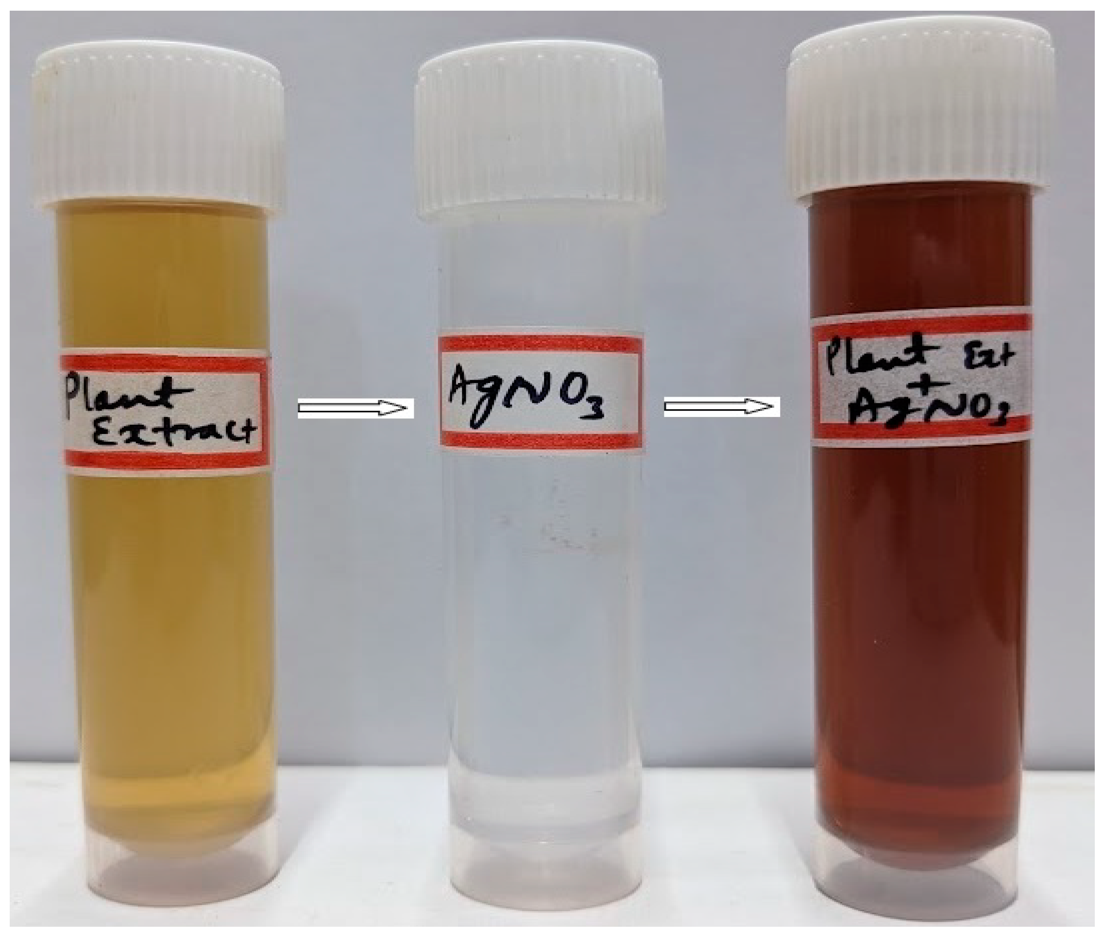
2.1. Biofabrication of Silver Nanoparticles

The C. hirsuta (Figure 1) leaf extract was blended with 1 mM silver nitrate solution in a 1:5 (v/v) ratio at the pH of 9.0 to fabricate the C-AgNPs. After an incubation period of 24 h, the color of the reaction suspension turns brown from light yellow, which depicts the proper synthesis of C-AgNPs (Figure 2). Because of phytochemicals’ reduction and capping mechanism in the leaf extract, Ag+ was reduced into Ago and capped and stabilized.

2.2. UV-Visible Spectroscopic Analysis

The preliminary characterization of the synthesis of AgNPs was conducted using UV-Vis spectrophotometry. The fabrication of AgNPs was indicated by a distinct peak between the wavelength ranging from 400 nm to 450 nm. The observed peak at a wavelength of 411 nm can be attained through the phenomenon of SPR (Figure 3). The observed data suggest that bioactive components are being released from the leaf extract of C. hirsuta. These components are responsible for facilitating the reduction and capping of NPs. The presence of enzymes in the aqueous solution enabled the reduction from (Ag+) ions to (Ago) ions in a colloidal solution, resulting in observable brown coloration.

2.3. FT-IR Analysis

In the region of 4000 to 500 cm−1, the FT-IR spectra of C-AgNPs displayed ten vibrational peaks, as opposed to eight in the spectrum of C. hirsuta leaf extract (Figure 4A,B). In the leaf extract and C-AgNPs spectra, a broad area peak switch to a longer wavelength of 3381 to 3394 cm−1 indicated a significant intermolecular O-H stretching of alcohol. The peak shift from 2921 to 2926 cm−1 indicated the occurrence of C-H stretching in alkane, whereas the peak of 2426 cm−1 in C-AgNPs indicated the presence of O=C=O-bonded carbon dioxide. The peak indicated aldehyde C=O stretching at 1734 cm−1. Peak redistribution from 1643 to 1617 cm−1 corresponds to the C=C stretching of alkene and-unsaturated ketones. The slight shift of the peak to 1383 cm−1 from 1381 cm−1 revealed the occurrence of C-H bending in alkane, while the vibrational peaks in leaf extract at 1284 and 1032 cm−1 indicated C-O stretching in aromatic esters and CO-O-CO stretching in anhydride groups.
Furthermore, the 839 and 776 cm−1 vibrational peaks revealed C=C bending alkenes and C-Cl stretching halo compounds, respectively. Halo compounds’ C-Br stretching is responsible for the peak shift to a longer wavelength of 613 to 618 cm−1. The peak at 514 cm−1 in C-AgNPs was finally correlated to the presence of C-I stretching of halo groups. The detected shifts in the vibrational peaks of the FTIR spectra depict the involvement of the functional compounds’ groups present in the phytochemicals in the formation of nanoparticles using the leaf extract.

2.4. HR-TEM Analysis

TEM analysis was utilized to provide insights into the size and morphology of the fabricated C-AgNPs. The electron microscopic observations provided evidence of the uniformity, good dispersion, and spherical morphology of the AgNPs that were synthesized, thereby suggesting their stabilization (Figure 5A). The spherical shape of the C-AgNPs was observed, with minimal scattering and low levels of aggregation, indicating a poly-dispersed nature. The size dispersion of the NPs was examined using Image J software, revealing a range of 5.36 to 87.65 nm (Figure 5B).

2.5. EDX Analysis

The EDX characterization was performed to determine the presence of different elemental mapping that was involved in the production of C-AgNPs. The spectra were generated through processing C-AgNPs at the energy range from 0 to 10 KeV. The elemental analysis indicated the predominant presence of silver, accounting for 36.3% of the composition. However, other elements were detected at varying proportions. Several extra other peaks were noticed in the spectrum, specifying the presence of various elements. These elements included carbon (22.94%), oxygen (28.24%), chlorine (3.87%), sodium (2.11%), potassium (2.28%), calcium (2.03%), and others, as depicted in Figure 6. These elements’ possible existence could be attributed to various phytochemicals in the leaf extracts of C. hirsuta, which may serve as both reducing and capping agents.

2.6. XRD Analysis of C-AgNPs

Powder X-ray diffraction analysis was conducted to validate the synthesis and characterize the properties of C-AgNPs. The XRD plot (Figure 7) illustrates the crystalline characteristics of C-AgNPs, as evidenced by the intensity of the X-ray scattering pattern observed at various angles. The diffraction angle, denoted as 2θ, represents the angle between the transmitted and reflected beams. The results of the observation revealed the presence of five prominent 2θ values peaks at 32.35°, 38.14°, 46.21°, 64.49°, and 77.44°, which can be attributed to the diffraction of X-rays by the 101, 111, 200, 220, and 311, crystallographic planes of silver. The face-centred cubic structure is observed as the characteristic pattern of C-AgNPs. The average size of C-AgNPs particles was estimated to be 66.27 nm based on Dubey Scherrer’s equation.

2.7. Zeta Potential and DLS Analysis

The zeta potential determines the colloidal nature, stability, and surface electric charge of the synthesized C-AgNPs. The zeta potential analysis indicated the surface charge of C-AgNPs to be −27.9 mV (Figure 8A). The C-AgNPs were well dispersed in a colloidal solution, and the high cationic nature conveyed strong electrostatic repulsion between the nanoparticles. From the DLS analysis, the size dispersion profile of nanoparticles depicted the mean average size of the particles as 76.5 nm due to agglomeration in a colloidal suspension (Figure 8B).

2.8. Thermo Gravimetric Analysis

C-AgNPs weight loss was correlated with temperature by TGA analysis. The interactions of phytochemicals with higher temperature break bonds and cause weight loss of C-AgNPs. C-AgNPs weighed 100% at room temperature, from which the temperature was increased to 800 °C at 10 °C/min. Initially, but at 100 °C and 300 °C, they lost weight by 92% and 73%, possibly due to moisture evaporation. Finally, at 700 °C, C-AgNPs weighed 60.7%, losing 39.3% due to the breakdown of chemical bonds in organic elements that stabilized them (Figure 9).

2.9. Antimicrobial Activity

The C-AgNPs exhibited moderate antimicrobial potential against tested clinical pathogens (Figure 10A–F). The maximum activity was found in P. aeruginosa with inhibitory zones of 16.8 ± 0.2, 20.5 ± 0.5, 22.3 ± 0.5 and 23.4 ± 1.0 mm at 25, 50, 75, and 100 µL of C-AgNPs suspensions. The minimum activity was found on S. aureus with 11.5 ± 0.3, 13.3 ± 0.3, 14.6 ± 0.5 and 16.3 ± 1.0 mm inhibition zones at the C-AgNPs volumes of 25, 50, 75, and 100 µL, respectively. Distilled water was taken as a negative, and streptomycin and amphotericin-B were utilized as positive controls. The microbial pathogens, namely E. coli, K. pneumoniae, B. subtilis, and C. glabrata, showed moderate inhibition by C-AgNPs at different volumes. The antimicrobial activity on test pathogens to varying volumes of C-AgNPs was depicted in the bar graph (Figure 11).

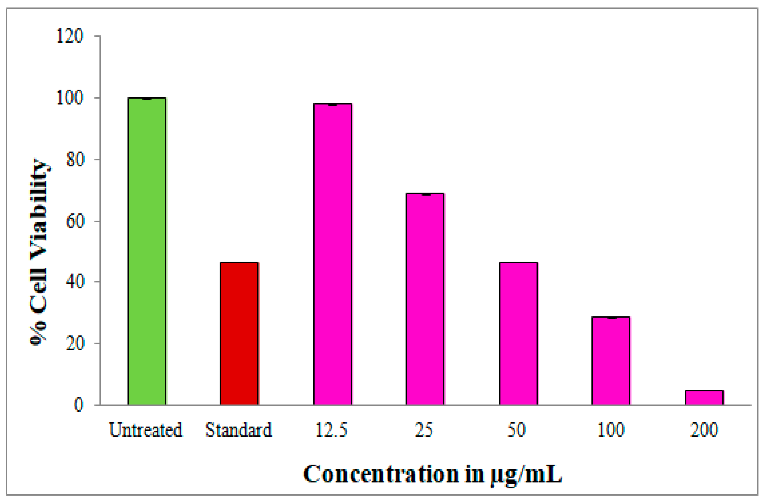
2.10. Evaluation of Cell Viability by MTT Assay

In-vitro anti-proliferative potential of C-AgNPs against the Caco-2 cell line was assessed, and made a comparison with untreated and standard drug 5-fluorouracil (30 µM/mL) (Figure S1). The results exhibited through MTT assay on the Caco-2 cell line depicted a concentration-dependent reduction in cell viability. The cell viability recorded 98.0%, 68.8%, 46.5%, 28.6%, and 4.8% at 12.5–200 µg/mL concentrations of C-AgNPs respectively. The maximum decrease in cell viability was measured as 4.8% at 200 µg/mL concentrations of C-AgNPs with the IC50 of 49.14 µg/mL (Figure 12). The MTT assay was performed with negative control having cells and medium without C-AgNPs, and 5-fluorouracil was used as a positive control (standard). The wells filled with only nanoparticles exhibited a slight difference in absorbance in a dose-dependent way, depicted in Figure 13; this is because of secondary metabolites from plant extract, which act as capping agents in the synthesized AgNPs. Nanoparticles play an essential role in redox reaction with MTT reagent solution to generate formazan crystals based on the difference in absorbance between treated and untreated cells.

2.11. Apoptosis Detection by Annexin V/PI Assay

The C-AgNPs with IC50 of 49.14 µg/mL were tested for apoptosis induction against the Caco-2 cell line. The untreated cells have not shown apoptosis on Caco-2 cells (Figure 14A); on the contrary, the standard 5-fluorouracil showed 11.5% of cell death and a viable cell population of 26.8%. The early and late apoptosis results obtained were 8.8% and 52.8%, respectively, for the standard anticancer drug (Figure 14B). C-AgNPs treated cells showed apoptosis after 24 h of incubation with an early apoptotic cell population of 2.6% and a late apoptotic population of 34.5%. The C-AgNPs induced 18.6% necrosis, and the viable cell population was 44.0% (Figure 14C). The cell cycle arrest analysis in M1 and M2 phases is shown in Figure 14D,F. The results showed that the cells with no treatment exhibited a cell viability of 99.6% for the M1 phase and 0.36% for M2, as depicted in Figure 14D. In Figure 14E, it is noticed that M1 showed 35.3% of the viable cells, while M2 expressed 64.63% of the apoptotic cells. Finally, in Figure 14F, it can be observed that 44.2% of viable cells in M1, where in M2 comprised 56.7% damaged cells.

3. Discussion

The current study reported the bio-fabrication of AgNPs derived from C. hirsuta aqueous leaf extract and their evaluation of in-vitro biological properties. In recent nano-science field findings, many aspects were utilized for synthesizing AgNPs from different plant parts. The initial finding of C-AgNPs synthesis was suggested by a discernible alteration in color within the combination of leaf extract and AgNO3 at a ratio of 1:5 (v/v). The transition in hue from a light yellow to a brown shade served as an indication of the SPR of metallic silver, consequently resulting in the formation of C-AgNPs. The synthesis of C-AgNPs hinted that leaf extract with various phytoconstituents acts as reducing and capping agents [30]. The ultraviolet (UV) absorbance peak observed for C-AgNPs was found to occur at a wavelength of 411 nm. This absorption band can be attributed to the SPR of the nanoparticles. Similarly, the involvement of Pyrostegia venusta and Passiflora vitifolia leaf extracts, which consist of various phytochemicals, has been suggested in the synthesis of AgNPs [18,31].
The characteristics feature of the functional group’s on the surface of C-AgNPs during the capping process of C. hirsuta phyto-constituents was analyzed using FT-IR measurements. The observed displacement of peaks in the FT-IR spectra of C-AgNPs can be attributed to the interaction between different phytochemical compounds and silver ions [32]. The leaf extract of C. hirsuta exhibited binding properties with various phytochemicals, including alkaloids, saponins, and certain acids, owing to functional groups like alcohols, amines, ketones, and others [33]. The C-AgNPs displayed functional groups consisting of primary and secondary amines, suggesting the involvement of proteins in the reduction and stabilization of metallic silver ion (Ag+). The reduction and stabilization of the AgNPs were achieved through nano-encapsulation using the leaf extract of C. hirsuta. Similarly, the results were in comparison with the study of Chakraborty et al. [34], where synthesized AgNPs from G. glauca leaf extract expressed the presence of functional groups like alkanes, aliphatic ethers, alkenes, aromatic phosphates, etc.
The HR-TEM images revealed the C-AgNPs to be spherical, polydispersed, and slightly agglomerated at specific locations. The particle sizes were measured to be between 5.36 and 87.65 nm. The AgNPs synthesized from D. indica exhibited a spherical shape and 10.0 to 23.24 nm of size ranges. The smallest particle size observed in earlier studies was 10.01 nm. According to the report, most of the AgNPs exhibited aggregation, although a minority was found to be dispersed and exhibited variations in size [35]. Previous research indicated that the AgNPs derived from the I. balsamina and L. camara leaf extracts showed spherical shape, with 10 to 30 nm size ranges and poly-dispersed nature [36].
The EDX spectra of C-AgNPs revealed that the sample contained 36.37% of Ag. Generating a significant peak at 3 KeV also suggested the reduction of Ag+ ions to Ag°. In addition to revealing the presence of carbon, sodium, chlorine etc., the EDX spectrum exhibited the identification of supplementary metallic elements. Similarly, the AgNPs derived from the R. serrata flower buds extract demonstrated a prominent signal for Ag and showed peaks corresponding to other elements. The detection of elemental peaks, aside from silver (Ag), can be configured to the existence of phytomolecules on the external surface of the nanoparticles, which exhibit a pivotal role in capping and stabilization. Furthermore, the peaks that indicate carbon and oxygen may attribute to the atmospheric moisture content [37].
X-ray diffractometry affirmed the face-centred cubic crystal form of C-AgNPs. The XRD patterns revealed reflection peaks at 32.35°, 38.14°, 46.21°, 64.49°, and 77.44° 2θ, which correspond to the 101, 111, 200, 220, and 311 Braggs plane faces, respectively. It was estimated that the average particle size of C-AgNPs was 66.27 nm. As a result, a prior investigation on the production of silver nanoparticles from C. sativus exhibited a comparable XRD pattern characterized by Bragg’s reflection indices. According to the studies, it was proposed that the additional peaks observed may be attributed to other crystalline phases associated with inorganic plant extract components present on the surface of the synthesized AgNPs [38].
The zeta potential analysis of the nanoparticles provides insights into their dispersion and surface electric charge stability. The observed zeta potential value of −27.9 mV for C-AgNPs suggests the presence of a significant surface charge, indicating the existence of a moderate repulsive force among the AgNPs. The value obtained by the zeta potential value suggested that the system’s stability was moderate. Likewise, the AgNPs obtained from the extract of O. sanctum leaves demonstrated a zeta potential of −55.0 mV, suggesting a higher degree of repulsion between the AgNPs and enhanced dispersion stability [39]. Based on prior studies, zeta potential values exceeding ±30 mV were classified as highly cationic or highly anionic. The zeta value that shows higher negative or positive represents stronger repulsion between the synthesized AgNPs, which leads to enhanced stability [40].
The DLS analysis of C-AgNPs was done to determine the size distribution. Additionally, the analysis tracked and computed the average particle size of 76.5 nm. The polydispersity index was measured to be 0.770. The obtained DLS spectrum provided evidence of the successful synthesis of AgNPs. The measurement of the size of C-AgNPs relied on the analysis of Brownian motion exhibited by nanoparticles in a colloidal suspension. The findings presented here are consistent with a previous study that reported a mean size of 73.14 nm for P. murex AgNPs [41].
The thermal stability behavior of C-AgNPs is demonstrated through TGA analysis, which involves measuring the percentage weight loss across a temperature range from 27 °C to 800 °C. During the initial stage, the weight of C-AgNPs was recorded as 100% at ambient temperature. Subsequently, as the temperature increased to 700 °C, the weight gradually decreased to 60.7%. This resulted in an overall weight loss of 39.3%. Previous studies have also documented comparable findings regarding the synthesized nanoparticles derived from Lagerstroemia speciosa and Salvia officinalis. These studies reported a noticeable decrease in the weight of the AgNPs, ranging from 31 °C to 900 °C [42,43].
Biosynthesized C-AgNPs exhibited concentration-dependent antimicrobial activity. In the present study, P. aeruginosa was more susceptible to C-AgNPs, whereas S. aureus exhibited the highest resistance among the microbial pathogens tested. Bhat et al. [44] found that AgNPs from Atrocarpus lakoocha showed strong antibacterial potential against S. pneumoniae and were less sensitive to S. aureus. A similar trend was observed by the AgNPs synthesized from Galphemia glauca leaf extract exhibited significant anti-microbial efficacy against tested pathogens and moderate efficacy against P. aeruginosa and C. glabrata, according to a separate study. This notable antimicrobial activity of AgNPs directly correlates with the surface-to-volume ratio, which enables a strong interaction with pathogens.
Moreover, the remarkable impact of silver nanoparticles on microorganisms can be attributed to their nanoscopic size and the electrostatic attraction between their relatively benign positive charge and the negatively charged cell membrane of the microbial organisms. This process induces changes in the physicochemical characteristics of the cells, leading to the disruption of their inherent physiological properties, including membrane permeability, cell respiration, and others [45,46]. Silver nanoparticles have been associated with the instigation of ROS. Moreover, the AgNPs create strong oxidative stress on DNA and different cellular components, disrupting crucial bacterial cell functions [47].
The bio-fabricated silver nanoparticles (C-AgNPs) exhibited a dose-dependent effect on inhibiting the proliferation of human colon adenocarcinoma cancer (Caco-2) cells. The treatment with the escalating various concentrations of C-AgNPs (12.5 to 200 µg/mL) to cancer cells yielded a noteworthy reduction in viable cells, declining from 98.0% to 4.8%. The untreated cells displayed a cell viability rate of 100%, while the standard drug 5-fluorouracil exhibited a cell viability rate of 46.5%—the C-AgNPs indicating a promising anticancer potential with an IC50 value of 49.14 µg/mL. Several previous studies have been reported to assess cytotoxicity without interference from control groups. The silver nanoparticles demonstrated minimal absorbance in the MTT assay. This phenomenon is due to the redox reaction involved in the MTT assay [48]. Similar efficacy of redox reaction was observed, where NP5-AgNPs treated with control samples exhibited minimal variation in absorbance of MTT salts, yielding comparable outcomes [49]. According to Zein et al. [22], the proliferation of Caco-2 cells was restricted by biosynthesized AgNPs derived from an extract of Eucalyptus camaldulensis leaves, with an IC50 concentration of 18 µg/mL. In their seminal study, Petrocelli et al. [47] discovered that eugenol and cinnamaldehyde exhibit a notable cell viability reduction, specifically in Caco-2 cells.
Furthermore, the authors identified these compounds as potent agents with anticancer properties. According to a report, it has been observed that fucoidan exhibits inhibitory effects on Caco-2 cells, with an IC50 value of 250 µg/mL [50]. In contrast to prior investigations about inhibitory techniques on Caco-2 cells, it was observed that all synthesized C-AgNPs exhibited significant anticancer properties.
The validation of the apoptosis was determined by employing the Annexin V-FITC/PI assay. The assay was performed using C-AgNPs, with an IC50 value of 49.14 µg/mL on the Caco-2 cell line. Apoptosis in cancer cells can be induced by the primary action that triggers the activation of the apoptotic signaling pathways by the anticancer drugs [23]. It is possible that a higher silver ion concentration could contribute to oxidative stress, which would then lead to an increase in reactive oxygen species and produce a cytotoxicity effect [51,52]. The strong interaction between the AgNPs with the DNA components within the cells directly correlates with the cytotoxicity potential. Specifically, silver nanoparticles may cause immediate oxygen species to become excited, thereby causing damage to a cellular component and ultimately leading to cell death [53].

4. Materials and Methods

4.1. Collection and Extraction of Plant Material

Cardamine hirsuta L. (C. hirsuta) was obtained from the university botanical garden of K.U.D, Karnataka, India. After washing and shade drying, the leaves were finely chopped. Grinders then powdered them. After cooling to room temperature, 10 g of powdered leaves were blended with 150 mL of deionized water, heated at 70 °C for 1 h, and filtered through Whatmann No. 1 filter paper. Before experimental use, the plant extract was stored at 4 °C [44].

4.2. Synthesis of C-AgNPs

Dissolving 0.16 g of AgNO3 (#MB156, Hi-media Pvt. Ltd., Mumbai, India) in 1 L of deionized water resulted in a 1 mM solution. In a clean conical flask, 100 mL of C. hirsuta leaf extract was minced with 500 mL of 1 mM AgNO3 solution at a volume-to-volume ratio 1:5 (pH 9.0). The first sign of C-AgNP synthesis was color change after 24 h dark incubation. Centrifugation at 10,000 rpm for 20 min and air-drying produced C-AgNPs [54].

4.3. Characterization of Synthesized C-AgNPs

The formation of C-AgNPs was monitored through spectroscopic measurement (UV-9600 A spectrophotometer, METASH, Shanghai, China) [55]. The stabilizing and capping agents of C-AgNPs were predicted using FT-IR analysis (NICOLET-6700, Thermo Fischer, Scientific, Waltham, MA, USA) [56]. HR-TEM analysis determined the size, distribution, and morphology of C-AgNPs (FEI Tecnai G2, F30, Beijing, China) [57]. For the elemental composition of C-AgNPs, EDX analysis was conducted [58]. The crystallinity, grain size, and phase variations of C-AgNPs were scrutinized by XRD analysis (Rigaku Miniflex 600, smart-Lab SE, Tokyo, Japan). Following Debye Scherrer’s equation, the average size of C-AgNPs was calculated [59,60].
D = 0.89λ/βCos𝜃
The zeta potential analysis and the dynamic light scattering analyzed the dispersion pattern and mean size of C-AgNPs (Horiba Scientific, SZ-100, Kyoto, Japan) [61]. The ability to withstand high temperature and decomposition via weight loss of C-AgNPs was examined using a thermo gravimetric analyzer (SDT Q 600, New Castle, DE, USA) [62,63].

4.4. Antimicrobial Activity of C-AgNPs

The antimicrobial potential of C-AgNPs was assessed by using the agar well diffusion method against six distinct microbial pathogens, namely, Staphylococcus aureus, Bacillus subtilis, Pseudomonas aeruginosa, Klebsiella pneumoniae, Escherichia coli, and Candida glabrata. C-AgNPs stock solution (1 mg/mL) was prepared using DMSO. Using sterile cotton swabs, 100 μL pathogen inoculum of 0.5 McFarland units was applied to the medium. Using a 6 mm sterile cork borer, the wells were filled with 25, 50, 75, and 100 μL of C-AgNPs. Streptomycin and amphotericin-B (1 mg/mL) were the positive controls for bacteria and yeast. The inhibition zones around each well were measured millimetres (mm) using an antibiotic zone scale after 16–20 h at 37 °C [44].

4.5. In-Vitro Anticancer Activity of C-AgNPs

4.5.1. MTT Cell Viability Assay

The human colon adenocarcinoma (Caco-2) cell line was procured from the National Centre for Cell Science, Pune and sub-cultured on DMEM (#AL111, Hi-media) supplemented with 10% FBS (#RM10432, Hi-media). The 96-well plate assay assessed C-AgNPs’ anti-cancer potential. MTT assay used a negative control of medium and cells without C-AgNPs and a positive control/standard of 30 µM/mL of 5-fluorouracil (#F6627, Sigma-Aldrich, St. Louis, MO, USA). A density of 20,000 Caco-2 cells per well was inoculated into 96-well plates and incubated for 24 h. The cells were treated with AgNP suspensions at 12.5 to 200 μg/mL and positive control for 24 h at 37 °C in a humidified chamber with 5% CO2. After incubation, the medium was removed and each well received 200 μL of MTT reagent (5 mg/mL) (#4060, Hi-media). Incubation continued for 3 h. The final step involved gently agitating 100 μL of DMSO to dissolve the MTT-formazan crystals. An ELISA reader (ELX-800, BioTek, Winooski, VT, USA) measured the absorbance at 570 nm [16,64]. Additionally, nanoparticle concentrations were tested on the MTT assay without cancer cells [48]. Using a concentration-response curve, the concentration of C-AgNPs needed to inhibit cell growth by 50% (IC50) was calculated. This formula calculated the cell viability percentage.
Percentage of cell viability = (O.D of treated cells/O.D of untreated cells) × 100

4.5.2. Apoptosis Detection Assay by Annexin-V/Propidium Iodide Labeling Method

The induction of apoptosis by C-AgNPs in Caco-2 cells was analyzed using the FITC Annexin-V and PI labeling technique. In 96 well plates, 20,000 Caco-2 cells were treated with the IC50 concentration of C-AgNPs for 24 h at 37 °C and 5% CO2. The negative control was untreated, while the positive control was 5-fluorouracil-treated. The cells were rinsed with PBS (#TL1006, Hi-media). After rinsing, the cells were exposed to 200 μL trypsin-EDTA for 3–4 min at 37 °C. Add 2 mL of culture medium and centrifuge at 300 rpm for 5 min. The supernatant liquid above the sediment was carefully removed. After two PBS washes, 5 μL of Annexin-V stained the cells. The cells were incubated in a dark chamber for 15 min. The cells were then gently vortexed with 5 μL of PI and 400 μL of 1× binding buffer. BD FACS Calibur flow cytometry analyzed the cells. BD Cell Quest Pro Ver.6.0 software was used to process and quantify experiment data [44,65].

4.6. Statistical Analysis

Each experiment was carried out in triplicates (n = 3), and the mean of those three parallel experiments constituted each final value. Each result was presented as mean ± Standard Deviation (±SD).

5. Conclusions

This study outlined a straightforward, environmentally friendly, cost-effective green synthesis method for producing silver nanoparticles from C. hirsuta leaf extract. By displaying a characteristic peak at 411 nm, the UV-Vis spectra affirmed the bio-fabrication of AgNPs. The mean average size of C-AgNPs, as determined by XRD, was 66.27 nm, confirming their FCC crystalline nature. The zeta potential value exhibited remarkable stability, and DLS measurements displayed that the average size of C-AgNPs is 76.5 nm. The HR-TEM analysis confirmed the randomly distributed spherical shape of C-AgNPs, while the TGA confirmed their excellent thermal stability. It is vital to understand the impact of nanoparticles’ physicochemical functions on their biological impacts. C-AgNPs have been observed to possess antimicrobial properties against a range of clinical pathogens and demonstrate anticancer effects specifically targeting the Caco-2 cell line. Furthermore, the flow cytometry technique was used to assess the occurrence of apoptosis in cancer cells. The confirmation of apoptosis induction by C-AgNPs was achieved through the Annexin-V-FITC assay, a commonly employed method for discerning between viable cells and those in the early or late stages of apoptosis. In summary, the findings of this study validate the significance of utilizing C. hirsuta leaf extract-mediated AgNPs as a potent biological agent. These nanoparticles offer a promising alternative to conventional drugs in combating multi-drug-resistant bacteria and cancer cells.

Supplementary Materials

The following supporting information can be downloaded at: https://www.mdpi.com/article/10.3390/inorganics11080322/s1, Figure S1: In-vitro anticancer activity of C-AgNPs from C. hirsuta leaf extract at different concentrations against Caco-2 cell line.

Author Contributions

Conceptualization, S.N.; methodology, H.H.M. and M.P.B.; software, H.H.M., K.P. and K.N.S.; validation, M.R., D.S.B. and S.N.; formal analysis, H.H.M. and M.P.B.; investigation, H.H.M. and K.N.S.; resources, A.I.A., D.S.B. and K.N.S. data curation, R.S.K., H.H.M., K.P. and M.R.; writing-original draft preparation, K.N.S., H.H.M. and M.P.B.; writing-review and editing, K.N.S. and S.N.; visualization, R.S.K. and K.P.; supervision, S.N.; project administration, R.S.K. and A.I.A. All authors have read and agreed to the published version of the manuscript.

Funding

The project was funded by Researchers Supporting Project number (RSP2023R231), King Saud University, Riyadh, Saudi Arabia.

Institutional Review Board Statement

Not applicable.

Informed Consent Statement

Not applicable.

Data Availability Statement

All data generated or analyzed during this study are included in this published article.

Acknowledgments

The authors are grateful to the P.G. Department of Studies in Botany for providing laboratory facility and instrumentation facilities for this work provided by Sophisticated Analytical Instrument Facility (SAIF) and University Scientific Instrumentation Centre (USIC), Karnatak University, Dharwad for the necessary facility.

Conflicts of Interest

The authors declare no conflict of interest.

References

  1. Rajeshkumar, S.; Bharath, L.V. Mechanism of plant-mediated synthesis of silver nanoparticles -a review on biomolecules involved, characterisation and antibacterial activity. Chem. Biol. Interact. 2017, 273, 219–227. [Google Scholar] [CrossRef] [PubMed]
  2. Chung, I.-M.; Park, I.; Seung-Hyun, K.; Thiruvengadam, M.; Rajakumar, G. Plant-mediated synthesis of silver nanoparticles: Their characteristic properties and therapeutic applications. Nanoscale. Res. Lett. 2016, 11, 40. [Google Scholar] [CrossRef] [PubMed] [Green Version]
  3. Saif, S.; Tahir, A.; Chen, Y. Green synthesis of iron nanoparticles and their environmental applications and implications. Nanomaterials 2016, 6, 209. [Google Scholar] [CrossRef] [PubMed] [Green Version]
  4. Mittal, A.K.; Chisti, Y.; Banerjee, U.C. Synthesis of metallic nanoparticles using plant extracts. Biotechnol. Adv. 2013, 31, 346–356. [Google Scholar] [CrossRef] [PubMed]
  5. Dwivedi, A.D.; Gopal, K. Biosynthesis of silver and gold nanoparticles using Chenopodium album leaf extract. Colloids Surf. A Physicochem. Eng. Asp. 2010, 369, 27–33. [Google Scholar] [CrossRef]
  6. Jha, A.K.; Prasad, K.; Kumar, V.; Prasad, K. Biosynthesis of silver nanoparticles using Eclipta leaf. Biotechnol. Prog. 2009, 25, 1476–1479. [Google Scholar] [CrossRef]
  7. Malik, P.; Shankar, R.; Malik, V.; Sharma, N.; Mukherjee, T.K. Green chemistry based benign routes for nanoparticle synthesis. J. Nanoparticles 2014, 2014, 302429. [Google Scholar] [CrossRef] [Green Version]
  8. Li, X.; Xu, H.; Chen, Z.-S.; Chen, G. Biosynthesis of nanoparticles by microorganisms and their applications. J. Nanomater. 2011, 2011, 270974. [Google Scholar] [CrossRef] [Green Version]
  9. Prathna, T.C.; Mathew, L.; Chandrasekaran, N.; Raichur, A.M.; Mukherjee, A. Biomimetic synthesis of nanoparticles: Science, technology & applicability. In Biomimetics Learning from Nature; Mukherjee, A., Ed.; InTech: Houston, TX, USA, 2010; ISBN 978-953-307-025-4. [Google Scholar]
  10. Ahmad, N.; Sharma, S.; Alam, M.K.; Singh, V.N.; Shamsi, S.F.; Mehta, B.R.; Fatma, A. Rapid synthesis of silver nanoparticles using dried medicinal plant of basil. Colloids Surf. B Biointerfaces 2010, 81, 81–86. [Google Scholar] [CrossRef]
  11. Panigrahi, S.; Kundu, S.; Ghosh, S.; Nath, S.; Pal, T. General method of synthesis for metal nanoparticles. J. Nanoparticle Res. 2004, 6, 411–414. [Google Scholar] [CrossRef]
  12. Iravani, S. Green synthesis of metal nanoparticles using plants. Green Chem. 2011, 13, 2638. [Google Scholar] [CrossRef]
  13. Li, S.; Shen, Y.; Xie, A.; Yu, X.; Qiu, L.; Zhang, L.; Zhang, Q. Green synthesis of silver nanoparticles using Capsicum annuum L. Extract. Green Chem. 2007, 9, 852. [Google Scholar] [CrossRef]
  14. Anandan, M.; Poorani, G.; Boomi, P.; Varunkumar, K.; Anand, K.; Chuturgoon, A.A.; Saravanan, M.; Gurumalleshprabu, H. Green synthesis of anisotropic silver nanoparticles from the aqueous leaf extract of Dodonaea viscosa with their antibacterial and anticancer activities. Process Biochem. 2019, 80, 80–88. [Google Scholar] [CrossRef]
  15. Uddin, A.K.M.R.; Siddique, M.A.B.; Rahman, F.; Ullah, A.K.M.A.; Khan, R. Cocos nucifera leaf extract mediated green synthesis of silver nanoparticles for enhanced antibacterial activity. J. Inorg. Organomet. Polym. Mater. 2020, 30, 3305–3316. [Google Scholar] [CrossRef]
  16. Gomathi, M.; Rajkumar, P.V.; Prakasam, A.; Ravichandran, K. Green synthesis of silver nanoparticles using Datura stramonium leaf extract and assessment of their antibacterial activity. Resour. Effic. Technol. 2017, 3, 280–284. [Google Scholar] [CrossRef]
  17. Jain, S.; Mehata, M.S. Medicinal plant leaf extract and pure flavonoid mediated green synthesis of silver nanoparticles and their enhanced antibacterial property. Sci. Rep. 2017, 7, 15867. [Google Scholar] [CrossRef] [Green Version]
  18. Bhat, M.P.; Kumar, R.S.; Almansour, A.I.; Arumugam, N.; Dupadahalli, K.; Rudrappa, M.; Shivapoojar, B.D.; Sathyanarayanaswamy, P.; Perumal, K.; Nayaka, S. Characterization, antimicrobial activity and anticancer activity of Pyrostegia venusta leaf extract-synthesized silver nanoparticles against COS-7 cell line. Appl. Nanosci. 2022, 13, 2303–2314. [Google Scholar] [CrossRef]
  19. Hembram, K.C.; Kumar, R.; Kandha, L.; Parhi, P.K.; Kundu, C.N.; Bindhani, B.K. Therapeutic prospective of plant-induced silver nanoparticles: Application as antimicrobial and anticancer agent. Artif. Cells Nanomed. Biotechnol. 2018, 46, 38–51. [Google Scholar] [CrossRef] [Green Version]
  20. Almeida, E.A.M.S.; Facchi, S.P.; Martins, A.F.; Nocchi, S.; Schuquel, I.T.A.; Nakamura, C.V.; Rubira, A.F.; Muniz, E.C. Synthesis and characterization of pectin derivative with antitumor property against Caco-2 colon cancer cells. Carbohydr. Polym. 2015, 115, 139–145. [Google Scholar] [CrossRef] [PubMed] [Green Version]
  21. Baran, A.; Keskin, C.; Irtegunkandemir, S. Rapid biosynthesis of silver nanoparticles by Celtis tournefortii LAM. leaf extract; investigation of antimicrobial and anticancer activities. KSU J. Agric. Nat. 2022, 25, 72–84. [Google Scholar] [CrossRef]
  22. Zein, R.; Alghoraibi, I.; Soukkarieh, C.; Salman, A.; Alahmad, A. In-vitro anticancer activity against Caco-2 cell line of colloidal nano silver synthesized using aqueous extract of Eucalyptus camaldulensis leaves. Heliyon 2020, 6, e04594. [Google Scholar] [CrossRef] [PubMed]
  23. Baran, A.; Fıratbaran, M.; Keskin, C.; Hatipoglu, A.; Yavuz, O.; Irtegunkandemir, S.; Adican, M.T.; Khalilov, R.; Mammadova, A.; Ahmadian, E.; et al. Investigation of antimicrobial and cytotoxic properties and specification of silver nanoparticles (AgNPs) derived from Cicer arietinum L. green leaf extract. Front. Bioeng. Biotechnol. 2022, 10, 855136. [Google Scholar] [CrossRef] [PubMed]
  24. Rudrappa, M.; Rudayni, H.A.; Assiri, R.A.; Bepari, A.; Basavarajappa, D.S.; Nagaraja, S.K.; Chakraborty, B.; Swamy, P.S.; Agadi, S.N.; Niazi, S.K.; et al. Plumeria alba—Mediated green synthesis of silver nanoparticles exhibits antimicrobial effect and anti-oncogenic activity against Glioblastoma U118 MG cancer cell line. Nanomaterials 2022, 12, 493. [Google Scholar] [CrossRef] [PubMed]
  25. Basumatary, S.; Narzary, H. Nutritional value, phytochemicals and antioxidant property of six wild edible plants consumed by the Bodos of North-East India. Mediterr. J. Nutr. Metab. 2017, 10, 259–271. [Google Scholar] [CrossRef]
  26. Narzary, H.; Islary, A.; Basumatary, S. Study of antimicrobial properties of six wild vegetables of medicinal value consumed by the Bodos of Assam, India. Med. Plants-Int. J. Phytomedicines Relat. Ind. 2018, 10, 363. [Google Scholar] [CrossRef]
  27. del Carmen Martinez-Ballesta, M.; Moreno, D.; Carvajal, M. The Physiological importance of glucosinolates on plant response to abiotic stress in Brassica. Int. J. Mol. Sci. 2013, 14, 11607–11625. [Google Scholar] [CrossRef] [Green Version]
  28. Fahey, J.W.; Zhang, Y.; Talalay, P. Broccoli sprouts: An exceptionally rich source of inducers of enzymes that protect against chemical carcinogens. Proc. Natl. Acad. Sci. USA 1997, 94, 10367–10372. [Google Scholar] [CrossRef]
  29. Robertson, J.D.; Rizzello, L.; Avila-Olias, M.; Gaitzsch, J.; Contini, C.; Magoń, M.S.; Renshaw, S.A.; Battaglia, G. Purification of nanoparticles by size and shape. Sci. Rep. 2016, 6, 27494. [Google Scholar] [CrossRef] [Green Version]
  30. Sytu, M.R.; Camacho, D. Green synthesis of silver nanoparticles (AgNPs) from Lenzites betulina and the potential synergistic effect of agnp and capping biomolecules in enhancing antioxidant activity. Bionanoscience 2018, 8, 835–844. [Google Scholar] [CrossRef]
  31. Dhanyakumara, S.B.; Raju, S.K.; Almansour, A.; Chakraborty, B.; Bhat, M.P.; Shashiraj, K.N.; Hiremath, H.; Perumal, K.; Nayaka, S. Bio-functionalized silver nanoparticles synthesized from Passiflora vitifolia leaf extract and evaluation of its antimicrobial, antioxidant and anticancer activities. Biochem. Eng. J. 2022, 187, 108517. [Google Scholar] [CrossRef]
  32. Chakraborty, B.; Bhat, M.P.; Basavarajappa, D.S.; Rudrappa, M.; Nayaka, S.; Kumar, R.S.; Almansour, A.I.; Karthikeyan, P. Biosynthesis and characterization of polysaccharide-capped silver nanoparticles from Acalypha indica L. and evaluation of their biological activities. Environ. Res. 2023, 225, 115614. [Google Scholar] [CrossRef] [PubMed]
  33. Supraja, N.; Prasad, T.N.V.K.V.; Avinash, B. Green synthesis and characterization of silver nanoparticles from Gymnema sylvestre leaf extract: Study of antimicrobial activities. Int. J. Curr. Microbiol. App. Sci. 2017, 6, 530–540. [Google Scholar] [CrossRef] [Green Version]
  34. Chakraborty, B.; Kumar, R.S.; Almansour, A.I.; Kotresha, D.; Rudrappa, M.; Pallavi, S.S.; Hiremath, H.; Perumal, K.; Nayaka, S. Evaluation of antioxidant, antimicrobial and antiproliferative activity of silver nanoparticles derived from Galphimia glauca leaf extract. J. King Saud Univ. Sci. 2021, 33, 101660. [Google Scholar] [CrossRef]
  35. Nayak, S.; Bhat, M.P.; Udayashankar, A.C.; Lakshmeesha, T.R.; Geetha, N.; Jogaiah, S. Biosynthesis and characterization of Dillenia indica -mediated silver nanoparticles and their biological activity. Appl. Organometal. Chem. 2020, 34, e5567. [Google Scholar] [CrossRef]
  36. Aritonang, H.F.; Koleangan, H.; Wuntu, A.D. Synthesis of silver nanoparticles using aqueous extract of medicinal plants’ (Impatiens balsamina and Lantana camara) fresh leaves and analysis of antimicrobial activity. Int. J. Microbiol. 2019, 2019, 8642303. [Google Scholar] [CrossRef] [Green Version]
  37. Shashiraj, K.N.; Nayaka, S.; Kumar, R.S.; Kantli, G.B.; Basavarajappa, D.S.; Gunagambhire, P.V.; Almansour, A.I.; Perumal, K. Rotheca serrata flower bud extract mediated bio-friendly preparation of silver nanoparticles: Their characterizations, anticancer, and apoptosis inducing ability against pancreatic ductal adenocarcinoma cell line. Processes 2023, 11, 893. [Google Scholar] [CrossRef]
  38. Nagaraja, S.K.; Kumar, R.S.; Chakraborty, B.; Hiremath, H.; Almansour, A.I.; Perumal, K.; Gunagambhire, P.V.; Nayaka, S. Biomimetic synthesis of silver nanoparticles using Cucumis sativus var. hardwickii fruit extract and their characterizations, anticancer potential and apoptosis studies against Pa-1 (Human Ovarian Teratocarcinoma) cell line via flow cytometry. Appl. Nanosci. 2022, 13, 3073–3084. [Google Scholar] [CrossRef]
  39. Rao, S.Y.; Kotakadi, V.S.; Prasad, T.N.V.K.V.; Reddy, A.V.; Saigopal, D.V.R. Green synthesis and spectral characterization of silver nanoparticles from lakshmitulasi (Ocimum sanctum) leaf extract. Spectrochim. Acta Part A Mol. Biomol. Spectrosc. 2013, 103, 156–159. [Google Scholar] [CrossRef]
  40. Paosen, S.; Saising, J.; Septama, A.W.; Voravuthikunchai, S.P. Green synthesis of silver nanoparticles using plants from Myrtaceae family and characterization of their antibacterial activity. Mater. Lett. 2017, 209, 201–206. [Google Scholar] [CrossRef]
  41. Anandalakshmi, K.; Venugobal, J.; Ramasamy, V. Characterization of silver nanoparticles by green synthesis method using Pedalium murex leaf extract and their antibacterial activity. Appl. Nanosci. 2016, 6, 399–408. [Google Scholar] [CrossRef] [Green Version]
  42. Shashiraj, K.N.; Hugar, A.; Kumar, R.S.; Rudrappa, M.; Bhat, M.P.; Almansour, A.I.; Perumal, K.; Nayaka, S. Exploring the antimicrobial, anticancer, and apoptosis inducing ability of biofabricated silver nanoparticles using Lagerstroemia speciosa flower buds against the Human Osteosarcoma (MG-63) cell line via flow cytometry. Bioengineering 2023, 10, 821. [Google Scholar] [CrossRef]
  43. Okaiyeto, K.; Hoppe, H.; Okoh, A.I. Plant-based synthesis of silver nanoparticles using aqueous leaf extract of Salvia officinalis: Characterization and its anti-plasmodial activity. J. Clust. Sci. 2021, 32, 101–109. [Google Scholar] [CrossRef] [Green Version]
  44. Bhat, M.P.; Kumar, R.S.; Rudrappa, M.; Basavarajappa, D.S.; Swamy, P.S.; Almansour, A.I.; Perumal, K.; Nayaka, S. Bio-inspired silver nanoparticles from Artocarpus lakoocha fruit extract and evaluation of their antibacterial activity and anticancer activity on human prostate cancer cell line. Appl. Nanosci. 2022, 13, 3041–3051. [Google Scholar] [CrossRef]
  45. Abbaszadegan, A.; Ghahramani, Y.; Gholami, A.; Hemmateenejad, B.; Dorostkar, S.; Nabavizadeh, M.; Sharghi, H. The Effect of charge at the surface of silver nanoparticles on antimicrobial activity against gram-positive and gram-negative bacteria: A preliminary study. J. Nanomater. 2015, 2015, 720654. [Google Scholar] [CrossRef] [Green Version]
  46. Riaz, M.; Mutreja, V.; Sareen, S.; Ahmad, B.; Faheem, M.; Zahid, N.; Jabbour, G.; Park, J. Exceptional antibacterial and cytotoxic potency of monodisperse greener AgNPs prepared under optimized pH and temperature. Sci. Rep. 2021, 11, 2866. [Google Scholar] [CrossRef]
  47. Petrocelli, G.; Farabegoli, F.; Valerii, M.C.; Giovannini, C.; Sardo, A.; Spisni, E. Molecules present in plant essential oils for prevention and treatment of colorectal cancer (CRC). Molecules 2021, 26, 885. [Google Scholar] [CrossRef]
  48. Oostingh, G.J.; Casals, E.; Italiani, P.; Colognato, R.; Stritzinger, R.; Ponti, J.; Pfaller, T.; Kohl, Y.; Ooms, D.; Favilli, F.; et al. Problems and challenges in the development and validation of human cell-based assays to determine nanoparticle-induced immunomodulatory effects. Part. Fibre Toxicol. 2011, 8, 8. [Google Scholar] [CrossRef] [PubMed] [Green Version]
  49. Rudrappa, M.; Kumar, R.S.; Nagaraja, S.K.; Hiremath, H.; Gunagambhire, P.V.; Almansour, A.I.; Perumal, K.; Nayaka, S. Myco-Nanofabrication of silver nanoparticles by Penicillium brasilianum NP5 and their antimicrobial, photoprotective and anticancer effect on MDA-MB-231 breast cancer cell line. Antibiotics 2023, 12, 567. [Google Scholar] [CrossRef]
  50. Narayani, S.S.; Saravanan, S.; Ravindran, J.; Ramasamy, M.S.; Chitra, J. In vitro anticancer activity of fucoidan extracted from Sargassum cinereum against Caco-2 cells. Int. J. Biol. Macromol. 2019, 138, 618–628. [Google Scholar] [CrossRef]
  51. Awad, M.; Ali, R.; Abd El-Monem, D.; El-Magd, M. Graviola leaves extract enhances the anticancer effect of cisplatin on various cancer cell lines. Mol. Cell. Toxicol. 2020, 16, 385–399. [Google Scholar] [CrossRef]
  52. Lekshmi, A.; Varadarajan, S.N.; Lupitha, S.S.; Indira, D.; Mathew, K.A.; Chandrasekharan, N.A.; Nair, M.; Prasad, T.; Sekar, H.; Gopalakrishnan, A.K.; et al. A quantitative real-time approach for discriminating apoptosis and necrosis. Cell Death Discov. 2017, 3, 16101. [Google Scholar] [CrossRef] [PubMed] [Green Version]
  53. Parasuraman, P.; Anju, V.T.; Lal, S.S.; Sharan, A.; Busi, S.; Kaviyarasu, K.; Arshad, M.; Dawoud, T.M.S.; Syed, A. Synthesis and antimicrobial photodynamic effect of methylene blue conjugated carbon nanotubes on E. coli and S. aureus. Photochem. Photobiol. Sci. 2019, 18, 563–576. [Google Scholar] [CrossRef]
  54. Nagaraja, S.K.; Niazi, S.K.; Bepari, A.; Assiri, R.A.; Nayaka, S. Leonotis nepetifolia flower bud extract mediated green synthesis of silver nanoparticles, their characterization, and in vitro evaluation of biological applications. Materials 2022, 15, 8990. [Google Scholar] [CrossRef]
  55. Mani, M.; Okla, M.K.; Selvaraj, S.; Ram Kumar, A.; Kumaresan, S.; Muthukumaran, A.; Kaviyarasu, K.; El-Tayeb, M.A.; Elbadawi, Y.B.; Almaary, K.S.; et al. A Novel biogenic Allium cepa leaf mediated silver nanoparticles for antimicrobial, antioxidant, and anticancer effects on MCF-7 cell line. Environ. Res. 2021, 198, 111199. [Google Scholar] [CrossRef] [PubMed]
  56. Moteriya, P.; Padalia, H.; Chanda, S. Green biosynthesis of silver nanoparticles using Psidium guajava L. leaf extract and antibacterial activity against some pathogenic microorganisms. J. Pharm. Res. 2014, 8, 1579–1585. [Google Scholar]
  57. Ntoumba, A.A.; Meva, F.E.; Ekoko, W.E.; Foko, L.P.K.; Hondt, E.N.; Schlüsener, C.; Moll, B.; Loe, G.E.; Kedi, P.B.E.; Fouda, J.Y.S.; et al. Biogenic synthesis of silver nanoparticles using Guava (Psidium guajava) leaf extract and its larvicidal action against Anopheles gambiae. J. Biomater. Nanobiotechnol. 2020, 11, 49–66. [Google Scholar] [CrossRef] [Green Version]
  58. Bhat, M.; Chakraborty, B.; Kumar, R.S.; Almansour, A.I.; Arumugam, N.; Kotresha, D.; Pallavi, S.S.; Dhanyakumara, S.B.; Shashiraj, K.N.; Nayaka, S. Biogenic synthesis, characterization and antimicrobial activity of Ixora brachypoda (DC) leaf extract mediated silver nanoparticles. J. King Saud Uni Sci. 2021, 33, 101296. [Google Scholar] [CrossRef]
  59. Gopinath, K.; Gowri, S.; Arumugam, A. Phytosynthesis of silver nanoparticles using Pterocarpus santalinus leaf extract and their antibacterial properties. J. Nanostr. Chem. 2013, 3, 68. [Google Scholar] [CrossRef]
  60. Rajesh, K.M.; Ajitha, B.; Reddy, Y.A.K.; Suneetha, Y.; Reddy, P.S. Assisted green synthesis of copper nanoparticles using Syzygium aromaticum bud extract: Physical, optical and antimicrobial properties. Optik 2018, 154, 593–600. [Google Scholar] [CrossRef]
  61. Jemal, K.; Sandeep, B.V.; Pola, S. Synthesis, characterization, and evaluation of the antibacterial activity of Allophylus serratus leaf and leaf derived callus extracts mediated silver nanoparticles. J. Nanomater. 2017, 2017, 4213275. [Google Scholar] [CrossRef] [Green Version]
  62. Wang, D.; Markus, J.; Wang, C.; Kim, Y.-J.; Mathiyalagan, R.; Aceituno, V.C.; Ahn, S.; Yang, D.C. Green synthesis of gold and silver nanoparticles using aqueous extract of Cibotium barometz root. Artif. Cells Nanomed. Biotechnol. 2017, 45, 1548–1555. [Google Scholar] [CrossRef] [PubMed] [Green Version]
  63. Dongargaonkar, A.A.; Clogston, J.D. Quantitation of surface coating on nanoparticles using thermo gravimetric analysis. In Characterization of Nanoparticles Intended for Drug Delivery; Methods in Molecular Biology; McNeil, S.E., Ed.; Springer: New York, NY, USA, 2018; Volume 1682, pp. 57–63. ISBN 978-1-4939-7350-7. [Google Scholar]
  64. Nakkala, J.R.; Mata, R.; Gupta, A.K.; Sadras, S.R. Biological activities of green silver nanoparticles synthesized with Acorous calamus rhizome extract. Eur. J. Med. Chem. 2014, 85, 784–794. [Google Scholar] [CrossRef] [PubMed]
  65. Homburg, C.; de Haas, M.; von dem Borne, A.; Verhoeven, A.; Reutelingsperger, C.; Roos, D. Human neutrophils lose their surface FC gamma RIII and acquire Annexin V binding sites during apoptosis in vitro. Blood 1995, 85, 532–540. [Google Scholar] [CrossRef] [PubMed] [Green Version]
Figure 1. Cardamine hirsuta L. plant habit with leaves.
Inorganics 11 00322 g001
Figure 2. Confirmation of biosynthesized silver nanoparticles (C-AgNPs) through visual observation.
Inorganics 11 00322 g002
Figure 3. UV-Vis. spectrum of C-AgNPs biosynthesized from C. hirsuta leaf extract.
Inorganics 11 00322 g003
Figure 4. FTIR spectrums of C. hirsuta leaf extract and biosynthesized C-AgNPs.
Inorganics 11 00322 g004
Figure 5. HR-TEM analysis: (A) HR-TEM micrograph showing C-AgNPs (scale = 50 nm); and (B) A spherical-shaped C-AgNPs synthesized from C. hirsuta leaf extract (scale = 5 nm).
Inorganics 11 00322 g005
Figure 6. EDX spectrum of biosynthesized C-AgNPs from C. hirsuta leaf extract.
Inorganics 11 00322 g006
Figure 7. XRD analysis spectra of biosynthesized C-AgNPs from C. hirsuta leaf extract.
Inorganics 11 00322 g007
Figure 8. (A) Zeta potential graph and (B) DLS analysis graph of synthesized C-AgNPs from C. hirsuta leaf extract.
Inorganics 11 00322 g008
Figure 9. TGA curve of synthesized C-AgNPs from C. hirsuta L. leaf extract.
Inorganics 11 00322 g009
Figure 10. Antimicrobial activity of biosynthesized C-AgNPs from C. hirsuta leaf extract against microbial pathogens: (A) E. coli; (B) K. Pneumoniae; (C) P. aeruginosa; (D) B. subtilis; (E) S. aureus; and (F) C. glabrata.
Inorganics 11 00322 g010
Figure 11. Bar graph showing antimicrobial efficacy of C-AgNPs from C. hirsuta leaf extract at different volumes.
Inorganics 11 00322 g011
Figure 12. Comparative cell viability percentage of Caco-2 cells treated with different volumes of C-AgNPs.
Inorganics 11 00322 g012
Figure 13. UV absorbance of C-AgNPs with MTT reagent excluding cancer cells.
Inorganics 11 00322 g013
Figure 14. Flow cytometric study of C-AgNPs showing from the quadrangular plot of Annexin-V/PI expression on Caco-2 cell line; (A) Untreated; (B) Standard; (C) C-AgNPs treated cell counts; (D) Cell cycle analysis of untreated cells; (E) Cell cycle analysis of Standard treated cells; (F) Cell cycle analysis of C-AgNPs treated cells.
Inorganics 11 00322 g014
Disclaimer/Publisher’s Note: The statements, opinions and data contained in all publications are solely those of the individual author(s) and contributor(s) and not of MDPI and/or the editor(s). MDPI and/or the editor(s) disclaim responsibility for any injury to people or property resulting from any ideas, methods, instructions or products referred to in the content.

