Article

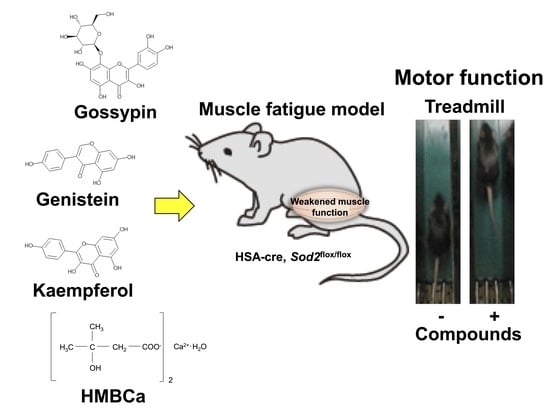
Natural Compounds That Enhance Motor Function in a Mouse Model of Muscle Fatigue

1
Aging Stress Response Research Project Team, National Center for Geriatrics and Gerontology, Obu 474-851, Aichi, Japan
2
Zenyaku Hanbai Co., Ltd., Toshima-ku 170-0013, Tokyo, Japan
*
Author to whom correspondence should be addressed.
Biomedicines 2022, 10(12), 3073; https://doi.org/10.3390/biomedicines10123073
Submission received: 31 October 2022 / Revised: 21 November 2022 / Accepted: 25 November 2022 / Published: 29 November 2022
(This article belongs to the Special Issue Musculoskeletal Diseases: From Molecular Basis to Therapy)

Abstract

:
Musculoskeletal disease can be a serious condition associated with aging that may lead to fractures and a bedridden state due to decreased motor function. In addition to exercise training to increase muscle mass, increasing muscle function with the intake of functional foods is an effective treatment strategy for musculoskeletal disease. Muscle-specific SOD2-deficient mice (muscle-Sod2-/-) show a severe disturbance in exercise in association with increased mitochondrial reactive oxygen species, as well as mitochondrial dysfunction and muscle damage. In the present study, to develop a therapeutic strategy for musculoskeletal disease, we searched for substances that enhanced motor function among functional compounds by in vivo screening using muscle-Sod2-/- mice as a muscle fatigue model. We administered 96 compounds, including antioxidants, to muscle-Sod2-/- mice and assessed their effects on treadmill performance. Among the administered compounds, gossypin, genistein, kaempferol, taxifolin, fumaric acid, β-hydroxy-β-methylbutyrate Ca, and astaxanthin, which are dietary functional food factors, increased forced running time in muscle-Sod2-/- mice. In addition, troglitazone, tempol, trolox, and MnTE-2-PyP, which are antioxidants, also significantly increased the running ability of muscle-Sod2-/- mice. These results suggest that the intake of functional foods with antioxidant activity can improve motor function. Muscle-Sod2-/- mice, as a muscle fatigue model, are suitable for the in vivo screening of functional substances that promote improvements in exercise and muscle performance.

Graphical Abstract

1. Introduction

Reactive oxygen species (ROS) cause tissue dysfunction by oxidizing the proteins, lipids, and nucleic acids that constitute living organisms. Oxidative damage markers, such as carbonylated protein, lipid peroxides, and oxidized nucleic acids, increase the aging process and are associated with the development of various diseases, suggesting a strong correlation between ROS and various tissue pathologies, including skeletal muscle pathologies. Actually, oxidative damage markers accumulate in aged rat skeletal muscle [1]. In humans, mitochondria derived from aged skeletal muscle also show an increase in the oxidized nucleic acid marker 8-OHdG, which causes respiratory dysfunction accompanied by a decrease in mitochondrial DNA [2]. The accumulation of ROS in skeletal muscle also contributes to the pathogenesis of muscular dystrophy, which causes progressive skeletal muscle wasting and degeneration [3]. These reports indicate that the redox imbalance caused by increased ROS induces muscle hypofunction.
Mammals have various antioxidant systems that protect tissues and cells from ROS. Superoxide dismutase (SOD) is an antioxidant enzyme that converts superoxide to hydrogen peroxide, which is further detoxified to water and O2 by catalase and GPx enzymes. SOD2 is constitutively and ubiquitously expressed in the mitochondria to regulate the redox balance in the cells of tissues. The loss of SOD2 induces mitochondrial redox imbalance by increasing the generation of superoxide, resulting in mitochondrial dysfunction in cells and several tissues, including the brain, heart, muscle, bone, and cartilage [4,5,6,7]. Muscle-specific SOD2-deficient (muscle-Sod2-/-) mice show a severe disturbance during exercise activity in association with increased oxidative damage and reduced ATP content [4]. Meanwhile, the administration of the antioxidant EUK8, which has superoxide dismutase and catalase activity, was shown to improve the motor activity of muscle-Sod2-/- mice [4]. Muscle-Sod2-/- mice can be used as a model of muscle fatigue to evaluate the effects of various compounds, including antioxidants, on the improvement of the motor function.
In this study, we evaluated the improvement effects of polyphenols, vitamins, amino acids, and other chemicals by an in vivo screening of the forced running ability of muscle-Sod2-/- mice.

2. Materials and Methods

2.1. Animals and Genotyping

The generation of muscle-Sod2-/- mice was described previously [4]. In our previous report, a 401-bp DNA fragment corresponding to the deleted allele was specifically amplified by PCR from the skeletal muscle of the muscle-Sod2-/- mice, whereas no fragments were amplified from other tissues in the muscle-Sod2-/- mice [4]. Western blot analyses also showed a specific loss of SOD2 expression in skeletal muscles, such as the tibialis anterior, gastrocnemius, soleus, and quadriceps, of muscle-Sod2-/- mice [4]. The muscle-Sod2-/- mice, which were all male (age: 6–9 months), were maintained under a 12 h light/12 h dark cycle with ad libitum access to water and chow. All genotyping of the HSA-Cre transgene and the SOD2lox/lox mice was performed by PCR using genomic DNA isolated from the tail tip. The primers used for identifying carriers of the HSA-Cre transgene were 5′-AAATACTCTGAGTCCAAACCGGGCCCC-3′ and 5′-CAGTGCGTTCGAACGCTAGAGCCTGTT-3′, while those used for genotyping SOD2 lox were 5′-TTAGGGCTCAGGTTTGTCCAGAA-3′, 5′-CGAGGGGCATCTAGTGGAGAA-3′, and 5′-AGCTTGGCTGGACGTAA-3′. The mice were maintained and studied according to protocols approved by the Tokyo Metropolitan Institute of Gerontology and the National Center for Geriatrics and Gerontology.

2.2. Substances

We selected the substances administered in this study from among the available substances, focusing on those with antioxidant, anti-inflammatory, and mitochondrial biosynthetic effects. The administered substances were all purchased from or provided by the following suppliers: Dojindo Laboratories (Kumamoto, Japan), FUJIFILM Wako Pure Chemical Corporation (Osaka, Japan), KOBAYASHI PERFUMERY CO., LTD. (Tokyo, Japan), Kyoto University (Kyoto, Japan), LKT Laboratories (St. Paul, MN, USA), and Merck (Darmstadt, Germany). Detailed information on the administered substances is provided in Table 1 and Table 2.

2.3. Administration

Many substances were administered by intraperitoneal (I. P.) injection, which is a simple procedure that can be performed with small amounts of expensive substances. The compounds used for I. P. administration were diluted in PBS and injected once into muscle-Sod2-/- mice at the dosages indicated in Table 1 and Table 2. Treadmill exercise was performed 24 h after administration. Some compounds used for oral administration were diluted in distilled water and administered once per day to muscle-Sod2-/- mice at the dosages and frequencies indicated in Table 1. A schematic illustration of the administration and treadmill experiments is shown in Figure 1. Mice that completed the treadmill exercise were used again for the evaluation of other compounds after a 2-week clearance period. The dosage of each substance was determined with reference to previous reports using experimental animals. To assess the safety, we administered each substance to wild-type mice and confirmed that no deaths or abnormalities occurred.

2.4. Treadmill Protocol

A treadmill apparatus (MK-680S/OP; Muromachi Kikai, Tokyo, Japan) was used to determine the endurance capacity for running. All mice were accustomed to the treadmill before the experiment, and the running test was carried out at 12 m/min with a 0° slope. An electrode at the back of the treadmill was activated to prevent mice from stopping naturally.

2.5. Statistical Analysis

The minimum number of mice in each experimental group was set at four for statistical analyses. Student’s t-test was used to compare the running times before and after treatment. p values of <0.05 were considered to indicate statistical significance. All data are expressed as the mean ± standard deviation (SD).

3. Results

In order to evaluate the improvement effect on motor function, we administered various compounds intraperitoneally or orally to muscle-Sod2-/- mice, and evaluated the changes in treadmill running time before and after these administrations. As evaluation compounds, we selected polyphenols, vitamins and their metabolites, carotenoids, amino acids, other phytochemicals, and other compounds with a focus on substances with antioxidant, anti-inflammatory, and mitochondrial biosynthetic effects (Table 1 and Table 2). In our previous report, we demonstrated that wild-type mice could complete a treadmill run for 2 h, whereas muscle-Sod2-/- mice ran for less than 10 min [4]. Each experimental group of muscle-Sod2-/- mice in this study averaged about 7–23 min of running time before administration (data not shown), indicating exercise intolerance similar to our previous reports. When the forced running time before and after administration was compared, we found 11 compounds that significantly increased running time. When the 11 compounds that increased the running time of muscle-Sod2-/- mice were divided into categories, they contained 7 dietary functional foods (gossyppin, genistein, kaempferol, taxifolin, fumaric acid, astaxanthin, and β-hydroxy-β-methylbutyrate (HMB) Ca) (Figure 2). The antioxidants troglitazone, tempol, trolox, and MnTE-2-PyP also improved the forced running time of muscle-Sod2-/- mice (Figure 3). In contrast, the intraperitoneal administration of citric acid, phosphocreatine, and nicotinamide significantly reduced the running time of muscle-Sod2-/- mice (Figure 4). These results indicate that muscle-Sod2-/- mice can be used as an exercise intolerance model for the in vivo screening of compounds that enhance physical function.

4. Discussion

In vivo screening identified 11 compounds that improved the physical function of muscle-Sod2-/- mice. Previously, we demonstrated that muscle-Sod2-/- mice exhibit exercise intolerance due to mitochondrial dysfunction caused by the accumulation of ROS in skeletal muscle and that the antioxidant EUK8 improves their running ability [4]. In this study, 10 of the 11 compounds that improved motor function were antioxidants, including polyphenols. Genistein (an isoflavone) and astaxanthin (a carotenoid classified as a xanthophyll) are major antioxidants. The administration of tempol, which has SOD activity, decreased oxidative markers along with the regulation of antioxidant enzyme expression in the skeletal muscle of a mouse model of Duchenne muscular dystrophy [8]. These results suggest that the reduction in oxidative damage in SOD2-deficient skeletal muscle is the main mechanism underlying the enhancement in motor function. Gossypin, a flavone isolated from Hibiscus vitifolious, has strong antioxidant activity in scavenging superoxide, hydroxyl radicals, DPPH radicals, and NO [9]. In addition, gossypin was also shown to have anti-inflammatory effects via the inhibition of NF-κB and its regulatory gene expression in in vitro studies using inflammatory stimuli and carcinogens [10]. Kaempferol is a flavonol (a type of flavonoid) that is abundantly contained in kale and broccoli. Kaempferol has ROS activity, scavenging superoxide, hydroxyl radical, and peroxynitrite [11,12,13]. In rat studies, the oral administration of the glycoside kaempferol 3-neohesperidoside increased the accumulation of glycogen by upregulating the muscle glucose uptake via the phosphoinositide 3-kinase and protein kinase C pathways [14], suggesting an additive effect—involving antioxidant activity and other mechanisms—on muscle function. Further analyses are needed to investigate the correlation between the enhancement in physical function and the level of oxidative damage in skeletal muscle in greater detail. The pharmacological effects on the motor function of substances may be associated with the pharmacokinetics after administration. In rats, astaxanthin accumulated in skeletal muscle at 24 h after ingestion, suggesting a direct action on skeletal muscle [15]. Human studies also suggest that HMB reaches various organs at nine hours after ingestion [16]. For the majority of this study, we analyzed animals at 24 h after I. P. administration, so the pharmacological action of the substances may have been enhanced if analyzed at the time point of peak pharmacokinetics. Indeed, despite having antioxidant activity, some compounds failed to improve motor function in muscle-Sod2-/- mice (Table 1 and Table 2). These substances have different pharmacokinetics, e.g., differences in the amount reaching muscle tissues and the stability, and may have their own appropriate concentrations, analysis timings, and administration methods.
Muscle-Sod2-/- mice exhibit mitochondrial dysfunction due to reduced complex II activity [4]. Some of the compounds that showed an improvement effect on the motor function of muscle-Sod2-/- mice in the present study are reported to enhance mitochondrial function. Taxifolin, a flavonoid contained in coniferous trees, reduced the intracellular ROS level against H2O2-induced pyroptosis and exhibited cytoprotective effects associated with increased mitochondrial membrane potential in rat heart cells [17]. In an in vitro study, taxifolin also decreased the lipid peroxidation levels in mitochondria derived from the livers of rats, suggesting that taxifolin may also enhance motor function due to the increased mitochondrial function in skeletal muscle [18]. Trolox, a water-soluble analog of vitamin E, which is known as a major antioxidant, induced the improvement in mitochondrial respiration and the suppression in ubiquitin–proteasome/autophagy activity by attenuating oxidative stress. This resulted in the prevention of muscle atrophy in skeletal muscle-specific transforming growth factor-β-activated kinase 1-deficient mice [19]. Fumaric acid, which is a type of dicarboxylic acid, is a constituent of the citric acid cycle and thus contributes to energy production in muscle.
The uptake of glucose into muscles in glycolysis affects the energy production capacity. Troglitazone, a type of thiazolidine with a tocopherol-like structure, is used as a hypoglycemic and anti-inflammatory medicine. In a glucose clamp test using Zucker fatty rats, troglitazone significantly increased the glycogen content in skeletal muscle [20]. These results suggest that the accelerated glucose uptake in muscles improves exercise capacity by increasing energy sources. HMBCa is a functional food material in which calcium is bound to HMB. HMB is a metabolite of leucine, an essential amino acid, which promotes muscle synthesis, inhibits muscle degradation, and acts as a source of components for muscle cell membranes [21]. Although HMB plays an important role in the maintenance of muscle function, the amount of HMB produced in the body is approximately 5% of the leucine intake [22]; thus, the direct ingestion of HMB is an effective method for increasing muscle function. In the present study, we performed the trials with a limited number of mice and did not examine biochemical markers in detail, thereby limiting the interpretation of the results obtained. In order to clarify the mechanisms through which the compounds evaluated in this study enhance motor function, we also need to examine other biochemical markers, such as ROS levels, ATP levels, and mitochondrial respiratory capacity, in detail.
Musculoskeletal disorders are a serious condition associated with aging, increasing the risk of a bedridden state due to decreased motor function. Muscle-Sod2-/- mice are very useful as a muscle fatigue model for the in vivo screening of reagents that enhance motor function. Utilizing this mouse model of muscle fatigue will contribute to the development of materials that can be applied to locomotive syndrome and frailty.

Author Contributions

S.S. and T.S. designed the study. S.S. and T.S. wrote the manuscript. S.S. performed the research. S.S. analyzed the data. S.S., K.W., D.S., T.A., and T.S. discussed the hypothesis and interpreted the data. S.S. and T.S. edited the article. T.S. coordinated and directed the project. All authors have read and agree to the published version of the manuscript.

Funding

This research was supported by funds from Zenyaku Kogyo CO., Ltd. [NCGG061503], the Program for the Promotion of Basic Research Activities for Innovative Biosciences from BRAIN (Bio-oriented Technology Research Advancement Institution) [H19-Y], and research funding for longevity science from the National Center for Geriatrics and Gerontology [grant number 19-50, 21-9]. The funding source was not involved in the study design; in the collection, analysis, or interpretation of the data; in the writing of the report; or in the decision to submit the article for publication.

Institutional Review Board Statement

All experiments involving animals were performed according to protocols approved by the animal care committee of the Tokyo Metropolitan Institute of Gerontology (G115, A11008, A11014) and the National Center for Geriatrics and Gerontology (G2-28, A4-47).

Informed Consent Statement

Not applicable.

Data Availability Statement

Not applicable.

Acknowledgments

We thank Tetsuro Horie, Toshihiko Toda, Yusuke Ozawa, Yoshihiro Noda, Chizuru Tsuda, Shin Ishikawa, and Hirotomo Kuwahara (Tokyo Metropolitan Institute of Gerontology) for the preparation of the mice.

Conflicts of Interest

The authors declare no conflict of interest in association with the present study.

Abbreviations

HMBβ-hydroxy-β-methylbutyrate
I. P.intraperitoneal
muscle-Sod2-/-muscle-specific SOD2 deficient
ROSreactive oxygen species
SODsuperoxide dismutase

References

  1. Hamilton, M.L.; Van Remmen, H.; Drake, J.A.; Yang, H.; Guo, Z.M.; Kewitt, K.; Walter, C.A.; Richardson, A. Does oxidative damage to DNA increase with age? Proc. Natl. Acad. Sci. USA 2001, 98, 10469–10474. [Google Scholar] [CrossRef] [PubMed] [Green Version]
  2. Short, K.R.; Bigelow, M.L.; Kahl, J.; Singh, R.; Coenen-Schimke, J.; Raghavakaimal, S.; Nair, K.S. Decline in skeletal muscle mitochondrial function with aging in humans. Proc. Natl. Acad. Sci. USA 2005, 102, 5618–5623. [Google Scholar] [CrossRef] [PubMed] [Green Version]
  3. Tidball, J.G.; Wehling-Henricks, M. The role of free radicals in the pathophysiology of muscular dystrophy. J. Appl. Physiol. 2007, 102, 1677–1686. [Google Scholar] [CrossRef] [PubMed] [Green Version]
  4. Kuwahara, H.; Horie, T.; Ishikawa, S.; Tsuda, C.; Kawakami, S.; Noda, Y.; Kaneko, T.; Tahara, S.; Tachibana, T.; Okabe, M.; et al. Oxidative stress in skeletal muscle causes severe disturbance of exercise activity without muscle atrophy. Free Radic. Biol. Med. 2010, 48, 1252–1262. [Google Scholar] [CrossRef] [PubMed]
  5. Shimizu, T.; Nojiri, H.; Kawakami, S.; Uchiyama, S.; Shirasawa, T. Model mice for tissue-specific deletion of the manganese superoxide dismutase gene. Geriatr. Gerontol. Int. 2010, 10 (Suppl. 1), S70–S79. [Google Scholar] [CrossRef]
  6. Kobayashi, K.; Nojiri, H.; Saita, Y.; Morikawa, D.; Ozawa, Y.; Watanabe, K.; Koike, M.; Asou, Y.; Shirasawa, T.; Yokote, K.; et al. Mitochondrial superoxide in osteocytes perturbs canalicular networks in the setting of age-related osteoporosis. Sci. Rep. 2015, 5, 9148. [Google Scholar] [CrossRef] [Green Version]
  7. Koike, M.; Nojiri, H.; Ozawa, Y.; Watanabe, K.; Muramatsu, Y.; Kaneko, H.; Morikawa, D.; Kobayashi, K.; Saita, Y.; Sasho, T.; et al. Mechanical overloading causes mitochondrial superoxide and SOD2 imbalance in chondrocytes resulting in cartilage degeneration. Sci. Rep. 2015, 5, 11722. [Google Scholar] [CrossRef] [Green Version]
  8. Hermes, T.A.; Mizobuti, D.S.; da Rocha, G.L.; da Silva, H.N.M.; Covatti, C.; Pereira, E.C.L.; Ferretti, R.; Minatel, E. Tempol improves redox status in mdx dystrophic diaphragm muscle. Int. J. Exp. Pathol. 2020, 101, 289–297. [Google Scholar] [CrossRef]
  9. Ganapaty, S.; Chandrashekhar, V.M.; Chitme, H.R.; Narsu, M.L. Free radical scavenging activity of gossypin and nevadensin: An in-vitro evaluation. Ind. J. Pharmacol. 2007, 39, 281–283. [Google Scholar] [CrossRef] [Green Version]
  10. Kunnumakkara, B.K.; Neir, A.S.; Ahn, K.S.; Pandey, M.K.; Yi, Z.; Liu, M.; Aggarwal, B.B. Gossypin, a pentahydroxy glucosyl flavone, inhibits the transforming growth factor beta-activated kinase-1-mediated NF-κB activation pathway, leading to potentiation of apoptosis, suppression of invasion, and abrogation of osteoclastogenesis. Blood 2007, 109, 5112–5121. [Google Scholar] [CrossRef]
  11. Wang, L.; Tu, Y.C.; Lian, T.W.; Hung, J.T.; Yen, J.H.; Wu, M.J. Distinctive antioxidant and antiinflammatory effects of flavonols. J. Agric. Food Chem. 2006, 54, 9798–9804. [Google Scholar] [CrossRef]
  12. Klaunig, J.E.; Kamendulis, L.M. The role of oxidative stress in carcinogenesis. Annu. Rev. Pharmacol. Toxicol. 2004, 44, 239–267. [Google Scholar] [CrossRef] [PubMed]
  13. Heijnen, C.G.; Haenen, G.R.; van Acker, F.A.; van der Vijgh, W.J.; Bast, A. Flavonoids as peroxynitrite scavengers: The role of the hydroxyl groups. Toxicol. In Vitro 2001, 15, 3–6. [Google Scholar] [CrossRef] [PubMed]
  14. Zanatta, L.; Rosso, A.; Folador, P.; Figueiredo, M.S.; Pizzolatti, M.G.; Leite, L.D.; Silva, F.R. Insulinomimetic effect of kaempferol 3-neohesperidoside on the rat soleus muscle. J. Nat. Prod. 2008, 71, 532–535. [Google Scholar] [CrossRef] [PubMed]
  15. Vukovich, M.D.; Slater, G.; Macchi, M.B.; Turner, M.J.; Fallon, K.; Boston, T.; Rathmacher, J. β-hydroxy-β-methylbutyrate (HMB) kinetics and the influence of glucose ingestion in humans. J. Nutr. Biochem. 2001, 12, 631–639. [Google Scholar] [CrossRef] [PubMed]
  16. Choi, H.D.; Kang, H.E.; Yang, S.H.; Lee, M.G.; Shin, W.G. Pharmacokinetics and first-pass metabolism of astaxanthin in rats. Br. J. Nutr. 2011, 105, 220–227. [Google Scholar] [CrossRef] [Green Version]
  17. Ye, Y.; Wang, X.; Cai, Q.; Zhuang, J.; Tan, X.; He, W.; Zhao, M. Protective effect of taxifolin on H2O2-induced H9C2 cell pyroptosis. J. Cent. South Univ. Med. Sci. 2017, 42, 1367–1374. [Google Scholar]
  18. Haraguchi, H.; Mochida, Y.; Sakai, S.; Masuda, H.; Tamura, Y.; Mizutani, K.; Tanaka, O.; Chou, W.H. Protection against oxidative damage by dihydroflavonols in Engelhardtia chrysolepis. Biosci. Biotechnol. Biochem. 1996, 60, 945–948. [Google Scholar] [CrossRef] [Green Version]
  19. Roy, A.; Sharma, A.K.; Nellore, K.; Narkar, V.A.; Kumar, A. TAK1 preserves skeletal muscle mass and mitochondrial function through redox homeostasis. FASEB BioAdvances 2020, 2, 538–553. [Google Scholar] [CrossRef]
  20. Oshida, Y.; Kako, M.; Nakai, N.; Shimomura, Y.; Li, L.; Sato, J.; Ohsawa, I.; Sato, Y. Troglitazone improves insulin-stimulated glucose utilization associated with an increased muscle glycogen content in obese Zucker rats. Endocr. J. 1999, 46, 723–730. [Google Scholar] [CrossRef] [Green Version]
  21. Wilson, J.M.; Fitschen, P.J.; Campbell, B.; Wilson, G.J.; Zanchi, N.; Taylor, L.; Wilborn, C.; Kalman, D.S.; Stout, J.R.; Hoffman, J.R.; et al. International Society of Sports Nutrition Position Stand: Beta-hydroxy-beta-methylbutyrate (HMB). J. Int. Soc. Sports Nutr. 2013, 10, 6. [Google Scholar] [CrossRef] [PubMed]
  22. Zanchi, N.E.; Gerlinger-Romero, F.; Guimaraes-Ferreira, L.; de Siqueira Filho, M.A.; Felitti, V.; Lira, F.S.; Seelaender, M.; Lancha, A.H., Jr. HMB supplementation: Clinical and athletic performance-related effects and mechanisms of action. Amino Acids 2011, 40, 1015–1025. [Google Scholar] [CrossRef] [PubMed]
Figure 1. Schematic illustration of the administration and treadmill experiments. We administrated substances by I. P. injection (top) or orally (bottom) to the muscle-Sod2-/- mice, followed by treadmill running.
Biomedicines 10 03073 g001
Figure 2. Functional factors that enhance the running activity of muscle-Sod2-/- mice. Positive effects of functional factors on the treadmill task performance of muscle-Sod2-/- mice (top) and structural formulas of each compound (bottom). * p < 0.05. Data are shown as the mean ± SD.
Biomedicines 10 03073 g002
Figure 3. Antioxidants that enhance the running activity of muscle-Sod2-/- mice. Positive effects of antioxidants on the treadmill task performance of muscle-Sod2-/- mice (top) and structural formulas of each compound (bottom). * p < 0.05. Data are shown as the mean ± SD.
Biomedicines 10 03073 g003
Figure 4. Compounds that reduce the running activity of muscle-Sod2-/- mice. Negative effects of some compounds on the treadmill task performance of muscle-Sod2-/- mice. * p < 0.05. Data are shown as the mean ± SD.
Biomedicines 10 03073 g004
Table 1. Positive effects of compounds on the running activity of muscle-Sod2-/- mice.
CompoundsSupplierDosage (mg/kg)AdministrationRelative Changet-Testn
Positive effects
AcacetinMerck50I. P.1.260.075
AesculinLKT Laboratories100I. P.1.410.365
AICARMerck500I. P.1.010.654
AlliinMerck10I. P.1.290.335
AllopurinolMerck100I. P.1.080.264
ApigeninMerck50I. P.1.030.635
ApocyninMerck300I. P.1.270.315
Ascorbic acidMerck500I. P.1.260.2910
AsparagineMerck500I. P.1.900.214
AstaxanthinMerck500Oral1.240.044
ATPMerck2000Oral1.070.964
AurapteneKyoto Univ.30I. P.1.240.124
BaicaleinMerck500I. P.1.440.175
Biochanin AMerck50I. P.1.070.685
Caffeic acidMerck50I. P.1.330.365
L-CarnitineMerck1690I. P.1.250.1314
ChrysinMerck250I. P.1.170.234
L-CitrullineMerck100I. P.1.180.475
CurcuminMerck150I. P.1.170.105
2,7-DichlorofluoresceinMerck500I. P.1.400.352
DipenteneMerck10I. P.1.030.784
EbselenMerck16I. P.1.440.194
EGCsMerck150Oral1.140.355
FenofibrateMerck1600I. P.1.250.585
Ferulic acidMerck50I. P.1.370.115
FisetinMerck50I. P.1.180.625
Folic acidMerck4I. P.1.210.1410
Fumaric acidMerck1000I. P.1.120.045
GenisteinMerck10 and 50I. P.1.450.0115
[6]-GingerolMerck10I. P.1.150.274
GlucoseMerck2000I. P.1.080.625
GlutamineMerck1000I. P.1.150.665
Glutamine monohydrateMerck1000I. P.1.100.475
Glutathione (GSH-MEE)Merck50I. P.1.060.495
GlycineMerck150I. P.1.240.375
GossypinMerck500I. P.1.650.0114
HistidineMerck800I. P.1.080.315
HMBCaKOBAYASHI
PERFUMERY
500 mg/kg × 5 timesOral1.440.0210
KaempferolMerck500I. P.1.420.034
LuteolinFUJIFILM100I. P.1.040.914
LysineMerck185I. P.1.350.105
MagnololMerck10I. P.1.050.955
Maleic acidMerck1000I. P.1.250.275
MethionineMerck187I. P.1.160.135
MevastatinMerck500I. P.1.180.064
Mn TE-2-PyPMerck10I. P.1.380.055
MorinMerck500I. P.1.320.055
MyricetinMerck500I. P.1.090.875
L-NAMEDojindo10I. P.1.860.145
α-NaphthoflavoneMerck250I. P.1.250.264
β-NaphthoflavoneMerck250I. P.1.190.435
NaringinMerck500I. P.1.120.755
NobiletinKyoto Univ.30I. P.1.490.194
1-OctanosanolMerck100I. P.1.080.144
OrnithineMerck1000I. P.1.040.575
(-)-2-Oxo-4-thiazolidinecarboxylic acidMerck50I. P.1.260.325
PrednisoloneMerck200I. P.1.200.275
Reduced lipoic acidMerck10I. P.1.250.375
RutinMerck50I. P.1.100.575
Sodium PyruvateMerck500I. P.1.210.285
Succinic acidMerck1000I. P.1.070.695
TaxifolinMerck500I. P.1.360.025
TempolMerck250I. P.1.360.045
L-TheanineMerck500I. P.1.560.144
ThreonineMerck800I. P.1.130.485
α-tocopherol acetateMerck500I. P.1.160.184
TroglitazoneMerck10I. P.1.410.015
TroloxMerck20I. P.1.410.015
Zn-protoporphyrin IXMerck10I. P.1.620.155
Table 2. Negative effects of compounds on the running activity of muscle-Sod2-/- mice.
CompoundsSupplierDosage (mg/kg)AdministrationRelative Changet-Testn
Negative effects
Aloe-emodinMerck10I. P.0.980.975
Bee pollenMerck100I. P.0.880.205
BilirubinMerck200I. P.0.870.315
CapsaicinMerck1I. P.0.980.554
β-caroteneMerck500I. P.0.710.634
Citric acidMerck500I. P.0.860.025
DaidzeinMerck50I. P.0.990.785
Docosahexaenoic acidMerck100I. P.0.810.214
Eicosapentaenoic acidMerck100I. P.0.860.095
18β-Glycyrrhetinic acidMerck10I. P.0.950.795
Glycyrrhizic acid ammonium saltMerck10I. P.0.950.617
IbuprofenMerck75I. P.0.870.674
Indore-3-carbinolMerck100I. P.0.920.694
Isosorbide dinitrateMerck30I. P.0.870.195
LimoninMerck10I. P.0.750.125
Linolenic acidMerck100I. P.0.870.915
N-acetyl-L-cysteineMerck500I. P.0.920.535
NicotinamideMerck200I. P.0.760.035
7-NitroindazoleMerck50I. P.0.970.515
PhosphocreatineMerck100I. P.0.770.015
QuercetinMerck3.3I. P.0.960.525
RiboflavinMerck100I. P.0.770.125
Rosmarinic acidMerck50I. P.0.990.625
SesaminMerck5I. P.0.880.285
Ursolic acidMerck100I. P.0.970.335
XanthophyllMerck5I. P.0.970.554
Publisher’s Note: MDPI stays neutral with regard to jurisdictional claims in published maps and institutional affiliations.

