
Reference Values for Cervical Muscle Strength in Healthy Women Using a Hand-Held Dynamometer and the Association with Age and Anthropometric Variables
 , , , and
Abstract
:1. Introduction
2. Materials and Methods
2.1. Study Design
2.2. Subjects
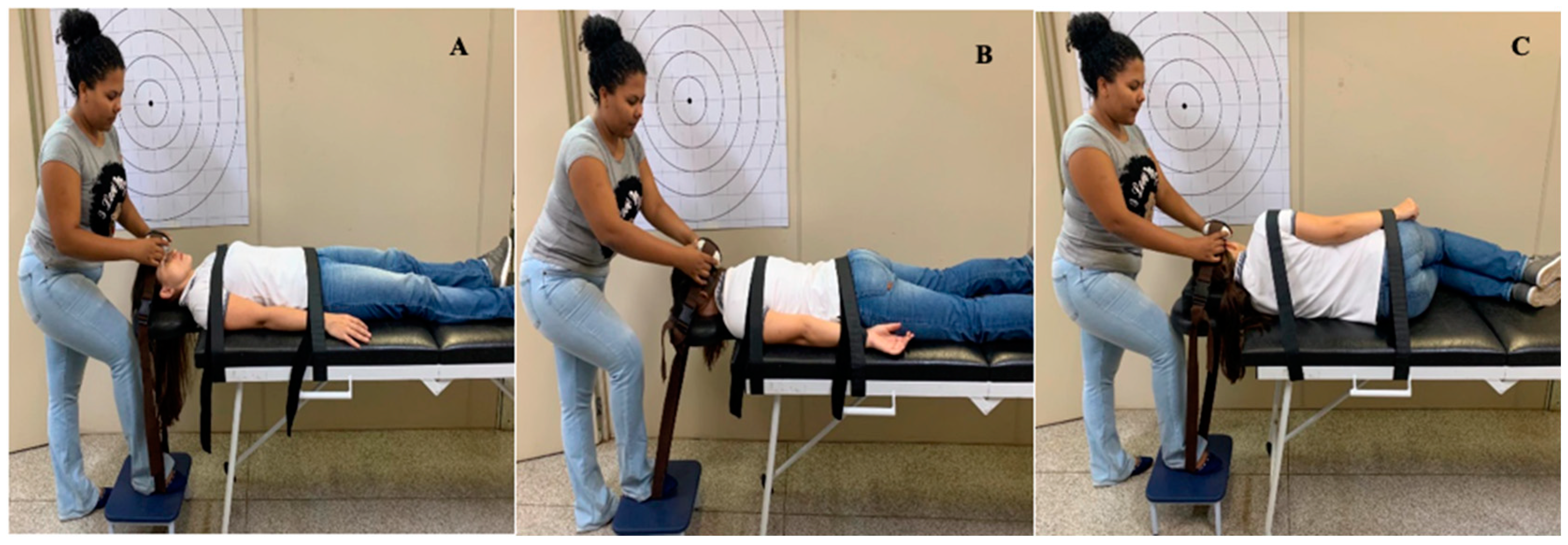
2.3. Procedures
2.4. Statistical Analysis
3. Results
4. Discussion
5. Conclusions
Author Contributions
Funding
Institutional Review Board Statement
Informed Consent Statement
Data Availability Statement
Conflicts of Interest
References
- Shin, D.W.; Shin, J.I.; Koyanagi, A.; Jacob, L.; Smith, L.; Lee, H.; Chang, Y.; Song, T.J. Global, regional, and national neck pain burden in the general population, 1990–2019: An analysis of the global burden of disease study 2019. Front. Neurol. 2022, 13, 955367. [Google Scholar] [CrossRef] [PubMed]
- GBD 2019 Diseases and Injuries Collaborators. Global burden of 369 diseases and injuries in 204 countries and territories, 1990–2019: A systematic analysis for the Global Burden of Disease Study 2019. Lancet 2020, 396, 1204–1222. [Google Scholar] [CrossRef] [PubMed]
- Armijo-Olivo, S.L.; Fuentes, J.P.; Major, P.W.; Warren, S.; Thie, N.M.; Magee, D.J. Is maximal strength of the cervical flexor muscles reduced in patients with temporomandibular disorders? Arch. Phys. Med. Rehabil. 2010, 91, 1236–1242. [Google Scholar] [CrossRef]
- Kapreli, E.; Vourazanis, E.; Billis, E.; Oldham, J.A.; Strimpakos, N. Respiratory dysfunction in chronic neck pain patients. A pilot study. Cephalalgia 2009, 29, 701–710. [Google Scholar] [CrossRef]
- Karaağaç, A.; Arslan, S.A.; Keskin, E.D. Assessment of pain, scapulothoracic muscle strength, endurance and scapular dyskinesis in individuals with and without nonspecific chronic neck pain: A cross-sectional study. J. Bodyw. Mov. Ther. 2023, 35, 261–267. [Google Scholar] [CrossRef]
- Chiu, T.T.; Lo, S.K. Evaluation of cervical range of motion and isometric neck muscle strength: Reliability and validity. Clin. Rehabil. 2002, 16, 851–858. [Google Scholar] [CrossRef]
- Dumas, J.P.; Arsenault, A.B.; Boudreau, G.; Magnoux, E.; Lepage, Y.; Bellavance, A.; Loisel, P. Physical impairments in cervicogenic headache: Traumatic vs. nontraumatic onset. Cephalalgia 2001, 21, 884–893. [Google Scholar] [CrossRef]
- Kumar, S.; Narayan, Y.; Prasad, N.; Shuaib, A.; Siddiqi, Z.A. Cervical electromyogram profile differences between patients of neck pain and control. Spine 2007, 32, E246–E253. [Google Scholar] [CrossRef] [PubMed]
- Ylinen, J.; Salo, P.; Nykanen, M.; Kautiainen, H.; Hakkinen, A. Decreased isometric neck strength in women with chronic neck pain and the repeatability of neck strength measurements. Arch. Phys. Med. Rehabil. 2004, 85, 1303–1308. [Google Scholar] [CrossRef]
- Ylinen, J.; Savolainen, S.; Airaksinen, O.; Kautiainen, H.; Salo, P.; Häkkinen, A. Decreased strength and mobility in patients after anterior cervical diskectomy compared with healthy subjects. Arch. Phys. Med. Rehabil. 2003, 84, 1043–1047. [Google Scholar] [CrossRef] [PubMed]
- Panjabi, M.M.; Cholewicki, J.; Nibu, K.; Grauer, J.; Babat, L.B.; Dvorak, J. Critical load of the human cervical spine: An in vitro experimental study. Clin. Biomech. 1998, 13, 11–17. [Google Scholar] [CrossRef] [PubMed]
- Li Causi, V.; Manelli, A.; Marini, V.G.; Cherubino, M.; Meccariello, L.; Mazzacane, M.; Ronga, M. Balance assessment after altering stimulation of the neurosensory system. Med. Glas. 2021, 18, 328–333. [Google Scholar]
- Collins, C.L.; Fletcher, E.N.; Fields, S.K.; Kluchurosky, L.; Rohrkemper, M.K.; Comstock, R.D.; Cantu, R.C. Neck strength: A protective factor reducing risk for concussion in high school sports. J. Prim. Prev. 2014, 35, 309–319. [Google Scholar] [CrossRef] [PubMed]
- Gilchrist, I.; Storr, M.; Chapman, E.; Pelland, L. Neck muscle strength training in the risk management of concussion in contact sports: Critical appraisal of application to practice. J. Athl. Enhanc. 2015, 4, 2. [Google Scholar]
- Catenaccio, E.; Mu, W.; Kaplan, A.; Fleysher, R.; Kim, N.; Bachrach, T.; Sears, M.Z.; Jaspan, O.; Caccese, J.; Kim, M.; et al. Characterization of neck strength in healthy young adults. PMR 2017, 9, 884–891. [Google Scholar] [CrossRef]
- Jordan, A.; Mehlsen, J.; Bulow, P.M.; Ostergaard, K.; Danneskiold-Samsoe, B. Maximal isometric strength of the cervical musculature in 100 healthy volunteers. Spine 1999, 24, 1343–1348. [Google Scholar] [CrossRef]
- Salo, P.K.; Ylinen, J.J.; Málkiá, E.A.; Kautiainen, H.; Hákkinen, A.H. Isometric strength of the cervical flexor, extensor, and rotator muscles in 220 healthy females aged 20 to 59 years. J. Orthop. Sports Phys. Ther. 2006, 36, 495–502. [Google Scholar] [CrossRef] [PubMed]
- Phillips, B.A.; Lo, S.K.; Mastaglia, F.L. Muscle force measured using “break” testing with a hand-held myometer in normal subjects aged 20 to 69 years. Arch. Phys. Med. Rehabil. 2000, 81, 653–661. [Google Scholar] [CrossRef]
- Staudte, H.W.; Duhr, N. Age- and sex-dependent force-related function of the cervical spine. Eur. Spine J. 1994, 3, 155–161. [Google Scholar] [CrossRef]
- Manderlier, A.; de Fooz, M.; Patris, S.; Berquin, A. Modifiable lifestyle-related prognostic factors for the onset of chronic spinal pain: A systematic review of longitudinal studies. Ann. Phys. Rehabil. Med. 2022, 65, 101660. [Google Scholar] [CrossRef]
- GBD 2016 Headache Collaborators. Global, regional, and national burden of migraine and tension-type headache, 1990–2016: A systematic analysis for the Global Burden of Disease Study 2016. Lancet Neurol. 2018, 17, 954–976. [Google Scholar] [CrossRef] [PubMed] [Green Version]
- Bueno, C.H.; Pereira, D.D.; Pattussi, M.P.; Grossi, P.K.; Grossi, M.L. Gender differences in temporomandibular disorders in adult populational studies: A systematic review and meta-analysis. J. Oral Rehabil. 2018, 45, 720–729. [Google Scholar] [CrossRef] [PubMed]
- Chiu, T.T.; Lam, T.H.; Hedley, A.J. Maximal isometric muscle strength of the cervical spine in healthy volunteers. Clin. Rehabil. 2002, 16, 772–779. [Google Scholar] [CrossRef]
- Cook, C.; Richardson, J.K.; Braga, L.; Menezes, A.; Soler, X.; Kume, P.; Zaninelli, M.; Socolows, F.; Pietrobon, R. Cross-cultural adaptation and validation of the Brazilian Portuguese version of the Neck Disability Index and Neck Pain and Disability Scale. Spine 2006, 31, 1621–1627. [Google Scholar] [CrossRef]
- Matsudo, S.; Araújo, T.; Matsudo, V.; Andrade, D.; Andrade, E.; Oliveira, L.C.; Braggion, G. International Physical Activity Questionnaire (IPAQ): Study of validity and reliability in Brazil. Rev. Bras. Atividade Física Saúde 2001, 6, 5–18. [Google Scholar]
- Carnevalli, A.P.; Bevilaqua-Grossi, D.; Oliveira, A.I.S.; Carvalho, G.F.; Fernández-de-las-Peñas, C.; Florencio, L.L. Intra-rater and inter-rater reliability of maximal voluntary neck muscle strength assessment using a hand-held dynamometer in women with headache and healthy women. J. Manip. Physiol. Ther. 2017, 41, 621–627. [Google Scholar] [CrossRef] [PubMed]
- Domholdt, E. Physical Therapy Research: Principles and Applications, 2nd ed.; Saunders: Philadelphia, PA, USA, 2000. [Google Scholar]
- Hildenbrand, K.J.; Vasavada, A.N. Collegiate and high school athlete neck strength in neutral and rotated postures. J. Strength Cond. Res. 2013, 27, 3173–3182. [Google Scholar] [CrossRef] [PubMed] [Green Version]
- Garces, G.L.; Medina, D.; Milutinovic, L.; Garavote, P.; Guerado, E. Normative database of isometric cervical strength in a healthy population. Med. Sci. Sports Exerc. 2002, 34, 464–470. [Google Scholar] [PubMed] [Green Version]
- Cleland, J.A.; Mintken, P.E.; Carpenter, K.; Fritz, J.M.; Glynn, P.; Whitman, J.; Childs, J.D. Examination of a clinical prediction rule to identify patients with neck pain likely to be benefit from thoracic spine thrust manipulation and a general cervical range of motion exercise: Multicenter randomized clinical trial. Phys. Ther. 2010, 90, 1239–1250. [Google Scholar] [CrossRef]
- Strimpakos, N. The assessment of the cervical spine. Part 2: Strength and endurance/fatigue. J. Bodyw. Mov. Ther. 2011, 15, 417–430. [Google Scholar] [CrossRef]
- Dvir, Z.; Prushansky, T. Cervical muscles strength testing: Methods and clinical implications. J. Manip. Physiol. Ther. 2008, 31, 518–524. [Google Scholar] [CrossRef] [PubMed]
- Pasquino, A.; Tomarchio, A.; De Cruto, E.; Conteduca, J.; Longo, D.; Russi, V.; Pica, G.; Meccariello, L.; Rollo, G. Comparing hand strength and quality life of locking plate versus intramedullary k wire for transverse midshaft metacarpal fractures. Med. Glas. 2021, 18, 316–321. [Google Scholar]
- Massy-Westropp, C.; Massy-Westropp, N.; Wechalekar, H. Normative values for pinch strength-relationship with joint hypermobility as measured with the Beighton criteria. J. Hand Surg. Glob. Online 2023, 17, 272–276. [Google Scholar] [CrossRef] [PubMed]
- Martins, J.; da Silva, J.R.; da Silva, M.R.B.; Bevilaqua-Grossi, D. Reliability and Validity of the Belt-Stabilized Handheld Dynamometer in Hip- and Knee-Strength Tests. J. Athl. Train. 2017, 52, 809–819. [Google Scholar] [CrossRef] [Green Version]


| Groups (n = 25) | Age (Years) | Height (m) | Weight (Kg) | BMI (Kg/m²) | Neck Length (cm) | Neck Circumference (cm) |
|---|---|---|---|---|---|---|
| 18–29 years | 22.0 (3.17) | 1.63 (0.07) | 64.28 (10.71) | 24.16 (3.91) | 14.76 (1.74) | 33.34 (1.92) |
| 30–39 years | 33.5 (3.1) | 1.65 (0.06) | 69.66 (13.64) | 25.57 (4.92) | 15.46 (1.45) | 33.98 (2.32) |
| 40–49 years | 43.4 (3.59) | 1.62 (0.06) | 70.44 (11.35) | 26.94 (4.67) | 14.48 (1.34) | 35.14 (3.02) |
| 50–60 years | 55.4 (3.83) | 1.63 (0.06) | 74.6 (14.53) * | 28.20 (5.01) * | 14.66 (0.94) | 36.16 (3.19) * |
| Normalized Strength (kgf/Kg) | Minimal (kgf) | Maximum (kgf) | Time to Reach the Peak (s) | ||
|---|---|---|---|---|---|
| 18–29 years (n = 25) | Flexion. | 0.08 (0.07–0.08) | 2.7 | 6.9 | 1.99 (1.71–2.26) |
| Extension | 0.15 (0.13–0.16) | 5.4 | 13.1 | 2.49 (2.27–2.70) | |
| RLF | 0.11 (0.10–0.12) | 3.8 | 12.6 | 2.24 (2.01–2.47) | |
| LLF | 0.11 (0.10–0.13) | 3.6 | 11.2 | 2.18 (1.96–2.40) | |
| 30–39 years (n = 25) | Flexion | 0.08 (0.07–0.09) | 3.0 | 14.4 | 2.24 (2.00–2.48) |
| Extension | 0.14 (0.13–0.15) | 5.8 | 13.5 | 2.53 (2.34–2.72) | |
| RLF | 0.10 (0.09–0.11) | 4.0 | 10.7 | 2.63 (2.19–3.00) | |
| LLF | 0.10 (0.10–1.11) | 3.6 | 9.4 | 2.41 (2.25–2.70) | |
| 40–49 years (n = 25) | Flexion. | 0.09 (0.08–0.10) | 2.2 | 13.9 | 2.29 (2.14–2.44) |
| Extension | 0.15 (0.14–0.16) | 8.2 | 13.2 | 2.39 (2.21–2.57) | |
| RLF | 0.10 (0.09–0.11) | 2.8 | 13.4 | 2.24 (2.06–2.42) | |
| LLF | 0.11 (0.10–0.12) | 3.6 | 14.2 | 2.34 (2.17–2.51) | |
| 50–60 years (n = 25) | Flexion | 0.08 (0.07–0.09) | 2.9 | 10.9 | 2.46 (2.29–2.63) * |
| Extension | 0.13 (0.13–0.14) | 7.1 | 12.6 | 2.51 (2.40–2.62) | |
| RLF | 0.10 (0.09–0.10) | 5.0 | 10.8 | 2.31 (2.11–2.50) | |
| LLF | 0.09 (0.00–0.10) | 4.8 | 11.4 | 2.34 (2.17–2.51) |
| Groups (n = 25) | Flexion (kgf) | Extension (kgf) | RLF (kgf) | LLF (kgf) |
|---|---|---|---|---|
| 18–29 years | 4.94 (4.45–5.42) | 9.30 (8.53–10.07) | 6.99 (6.21–7.76) | 7.19 (6.44–7.93) |
| 30–39 years | 5.53 (4.65–6.41) | 9.49 (8.71–10.27) | 7.02 (6.40–7.64) | 7.16 (6.60–7.72) |
| 40–49 years | 6.33 (5.41–7.25) | 10.05 (9.56–10.55) | 7.24 (6.40–8.08) | 7.38 (6.53–8.24) |
| 50–60 years | 5.77 (5.06–6.47) | 9.85 (9.25–10.44) | 6.95 (6.36–7.53) | 6.94 (6.32–7.57) |
| ANOVA p value | 0.094 | 0.402 | 0.941 | 0.860 |
| Age (Years) | Height (m) | Weight (Kg) | BMI (Kg/m²) | Neck Length (cm) | Neck Circumference (cm) | ||
|---|---|---|---|---|---|---|---|
| Flexion. | r | 0.14 | 0.06 | 0.55 * | 0.54 * | 0.11 | 0.59 * |
| p value | 0.170 | 0.568 | <0.001 | <0.001 | 0.274 | <0.001 | |
| Extension | r | 0.11 | 0.07 | 0.46 * | 0.43 * | 0.09 | 0.36 * |
| p value | 0.283 | 0.494 | <0.001 | <0.001 | 0.394 | <0.001 | |
| RLF | r | 0.01 | 0.19 | 0.28 * | 0.22 * | 0.15 | 0.31 * |
| p value | 0.946 | 0.057 | 0.004 | 0.029 | 0.139 | 0.002 | |
| LLF | r | −0.11 | 0.13 | 0.34 * | 0.29 * | 0.19 | 0.36 * |
| p value | 0.282 | 0.213 | 0.001 | 0.003 | 0.057 | <0.001 |
| Flexion | Extension | ||||||
|---|---|---|---|---|---|---|---|
| Phillips et al. [18] (n = 98) | Strength (N) | Current Study (n = 100) | Strength (N) a | Staudte and Duhr [19] (n = 163) | Strength (N) | Current Study (n = 100) | Strength (N) a |
| 20–29 years (n = 20) | 82 (13) | 18–29 years (n = 25) | 48(12) | 14–24 years (n = 75) | 68.8 (24) | 18–29 years (n = 25) | 91.2 (19.3) |
| 30–39 years (n = 18) | 86 (13) | 30–39 years (n = 25) | 54 (22) | 25–34 years (n = 27) | 76.5 (25.6) | 30–39 years (n = 25) | 93.0 (19.5) |
| 40–49 years (n = 20) | 78 (14) | 40–49 years (n = 25) | 62(23) | 35–44 years (n = 14) | 70.2 (19.4) | 40–49 years (n = 25) | 98.5 (12.4) |
| 50–59 years (n = 20) | 72 (15) | 50–60 years (n = 25) | 57 (18) | 45–54 years (n = 8) | 71.9 (17.1) | 50–60 years (n = 25) | 96.6 (14.0) |
| 60–69 years (n = 20) | 66 (13) | -- | -- | 55–64 years (n = 6) | 56.1 (19.8) | -- | -- |
| 65–74 years (n = 10) | 57.2 (16.9) | -- | -- | ||||
| >74 years (n = 3) | 44.6 (24.7) | -- | -- | ||||
Disclaimer/Publisher’s Note: The statements, opinions and data contained in all publications are solely those of the individual author(s) and contributor(s) and not of MDPI and/or the editor(s). MDPI and/or the editor(s) disclaim responsibility for any injury to people or property resulting from any ideas, methods, instructions or products referred to in the content. |
© 2023 by the authors. Licensee MDPI, Basel, Switzerland. This article is an open access article distributed under the terms and conditions of the Creative Commons Attribution (CC BY) license (https://creativecommons.org/licenses/by/4.0/).
Share and Cite
Gorla, C.; Martins, T.d.S.; Florencio, L.L.; Pinheiro-Araújo, C.F.; Fernández-de-las-Peñas, C.; Martins, J.; Bevilaqua-Grossi, D. Reference Values for Cervical Muscle Strength in Healthy Women Using a Hand-Held Dynamometer and the Association with Age and Anthropometric Variables. Healthcare 2023, 11, 2278. https://doi.org/10.3390/healthcare11162278
Gorla C, Martins TdS, Florencio LL, Pinheiro-Araújo CF, Fernández-de-las-Peñas C, Martins J, Bevilaqua-Grossi D. Reference Values for Cervical Muscle Strength in Healthy Women Using a Hand-Held Dynamometer and the Association with Age and Anthropometric Variables. Healthcare. 2023; 11(16):2278. https://doi.org/10.3390/healthcare11162278
Chicago/Turabian StyleGorla, Camila, Taís de Souza Martins, Lidiane Lima Florencio, Carina Ferreira Pinheiro-Araújo, César Fernández-de-las-Peñas, Jaqueline Martins, and Débora Bevilaqua-Grossi. 2023. "Reference Values for Cervical Muscle Strength in Healthy Women Using a Hand-Held Dynamometer and the Association with Age and Anthropometric Variables" Healthcare 11, no. 16: 2278. https://doi.org/10.3390/healthcare11162278