Article

Polyphenol Composition of Extracts of the Fruits of Laserpitium Krapffii Crantz and Their Antioxidant and Cytotoxic Activity

by
Anna Bogucka-Kocka
1,
Natalia Vorobets
2,
Małgorzata Chrząszcz
3,
Wioleta Pietrzak
3 and
Katarzyna Szewczyk
3,*
1
Department of Biology and Genetics, Medical University of Lublin, 4a Chodźki Str., 20-093 Lublin, Poland
2
Department of Pharmacognosy and Botany, Faculty of Pharmacy, Danylo Halytsky Lviv National Medical University, 69 Pekarska Str., Lviv 79010, Ukraine
3
Department of Pharmaceutical Botany, Medical University of Lublin, 1 Chodźki Str., 20-093 Lublin, Poland
*
Author to whom correspondence should be addressed.
Antioxidants 2019, 8(9), 363; https://doi.org/10.3390/antiox8090363
Submission received: 23 June 2019 / Revised: 19 August 2019 / Accepted: 27 August 2019 / Published: 1 September 2019
(This article belongs to the Special Issue Antioxidant and Anti-inflammatory Properties of Plants Extract)

Abstract

:
During inflammation, reactive oxygen species (ROS) are produced in large amounts, causing oxidative stress. Several studies confirm that plant extracts rich in phenolic compounds may inhibit ROS production. For that reason, the aim of this work is the qualitative and quantitative analysis of phenolic acids and flavonoids in the etheric (LAEN) and methanolic (LAM) extracts of the fruits of Laserpitium krapffii Crantz, as well as their antioxidative and cytotoxic properties. Liquid chromatography–electrospray tandem mass spectroscopy (LC-ESI-MS/MS) enabled the identification of 12 phenolic acids and nine flavonoids. Both tested extracts scavenged ROS in a concentration-dependent manner. Stronger activity was observed for the methanolic extract. The cytotoxic effect of both extracts in increasing concentrations on five types of cancer cell lines was also investigated. The cytotoxicity was estimated using trypan blue vital staining. It was found that the analyzed extracts induced the apoptosis of the cells of all the tested cell lines. In conclusion, our results present that the fruits of L. krapffii can be a source of valuable compounds with protective effects against oxidative damage.

1. Introduction

Laserpitium krapffii Crantz (syn. L. alpinum Waldst. and Kit.) is a perennial herbaceous plant that is commonly known as Krapf’s laserwort. The species, belonging to the Apiaceae family, is widely spread throughout the northern regions of the Balkan Peninsula, and the southern and eastern parts of the Alps [1].
Reports regarding the chemical composition and biological activities of L. krapffii are limited. Primarily data indicated the content of only sesquiterpene lactones [2,3] and essential oils [4] in this plant.
The main group of secondary metabolites occurring in the Laserpitium L. genus is sesquiterpene lactones, which are mainly of the slovanolide-type class of guaianolides [3,5]. The exception is L. krapffii; these compounds are also isolated from the herb, fruits, and underground parts of L. siler [3,6], the rhizomes and roots of L. orchidanum and L. zerney [5], L. archangelica [7], and L. prutenicum [8]. The polyphenols isolated from the ethanolic extract of L. latifolium leaves included astragalin, avicularin, isoquercitrin, quercitrin, rutin, chlorogenic, and neochlorogenic acid; other unidentified quercetin, isorhamnetin, and kaempferol derivatives were also isolated [9]. Chemical studies of various Laserpitiums pecies have also shown the presence of daucane derivatives [10,11], phenylpropanoid derivatives [10], and essential oils [4,12,13,14].
Some Laserpitium species have been used in traditional European medicine as diuretics for treating liver and heart dysfunctions, gastrointestinal disorders, pulmonary tuberculosis, and rheumatism [9,15]. It has been reported that the extracts and isolated compounds from L. zernyi Hayek and L. ochridanum Micevski have cytotoxic activities on two human breast cancer cell lines [5]. Moreover, trilobolide and its acetoxy analog obtained from L. siler L. and L. archangelica Wulf. in Jacq. are strong activators of cytokine secretion [16], and daucane esters isolated from L. latifolium have anti-inflammatory potential [11]. Tirillini et al. [17] noticed that the oil from L. garganicum has a strong antifungal activity. The essential oil from the underground parts of L. zernyi and L. ochridanum have a significant antinociceptive effect and reduced paw edema [18].
Due to the biological importance of Laserpitium species and the negligible current knowledge about the chemical components and biological activity among these plants, the aim of the present study was to evaluate the cytotoxic and antioxidant properties of the extracts of the fruits of L. krapffii. Furthermore, the phenolic acids and flavonoids content in the investigated samples was also determined.

2. Materials and Methods

2.1. Plant Material

The fruits of Laserpitium krapffii Crantz were gathered at the top of the Sheshul Montenegro mountain ridge in the Rakhiv region of Zakarpatska oblast’ (Transcarpathian region) (Ukraine). The plant material was dried in the air, in shade, and at an average temperature 26.0 ± 0.5 °C [19].

2.2. Chemicals and Reagents

LC grade acetonitrile, ascorbic acid, 2,2-diphenyl-1-picrylhydrazyl radical (DPPH), 2,2′-azino-bis-(3-ethyl-benzthiazoline-6-sulfonic acid) (ABTS•+), Folin–Ciocalteu reagent, nitrobluetetrazolium (NBT), xanthine, xanthine oxidase, and 5,5-dithiobis-(2-nitrobenzoic acid) (DTNB) were obtained from Sigma-Aldrich Fine Chemicals (St. Louis, MO, USA). Phosphate-buffered saline (PBS) was obtained from Gibco (Carlsbad, CA, USA). Reference compounds of phenolic acids and flavonoids were purchased from ChromaDex (Irvine, CA, USA). Water and formic acid for LC analysis were from Merck (Darmstadt, Germany). Sodium hypochlorite (NaOCl), ethylenediaminetetraacetic acid (EDTA), and all the other chemicals were of analytical grade and were purchased from the Polish Chemical Reagent Company (POCH, Gliwice, Poland).

2.3. Extraction Method

First, 50.0 g of dried and powdered fruits were extracted successively in the filter paper with petroleum ether (bp 45–60 °C; 300 mL) for over 40 h in a Soxhlet apparatus. Etheric extract was filtered and evaporated under reduced pressure, and then subjected to lyophilization using a vacuum concentrator (Free Zone 1 apparatus; Labconco, Kansas City, KS, USA) affording 4.2 g of dried fraction (LAEN). In the next stage, dried fruits (after petroleum ether extraction) were sonicated with a mixture of methanol–acetone–water (3:1:1, v/v/v) (3 × 100 mL) at a controlled temperature (40 ± 2 °C) for 45 min. Combined methanolic extracts were filtered, concentrated under reduced pressure, and lyophilized in a vacuum concentrator to obtain 11.3 g of dried residue (LAM).

2.4. Solid Phase Extraction (SPE)

Both crude extracts (LAEN, LAM) were purified using an Solid Phase Extraction (SPE) system (Baker spe-12G™, J.T. Baker, Germany) and previously conditioned Bakerbond C-18 column (Octadecyl, 500 mg, J.T. Baker, Phillipsburg, USA). Then, 5 mL of extracts in 30% methanolic were eluted with 5 mL of water, followed by 5 mL of 50% and finally 5 mL of 80% methanol. The obtained extracts were evaporated to dryness and used for LC-MS/MS analysis.

2.5. Total Phenolic, Flavonoid, and Phenolic Acids Content

The total phenolic (TPC) and total flavonoid content (TFC) were determined using the colorimetric methods previously described [20]. The absorbance was measured at 680 nm and 430 nm, respectively, using an ELISA Reader. The total phenolic concentration was calculated from the calibrated curve (R2 = 0.9977), using gallic acid in a concentration of 0.001 to 0.008 mg/mL as a standard. The results were expressed as mg of gallic acid equivalent (GAE) per 1 g of dry extract (DE). TFC was calculated from the calibrated curve (R2 = 0.9999), using quercetin in a concentration of 0.05 to 0.20 mg/mL as a standard. The results were expressed as mg of quercetin equivalent (QE) per 1 g of DE. The total phenolic acids content (TPAC) was performed using the method with Arnov’s reagent, as described in Polish Pharmacopoeia IX [19]. The absorbance was measured at 490 nm. The total phenolic acids content was calculated from the calibrated curve (R2 = 0.9964), using caffeic acid in a concentration of 3.36 to 23.52 μg/mL. The results were expressed as mg of caffeic acid equivalent (CAE) per 1 g of DE.

2.6. LC-ESI-MS/MS Analysis

Phenolic acids and flavonoids contents were analyzed by liquid chromatography coupled with electrospray ionization tandem mass spectrometry (LC-ESI-MS/MS). For the chromatographic separation of the obtained extracts, an Agilent 1200 Series HPLC system (Agilent Technologies, Santa Clara, CA, USA) equipped with a binary gradient solvent pump, a degasser, an autosampler, and a column oven was used. The separation of analyzed compounds was performed with a Zorbax SB-C18 analytical column (2.1 × 100 mm × 1.8 µm; Agilent Technologies, Palo Alto, CA, USA). The column temperature was carried at 25 °C. Elution was conducted using solvent A (0.1% HCOOH in water) and solvent B (0.1% HCOOH in acetonitrile). The following gradient elution at a flow rate of 300 µL/min and 3-µL injection volume was used: 0–2 min—20% B; 3–4 min—25% B; 5–6 min—35% B; 5–6 min—35% B; 8–12 min—65% B; 14–16 min—80% B; and 20–28 min—20% B.
MS detection was made in a 3200 QTRAP Mass spectrometer (AB Sciex, Framingham, MA, USA) with an electrospray ionization source (ESI) and a triple quadrupole-ion trap mass analyzer that was monitored by the Analyst 1.5 software. The ESI worked in the negative-ion mode, and the optimum values of the source parameters were: capillary temperature 450 °C, curtain gas 30 psi, nebulizer gas 50 psi, source voltage −4500 V for the phenolic acids and flavonoid glycosides, and capillary temperature 550 °C, curtain gas 20 psi, nebulizer gas 30 psi, and source voltage −4500 V for the analysis of flavonoid aglycones. Nitrogen was applied as collision and curtain gas. For each compound, the optimum conditions of multiple reaction mode (MRM) were established in the infusion mode. The analytes were identified by comparing the retention time and m/z values obtained by MS and MS2 with the mass spectra from corresponding references checked under the same conditions. The calibration curves from MRM mode were used for quantification of all samples. The identified compounds were quantified on the basis of their peak areas, and comparison with a calibration curve was obtained with the corresponding standards. Linearity ranges for the calibration curves were determined. The limits of detection (LOD) and quantification (LOQ) for all the samples were specified at signal-to-noise ratios of 3:1 and 10:1, respectively, using the injection of a series of dilute solutions with known concentrations [21]. Details of the conditions of LC-ESI-MS/MS analysis are shown in Table 1 and Table 2.

2.7. Cell Lines and Cell Culture

Human acute promyelocytic leukemia cell lines HL-60 (CCL 240), HL-60/MX1 (CRL–2258), and HL-60/MX2 (CRL–2257), and acute lymphoblastic leukemia cell lines CEM/C1 (CRL-2265) and CCRF/CEM (CCL–119) were used in this research. Cell lines were from the American Type Culture Collection (ATCC®) 10801, University Boulevard Manassas, VA 20110, USA. Details regarding all the cell lines used in our study have been described previously by Kubrak et al. [22]. The cells were kept in RPMI 1640 medium (Biomed, Lublin, Poland) with 10% fetal bovine serum (FBS) (PAA Laboratories) for HL-60/MX1, HL-60/MX2, CEM/C1, and CCRF/CEM, and 20% FBS for HL-60 cell lines, streptomycin, and penicillin (100 U/mL PAA Laboratories), and 2.5 µg/mL amphotericin B (Gibco, Carlsbad, USA) at 37 °C in a humidified atmosphere of 5% CO2.

2.8. Analysis of Cell Viability

The cells of all the lines were put on 12-well plates (Sarstedt GesmbH, Wiener Neudorf, Austria) at an initial density of 1 × 106 cells/mL. After incubation (24 h at 37 °C), the cell suspension was stimulated with investigated extracts at concentrations ranging from 1 to 1000 μg/mL for etheric extracts and from 1 to 5000 μg/mL for methanolic extracts. Then, 1 mL of cell suspension was centrifuged at 900 rpm for 5 min, the supernatant was discarded, and the cells were resuspended in 50 μL of PBS. Then, 10 μL of cell suspension was taken and mixed with 10 μL of 0.4% solution of Trypan blue reagent (Bio-Rad, Hercules, CA, USA). The samples were incubated for 5 min. Cell viability was measured using TC 10™ Automated Cell Counter (Bio-Rad). The experiment was done in triplicate. The IC50 (half-maximal inhibitory concentration, the inhibitor concentration when cell viability is 50%) values of both extracts were determined using MS Excel.

2.9. Antioxidant Activity Cell-Free Assays

All the assays were made using 96-well microplates (Nunclon, Nunc, Roskilde, Denmark) and were measured in an Elisa Reader Infinite Pro 200F (Tecan Group Ltd., Männedorf, Switzerland).
First, 2,2-diphenyl-1-picryl-hydrazyl (DPPH) free radical scavenging activity of the extracts and the reference compound ascorbic acid was tested using a previously described method [23,24]. The DPPH absorbance decreasing induced by the samples was monitored at 517 nm. The second method used was 2,2′-azinobis[3-ethylbenzthiazoline]-6-sulfonic acid (ABTS•+) decolorization assay [25]. The absorbance was measured at 734 nm. Gallic acid was used as a positive control.
The hypochlorous acid (HOCl)-scavenging effect of the extracts and the reference compound ascorbic acid was studied by means of 5-thio-2-nitrobenzoic acid (TNB) as a reductant oxidized by HOCl into 5,5-dithiobis (2-nitrobenzoic acid) according to the method described earlier by Czerwińska et al. [26]. The absorbance was monitored at 412 nm. The scavenging of superoxide anion (O2) was determined by means of a xanthine–xanthine oxidase system with the NBT reduction assay according to the previously reported procedure of Kiss et al. [27]. Quercetin was used as a positive control. To estimate the samples influenced on the O2 production by direct interaction with xanthine oxidase, enzyme activity was made by monitoring the uric acid formation at 295 nm [28].
All the results were expressed as the IC50 values of the extracts on grounds on concentration–inhibition curves.

2.10. Statistical Analysis

All the results were expressed as means ± standard error of the mean (SEM) of three independent experiments. One-way ANOVA with Tukey’s post hoc test was used for the statistical analysis of significance of differences between means. P values below 0.05 were accepted as statistically significant. All the investigations were done by means of Statistica 10.0 (StatSoft Poland, Cracow, Poland).

3. Results and Discussion

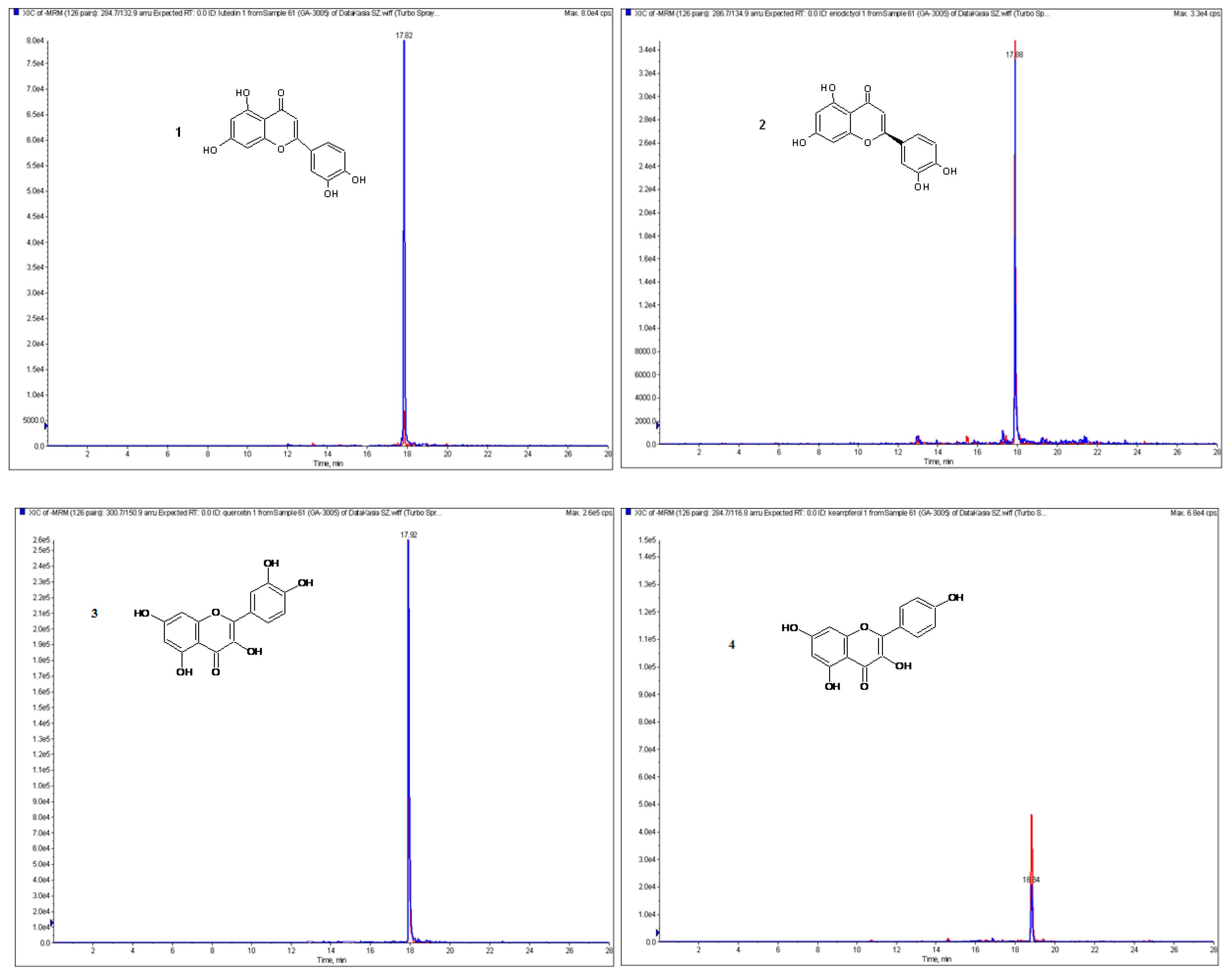
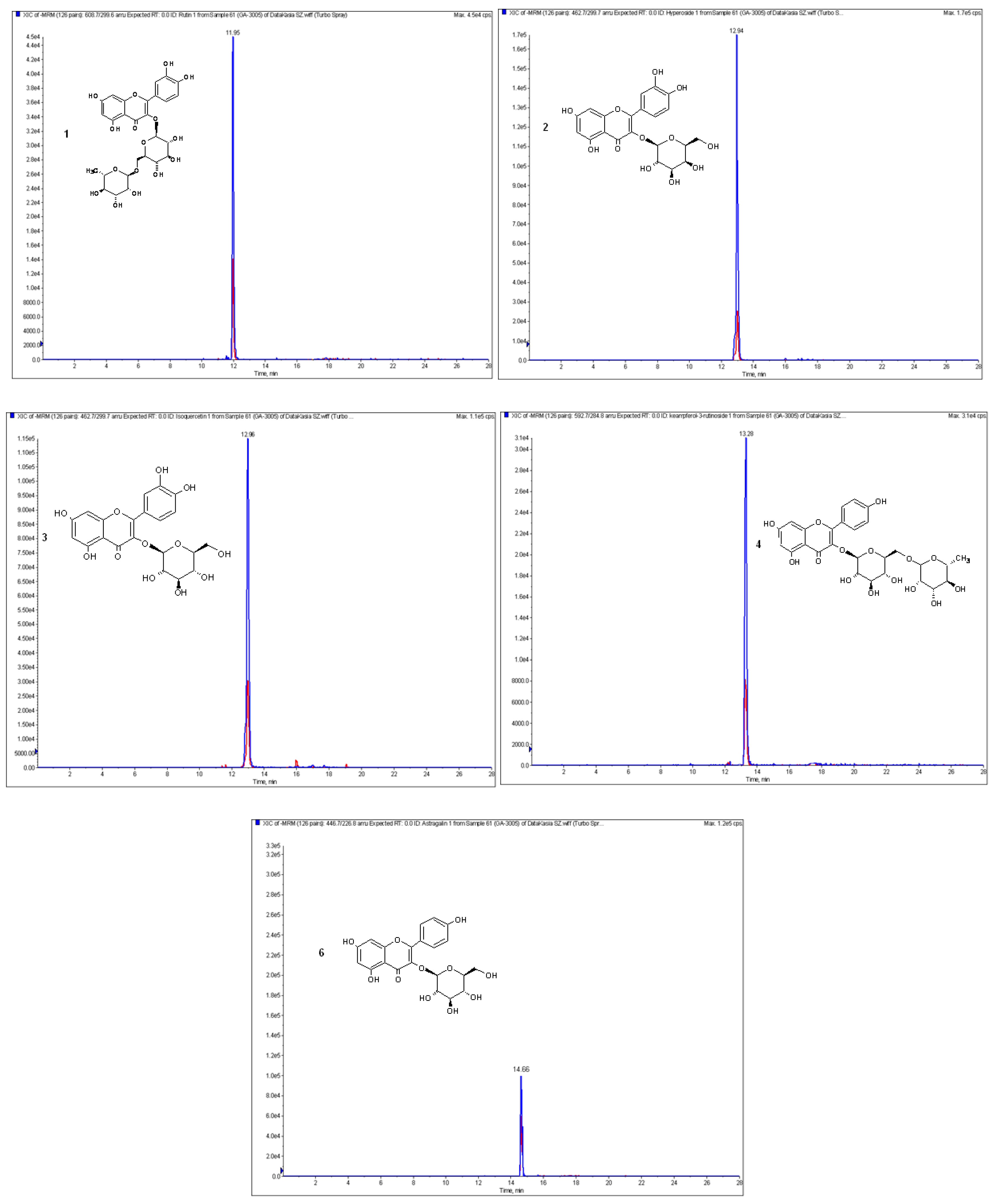
3.1. Polyphenol Composition

There are only a few reports in the literature about the composition and biological activity of Laserpitium species [2,3,4,5,9,10,11,12,14,15,16,17,18]. To date, the polyphenol composition of fruits of L. krapffii have not been investigated. Therefore, the first stage of our study was to examine the total content of these compounds in the etheric and methanolic extracts of fruits of L. krapffii. The amounts of phenolic compounds in the tested extracts were proportional to the intensity of absorption measured spectrophotometrically. The analysis indicated that the methanolic extract contains much larger quantities of total phenolic, flavonoids, and phenolic acids content. The obtained results are shown in Table 3.
The next purpose of our study was the qualitative and quantitative analysis of phenolic acids and flavonoids in obtained extracts. The optimized LC-ESI-MS/MS procedure allowed the identification of 12 phenolic acids and nine flavonoids in the etheric and methanolic extracts from the fruits of L. krapffii. The chromatograms of the flavonoid aglycones and glycosides in methanolic extract are shown in Figure 1 and Figure 2, respectively. The amounts of all the compounds, which were quantified by a comparison of peak areas with the calibration curves of the corresponding references, are shown in Table 4. Both extracts did not differ significantly in the composition of polyphenols except for the absence of 3-hydroxybenzoic acid in the etheric extract. Gallic acid, eriodictyol, kaempferol, and nicotiflorin were identified in quantifiable amounts only in the methanolic extract.
Protocatechuic (LAM—195.93 ± 0.47 μg and LAEN—58.10 ± 1.16 μg per g of dry extract) and vanillic (LAM—34.13 ± 0.08 μg and LAEN—12.45 ± 0.21 μg/g DE) acids were the most abundant among the phenolic acids in both investigated extracts. In the methanolic extract, the large amount of 4-hydroxybenzoic (24.4 ± 0.03 μg/g DE), p-coumaric (24.3 ± 0.38 μg/g DE), and caffeic (20.55 ± 0.38 μg/g DE) acids was also observed.
Among the flavonoid aglycones, a large amount of quercetin (13.92 ± 0.03 μg/g of dry LAEN extract and 22.18 ± 0.09 μg/g of dry LAM extract) was found. The highest yields of flavonoid glycosides was obtained for the methanolic extract. Isoquercetin, nicotiflorin, and astragalin were presented in the greatest quantities in this extract.
To the best of our knowledge, L. krapffii was investigated for the first time in terms of phenolic acids and flavonoids quality and quantity. However, from the leaves of L. latifolium, astragalin, avicularin, isoquercitrin, quercitrin, rutin, chlorogenic and neochlorogenic acid, and other unidentified quercetin, isorhamnetin, and kaempferol derivatives were previously isolated [9].
Since plant phenolic acids and flavonoids have been noticed to possess a wide spectrum of biological activities including anti-inflammatory, antimutagenic, antioxidant, antitumor, and anticarcinogenic properties [29,30,31], the antioxidant and cytotoxic activity of the etheric and methanolic extracts obtained from the fruits of L. krapffii were also evaluated.

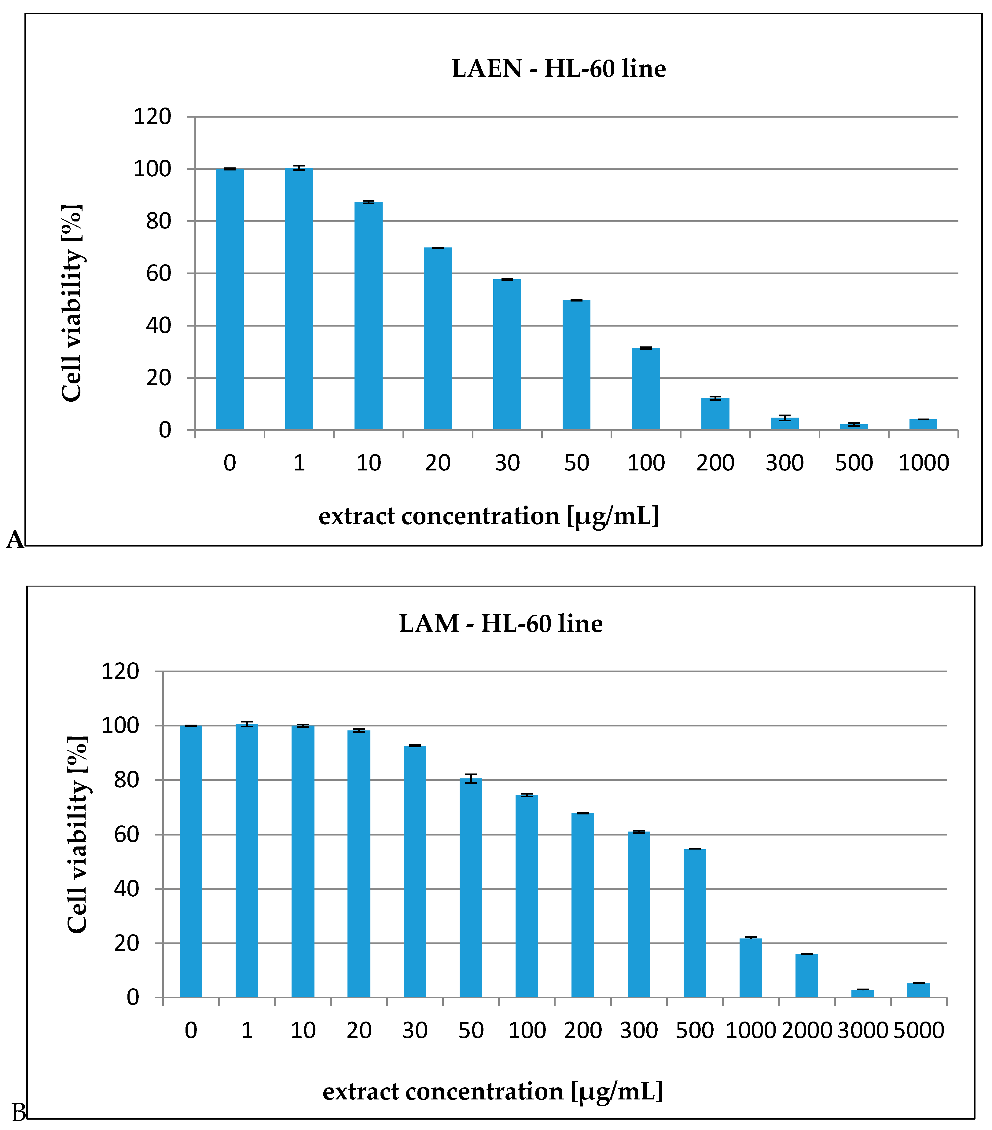
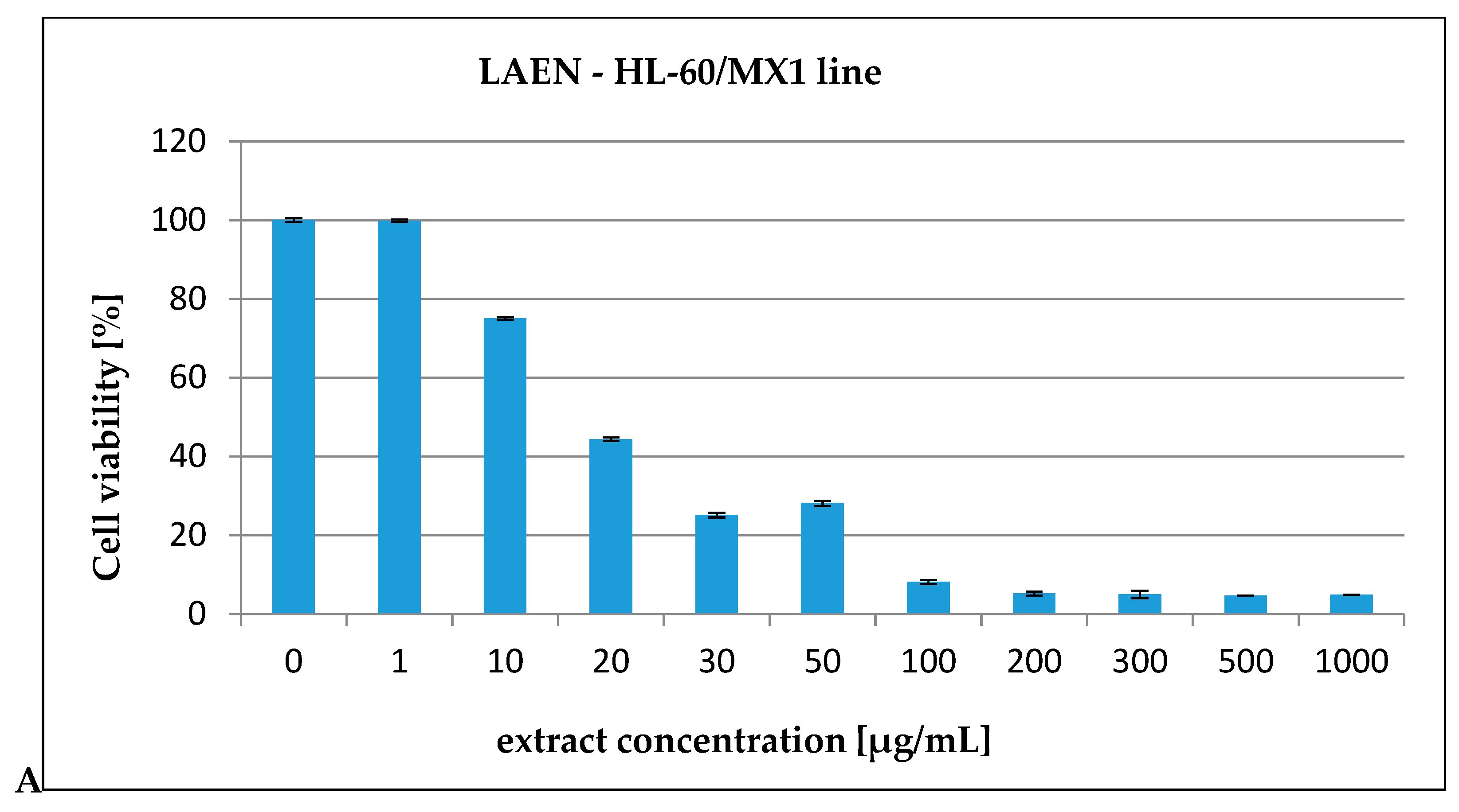
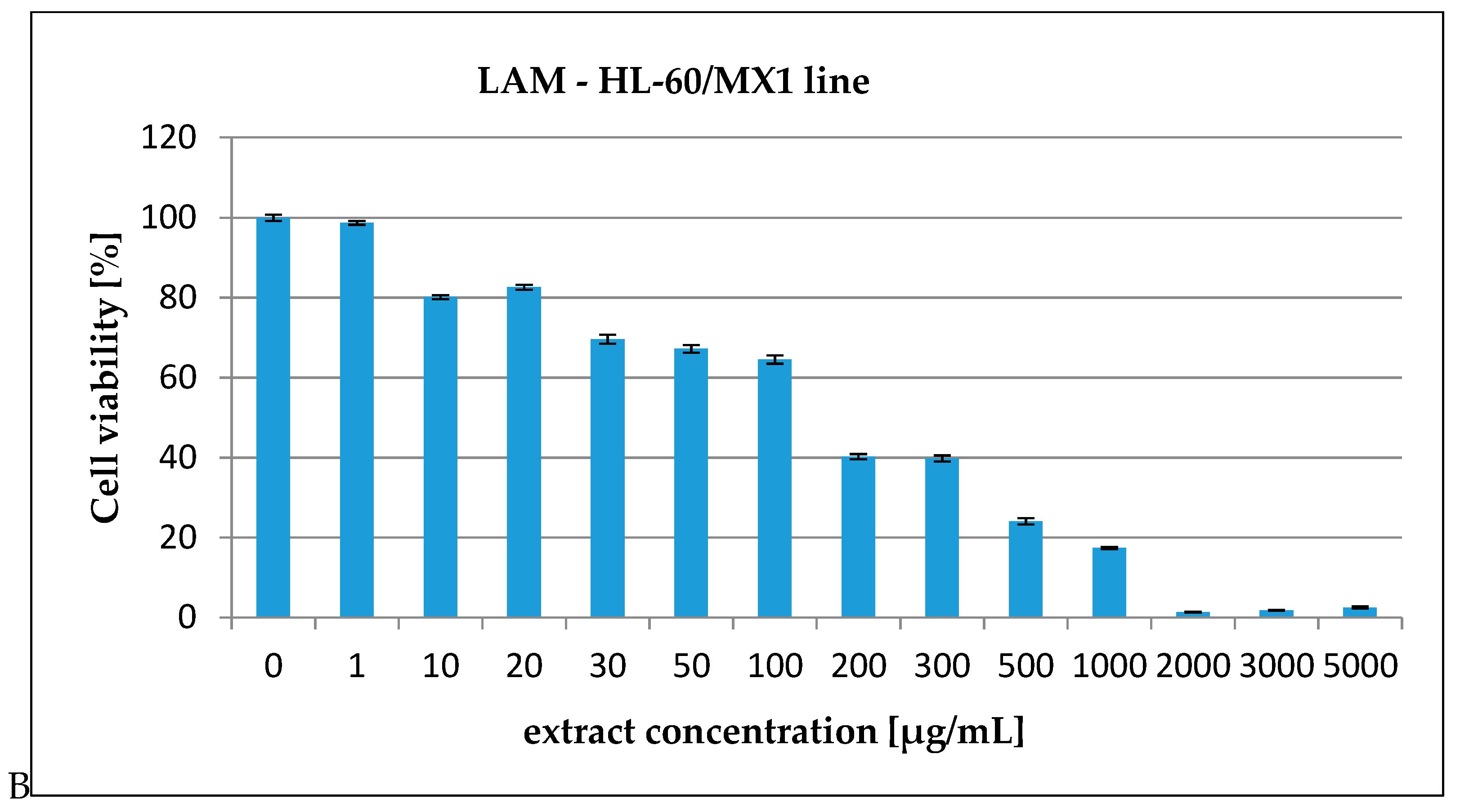
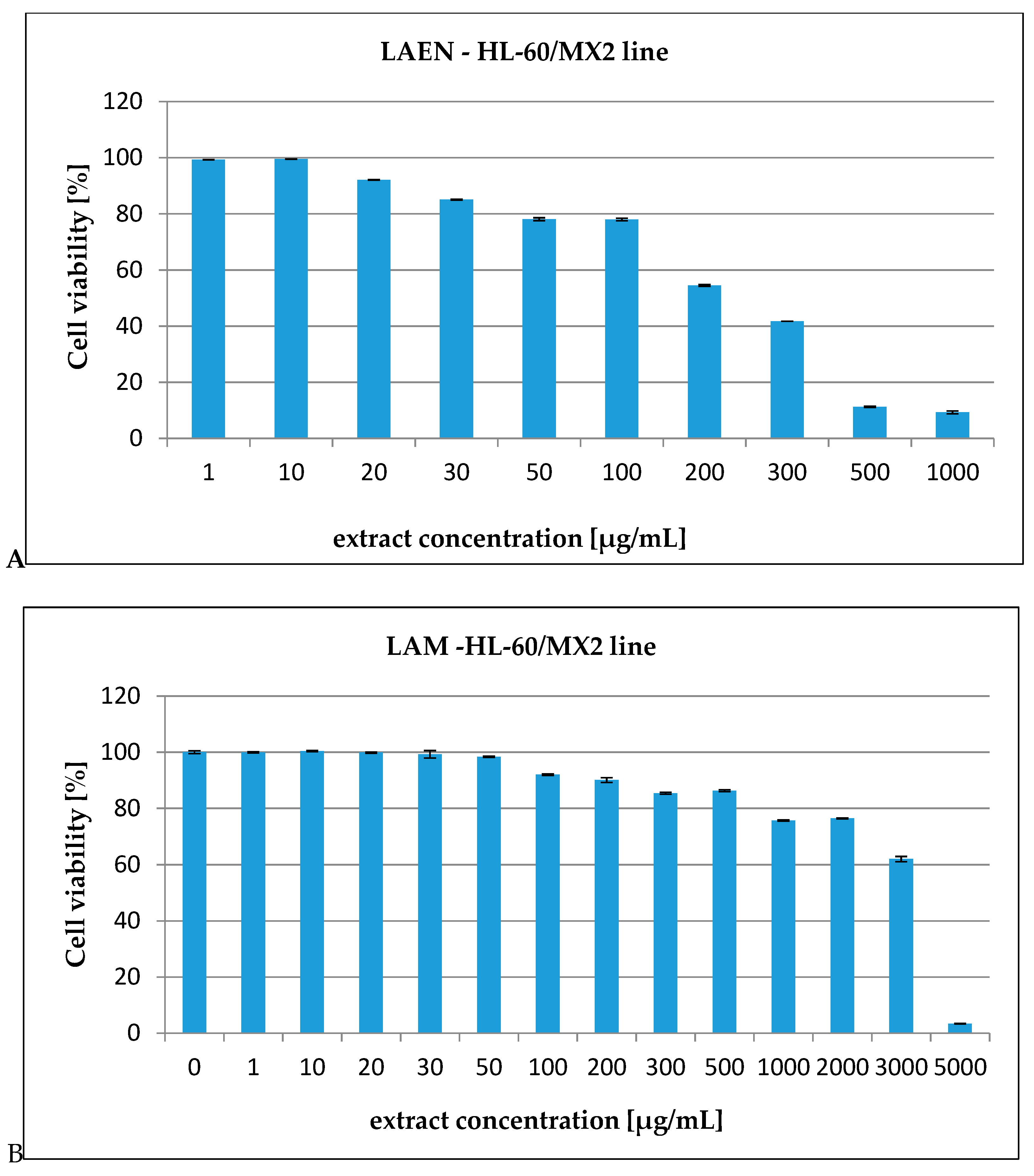
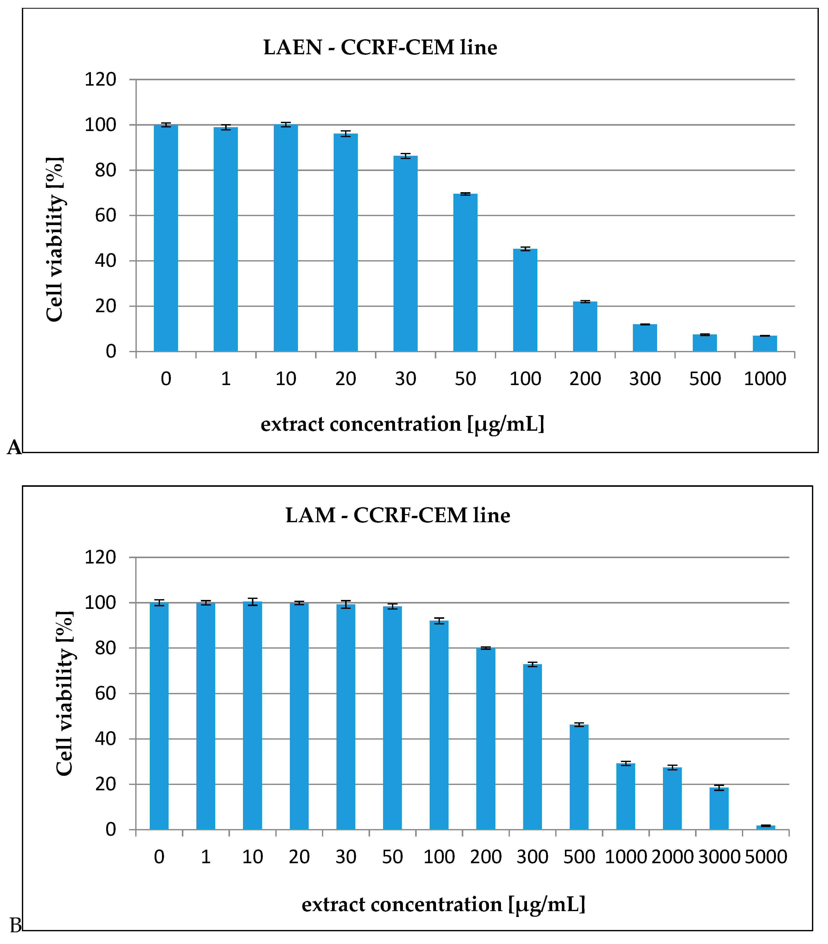
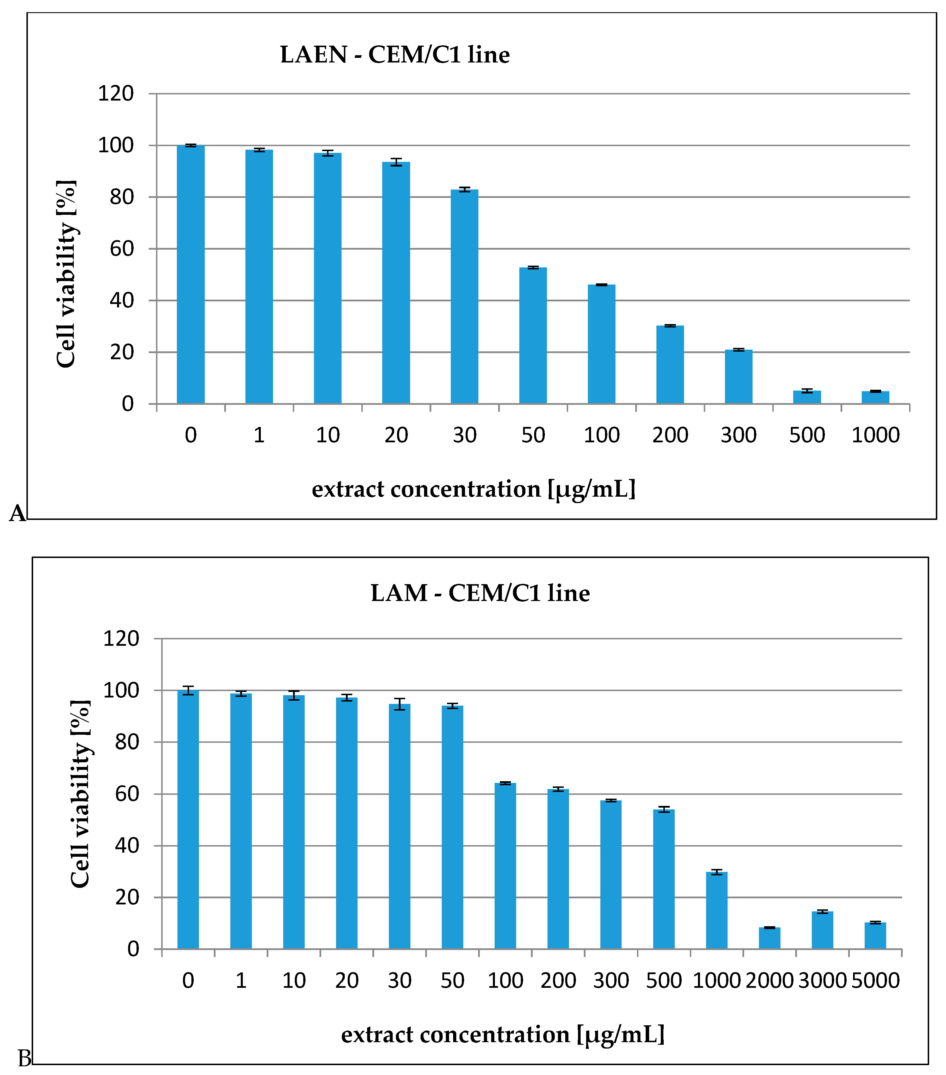
3.2. In Vitro Cytotoxicity Assay

The process of induction of neoplastic changes is associated with the appearance and accumulation of numerous mutations in the genetic material that caused the acquisition of unlimited division potential by phenotype cells. Such cells start to divide without limitations and lose their ability to die, creating a cancer. One way to eliminate cancer cells is to kill them by starting the process of apoptosis. The occurrence of multidrug resistance, inadequate efficacy, and numerous complications of current therapies motivate the search for new drugs. Plants are one of the basic sources of anti-cancer compounds [22,32].
In our report, the effect of the etheric and methanolic extracts in increasing concentrations on five types of cancer cell lines, HL-60 (Figure 3), HL-60/MX1 (Figure 4), HL-60/MX2 (Figure 5), CEM/C1 (Figure 6), and CCRF/CEM (Figure 7) was investigated. The cytotoxicity was estimated using trypan blue vital staining. The cells of all the cancer lines exposed to the examined samples showed various cytotoxicity depending on the IC50 value.
Based on the obtained results, it was found that the analyzed extracts from the fruits of L. krapffii induce apoptosis of the cells of all the tested cell lines. The results, which are given in Table 5, showed that the etheric extract (LAEN) significantly inhibited HL-60/MX1 human leukemia cells, and was the most potent sample with an IC50 value of 31.08 μg/mL. Moreover, this extract showed a moderate cytotoxicity against HL-60 with IC50 = 72.19 μg/mL and against CEM/C1 with IC50 = 99.40 μg/mL. The etheric extract showed also relatively high cytotoxicity against CCRF/CEM (IC50 = 172.58 μg/mL).
The weak cytotoxic activity was found in the methanolic extract from the fruits of L. krapffii. The IC50 values for the LAM extract ranged from 205.72 μg/mL for the HL-60/MX1 line to 2848.31 μg/mL for the HL-60/MX2 line.
The presence of the large amounts of protocatechuic acid and quercetin may be responsible for the cytotoxic activity of the studied extracts. It has been reported that protocatechuic acid induces apoptosis in HL-60 human leukemia cells [33].
The published reports on the cytotoxic and antitumor activities of Laserpitium species are limited, and they focus on only a few species: Laserpitium latifolium L., L. zernyi Hayek, and L. ochridanum Micevski. The cytotoxic activities of chloroform extracts of dried roots and rhizomes of L. zernyi and L. ochridanum were tested against MCF 7/6 and MCF 7/AZ (human breast cancer) cell lines. The extracts exerted cytotoxic activities with the IC50 values ranging from 65.21 to 348.25 μg/mL. The obtained results showed that the extract of L. ochridanum was most potent in the MTT test with IC50 values of 65.21 μg/mL and 66.09 μg/mL in the MCF 7/AZ and MCF 7/6 cell lines, respectively [5].
Despite the use of different Laserpitium species and various cell lines as well as different research assays, the IC50 values obtained in our work and previous works are similar.

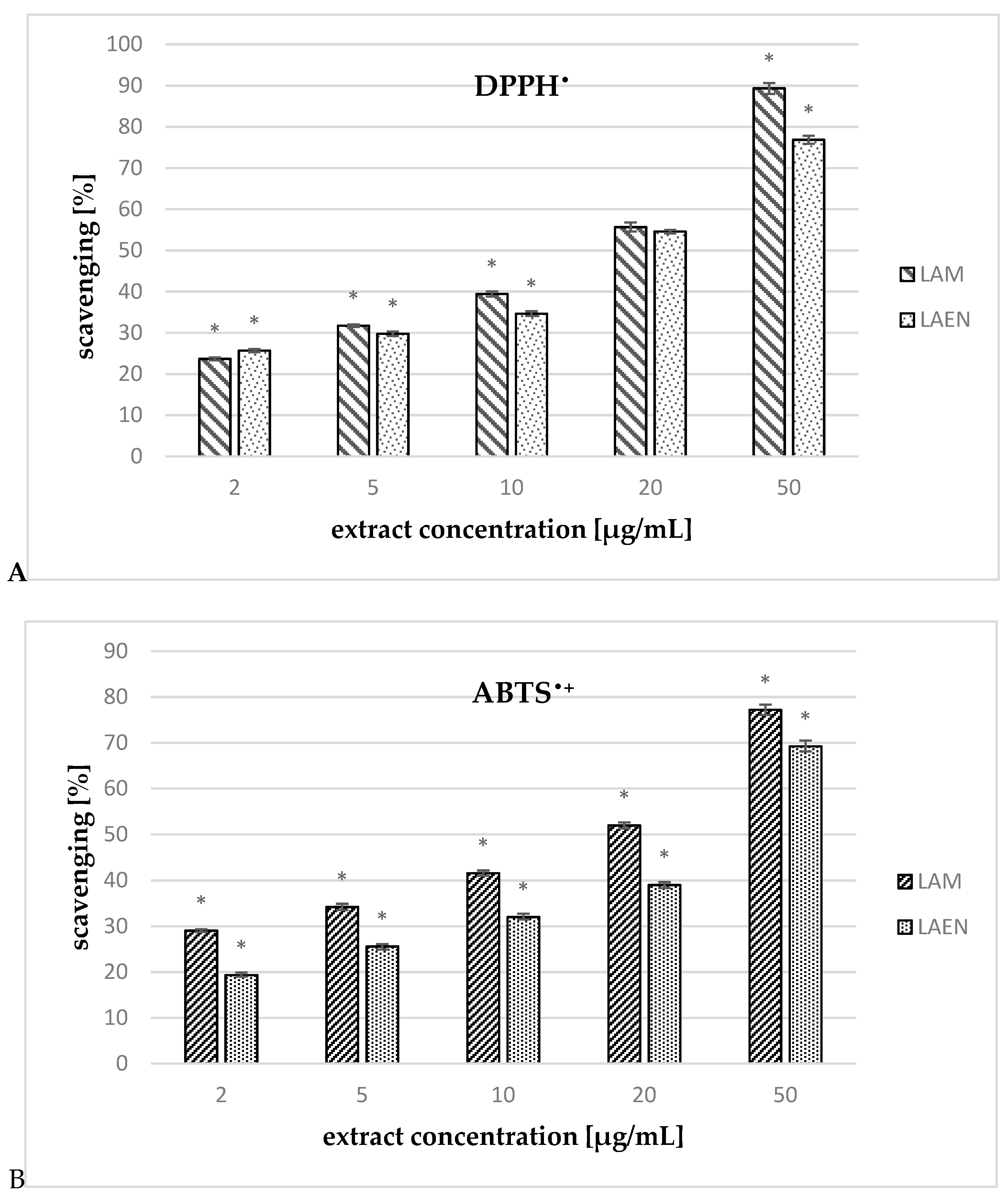
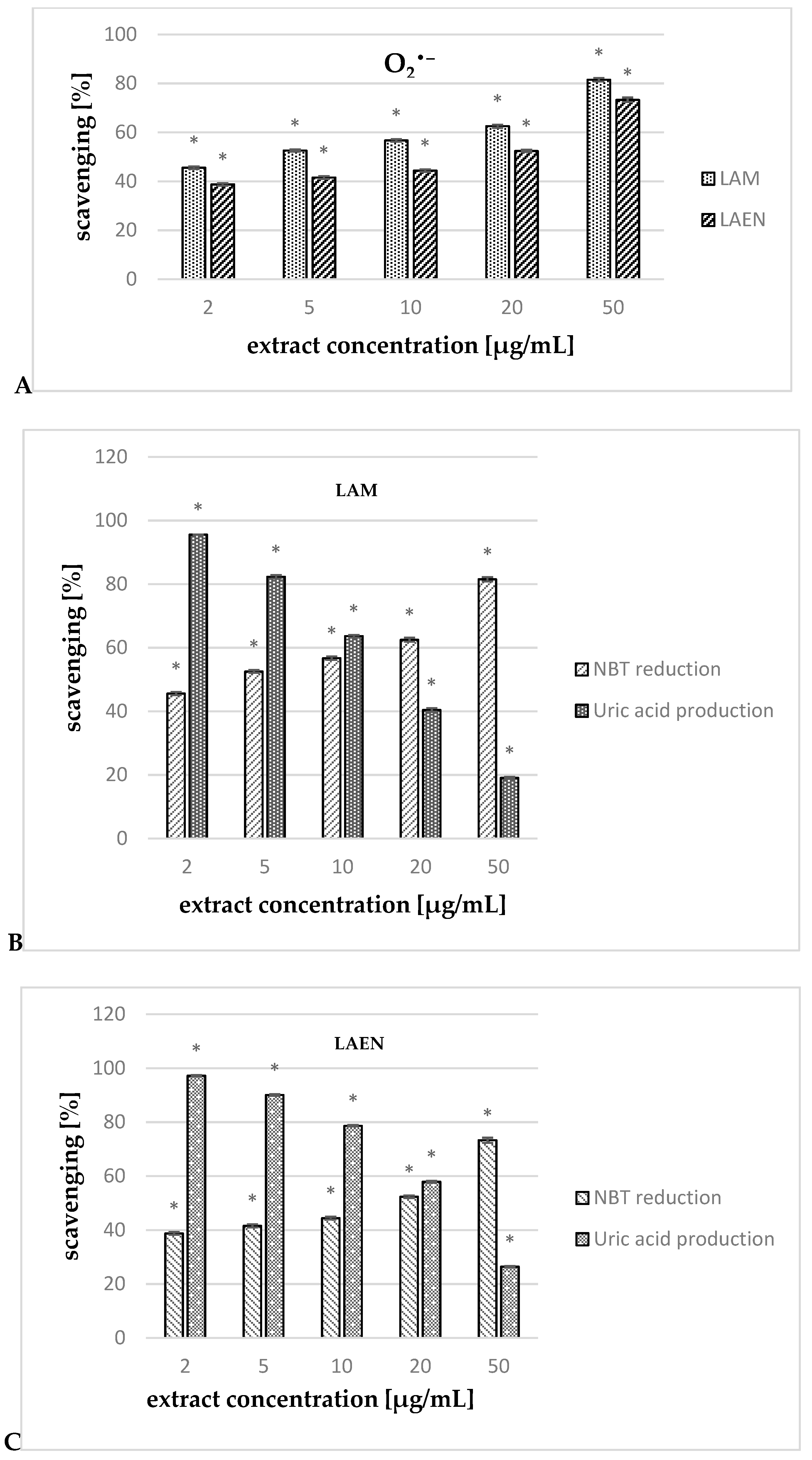
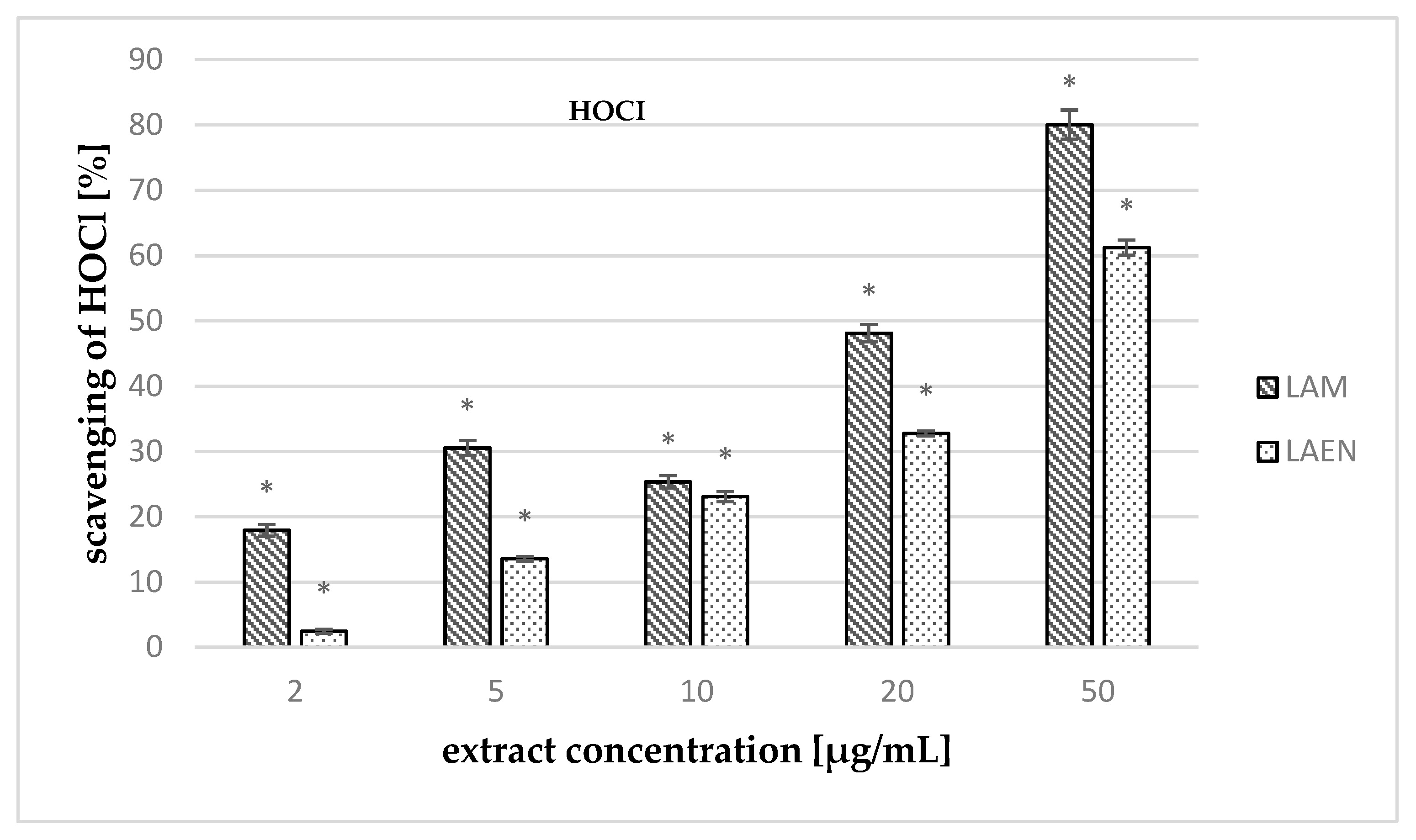
3.3. Antioxidant Activity

In our research, the antioxidant activity of etheric and methanolic extracts of the fruits of L. krapffii was determined for the first time. We used four colorimetric methods to determine the activity of both tested extracts against synthetic radicals DPPH and ABTS•+, xanthine/xanthine oxidase (generating O2), as well as against HOCl. The measurement of antioxidant activities was performed on a microplate scale in cell-free systems.
The extracts examined in the study exhibited high scavenging ability in a concentration-dependent manner (Figure 8). For comparison, the radical scavenging activity of ascorbic acid was measured in the same conditions. The higher DPPH scavenging activity was demonstrated for the methanolic extract (LAM). The IC50 value for methanolic extract was almost one and half times lower than the for ascorbic acid (Table 6).
The percentage of ABTS•+ radicals reduction for extracts was measured at λ = 734 nm after 10 min of incubation (experimentally determined). In this time, both the analyzed extracts and the reference compound reached a constant antioxidant value. In comparison to gallic acid, which is a strong antioxidant compound [34], both tested extracts of L. krapffii showed weak antioxidant properties. The IC50 values for the methanolic (20.69 μg/mL) and etheric (30.45 μg/mL) extracts were much higher than that for gallic acid, which was used as a positive control (0.75 μg/mL).
In the study, we also evaluated the ability of the etheric and methanolic extracts of fruits of L. krapffii to scavenge O2 generated by the xanthine/xanthine oxidase system. Both extracts scavenge O2 in a concentration-dependent manner (Figure 9A). At all the concentrations studied, LAM showed statistically (p < 0.05) stronger activity than the etheric extract did. The IC50 value for the methanolic extract (3.28 μg/mL) was almost five times lower than that for the etheric extract (17.31 μg/mL), and almost two times lower in comparison to the quercetin (5.72 μg/mL). Additionally, both extracts strongly inhibited uric acid production (for LAEN—97.23–26.41%, and for LAM—95.56–19.06%), confirming that their activity in a xanthine/xanthine oxidase system is related to the scavenging effect against O2 and the inhibition of the enzyme activity (Figure 9B,C).
The measurement of activity against HOCl (Figure 10) showed that the extracts tested have a high scavenging ability of this radical. In the concentrations studied (2–50 μg/mL), the methanolic extract had stronger ability (17.91–80.04%) than the etheric one (2.45–61.23%), and the IC50 values were 25.09 μg/mL and 38.17 μg/mL, respectively. Moreover, the IC50 values for both extracts were lower than that for ascorbic acid, which was used as a reference compound (49.56 μg/mL).
The obtained lower IC50 values for the methanolic extract in all the antioxidant assays may be connected with higher phenolic compounds content, as it is well known that flavonoids and phenolic acids are the most popular natural components responsible for the antioxidant activity of plants.

4. Conclusions

In the literature, there are no reports concerning the biological activities of extracts of Laserpitium krapffii. In this study, it was found that both extracts of the fruits of L. krapffii have high cytotoxic and antioxidant potential, and can be a source of valuable compounds with protective effects against oxidative damage. The inhibiting influence of both extracts on ROS production may reduce oxidative modifications of biomolecules and tissue dysfunctions during chronic degenerative diseases.

Author Contributions

A.B.-K., N.V. and K.S. designed research plan; A.B.-K., K.S., M.C. and W.P. carried out experiments and analyzed data; K.S. and A.B.-K. interpreted results; K.S. wrote the manuscript.

Funding

This research received no external funding.

Conflicts of Interest

The authors declare no conflict of interest.

References

  1. Tutin, T.G. Laserpitium L. In Flora Europaea 2; Tutin, T.G., Heywood, V.H., Burges, N.A., Moore, D.M., Valentine, D.H., Walters, S.M., Webb, D.A., Eds.; Cambridge University Press: Cambridge, UK, 1968; Volume 2, pp. 368–370. [Google Scholar]
  2. Djermanovic, M.; Mladenovic, S.; Ristic, N.; Stefanovic, M. Sesquiterpene lactones from Laserpitium alpinum (Umbelliferae). Int. Conf. Chem. Biotechnol. Biol. Act. Nat. Prod. 1981, 3, 317–320. [Google Scholar]
  3. Milosavljević, S.; Bulatović, V.; Stefanović, M. Sesquiterpene lactones from Yugoslavian wild growing plant families Asteraceae and Apiaceae. J. Serb. Chem. Soc. 1999, 64, 397–442. [Google Scholar] [CrossRef]
  4. Adcock, J.W.; Betts, T.J. A chemotaxonomic survey of essential oil constituents in the tribe Laserpitieae (F. Umbelliferae). Planta Med. 1974, 26, 52–64. [Google Scholar] [CrossRef] [PubMed]
  5. Popović, V.; Heyerick, A.; Petrović, S.; Calenbergh, S.V.; Karalić, I.; Niketić, M.; Deforce, D. Sesquiterpene lactones from the extracts of two Balkan endemic Laserpitium species and their cytotoxic activity. Phytochemistry 2013, 87, 102–111. [Google Scholar] [CrossRef] [PubMed]
  6. Holub, M.; Motl, O.; Samek, Z. Terpenes. CCIV. Structure of two sesquiterpenic lactones, isomontanolide, and acetylisomontanolide from Laserpitium siler. Collect. Czech. Chem. Commun. 1972, 37, 1186–1194. [Google Scholar] [CrossRef]
  7. Holub, M.; Samek, Z. Structure of archangelolide, a sesquiterpenic lactone from Laserpitium archangelica. Collect. Czech. Chem. Commun. 1973, 38, 731–738. [Google Scholar] [CrossRef]
  8. Bohlman, F.; Zdero, C. Prutenin, 4-Angeloyloxypruteninone, and 4-Acetoxy-pruteninone und-isopruteninone—4 new sesquiterpene lactones from Laserpitium prutenicum L. Chem. Ber. 1971, 104, 1611–1615. [Google Scholar] [CrossRef]
  9. Vereskovskii, V.V.; Kuznetsova, Z.P.; Loznukho, I.V.; Sokolov, I.V.; Osokin, D.M. Phenolic compounds of Laserpitium latifolium. Khim. Prir. Soedin. 1992, 6, 724–726. [Google Scholar] [CrossRef]
  10. Popović, V.; Heyerick, A.; Petrović, S.; Calenbergh, S.; Karalić, I.; Deforce, D.; Niketić, M. Cytotoxic activity of Laserpitium latifolium L. extract and its daucane and phenylpropanoid constituents. Rec. Nat. Prod. 2013, 7, 245–249. [Google Scholar]
  11. Popović, V.; Goeman, J.; Thommis, J.; Heyerick, A.; Caroen, J.; Van der Eycken, J.; De Bosscher, K. Daucane esters from laserwort (Laserpitium latifolium L.) inhibit cytokine and chemokine production in human lung epithelial cells. Phytomedicine 2017, 26, 28–36. [Google Scholar] [CrossRef]
  12. Baser, K.H.C.; Duman, H. Composition of the essential oil of Laserpitium petrophilum Boiss. et Heldr. J. Essent. Oil Res. 1997, 9, 707–708. [Google Scholar] [CrossRef]
  13. Chizolla, R.; Novak, J.; Franz, C. Fruit of Laserpitium siler grown in France. J. Essen. Oil Res. 1999, 11, 197–198. [Google Scholar] [CrossRef]
  14. Popović, V.; Petrović, S.; Milenković, M.; Drobaca, M. Composition and antimicrobial activity of the essential oils of Laserpitium latifolium L. and L. ochridanum Micevski (Apiaceae). Chem. Biodivers. 2015, 12, 170–177. [Google Scholar] [CrossRef] [PubMed]
  15. Hegi, G. Siler Miller, Laserpitium, L. In Illustrierte Flora von Mitteleuropa, Band V (2); Beger, H., Ed.; A. Pichler’s Witwe & Sohn: Wien, Austria, 1906; pp. 1467–1501. [Google Scholar]
  16. Harmatha, J.; Buděšínský, M.; Vokáč, K.; Kostecká, P.; Kmoníčková, E.; Zídek, Z. Trilobolide and related sesquiterpene lactones from Laser trilobum possessing immunobiological properties. Fitoterapia 2013, 89, 157–166. [Google Scholar] [CrossRef] [PubMed]
  17. Tirillini, B.; Pagiotti, R.; Angelini, P.; Pintore, G.; Chessa, M.; Menghini, L. Chemical composition and fungicidal activity of the essential oil of Laserpitium garganicum from Italy. Chem. Nat. Comp. 2009, 45, 103–105. [Google Scholar] [CrossRef]
  18. Popović, V.; Petrović, S.; Tomić, M.; Stepanović-Petrović, R.; Micov, A.; Pavlović-Drobac, M.; Couladis, M.; Niketić, M. Antinociceptive and anti-edematous activities of the essential oils of two Balkan endemic Laserpitium species. Nat. Prod. Commun. 2014, 9, 125–128. [Google Scholar] [PubMed]
  19. Polish Pharmacopoeia IX. PTFarm; Polish Pharmaceutical Society: Warsaw, Poland, 2011; p. 150.
  20. Olech, M.; Nowak, R. Influence of different extraction procedures on the antiradical activity and phenolic profile of Rosa rugosa petals. Acta Pol. Pharm. Drug Res. 2012, 69, 501–507. [Google Scholar]
  21. Nowacka, N.; Nowak, R.; Drozd, M.; Olech, M.; Łoś, R.; Malm, A. Antibacterial, antiradical potential and phenolic compounds of thirty-one polish mushrooms. PLoS ONE 2015, 10, e0140355. [Google Scholar] [CrossRef]
  22. Kubrak, T.; Bogucka-Kocka, A.; Komsta, Ł.; Załuski, D.; Bogucki, J.; Gałkowski, D.; Kaczmarczyk, R.; Feldo, M.; Cioch, M.; Kocki, J. Modulation of multidrug resistance gene expression by coumarin derivatives in human leukemic cells. Oxid. Med. Cell. Longev. 2017, 2017, 5647281. [Google Scholar] [CrossRef] [PubMed]
  23. Choi, C.W.; Kim, S.C.; Hwang, S.S.; Choi, B.K.; Ahn, H.J.; Lee, M.Y.; Park, S.H.; Kim, S.K. Antioxidant activity free radical scavenging capacity between Korean medicinal plants flavonoids by assay-guided comparison. Plant Sci. 2002, 163, 1161–1168. [Google Scholar] [CrossRef]
  24. Olech, M.; Nowacka-Jechalke, N.; Masłyk, M.; Martyna, A.; Pietrzak, W.; Kubiński, K.; Załuski, D.; Nowak, R. Polysaccharide-rich fractions from Rosa rugosa Thunb.—Composition and chemopreventive Potential. Molecules 2019, 24, 1354. [Google Scholar] [CrossRef] [PubMed]
  25. Re, R.; Pellegrini, N.; Proteggente, A.; Pannala, A.; Yang, M.; Rice-Evans, C. Antioxidant activity applying an improved ABTS· radical cation decolorization assay. Free Radic. Biol. Med. 1999, 26, 1231–1237. [Google Scholar] [CrossRef]
  26. Czerwińska, M.; Kiss, A.K.; Naruszewicz, M. A comparison of antioxidant activities of oleuropein its dialdehydic derivative from olive oil, oleacein. Food Chem. 2012, 131, 940–947. [Google Scholar] [CrossRef]
  27. Kiss, A.K.; Filipek, A.; Czerwińska, M.; Naruszewicz, M. Oenothera paradoxa defatted seeds extract its bioactive component penta-O-galloyl-β-d-glucose decreased production of reactive oxygen species inhibited release of leukotriene B4, interleukin-8, elastase, myeloperoxidase in human neutrophils. J. Agric. Food Chem. 2010, 58, 9960–9966. [Google Scholar] [CrossRef] [PubMed]
  28. Schepetkin, I.A.; Kirpotina, L.N.; Jakiw, L.; Khlebnikov, A.I.; Blaskovich, C.L.; Jutila, M.A.; Quinn, M.T. Immunomodulatory activity of oenothein B isolated from Epilobium angustifolium. J. Immunol. 2009, 83, 6754–6766. [Google Scholar] [CrossRef]
  29. Farhoosh, R.; Johnny, S.; Asnaashari, M.; Molaahmadibahraseman, N.; Sharif, A. Structure-antioxidant activity relationships of o-hydroxyl, o-methoxy, and alkyl ester derivatives of p-hydroxybenzoic acid. Food Chem. 2016, 194, 128–134. [Google Scholar] [CrossRef]
  30. Middleton, E.J.; Kandaswami, C.; Theoharides, T.C. The effects of plant flavonoids on mammalian cells: Implications for inflammation, heart disease, and cancer. Pharmacol. Rev. 2000, 52, 673–751. [Google Scholar]
  31. Szewczyk, K.; Grzywa-Celińska, A. Antioxidant and cytotoxic activities of phenolic acida and their role in the anticancer therapies. In Phenolic Acids: Properties, Food Sources and Health Effects, 1st ed.; Flores, A., Ed.; Nova Publishers: New York, NY, USA, 2016; pp. 61–104. [Google Scholar]
  32. Stańczyk, M.; Majsterek, I. Apoptoza-cel ukierunkowanej terapii przeciwnowotworowej. Postepy Biol. Komorki 2008, 35, 467–484. [Google Scholar]
  33. Jaouad, A.; Romero-Jiménez, M.; Fernández-Bedmar, Z.; Villatoro-Pulido, M.; Analla, M.; Alonso-Moraga, A.; Muñoz-Serrano, A. Antigenotoxicity, cytotoxicity, and apoptosis induction by apigenin, bisabolol, and protocatechuic acid. J. Med. Food. 2011, 14, 276–283. [Google Scholar]
  34. Nenadis, N.; Lazaridou, O.; Tsimidou, M.Z. Use of reference compounds in antioxidant activity assessment. J. Agric. Food Chem. 2007, 55, 5452–5460. [Google Scholar] [CrossRef]
Figure 1. The chromatograms in multiple reaction mode (MRM) of flavonoid aglycones occuring in the methanolic extract of L. krapffii fruits: 1-luteolin; 2-eriodictyol; 3-quercetin; 4-kaempferol.
Antioxidants 08 00363 g001
Figure 2. The chromatogram in MRM of flavonoid glycosides occurring in the methanolic extract of L. krapffii fruits: 1-rutin; 2-hyperoside; 3-isoquercetin; 4-kaempferol-3-O-rutinoside; 6-astragalin.
Antioxidants 08 00363 g002
Figure 3. The valuation of the HL-60 cell line viability exposed to 24 h of increasing concentrations of: (A) — LAEN extract and (B) — LAM extract.
Antioxidants 08 00363 g003
Figure 4. The valuation of the HL-60/MX1 cell line viability exposed to 24 h of increasing concentrations of: (A)—LAEN extract and (B)—LAM extract.
Antioxidants 08 00363 g004aAntioxidants 08 00363 g004b
Figure 5. The valuation of the HL-60/MX2 cell line viability exposed to 24 h of increasing concentrations of: (A)—LAEN extract and (B)—LAM extract.
Antioxidants 08 00363 g005
Figure 6. The valuation of the CCRF-CEM cell line viability exposed to 24 h of increasing concentrations of: (A)—LAEN extract and (B)—LAM extract.
Antioxidants 08 00363 g006
Figure 7. The valuation of the CEM/C1 cell line viability exposed to 24 h of increasing concentrations of: (A)—LAEN extract and (B)—LAM extract.
Antioxidants 08 00363 g007
Figure 8. Antioxidant activity of the etheric (LAEN) and methanolic (LAM) extracts against: (A) 2,2-diphenyl-1-picrylhydrazyl radical (DPPH), and (B) 2,2′-azino-bis-(3-ethyl-benzthiazoline-6-sulfonic acid) (ABTS). Data are expressed as means ± SEM; obtained in three independent experiments, assayed in triplicate. Statistical significance of differences was calculated between both extracts; * p < 0.05.
Antioxidants 08 00363 g008
Figure 9. Scavenging effect of the etheric (LAEN) and methanolic (LAM), in the concentration range of 2 to 50 μg/mL, on O2 scavenging [%] in the xanthine/xanthine oxidase system (A). Effects of the LAM (B) and LAEN (C) on the xanthine/xanthine oxidase system [%]: nitrobluetetrazolium (NBT) reduction determines the scavenging of O2; uric acid production determines the oxidase inhibition. Statistical significance of differences was calculated between both extracts or between uric acid production and NBT reduction; * p < 0.05.
Antioxidants 08 00363 g009
Figure 10. Scavenging effects of the etheric (LAEN) and methanolic (LAM) on hypochlorous acid (HOCl)-mediated oxidation of 5-thio-2-nitrobenzoic acid (TNB). Data are expressed as means ± SEM, obtained in at least three independent experiments, and assayed in triplicate. Statistical significance of differences was calculated between both extracts; * p < 0.05.
Antioxidants 08 00363 g010
Table 1. Liquid chromatography–electrospray tandem mass spectroscopy (LC-ESI-MS/MS) analytical results of phenolic acids and flavonoids investigated in the extracts, which were confirmed by comparison with reference compounds.
CompoundRetention Time [min][M − H] [m/z]Fragment Ions [m/z]Collision Energy [eV]
Gallic acid5.07168.778.9
124.9
−36
−14
Protocatechuic acid8.23152.980.9
107.8
−26
−38
4-Hydroxybenzoic acid11.27136.892.9
107.9
−18
−18
Syringic acid11.41196.9122.8
181.9
−24
−12
Gentisic acid11.43352.980.0
96.9
−110
−52
Vanilic acid11.49166.8107.9
123.0
−18
−12
Caffeic acid11.68178.788.9
134.9
−46
−16
Rutin11.99608.7299.6
270.9
−46
−60
3-Hydroxybenzoic acid12.08136.993.0
75.0
−16
−48
Hyperoside12.80462.7299.7
254.7
−28
−42
Isoquercetin13.00462.7299.7
270.7
−30
−44
Nicotiflorin13.31592.7284.8
226.7
−38
−68
p-Coumaric acid14.18162.893.0
119.0
−44
−14
Astragalin14.66446.7226.8
254.8
−54
−40
Ferulic acid14.84192.8133.9
177.9
−16
−12
o-Coumaric acid17.17162.8119.0
93.0
−14
−46
Luteolin17.54284.7132.9
150.9
−38
−26
Eriodictyol17.87286.7134.9
150.9
−32
−18
Quercetin17.91300.7150.9
178.8
−26
−20
Salicylic acid18.06136.975.0
93.0
−48
−16
Kaempferol18.56284.7116.8
93.0
−46
−52
Table 2. Analytical results of LC-MS/MS quantitative method for determination of flavonoids and phenolic acids. Limit of detection (LOD), limit of quantification (LOQ) and calibration curve parameters.
CompoundLOD [ng/mL]LOQ [ng/mL]R2Linearity Range [ng/mL]
Gallic acid33.395.00.9987167–3300
Protocatechuic acid17.034.00.999734–3470
4-Hydroxybenzoic acid17.434.70.999369.4–3470
Syringic acid167.0666.00.9993666–11,100
Gentisic acid 1.73.30.99973.3–330
Vanillic acid100.0250.00.9997330–33,000
Caffeic acid60.0160.00.9990175–3500
Rutin120.0300.00.99852000–25,000
3-Hydroxybenzoic acid 33.3334.00.9994334–6670
Hyperoside150.0200.00.99871000–25,000
Isoquercetin 150.0300.00.99872000–25,000
Nicotiflorin60.0120.00.9991120–50,000
p-Coumaric acid7.318.10.999618.1–1820
Astragalin100.0200.00.99781200–24,000
Ferulic acid17.434.70.999469.4–11,600
o-Coumaric acid7.318.10.999618.1–1820
Luteolin25.040.00.998940–4000
Eriodictyol10.015.00.998215–5000
Quercetin5.010.00.998020–3000
Salicylic acid3.316.50.998916.5–1650
Kaempferol20.033.00.998933–20,000
Table 3. The total phenolic (TPC) and total flavonoid (TFC) content in the etheric (LAEN) and methanolic (LAM) extracts from L. krapffii fruits. The results are expressed as mg of gallic acid equivalent (GAE) and quercetin equivalent (QE) per 1 g of dry extract (DE). Values are presented in mean ± SEM, n = 3.
ExtractTPC
[mg GAE/g DE]
TFC
[mg QE/g DE]
TPAC
[mg CAE/g DE]
LAEN5.53 ± 0.580.41 ± 0.171.19 ± 0.06
LAM14.53 ± 0.601.22 ± 0.387.98 ± 0.07
Table 4. Content of phenolic acids and flavonoid aglycones and glycosides in the etheric (LAEN) and methanolic (LAM) extracts from L. krapffii fruits. Values are presented in means ± SEM, n = 3. nd—not detected; BQL—peak detected, concentration lower than the LOQ but higher than the LOD.
CompoundLAENLAM
Phenolic Acid/Flavonoid (μg per g of Dry Extract)
Gallic acidBQL1.19 ± 0.05
Protocatechuic acid58.10 ± 0.67195.93 ± 0.27
4-Hydroxybenzoic acid5.76 ± 0.1724.4 ± 0.02
Syringic acid1.60 ± 0.059.02 ± 0.02
Gentisic acid1.53 ± 0.138.33 ± 0.14
Vanillic acid12.45 ± 0.1234.13 ± 0.05
Caffeic acid5.34 ± 0.0820.55 ± 0.22
Rutin 3.17 ± 0.0211.05 ± 0.42
3-Hydroxybenzoic acidnd0.73 ± 0.08
Hyperoside1.89 ± 0.005.12 ± 0.08
Isoquercetin3.05 ± 0.0622.53 ± 0.01
NicotiflorinBQL15.36 ± 0.02
p-Coumaric acid6.65 ± 0.1824.3 ± 0.22
Astragalin0.74 ± 0.0315.51 ± 0.01
Ferulic acid0.75 ± 0.052.91 ± 0.08
o-Coumaric acid2.56 ± 0.059.58 ± 0.32
Luteolin0.16 ± 0.000.86 ± 0.08
EriodictyolBQL0.17 ± 0.02
Quercetin13.92 ± 0.0122.18 ±0.05
Salicylic acid3.83 ± 0.2413.35 ± 0.25
KaempferolBQL0.04 ± 0.00
Table 5. The extrapolated IC50 values for the HL-60, HL-60/MX1, HL-60/MX2, CEM/C1, and CCRF/CEM line cells.
-IC50 [µg/mL]
LAENLAM
HL-6072.19757.77
HL-60/MX131.08205.72
HL-60/MX2248.432848.31
CEM/C199.40513.03
CCRF/CEM172.58671.90
Table 6. The IC50 values established in antioxidant assays. Data are expressed as mean ± SEM, n = 3. AA—ascorbic acid, GA—gallic acid, Q quercetin.
AssayAntioxidant Activity [IC50 in μg/mL of Extract]
LAENLAMAAGAQ
DPPH22.70 ± 0.2318.93 ± 0.3425.01 ± 0.14--
ABTS•+30.45 ± 0.4720.69 ± 0.43-0.75 ± 0.01-
O2•−18.35 ± 0.163.28 ± 0.12--5.72 ± 0.02
HOCl38.17 ± 0.0225.09 ± 0.1049.56 ± 1.35--

