Article

Antioxidant Activity of Graptopetalum paraguayense E. Walther Leaf Extract Counteracts Oxidative Stress Induced by Ethanol and Carbon Tetrachloride Co-Induced Hepatotoxicity in Rats

1
Department of Nutrition and Health Sciences, Kainan University, Taoyuan 33857, Taiwan
2
Department of Food and Nutrition, Providence University, Taichung 43301, Taiwan
3
Graduate Institute of Veterinary Pathobiology, National Chung Hsing University, Taichung 40227, Taiwan
*
Author to whom correspondence should be addressed.
Antioxidants 2019, 8(8), 251; https://doi.org/10.3390/antiox8080251
Submission received: 26 June 2019 / Revised: 24 July 2019 / Accepted: 25 July 2019 / Published: 28 July 2019
(This article belongs to the Special Issue Phytochemical Antioxidants and Health)

Abstract

:
(1) Background: Graptopetalum paraguayense E. Walther is a traditional Chinese herbal medicine. In our previous study, 50% ethanolic G. paraguayense extracts (GE50) demonstrated good antioxidant activity. (2) Methods: To investigate the hepatoprotective effects of GE50 on ethanol and carbon tetrachloride (CCl4) co-induced hepatic damage in rats, Sprague–Dawley rats were randomly divided into five groups (Control group; GE50 group, 0.25 g/100 g BW; EC group: Ethanol + CCl4, 1.25 mL 50% ethanol and 0.1 mL 20% CCl4/100 g BW; EC + GE50 group: Ethanol + CCl4 + GE50; EC + silymarin group: ethanol + CCl4 + silymarin, 20 mg/100 g BW) for six consecutive weeks. (3) Results: Compared with the control group, EC group significantly elevated the serum aspartate aminotransferase (AST), alanine aminitransferase (ALT), and lactate dehydrogenase (LDH). However, GE50 or silymarin treatment effectively reversed these changes. GE50 had a significant protective effect against ethanol + CCl4 induced lipid peroxidation and increased the levels of glutathione (GSH), vitamin C, E, total antioxidant status (TAS), and the activities of superoxide dismutase (SOD), glutathione peroxidase (GPx), catalase (CAT), and glutathione S-transferases (GST). Furthermore, in EC focal group, slight fat droplet infiltration was observed in the livers, while in the GE50 or silymarin treatment groups, decreased fat droplet infiltration. HPLC phytochemical profile of GE50 revealed the presence of gallic acid, flavone, genistin, daidzin, and quercetin. (4) Conclusions: The hepatoprotective activity of GE50 is proposed to occur through the synergic effects of its chemical component, namely, gallic acid, flavone, genistin, daidzin, and quercetin. Hence, G. paraguayense can be used as a complementary and alternative therapy in the prevention of alcohol + CCl4-induced liver injury.

1. Introduction

The liver, being a dynamic and vital organ, actively participates in multi-metabolic functions of foods, drugs, chemicals, biologicals, and xenobiotics, as well as detoxification of viral and bacterial products. These models, induced by toxins such as carbon tetrachloride (CCl4), dimethylnitrosamine (DMN), acetaminophen, or thioacetamide, can represent chronic or acute/fulminant hepatitis. Experimentally induced cirrhotic response in rat by CCl4 is shown to be similar to liver cirrhosis in the humans. Hepatotoxicity of CCl4 is largely due to its degraded metabolites trichloromethyl (CCl3) and trichloromethyl peroxyl (CCl3O2) formed by hepatic microsomal enzyme [1,2]. The hepatotoxicity of CCl4 is considered to be mediated by highly reactive trichloromethyl free radical (CCl3•) and/or peroxyl radical (CCl3OO•) activated forms of CCl4 formed by the action of the cytochrome P450 system, including CYP2B1, CYP2B2, CYP2E1, and CYP3A [3]. Liver injury induced by CCl4 has been widely used as an experimental model to screen medicine drugs [4,5,6].
Alcoholic liver disease (ALD), one of the most common causes of chronic liver disease in the world, is mainly caused by the excessive intake of alcohol. Its misuse represents a major risk factor for many organs, including the heart, brain, pancreas, and particularly, the liver. Oxidative stress associated with alcohol toxicity is mainly caused by reactive oxygen species (ROS) generated by the mitochondrial respiratory chain [7]. Oxidative stress is considered to be a key risk factor in the development of hepatic diseases. Ethanol is metabolized into acetaldehyde by alcohol metabolizing enzymes, including alcohol dehydrogenase (present in cytosol), CYP2E1 (present in microsomes), and catalase (CAT) (present in peroxisomes). The acetaldehyde is further oxidized into acetic acid by aldehyde dehydrogenase in the mitochondria [8]. Chronic alcohol consumption induces an increase in cellular nicotinamide adenine dinucleotide hydrate concentration and acetaldehyde dehydrogenase activity, which leads to severe free fatty acid overload, triglyceride accumulation, and subsequent steatosis in hepatic tissue [9]. Alcohol consumption is a common cause of death in adults, and CCl4 is a commonly used model for the hepatoprotective activity of drugs.
Plant phenols, such as flavonoids and anthocyanins, are widely distributed in the human diet through vegetables, fruits, cereals, beans, coffee, tea, natural herbs, and extracts, and they have been found to possess significant antioxidant activities. Silymarin is the extract of Silybum marianum and consists of seven flavonoglignans (silibinin, isosilibinin, silychristin, isosilychristin, and silydianin) and a flavonoid (taxifolin). Silybum marianum is one of the oldest and thoroughly researched plants in the treatment of liver diseases. Silymarin, found in milk thistle, also inhibits CYP2E1. Silymarin demonstrated potent antioxidative, anti-inflammatory, and immunomodulatory activities against liver diseases in various animal models [10,11]. Silymarin, a clinical antifibrotic agent, is widely accepted and used for treating liver diseases.
Graptopetalum paraguayense E. Walther is a traditional Chinese herbal medicine belonging to the Crassulaceae family. In Taiwan, G. paraguayense is a medicinal plant that is regarded as a vegetable with health benefits. The general composition of G. paraguayense contains 95–96% water, 0.82% dietary fiber, 0.54% protein, 0.52% fat, and the total phenolic compounds and anthocyanins levels are in the range of 11—34 mg gallic acid equivalent/g and 0.03–1.29 μmol/g [12]. It has also demonstrated dose-dependent ACE inhibitory activity, and the kinetics of ACE inhibition reveal that the G. paraguayense extracts are mixed-type inhibitors [13]. Our previous study also demonstrated that the leaf extracts of G. paraguayense are safe and have a potential antioxidative activity [14,15,16]. The previous studies have shown that G. paraguayense plays neuroprotective effects of brain injury in ischemic rats [17] and reduces oxidative stress in hypercholesterolemia patients [18]. Furthermore, according to the Chinese prescription, G. paraguayense is able to alleviate hepatic disorders. In our previous studies, we have shown that G. paraguayense have hepatoprotective effects in human hepatoma cell line-HepG2 by induced G1 phase arrest and apoptosis [14]. In vivo, G. paraguayense can attenuate toxin-induced hepatic damage and fibrosis, while in vitro, it can inhibit HSC and Kupffer cell activation. Duh et al. suggested that G. paraguayense exerts hepatoprotection through antioxidative and anti-inflammatory properties and can protect against CCl4-induced oxidative stress and liver injury [19]. The treatment of a rat model with diethylnitrosamine (DEN)-induced liver cancer with G. paraguayense extracts decreased hepatic collagen contents and inhibited tumor growth [20]. The abovementioned suggest that G. paraguayense could be considered as a complementary therapy agent for liver disease [21]. In this study, we investigated the protective effects of G. paraguayense extracted with 50% ethanol (GE50) against ethanol + CCl4 (EC)-induced hepatic damage in rats. Silymarin, an antioxidant flavonoid, was used as the control preventive agent in our experiment.

2. Materials and Methods

2.1. Preparation of GE50 Extract of G. Paraguayense

The 50% ethanolic extract of G. paraguayense E. Walther (GE50) was prepared according to the previously described procedures [12]. The G. paraguayense E. Walther was grown in a pot, and the leaves were cleaned, washed, cut into small pieces, and then freeze-dried by a vacuum freeze-dryer. Each 20 g of freeze-dried leaves was extracted with 700 mL of 50% ethanol at 85 °C for 3 h. The decoction was filtered and dried using a vacuum freeze-dryer. The GE50 extracts were sealed in plastic bottles and stored at −70 °C until use.

2.2. Animal Treatment

Fifty male weanling Sprague–Dawley (SD) rats were obtained and fed commercial chow diets (Fwusow Industry Co., LTD, Taiwan). They were randomly divided into five groups, each containing ten animals. The control group was gavaged with 1.25 mL of normal saline daily for six weeks. The GE50 group was gavaged with GE50, dissolved in 1.25 mL normal saline, at a dose of 0.25 g/100 g BW for six weeks. The ethanol + CCl4 (EC) group was gavaged with 50% ethanol 1.25 mL 50% ethanol/100 g BW (equal to 0.5 g ethanol/100 g BW) and 0.1 mL of 20% CCl4 in olive oil twice a week and administered 1.25 mL of normal saline daily for six weeks. The ethanol + CCl4 + GE50 (EC + GE50) group and ethanol + CCl4 + silymarin (EC + silymarin) group were gavaged with GE50 (0.25 g/100 g BW) and silymarin (20 mg/100 g BW), respectively, daily for six weeks and received ethanol/CCl4 in the same manner as EC group. The control and GE50 groups, which are not administered CCl4, received 0.1 mL of olive oil/100 g BW at the same time points. The animals were kept under standard laboratory conditions of light/dark cycle, a temperature of 22 ± 2 °C and humidity of 50 ± 10%. This animal research and all the procedures were reviewed and approved by the Animal Research Ethics Committee at Providence University, Taichung, Taiwan (Approval No: 20071210-A05).

2.3. Serum and Liver Tissue Preparation

After six weeks of feeding, the blood was collected. The serum was analyzed for aspartate aminotransferase (AST), alanine aminotransferase (ALT), lactate dehydrogenase (LDH), and total antioxidant status (TAS). The livers were homogenized in an ice-cold phosphate buffer (0.05 M, pH 7.4) using a Potter-Elvehjem-type homogenizer with a Teflon pestle. One portion of this tissue homogenate (0.3 g/mL) was used for assaying the levels of malondialdehyde (MDA), vitamin C, vitamin E, and reduced glutathione (GSH). After centrifuged at 12,000× g and 4 °C for 10 min. The resulting supernatant was used to determine the activities of superoxide dismutase (SOD), glutathione peroxidase (GPx), CAT, and glutathione S-transferase (GST).

2.4. Determination of AST, ALT, LDH, and TAS Serum Levels in Rats

The levels of AST and ALT in the serum samples were determined by enzymatic methods using an automatic analyzer at a commercial analytical service center (Lian-Ming Co., Taiwan, ROC). The levels of LDH and TAS were determined using commercial kits from Randox Laboratories Ltd. (Antrim, UK).

2.5. Measurement of MDA and GSH Levels, and GPx, SOD, and CAT, and GST Activities

To measure activities in the liver, MDA, GSH, GPx, SOD, and CAT were performed in accordance with our previously reported procedures. Tissue MDA levels were used to spectrophotometrically estimate thiobarbituric acid-reactive substances (TBARS) at 535 nm. GSH contents were measured by HPLC. SOD activity was determined spectrophotometrically at 325 nm. One unit of SOD activity was defined as half the rate of reduction of pyrogallol autoxidation over a 1-min period at 15-s intervals. GPx activity was determined by an enzyme coupled method with glutathione reductase (GR) using cumene hydroperoxide as the substrate at 30 °C. The rate of decrease in the NADPH concentration was observed at 340 nm over a 3-min period at 30-s intervals. One unit of GPx activity was defined as the amount of enzyme that catalyzed the oxidation of 1 μmol of NADPH/min/mL. CAT activity was determined using H2O2 as the substrate. The rate of H2O2 dismutated to H2O and O2 was proportional to the CAT activity. The decrease in the amount of H2O2 was observed at 240 nm over a 1-min period at 15-s intervals. One unit of CAT activity was defined as 1 mmole H2O2 remaining per minute [22]. GST activity was determined at 340 nm by an enzyme-coupled method with glutathione-1-chloro-2,4-dinitrobenzene (CDNB) as the substrate at 25 °C [23]. One unit of GST activity was defined as the amount of enzyme that catalyzed the formation of glutathione-CDNB/min/ml. The protein content of the tissue cytosols was determined based on the Biuret reaction using a BCA kit. The specific activity of the enzyme was expressed as unit/mg protein.

2.6. Measurement of Antioxidants

The liver vitamin C content was stabilized by MPA and cysteine solution and determined by HPLC [24]. The vitamin E standard and the tissue cytosols were diluted in methanol solution containing 0.25% BHT and 0.2% ascorbate before HPLC analysis. The tissue vitamin E and GSH contents were measured by HPLC [25,26]. The tissue GSH was reduced by dithiothrietol and the monobromobimane derivative was produced before the HPLC analysis. As the eluent, 30 mM TBA in methanol solution was used at a flow rate of 1 mL/min.

2.7. Histopathology

The liver tissue of rats was immobilized in 10% formalin solution for 24 h. Then, the liver tissue was dehydrated, made transparent, waxed, embedded, and sectioned. Liver tissues were trimmed to 2-mm thickness. Then, the liver tissue was stained with hematoxylin and eosin (H&E).

2.8. Characterization of Phenolic Compounds in GE50 Extracts

Determine the polyphenolic compounds in GE50 extracts, HPLC analysis was performed in accordance with our previously described method with modifications [16,27]. GE50 was dissolved in 50% ethanol, filtered, and analyzed by HPLC. Peak areas and concentrations were determined. The identification of each compound was based on a combination of retention time and spectral matching by comparison with those of known standards.

2.9. Statistical Analysis

Data are expressed as mean ± standard deviation (SD). Statistical analyses were conducted using SPSS (v.16.0; SPSS, USA). One-way ANOVA and Scheffe’s method were used to analyze the differences between the means. Differences with p < 0.05 were considered statistically significant.

3. Results

3.1. Effects of GE50 on Body Weight Gain, Feed Efficiency, and Liver Index in Ethanol + CCl4 -Treated Group

Table 1 shows the daily body weight gain, feed efficiency, and liver index of the rats in each group. The effects of GE50 on the body weight were evaluated. Silymarin, a polyphenolic flavonoid, was used in this study as a reference drug. Significant weight loss was observed in ethanol + CCl4 (EC)-treated group. The change in body weight was highest in the EC-treated group (3.56 ± 0.66 g/day/rat) compared with the control (5.58 ± 0.66 g/day/rat), followed by the GE50 (5.92 ± 0.81 g/day/rat), EC + GE50 (4.68 ± 0.59 g/day/rat), and EC + silymarin (3.56 ± 0.74 g/day/rat) groups. The change in feed efficiency was the highest in EC-treated group compared with the other groups. The relative liver weight in the EC-treated group was the highest compared with the other groups. The administration of GE50 or silymarin over six weeks significantly reversed the ethanol + CCl4 effects, inducing body weight gain and improving feed efficiency.

3.2. Effects of GE50 on Ethanol + CCl4 -Induced Hepatotoxicity

To evaluate possible hepatocellular damage caused by ethanol and CCl4, the activities of AST and ALT were assessed. Ethanol and CCl4 co-treatment significantly increased the activity of these enzymes in plasma, indicating intense hepatic damage. As shown in Table 2, the serum AST, ALT, LDH, and TAS levels were measured. A significantly higher serum levels of AST, ALT, and LDH were observed in the EC group than in the control group (AST: 184.40 ± 25.5 vs. 103.98 ± 14.0 U/L; ALT: 66.89 ± 4.9 vs. 44.10 ± 5.4 U/L; LDH: 563.04 ± 103.7 vs. 273.82 ± 94.9 U/L). However, EC + GE50 treatment efficiently decreased the AST level to 110.95 ± 14.0 U/L, ALT level to 50.01 ± 4.2, and LDH level to 403.20 ± 79.4 U/L. Similar results were obtained when the rats were treated with silymarin, which is a known hepatoprotective chemical.
The serum TAS level in rats showed a significantly decreased in the EC group compared to the control group (0.15 ± 0.1 vs. 0.39 ± 0.1 nmole/L). The EC + GE50 treatment reversed the TAS level to 0.28 ± 0.1 nmole/L. GE50 treatment alone did not affect the serum AST and ALT levels.

3.3. Histological Analyses

The hepatoprotective effects were confirmed by histological examinations. Hepatic steatosis represents an excess accumulation of fat in hepatocytes. To assess the degree of fatty liver, we examined the accumulation of hepatic triglyceride by H&E staining of the rat liver. Ethanol administration caused degenerative morphological changes, which were exhibited by fat droplets in the liver sections.
The results of H&E staining indicated that ethanol and CCl4 co-administration presented extensive changes in the liver morphology, including marked enlarged areas of hepatocellular degeneration and infiltration inflammatory cells. No histological abnormality was observed in the control group. As illustrated in Figure 1, ethanol + CCl4 (EC)-induced injury included increased vacuole formation, neutrophil infiltration, and necrosis. Liver section from the EC-treated group showed highly deformed liver architecture with fatty lesion due to intensive fatty infiltration (FI) and signs of necrosis (N). EC + GE50 group and EC + silymarin group demonstrated improved hepatocellular architecture with intact nucleus (IN) and normal sinusoids (NS).

3.4. Effects of GE50 on Hepatic MDA, Vitamin C, Vitamin E, and GSH Levels in Ethanol + CCl4-Treated Group

In our study, the hepatic MDA levels were significantly elevated in the ethanol + CCl4 (EC) group (2.40 ± 0.13 nmol/mg protein) compared with that in the control (1.28 ± 0.21 nmol/mg protein) and GE50 (1.19 ± 0.21 nmol/mg protein) groups (p < 0.05). In contrast, in the EC + GE50 group and EC + silymarin group, hepatic MDA levels were markedly decreased compared with the EC group (Figure 2). This observation indicated that the plant extracts may provide phytochemicals that inhibit lipid peroxidation in the rat liver.
Vitamin C level was significantly decreased in the EC group (6.37 ± 0.87 nmol/mg protein) compared with the control group (8.97 ± 1.26 nmol/mg protein). Vitamin E level was also significantly decreased in the EC group (0.22 ± 0.03 nmol/mg protein) compared with the control group (0.36 ± 0.02 nmol/mg protein). GSH level was significantly decreased in the EC group (22.30 ± 3.62 nmol/mg protein) compared with the control group (32.78 ± 2.55 nmol/mg protein). However, EC + GE50 treatment reversed the vitamin C level to 8.55 ± 0.68 nmol/mg protein, vitamin E level to 0.33 ± 0.04 nmol nmol/mg protein, and GSH level to 27.41 ± 3.45 nmol/mg protein. The decline of GSH level in the EC group might be due to its excessively generated quantity of free radicals leading to hepatic injury.

3.5. Effects of GE50 on Antioxidant Enzymatic Activities in Ethanol + CCl4-Treated Group

The activities of SOD, GPx, CAT, and GST were measured to evaluate the antioxidant effects of GE50 (Figure 3). The SOD activity significantly decreased in the ethanol + CCl4 (EC) group (3.27 ± 0.11 unit/mg protein) compared with the control (3.95 ± 0.11 unit/mg protein) and GE50 (3.91 ± 0.19 unit/mg protein) groups (p < 0.05). The GPx activity significantly decreased in the EC group (627.25 ± 79.43 unit/mg protein) compared with the control (711.73 ± 37.97 unit/mg protein) and GE50 (702.96 ± 37.71 unit/mg protein) groups. The CAT activity significantly decreased in the EC group (19.61 ± 1.11 unit/mg protein) compared with the control (24.11 ± 1.44 unit/mg protein) and GE50 (23.87 ± 0.91 unit/mg protein) groups. The GST activity significantly decreased in the EC group (908.03 ± 88.92 unit/mg protein) compared with the control (1079.68 ± 41.13 unit/mg protein) and GE50 (1045.72 ± 52.27 unit/mg protein) groups (p < 0.05). GE50 treatment successfully recovered these enzymes levels to almost normal levels. The effect of GE50 was similar to silymarin, which has been previously shown to have a significant protective effect in rats.

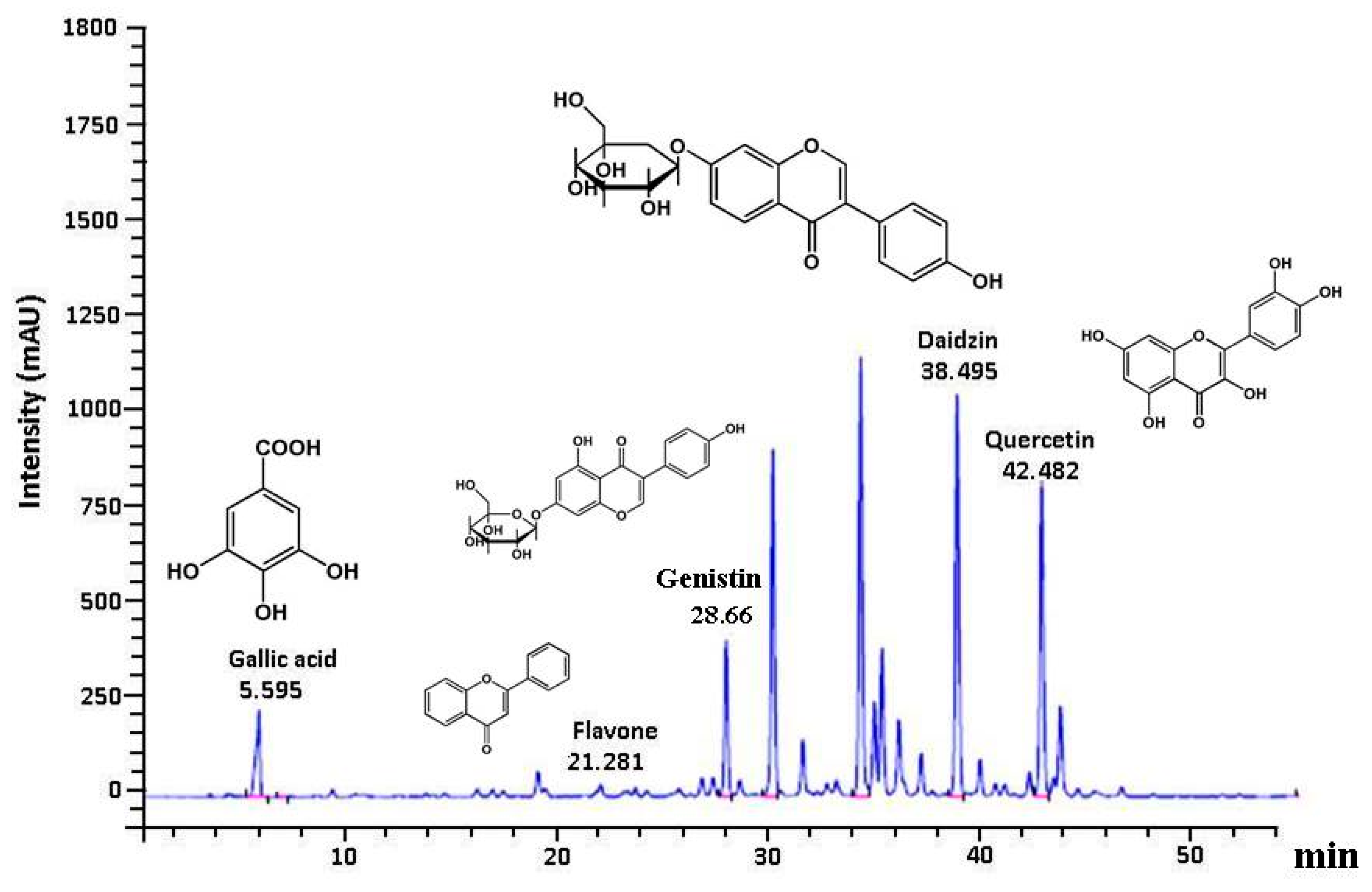
3.6. Polyphenolic Profile in GE50

The HPLC chromatogram showed that gallic acid, quercetin, genistin, and daidzin were the major components among organic molecules found in GE50, which had maximum absorbance at 270 nm. G. paraguayense E. Walther mainly contained flavonoids that were identified in our study (Figure 4). It has also been reported that G. paraguayense E. Walther contains various antioxidants, such as gallic acid, quercetin, genistin, and daidzin [16,17]. The protective effects of GE50 may be attributed to the presence of polyphenolic compounds such as gallic acid, flavone, genistin, daidzin, and quercetin. The pharmacological fundamental constituents of the plant are flavonoids.

4. Discussion

CCl4 is a typical hepatotoxic substance, and its mechanism of action is complex. CCl4-mediated hepatotoxicity was chosen as the experimental model. Due to ethanol and CCl4 toxicity, relative liver weight in the ethanol + CCl4 (EC)-treated group was higher than that in the control group (Table 1). Liver weight is known to increase due to hepatic damage inflicted by trichloromethyl radical, as well as consequent liver fibrosis and hypertrophy. Changes in body and liver weight after ethanol and CCl4 intoxication provides direct evidence of the overall hepatic damage [28,29]. The liver, the largest and the most metabolically complex organ in the body, is involved in the storage and biosynthesis metabolism. It is also responsible for detoxification and metabolic homeostasis. Ethanol and CCl4 resulted in loss of body weight, which is considered a putative indicator of health. We demonstrated that GE50 markedly ameliorated ethanol + CCl4 (EC)-induced chronic hepatitis in test rats, accompanied by reduced relative liver weight. Similar results were obtained when the rats were treated with silymarin, a commercial hepatoprotective agent. Silymarin was used as the positive control in this study.
AST and ALT are aminotransferases linked with liver parenchymal cells. If the hepatocellular plasma membrane is damaged, these will leak out from the cytosol into the bloodstream. Serum AST and ALT levels markedly increased in the ethanol + CCl4 (EC)-treated group, indicating altered permeability of membranes and hepatotoxicity. The results revealed that the serum AST and ALT levels significantly decreased after treatment with GE50. These results demonstrate the hepatoprotective effect of GE50 against ethanol + CCl4-induced liver injury in rats. Damage to the liver cells results in elevations of the both ALT and AST, which have been widely accepted as major biomarkers to assess the hepatic injury [30].
Alcoholic hepatitis should at least include inflammation, steatosis, fibrosis, and cell damage. The activity of alcohol dehydrogenase and aldehyde dehydrogenase causes a reduction in NAD+/NADH ratio, which is the process that causes a reduced mitochondrial capacity to metabolize lipids [11,31]. Otherwise, CCl4-induced liver injury is characterized by oxidative stress and activated hepatic macrophage, leading to hepatocyte damage and death [32]. Chronic alcohol consumption increases cellular nicotinamide adenine dinucleotide hydrate concentration and acetaldehyde dehydrogenase activity, which leads to severe free fatty acid overload, triglyceride accumulation, and subsequent hepatic steatosis [9]. Liver injury can lead to the transfer of fatty acids to the liver, resulting in an increase in the TG content in the liver. These results demonstrate that GE50 treatment significantly alleviated the degree of liver injury.
Lipid peroxidation is one of the major characteristics of CCl4-induced hepatotoxicity [33]. MDA, the end product of lipid peroxidation, is widely used as a marker of lipid peroxidation injury. Antioxidants, such as N-acetyl-cysteine, α-tocopherol, phenols, selenium, and vitamin C and E, have been proposed and used as therapeutic agents in liver damage [34]. Vitamin E is believed to be the most important lipophilic antioxidant in biological tissues. Cheeseman et al. demonstrated that an increased vitamin E level in liver protects from acute CCl4-induced damage by preventing lipid peroxidation [35].
Astrocytes contain one of the highest cytosolic concentrations of GSH amongst mammalian cells. GSH is the major non-protein thiol that plays a vital role in maintaining the antioxidant defense mechanism in the body. GSH levels in the liver dropped in CCl4-treated mice. The depletion of GSH may also be a consequence of liver damage. Ethanol inhibits the synthesis of reduced GSH. Moreover, acetaldehyde promotes GSH depletion, free radical-mediated toxicity, and lipid peroxidation [36]. The hepatoprotective effects of some compounds, such as silymarin, may be attributable to its ability to increase cellular GSH [37]. The increase in hepatic GSH by GE50 may be one of the ways in which G. paraguayense protects the liver against ethanol and CCl4 co-induced hepatotoxicity in rats.
CCl4 initiates lipid peroxidation, as well as reduces tissue GPx, GR, CAT, and SOD activities. In experimental animals, the induction of an SOD–CAT-insensitive free radical species by diet and alcohol facilitates liver damage [38]. The GST family represents one of the main detoxification systems in the hepatocytes. GST regulates apoptosis by influencing the lipid peroxidation pathway [39]. GPx and GSH are well-known reductants that metabolize toxic chemicals, drugs, and xenobiotics. In general, the liver can combat the free radical damage by biotransformation of these toxic agents in less reactive compounds through cytochrome P-450 and GPx [40]. CAT plays a role in the metabolism of ethanol. In addition to alcohol dehydrogenase and CYP2E1, CAT is involved in the metabolism of ethanol in the body.
The treatment of SD rats with alcohol and CCl4 caused the development of significant hepatocellular damage, as was evident from a marked increase in the serum activities of AST, ALT, and LDH compared with the control rats (Table 2). We observed a large number of inflammatory cell infiltration in the ethanol + CCl4 (EC) co-treatment group (Figure 1). Rats treated with the GE50 had lower concentration of MDA in the liver cells. Alcohol + CCl4 (EC) co-treatment also caused a considerable increase (p < 0.05) in hepatic MDA formation (Figure 2) and simultaneously induced a marked depletion (p < 0.05) in vitamin C, vitamin E, GSH, and SOD, GPx, CAT, and GST levels in the liver (Figure 3) compared with the control rats. Our studies showed a decrease in food intake and increase in oxidative stress in rats co-treated ethanol and CCl4. Our results demonstrated that GE50 significantly enhanced the GSH, SOD, CAT, and GR levels, and may contribute to the important mechanisms underlying the liver preventive effects.
Plant flavonoid compounds are a gifted class of so-called “nutraceuticals” that include the ability to protect hepatic damage [41,42,43]. Phytochemicals are naturally occurring chemicals in plant that promote the prevention and treatment of various diseases. Plants are a good source of useful hepatoprotective agents that can modulate the activities of free radicals [41]. A significant decrease in lipid peroxides in liver tissues following co-treatment with CCl4 and antioxidants indicated the protective effects of polysaccharide from Angelica and Astragalus [44] and Fagonia schweinfurthii [45] through the scavenging of free radicals produced by CCl4. Several reports have shown that Antrodia camphorate, also known as Antrodia cinnamomea (Niuchangchih), is a precious fungus grown in Taiwan. It has been reported to prevent ethanol-, CCl4-, and cytokine-induced liver injury, ameliorate fatty liver and liver fibrosis, and inhibit hepatoma cells [46,47,48]. In our study, GE50 and Niuchangchih were identical to the positive drug Silymarin.
The structural characteristics of plant polyphenols provide them with strong antioxidant and free radical scavenging abilities. Hepatoprotection using edible phytochemicals is a novel strategy for the treatment of various hepatic dysfunctions. Gallic acid (3,4,5-trihydroxybenzoic acid), a phenolic acid with strong antioxidant effect, is abundant in tea leaves, as well as white, red, and black mulberry, blackberry, raspberry, strawberry, dragon fruit, guava, mangosteen, papaya, and other plants. Gallic acid downregulated CYP2E1 expression in liver tissues while increasing SOD activities. These results support its ability to regulate the antioxidant enzymes activities and inhibit lipid peroxidation. Many studies have demonstrated its hepatoprotective effects [49,50,51].
Isoflavones are dietary phytoestrogens occurring naturally in legumes such as soybeans. Two major isoflavones found in soybean are daidzin and genistin, respectively. In soy and soy products, 95–99% of genistein exists in the conjugated form genistin (glycoside). Many studies demonstrated that daidzein and genistein alleviate hepatic damage [52,53].
Quercetin (3,5,7,30,40-pentahydroxyflavone) is a major flavonoid found in fruits, vegetables, and red wine and displays several healthy properties, including antioxidative, anti-inflammatory, anti-apoptotic, and hepatoprotective activities against various hepatic ailments. Several studies examined the protective effects of quercetin on chronic ethanol-induced liver injury [43,54]. Quercetin ameliorated lipid metabolism and ethanol-induced liver damage by inducing antioxidant enzymes, increasing GSH levels, and reducing CYP2E1 activity [55]. Quercetin, a well-known flavonoid, ameliorates CCl4 and ethanol-induced liver injury in vivo by alleviating oxidative stress [56,57,58]. The antioxidant and antifibrotic effect of GE50 may be due to presence of quercetin. In our previous study, we also demonstrated that GE50 contains high levels of quercetin. The major components isolated were quercetin 3-O-[6′’-(3-hydroxyl-3-methylglutaroyl)]-β-d- glucopyranoside and quercetin 3-O-[6´´-(3-hydroxyl-3-methylglutaroyl)-2´´-acetyl]- β-d-glucopyranoside [16]. Quercetin also improves the antioxidant defense mechanism by increasing the levels of enzymatic and nonenzymatic antioxidants in cells, thus reducing oxidative stress.
It is, therefore, reasonable to assume that the hepatoprotective activities of GE50 is attributed to its gallic acid, flavone, genistin, daidzin, and quercetin components that most possibly act synergistically. G. paraguayense is an edible vegetable that has also been used in traditional Taiwanese folk medicine for protection against liver injury.

5. Conclusions

In conclusion, the results of our study indicate that GE50 enhances hepatic antioxidant enzyme activities, as well as inhibits lipid peroxidation in ethanol and CCl4 (EC) co-induced liver injury, and its effect is similar to that of silymarin. The hepatoprotective activity of GE50 is proposed to occur through the synergic effects of its chemical component, namely gallic acid, flavone, genistin, daidzin, and quercetin. These results confirm that the in vivo hepatoprotective activity of GE50 may be associated with the phenolic phytochemicals present in the extract, which are known for their antioxidant potential. GE50 can be used as a functional food or even a pharmacological agent for the prevention of liver diseases.

Author Contributions

Conceived and designed the experiments: S.-T.C.; Performed the experiments: H.-C.P., J.-W.L., S.-J.C.; Analyzed the data: H.-C.P., S.-J.C., W.-W.C.; Wrote the manuscript: W.-W.C.; Reviewed the manuscript and coordinated the submission processes: S.-T.C. All authors had reviewed and approved the final version of manuscript for submission.

Funding

This research received no external funding. The authors are grateful for financial support from the Ministry of Science and Technology of the Republic of China (NSC98-2313-B-235-002-MY3, NSC102-2313-B-126-004-MY3).

Conflicts of Interest

The authors declare no conflict of interest.

References

  1. Zhang, H.Y.; Wang, H.L.; Zhong, G.Y.; Zhu, J.X. Molecular mechanism and research progress on pharmacology of traditional Chinese medicine in liver injury. Pharm. Biol. 2018, 56, 594–611. [Google Scholar] [CrossRef] [PubMed] [Green Version]
  2. Ingawale, D.K.; Mandlik, S.K.; Naik, S.R. Models of hepatotoxicity and the underlying cellular, biochemical and immunological mechanism(s): A critical discussion. Environ. Toxicol. Pharmacol. 2014, 37, 118–133. [Google Scholar] [CrossRef] [PubMed]
  3. Stoyanovsky, D.A.; Cederbaum, A.I. Thiol oxidation and cytochrome P450-dependent metabolism of CCl4 triggers Ca2+ release from liver microsomes. Biochemistry 1996, 35, 15839–15845. [Google Scholar] [CrossRef] [PubMed]
  4. Chao, W.W.; Lin, B.F. Hepatoprotective diterpenoids isolated from Andrographis paniculata. Chin. Med. 2012, 3, 136–143. [Google Scholar] [CrossRef]
  5. Wang, M.Y.; Srinivasan, M.; Dasari, S.; Narvekar, P.; Samy, A.L.P.; Dontaraju, V.S.; Peng, L.; Anderson, G.L.; Munirathinam, G. Antioxidant activity of Yichun Blue Honeysuckle (YBHS) berry counteracts CCl4-induced toxicity in liver injury model of mice. Antioxidants 2017, 6, 50. [Google Scholar] [CrossRef] [PubMed]
  6. Wang, R.; Yang, Z.; Zhang, J.; Mu, J.; Zhou, X.; Zhao, X. Liver injury induced by carbon tetrachloride in mice is prevented by the antioxidant capacity of Anji white tea polyphenols. Antioxidants 2019, 8, 64. [Google Scholar] [CrossRef] [PubMed]
  7. Albano, E. Oxidative mechanisms in the pathogenesis of alcoholic liver disease. Mol. Asp. Med. 2008, 29, 9–16. [Google Scholar] [CrossRef]
  8. Das, M.; Basu, S.; Banerjee, B.; Sen, A.; Jana, K.; Datta, G. Hepatoprotective effects of green Capsicum annum against ethanol induced oxidative stress, inflammation and apoptosis in rats. J. Ethnopharmacol. 2018, 227, 69–81. [Google Scholar] [CrossRef]
  9. Choi, Y.; Abdelmegeed, M.A.; Song, B.J. Preventive effects of indole-3-carbinol against alcohol-induced liver injury in mice via antioxidant, anti-inflammatory, and anti-apoptotic mechanisms: Role of gut-liver-adipose tissue axis. J. Nutr. Biochem. 2018, 55, 12–25. [Google Scholar] [CrossRef]
  10. Hellerbrand, C.; Schattenberg, J.M.; Peterburs, P.; Lechner, A.; Brignoli, R. The potential of silymarin for the treatment of hepatic disorders. Clin. Phytosc. 2016, 2, 7–20. [Google Scholar] [CrossRef]
  11. Federico, A.; Dallio, M.; Loguercio, C. Silymarin/silybin and chronic liver disease: A marriage of many years. Molecules 2017, 22, 191. [Google Scholar] [CrossRef] [PubMed]
  12. Chung, Y.C.; Chen, S.J.; Hsu, C.K.; Chang, C.T.; Chou, S.T. Studies on the antioxidative activity of Graptopetalum paraguayense E. Walther. Food Chem. 2005, 91, 419–424. [Google Scholar] [CrossRef]
  13. Chen, S.J.; Chang, C.T.; Chung, Y.C.; Chou, S.T. Studies on the inhibitory effect of Graptopetalum paraguayense E. Walther extracts on the angiotensin converting enzyme. Food Chem. 2007, 100, 1032–1036. [Google Scholar] [CrossRef]
  14. Chen, S.J.; Chung, J.G.; Chung, Y.C.; Chou, S.T. In vitro antioxidant and antiproliferative activity of the stem extracts from Graptopetalum paraguayense. Am. J. Chin. Med. 2008, 36, 369–383. [Google Scholar] [CrossRef] [PubMed]
  15. Chung, Y.C.; Chou, S.T.; Jhan, J.K.; Liao, J.W.; Chen, S.J. In vitro and in vivo safety of aqueous extracts of Graptopetalum paraguayense E. Walther. J. Ethnopharmacol. 2012, 140, 91–97. [Google Scholar] [CrossRef]
  16. Liu, H.Y.; Peng, H.Y.; Hsu, S.L.; Jong, T.T.; Chou, S.T. Chemical characterization and antioxidative activity of four 3-hydroxyl-3-methylglutaroyl (HMG)-substituted flavonoid glycosides from Graptopetalum paraguayense E. Walther. Bot. Stud. 2015, 56, 8–16. [Google Scholar] [CrossRef]
  17. Kao, T.K.; Ou, Y.C.; Raung, S.L.; Chen, W.Y.; Yen, Y.J.; Lai, C.Y.; Chou, S.T.; Chen, C.J. Graptopetalum paraguayense E. Walther leaf extracts protect against brain injury in ischemic rats. Am. J. Chin. Med. 2010, 38, 495–516. [Google Scholar] [CrossRef]
  18. Lin, Y.L.; Peng, H.Y.; Hsieh, H.M.; Lin, C.H.; Chou, S.T. Effects of Graptopetalum paraguayense consumption on serum lipid profiles and antioxidative status in hypercholesteremic subjects. J. Sci. Food Agric. 2011, 91, 1230–1235. [Google Scholar]
  19. Duh, P.D.; Lin, S.L.; Wu, S.C. Hepatoprotection of Graptopetalum paraguayense E. Walther on CCl4-induced liver damage and inflammation. J. Ethnopharmacol. 2011, 134, 379–385. [Google Scholar] [CrossRef]
  20. Hsu, W.H.; Chang, C.C.; Huang, K.W.; Chen, Y.C.; Hsu, S.L.; Wu, L.C.; Tsou, A.P.; Lai, J.M.; Huang, C.Y. Evaluation of the medicinal herb Graptopetalum paraguayense as a treatment for liver cancer. PLoS ONE 2015, 10, e0121298. [Google Scholar] [CrossRef]
  21. Su, L.J.; Chang, C.C.; Yang, C.H.; Hsieh, S.J.; Wu, Y.C.; Lai, J.M.; Tseng, T.L.; Huang, C.Y.F.; Hsu, S.L. Graptopetalum paraguayense ameliorates chemical-induced rat hepatic fibrosis in vivo and inactivates stellate cells and kupffer cells in vitro. PLoS ONE 2013, 8, e53988. [Google Scholar] [CrossRef] [PubMed]
  22. Chou, S.T.; Lai, C.C.; Lai, C.P.; Chao, W.W. Chemical composition, antioxidant, anti-melanogenic and anti-inflammatory activities of Glechoma hederacea (Lamiaceae) essential oil. Ind. Crops Prod. 2018, 122, 675–685. [Google Scholar] [CrossRef]
  23. Habig, W.H.; Pabst, M.J.; Jakoby, W.B. Glutathione S-transferase: The first enzymatic step in mercapturic acid formation. J. Biol. Chem. 1974, 249, 7130–7139. [Google Scholar] [PubMed]
  24. Mitton, K.P.; Trevithick, J.R. High-performance liquid chromatography-electrochemical detection of antioxidants in vertebrate lens: Glutathione, α-tocopherol and ascorbate. Meth. Enzymol. 1994, 233, 523–539. [Google Scholar] [PubMed]
  25. Chou, S.T.; Ko, L.E.; Yang, C.S. Determination of tissue α- tocopherol in senescence-accelerated mice by high-performance liquid chromatography with fluorimetric detection. Anal. Chim. Acta 2000, 419, 81–86. [Google Scholar] [CrossRef]
  26. Yang, C.S.; Chou, S.T.; Lin, L.; Tsai, P.J.; Kuo, J.S. Effect of ageing on human plasma glutathione concentrations as determined by high-performance liquid chromatography with fluorimetric detection. J. Chromatogr. B 1995, 674, 23–30. [Google Scholar] [CrossRef]
  27. Li, Q.; Jia, Y.; Xu, L.; Wang, X.; Shen, Z.; Liu, Y.; Bi, K. Simultaneous determination of protocatechuic acid, syringin, chlorogenic acid, caffeic acid, liriodendrin and isofraxidin in Acanthopanax senticosus Harms by HPLC-DAD. Biol. Pharm. Bull. 2006, 29, 532–534. [Google Scholar] [CrossRef]
  28. Chang, Y.Y.; Liu, Y.C.; Kuo, Y.H.; Lin, Y.L.; Wu, Y.H.S.; Chen, J.W.; Chen, Y.C. Effects of antrosterol from Antrodia camphorata submerged whole broth on lipid homeostasis, antioxidation, alcohol clearance, and antiinflammation in livers of chronic-alcohol fed mice. J. Ethnopharmacol. 2017, 202, 200–207. [Google Scholar] [CrossRef]
  29. Tiwary, B.K.; Dutta, S.; Dey, P.; Hossain, M.; Kumar, A.; Bihani, S.; Nanda, A.K.; Chaudhuri, T.K. Radical scavenging activities of Lagerstroemia speciosa (L.) Pers. Petal extracts and its hepatoprotection in CCl4-intoxicated mice. BMC Complement. Altern. Med. 2017, 17, 55–67. [Google Scholar] [CrossRef]
  30. Kim, T.W.; Lee, D.R.; Choi, B.K.; Kang, H.K.; Jung, J.Y.; Lim, S.W.; Yang, S.H.; Suh, J.W. Hepatoprotective effects of polymethoxyflavones against acute and chronic carbon tetrachloride intoxication. Food Chem. Toxicol. 2016, 91, 91–99. [Google Scholar] [CrossRef]
  31. Bishayee, A.; Mandal, A.; Atterjee, M. Prevention of alcohol-carbon tetrachloride-induced signs of early hepatotoxicity in mice by Trianthema portulacastrum L. Phytornedicine 1996, 3, 155–161. [Google Scholar] [CrossRef]
  32. McCay, P.B.; Lai, E.K.; Poyer, J.L.; DuBose, C.M.; Janzen, E.G. Oxygen- and carboncentered free radical formation during carbon tetrachloride metabolism. Observation of lipid radicals in vivo and in vitro. J. Biol. Chem. 1984, 259, 2135–2143. [Google Scholar]
  33. Basu, S. Carbon tetrachloride-induced lipid peroxidation: Eicosanoid formation and their regulation by antioxidant nutrients. Toxicology 2003, 189, 113–127. [Google Scholar] [CrossRef]
  34. Turkodgan, M.K.; Agaoglu, Z.; Yener, Z.; Sekeroglu, R.; Akkan, H.A.; Avci, M.E. The role of antioxidant vitamins (C and E), selenium and nigella sativa in the prevention of liver fibrosis and cirrhosis in rabbits: New hopes. Dtsch. Tierarztl. Wochenschr. 2001, 108, 71–73. [Google Scholar]
  35. Cheeseman, K.H.; Davies, M.J.; Emery, S.; Maddix, S.P.; Slater, T.F. Effects of alpha-tocopherol on carbon tetrachloride metabolism in rat liver microsomes. Free Radic. Res. Commun. 1987, 3, 325–330. [Google Scholar] [CrossRef]
  36. McBean, G. Cysteine, glutathione, and thiol redox balance in astrocytes. Antioxidants 2017, 6, 62. [Google Scholar] [CrossRef]
  37. Fraschini, F.; Demartini, G.; Esposti, D. Pharmacology of silymarin. Clin. Drug Investig. 2002, 22, 51–65. [Google Scholar] [CrossRef]
  38. Koch, O.; Farre, S.; De Leo, M.E.; Palozza, P.; Palazzotti, B.; Borrelo, S.; Palombini, G.; Cravero, A.; Galeotti, T. Regulation of manganese superoxide dismutase (MnSOD) in chronic experimental alcoholism: Effects of vitamin E supplemented and -deficient diets. Alcohol Alcohol. 2000, 35, 159–163. [Google Scholar] [CrossRef]
  39. Hayes, P.C.; Bouchier, J.A.D.; Beckett, G.J. Glutathione S-transferase in humans in heath and disease. Gut 1991, 32, 813–818. [Google Scholar] [CrossRef]
  40. Hiraganahalli, D.; Chandrasekaran, C.; Dethe, S.; Mundkinajeddu, D.; Pandre, M.; Balachandran, J. Hepatoprotective and antioxidant activity of standardized herbal extracts. Pharm. Mag. 2012, 8, 116–123. [Google Scholar]
  41. Kerimi, A.; Williamson, G. At the interface of antioxidant signaling and cellular function: Key polyphenol effects. Mol. Nutr. Food Res. 2016, 60, 1770–1788. [Google Scholar] [CrossRef]
  42. Sun, X.; Yamasaki, M.; Katsube, T.; Shiwaku, K. Effects of quercetin derivatives from mulberry leaves: Improved gene expression related hepatic lipid and glucose metabolism in short-term high-fat fed mice. Nutr. Res. Pract. 2015, 9, 137–143. [Google Scholar] [CrossRef] [Green Version]
  43. Miltonprabu, S.; Tomczyk, M.; Skalicka-Woaniak, K.; Rastrelli, L.; Daglia, M.; Nabavi, S.F.; Alavian, S.M.; Nabavi, S.M. Hepatoprotective effect of quercetin: From chemistry to medicine. Food Chem. Toxicol. 2017, 108, 365–374. [Google Scholar] [CrossRef]
  44. Pu, X.; Fan, W.; Yu, S.; Li, Y.; Ma, X.; Liu, L.; Ren, J.; Zhang, W. Polysaccharides from Angelica and Astragalus exert hepatoprotective effects against carbon-tetrachloride-induced intoxication in mice. Can. J. Physiol. Pharmacol. 2015, 93, 39–43. [Google Scholar] [CrossRef]
  45. Pareek, A.; Godavarthi, A.; Issarani, R.; Nagori, B.P. Antioxidant and hepatoprotective activity of Fagonia schweinfurthii (Hadidi) Hadidi extract in carbon tetrachloride induced hepatotoxicity in HepG2 cell line and rats. J. Ethnopharmacol. 2013, 150, 973–981. [Google Scholar] [CrossRef]
  46. Ao, Z.H.; Xu, Z.H.; Lu, Z.M.; Xu, H.Y.; Zhang, X.M.; Dou, W.F. Niuchangchih (Antrodia camphorata) and its potential in treating liver diseases. J. Ethnopharmacol. 2009, 121, 194–212. [Google Scholar] [CrossRef]
  47. Li, Z.W.; Kuang, Y.; Tang, S.N.; Li, K.; Huang, Y.; Qiao, X.; Yu, S.W.; Tzeng, Y.M.; Lo, J.Y.; Ye, M. Hepatoprotective activities of Antrodia camphorata and its triterpenoid compounds against CCl4-induced liver injury in mice. J. Ethnopharmacol. 2017, 206, 31–39. [Google Scholar] [CrossRef]
  48. Wu, Y.; Tian, W.J.; Gao, S.; Liao, Z.J.; Wang, G.H.; Lo, J.M.; Lin, P.H.; Zeng, D.Q.; Qiu, D.R.; Liu, X.Z.; et al. Secondary metabolites of petri-dish cultured Antrodia camphorata and their hepatoprotective activities against alcohol-induced liver injury in mice. Chin. J. Nat. Med. 2019, 17, 33–42. [Google Scholar] [CrossRef]
  49. Wang, J.; Tang, L.; White, J.; Fang, J. Inhibitory effect of gallic acid on CCl4-mediated liver fibrosis in mice. Cell Biochem. Biophys. 2014, 69, 21–26. [Google Scholar] [CrossRef]
  50. Chen, Y.; Zhou, Z.; Mo, Q.; Zhou, G.; Wang, Y. Gallic acid attenuates dimethylnitrosamine-induced liver fibrosis by alteration of Smad phosphoisoform signaling in rats. BioMed Res. Int. 2018, 2018, 1682743. [Google Scholar] [CrossRef]
  51. Hsu, W.H.; Liao, S.C.; Chyan, Y.J.; Huang, K.W.; Hsu, S.L.; Chen, Y.C.; Siu, M.L.; Chang, C.C.; Chung, Y.S.; Huang, C.Y.F. Graptopetalum paraguayense inhibits liver fibrosis by blocking TGF-β signaling in vivo and in vitro. Int. J. Mol. Sci. 2019, 20, 2592. [Google Scholar] [CrossRef]
  52. Chiang, C.M.; Wang, D.S.; Chang, T.S. Improving free radical scavenging activity of soy isoflavone glycosides daidzin and genistin by 30-hydroxylation using recombinant Escherichia coli. Molecules 2016, 21, 1723. [Google Scholar] [CrossRef]
  53. Zhao, L.; Wang, Y.; Liu, J.; Wang, K.; Guo, X.X.; Ji, B.; Wu, W.; Zhou, F. Protective effects of genistein and puerarin against chronic alcohol-induced liver injury in mice via antioxidant, anti-inflammatory, and anti-apoptotic mechanisms. J. Agric. Food Chem. 2016, 64, 7291–7297. [Google Scholar] [CrossRef]
  54. Qiu, P.; Dong, Y.; Zhu, T.; Luo, Y.Y.; Kang, X.J.; Pang, M.X.; Li, H.Z.; Xu, H.; Gu, C.; Pan, S.H.; et al. Semen hoveniae extract ameliorates alcohol-induced chronic liver damage in rats via modulation of the abnormalities of gut-liver axis. Phytomedicine 2019, 52, 40–50. [Google Scholar] [CrossRef]
  55. Surapaneni, K.M.; Jainu, M. Comparative effect of pioglitazone, quercetin and hydroxy citric acid on the status of lipid peroxidation and antioxidants in experimental non-alcoholic steatohepatitis. J. Physiol. Pharmacol. 2014, 65, 67–74. [Google Scholar]
  56. Kemelo, M.K.; Pierzynova, A.; Canova, N.K.; Kucera, T.; Farghali, H. The involvement of sirtuin 1 and heme oxygenase 1 in the hepatoprotective effects of quercetin against carbon tetrachloride-induced sub-chronic liver toxicity in rats. Chem. Biol. Interact. 2017, 269, 1–8. [Google Scholar] [CrossRef]
  57. Ma, J.Q.; Li, Z.; Xie, W.R.; Liu, C.M.; Liu, S.S. Quercetin protectsmouse liver against CCl4-induced inflammation by the TLR2/4 and MAPK/NF-κB pathway. Int. Immunopharmacol. 2015, 28, 531–539. [Google Scholar] [CrossRef]
  58. Zakarua, Z.A.; Mahmood, N.D.; Omar, M.H.; Taher, M.; Basir, R. Methanol extract of Muntingia calabura leaves attenuates CCl4-induced liver injury: Possible synergistic action of flavonoids and volatile bioactive compounds on endogenous defence system. Pharm. Biol. 2019, 57, 335–344. [Google Scholar] [CrossRef]
Figure 1. Effects of oral administration of GE50 over six consecutive weeks on histopathological changes in the livers of rats treated with ethanol + CCl4. Hematoxylin/Eosin staining (H&E), 400×. (A) Liver section from the control group demonstrating normal hepatic architecture with intact nucleus (IN) and normal sinusoids (NS); (B) GE50; (C) Section of ethanol + CCl4 (EC)-induced damaged liver showing highly deformed hepatic architecture with fatty lesion due to fatty infiltration (FI) and necrosis (N); (D) EC+ GE50; (E) Liver section from EC + silymarin-treated group showing improved hepatocellular architecture with IN and NS.
Antioxidants 08 00251 g001
Figure 2. Effects of oral administration of GE50 over six consecutive weeks on malondialdehyde (MDA) (A), vitamin C (B), vitamin E (C), glutathione (GSH) (D) levels in the livers of rats treated with ethanol + CCl4. The data are presented as mean ± S.D. of 10 rats. One-way ANOVA and Scheffe’s method were used to analyze the differences between the means. a–c Mean values with different letters in the same row are significantly different (p < 0.05) according to Duncan’s multiple-range test. Control group; GE50 group; EC group: Ethanol + CCl4; EC + GE50 group: Ethanol + CCl4 + GE50; EC + silymarin group: ethanol + CCl4 + silymarin.
Antioxidants 08 00251 g002
Figure 3. Effects of oral administration of GE50 over six consecutive weeks on superoxide dismutase (SOD) (A), glutathione peroxidase (GPx), (B), catalase (CAT), (C), and glutathione S-transferases (GST) (D) activities in the livers of rats treated with ethanol + CCl4. The data are presented as mean ± S.D. of 10 rats. One-way ANOVA and Scheffe’s method were used to analyze the differences between the means. a–c Mean values with different letters in the same row are significantly different (p < 0.05) according to Duncan’s multiple-range test. Control group; GE50 group; EC group: Ethanol + CCl4; EC + GE50 group: Ethanol + CCl4 + GE50; EC + silymarin group: ethanol + CCl4 + silymarin.
Antioxidants 08 00251 g003
Figure 4. HPLC phytochemical profile of GE50 extract detected at 270 nm. GE50 chemical profile was identified by relative retention times using authentic standards. Key to peak identities: Gallic acid, flavone, genistin, daidzin, and quercetin.
Antioxidants 08 00251 g004
Table 1. Effects of oral administration of 50% ethanolic Graptopetalum paraguayense E. Walther (GE50) over six consecutive weeks on body weight gain, feed efficiency, and relative liver weight in rats treated with ethanol + carbon tetrachloride (CCl4).
GroupsDaily Body Weight Gain (g/Day/Rat)Feed Efficiency (g Gain/g Feed)Relative Liver Weight (g/100 g Body Weight)
Control5.58 ± 0.66 a0.21 ± 0.02 a2.96 ± 0.14 c
GE505.92 ± 0.81 a0.21 ± 0.02 ab3.05 ± 0.19 bc
Ethanol + CCl4 (EC)3.56 ± 0.66 c0.15 ± 0.01 c3.30 ± 0.11 a
EC + GE504.68 ± 0.59 b0.20 ± 0.01 b3.10 ± 0.12 b
EC + silymarin4.32 ± 0.74 b0.20 ± 0.02 ab3.17 ± 0.11 b
The data are presented as mean ± S.D. of 10 rats. One-way ANOVA and Scheffe’s method were used to analyze the differences between the means. a–c Mean values with different letters in the same row are significantly different (p < 0.05) according to Duncan’s multiple-range test. Control group; GE50 group, 0.25 g/100 g BW; EC group: Ethanol + CCl4, 1.25 mL 50% ethanol and 0.1 mL 20% CCl4/100 g BW; EC + GE50 group: Ethanol + CCl4 + GE50; EC + silymarin group: ethanol + CCl4 + silymarin, 20 mg/100 g BW.
Table 2. Effects of oral administration of GE50 over six consecutive weeks on serum aspartate aminotransferase (AST), alanine aminotransferase (ALT), and lactate dehydrogenase (LDH) levels, and total antioxidant status (TAS) in rats treated with ethanol + CCl4.
GroupsAST (U/L)ALT (U/L)LDH (U/L)TAS (nmole/L)
Control103.98 ± 14.0 c44.10 ± 5.4 c273.82 ± 94.9 c0.39 ± 0.1 a
GE5097.25 ± 19.8 c44.28 ± 3.8 c218.39 ± 35.2 c0.37 ± 0.1 a
Ethanol + CCl4 (EC)184.40 ± 25.5 a66.89 ± 4.9 a 563.04 ± 103.7 a0.15 ± 0.1 c
EC + GE50110.95 ± 14.0 bc50.01 ± 4.2 b403.20 ± 79.4 b0.28 ± 0.1 b
EC + silymarin122.28 ± 24.2 b42.05 ± 6.6 c347.13 ± 43.9 b0.26 ± 0.1 b
The data are presented as mean ± S.D. of 10 rats. One-way ANOVA and Scheffe’s method were used to analyze the differences between the means. a–c Mean values with different letters in the same row are significantly different (p < 0.05) according to Duncan’s multiple-range test. Control group; GE50 group; EC group: Ethanol + CCl4; EC + GE50 group: Ethanol + CCl4 + GE50; EC + silymarin group: ethanol + CCl4 + silymarin.

