
Flavonoids from Selaginella doederleinii Hieron and Their Antioxidant and Antiproliferative Activities
by
Felix Wambua Muema
1,2,3,4,†, 
Ye Liu
1,2,3,†,
Yongli Zhang
1,2,3,
Guilin Chen
1,2,3 and
Mingquan Guo
1,2,3,4,* 1
CAS Key Laboratory of Plant Germplasm Enhancement and Specialty Agriculture, Wuhan Botanical Garden, Chinese Academy of Sciences, Wuhan 430074, China
2
Sino-Africa Joint Research Center, Chinese Academy of Sciences, Wuhan 430074, China
3
Innovation Academy for Drug Discovery and Development, Chinese Academy of Sciences, Shanghai 201203, China
4
University of Chinese Academy of Sciences, Beijing 100049, China
*
Author to whom correspondence should be addressed.
†
These authors contributed equally to this work.
Antioxidants 2022, 11(6), 1189; https://doi.org/10.3390/antiox11061189
Submission received: 16 May 2022
/
Revised: 2 June 2022
/
Accepted: 9 June 2022
/
Published: 17 June 2022
(This article belongs to the Special Issue Flavonoids and Chronic Diseases)
Abstract
:Selaginella doederleinii Hieron. (S. doederleinii) is a traditional herb that is widely used in China to treat several ailments, but mainly cancer. Studies have been carried out to determine the phytochemicals ascribed to its pharmacological activity. However, both phytochemical and pharmacological profiles have not been fully explored as few compounds have been reported. This study evaluated the flavonoid content of the ethanol extract and its four fractions (petroleum ether, dichloromethane, ethyl acetate, and n-butanol) together with their antioxidant activity (DPPH and FRAP assays). Further, the antiproliferative activity was evaluated. Two new secondary metabolites (1 and 3) were isolated from S. doederleinii, which comprised of an apigenin skeleton with a phenyl attached at C-8 of ring A and an acetyl group. Additionally, other known metabolites 2 and 4–16 were isolated, whereby compounds 2, 4, 5, 8, 12, 15, and 16 were reported for the first time in this species. These compounds were evaluated for their antioxidative potentials by both DPPH and FRAP assays, and for their antiproliferative activities by the MTT assay on three human cancer cell lines: colon cancer (HT-29), cervical cancer (HeLa), and lung cancer (A549). Compound 7 exhibited the best activity on the three cancer cell lines (HT-29, HeLa, A549) by inhibiting the rate of growth of the cancer cells in a dose-dependent manner with IC50 values of 27.97, 35.47, and 20.71 µM, respectively. The structure–activity relationship of the pure compounds was highlighted in this study. Hence, the study enriched both the phytochemical and pharmacological profiles of S. doederleinii.
1. Introduction
Cancer has persistently remained a global health concern by claiming many human lives [1]. In some developed countries, cancer incidences and the rate of mortality for many cancers have been reported to be decreasing. However, in developing countries, both morbidity and mortality rates are escalating at an alarming rate [2]. Screening and developing new anticancer chemotherapeutic drugs have remained an urgent approach in cancer management and mitigation [3]. Additionally, since a number of these phytochemicals’ solubility in water is poor, studies on the administration of plant extracts and pure isolated compounds to the delivery system are imperative. This would amplify their oral bioavailability and control the release of the drug payloads [4].
On the other hand, reactive oxygen species (ROS) are implicated in detrimental body health. They are chemical molecules that enclose oxygen in the form of superoxide hydroxyl radicals, peroxides, singlet oxygen, and hydrogen peroxide, which are generated by biological reactions in humans [5]. The production of ROS is usually in concentrations of picomolar and, when produced in excess amounts, they are neutralized in the body by the antioxidant system [6]. However, sometimes an imbalance between the ROS production and antioxidant protection system happens, hence there is oxidative stress [5], which potentially causes diseases such as diabetes and cancers [7,8]. To mitigate this, the intake of antioxidants that directly scavenge the free radicals or turn on molecules and enzymes that stimulate redox homeostasis can protect body cells from ROS-induced damage [9,10]. Natural antioxidants have increasingly attracted research focus as synthetic antioxidants have been associated with adverse effects on humans [11]. Additionally, anticancer agents derived from natural resources have been developed and approved to treat various types of cancer [12]. Therefore, phytochemicals are plant-based chemical constituents that occur naturally [13]. More than ten thousand phytochemicals have been identified and found to be of health benefits to humans in treating and reducing the risk of infection from several diseases [14,15]. Therefore, many researchers are focusing on naturally occurring phytochemicals for cancer treatment and prevention [3].
Selaginella doederleinii Hieron., commonly referred to as “da ye cai” and “shi shang bai” [16] in China, belongs to the genus Selaginella and family Selaginellaceae [17,18]. In China, the herb is distributed in the Guangxi Zhuang Autonomous Region and Yunnan and Guizhou provinces [19,20]. Traditionally, S. doederleinii has been used to treat cardiovascular disease [21], cancer [22,23], sore throat, and rheumatoid arthritis [24]. The decoction from this plant is normally prepared by boiling the whole plant in water. Owing to its traditional uses, studies evaluating its phytochemical and pharmacological properties have been carried out. The phytochemical studies of this species have revealed that it is composed of mainly biflavonoids [18,25], lignans [21], and alkaloids [26]; as well, Zou et al. [19] reported eight uncommon triflavonoids. Continued phytochemical research has led to the isolation of compounds with a unique apigenin skeleton structure and a phenyl attached at C-8 of ring A of the apigenin skeleton and flavonoids [27]. Pharmacological studies of S. doederleinii have revealed its antiproliferative [28,29,30], antioxidant [31,32], and anti-Alzheimer [19] activities. However, both phytochemical and pharmacological profiles of S. doederleinii have not been fully explored. More studies on the isolation and identification of novel compounds with significant biological activities are regarded as necessary as only a few compounds have been reported.
Therefore, this study aimed to explore the phytochemical constituents of S. doederleinii and evaluate its antioxidant and antiproliferative activities. To this end, the antioxidant potential of the ethanol extract, its fractions, and isolated compounds were evaluated by DPPH (2,2-diphenyl-1-picrylhydrazyl) and FRAP (ferric reducing antioxidant power) assays. The antiproliferative activities for both the ethanol extract, its fractions, and isolated compounds were evaluated by MTT (3-(4, 5-dimethylthiazol-2-yl)-2, 5-diphenyltetrazolium bromide) assay on three different human cancer cell lines: HT-29, Hela, and A549. Additionally, the study highlighted the structure–activity relationship of the evaluated compounds.
2. Materials and Methods
2.1. Plant Materials
S. doederleinii plant material was acquired from Bozhou Dianshitang Pharmaceutical Sales Co., Ltd., which were collected from Zhaotong City (China) and authenticated by Prof. Guangwan Hu from the Key Laboratory of Plant Germplasm Enhancement and Specialty Agriculture of Wuhan Botanical Garden, Chinese Academy of Sciences, Wuhan, China. A botanical specimen (20190710) was deposited at the herbarium of the institute.
2.2. Experimental Reagents and Instruments
Ethanol, petroleum ether, ethyl acetate, dichloromethane, and n-butanol were acquired from SinoPharm Chemical Reagent Co. Ltd. (Shanghai, China) and the HPLC-grade solvents (methanol, formic acid, and acetonitrile) were purchased from TEDIA Company Inc. (Fairfield, CA, USA). Chromatographic gels of ODS (YMC, Tokyo, Japan) and silica gel (Qingdao Marine Chemical Inc., Qingdao, China) were acquired. YMC-Pack ODS-A C18 (YMC, Tokyo, Japan) was used. 1,3,5-tri(2-pyridyl)-2,4,6-triazine (TPTZ), 2,2-diphenyl-1-picrylhydrazyl (DPPH), and vitamin C were purchased from Sigma-Aldrich Co. (St. Louis, MO, USA). Human cancer cell lines, human colon adenocarcinoma (HT-29), human cervical cancer (HeLa), and human lung adenocarcinoma (A549) were procured from American Type Culture Collection (ATCC, Manassas, VA, USA). HPLC Separation and purification were done with an Agilent 1100 series system with a YMC-Pack column (ODS-A, 250 × 10.0 mm I.D). NMR (1D and 2D) analysis were carried out on a Bruker-Avance-600 NMR spectrometer (Bruker, Karlsruhe, Germany). TMS was used as an internal standard. OD values were obtained on a Tecan Infinite M200 PRO multi-functional microplate reader (Männedorf, Switzerland). Ultra-pure water for HPLC was obtained from our laboratory using a Milli-Q system (Millipore, Billerica, MA, USA), Millipore membranes (0.22 µm).
2.3. Extraction and Separation
The dried plant materials (8.0 kg) were extracted by maceration with 75% ethanol (4 times, 3 days/time) at room temperature. The ethanol extract was evaporated under reduced pressure to obtain a residue (638.2 g). The obtained ethanol extract was then suspended in water for liquid–liquid extraction and successively extracted with petroleum ether (PE), dichloromethane (DCM), ethyl acetate (EA), and n-butanol (n-BuOH) to obtain their corresponding fractions.
The DCM extract (169.9 g) was subjected to an MCI gel column (MCI gel, 70–150 µm) to eliminate the dark color by eluting with MeOH-water at a ratio of 50:50, 80:20, and 100:0, and obtained four fractions (Fr. 1–4). Fr. 3 was separated by silica gel column chromatography (200–300 mesh) and passed with PE: DCM (2:1–1:3), 100% DCM, DCM: MeOH (20:1–1:1), and MeOH to obtain 13 fractions: A-M. Fr. G was further separated with MPLC (ODS C18, 5 µm) and eluted with MeOH-water in a ratio from 40:60 to 90:10 for 10.0 mL/min to give 12 subfractions: (Fr. G1-G12). Fr. F4 was separated on RP-HPLC (71% ACN-H2O, 2.5 mL/min, 280 nm) to obtain compound 14 (0.6 mg) and Fr. F5 (79% ACN-H2O, 2.5 mL/min, 280 nm) was separated to obtain compound 5 (0.7 mg). Fr. G4 was separated on RP-HPLC (77% MeOH-H2O, 2.5 mL/min, 280 nm) to obtain 9 peaks, which were further purified (68% ACN-H2O, 2.5 mL/min, 280 nm) to obtain compounds 3 (1.0 mg), 4 (1.0 mg), and 13 (1.4 mg). Fr. G6 was purified by RP-HPLC (72% ACN-H2O, 2.5 mL/min, 280 nm) to give compound 1 (1 mg), 8 (2.1 mg), and 12 (1 mg), and Fr. G7 (70% ACN-H2O, 2.5 mL/min, 280 nm) gave compounds 6 (1 mg), 15 (1.0 mg) and 16 (1.2 mg). Fr. G8 was purified by RP-HPLC (85% MeOH-H2O, 2.5 mL/min, 280 nm) to give compounds 7 (0.8 mg) and 9 (1.8 mg). Fr. G10 was purified (80% ACN-H2O, 2.5 mL/min, 280 nm) to obtain compound 11 (1.9 mg). Fr. H4 was separated (65–70%, MeOH-25 min, 3 mL/min), obtaining compounds 2 (0.8 mg) and 10 (0.5 mg).
2.4. Determination of the Total Flavonoid Content (TFC)
The TFC analysis was evaluated using the colorimetric method as described [33,34], with some modifications. Briefly, 80 µL of a diluted sample solution was mixed with NaNO2 (80 µL 5% w/v) solution and then shaken for 6 min. AlCl3 (80 µL 10% w/v) was added and allowed to stand for 6 min. Then, NaOH (400 µL 4% w/v) solution was added and allowed to react for 15 min. Afterward, the absorbance of the reaction mixture was read at 510 nm with a UV/VIS spectrophotometer (UV-11000, MAPADA, Shanghai, China) with methanol used as the blank. The TFC of each sample was evaluated in triplicate and expressed as rutin equivalents, which were determined from a rutin calibration curve (100–600 µg/mL), and the results were expressed as mg RE/g sample.
2.5. In Vitro Antioxidant Assays
2.5.1. DPPH (2,2-diphenyl-1-picrylhydrazyl) Assay
The DPPH assay of S. doederleinii ethanol extract and four fractions was assessed as described in [35,36], with some minor modifications. Firstly, the DPPH solution was prepared with methanol at a concentration of 0.1 mM. Then, 10 µL of prepared samples and standards (vitamin C and BHT) of 9.375–250 µg/mL were added to 190 µL of the DPPH solution in each well of a 96-well plate. The mixture was shaken and incubated in darkness for 30 min. The absorbance of the reaction mixture was then taken at 517 nm using a multifunctional microplate reader (Tecan, Infinite M20PRO, Switzerland) with methanol being used as the blank. The analysis was done in triplicates and the results were expressed as the inhibition rate (%) and IC50 values. The DPPH radical scavenging activity was then calculated and expressed as follows:
where A0 is the control absorbance and A1 is the sample/standard control absorbance.
DPPH radical scavenging activity (%) = [(A0 − A1/A0)] × 100%
2.5.2. Ferric Reducing Antioxidant Power (FRAP) Assay
This assay was evaluated on the ethanol extract and its fractions (PE, DCM, EA, and n-BuOH) of S. doederleinii according to the reported method, with some slight modifications [37]. Firstly, a working solution, FRAP reagent comprised of 300 mM acetate buffer of pH 3.6, 20 mM FeCl3∙6H2O solution, and 10 mM TPTZ (2,4,6-tri(2-pyridyl)-S-triazine) solution in a ratio of 10:1:1 (v/v/v), was used. The working solution was then heated to 37 °C before use and 190 µL of FRAP working solution was mixed with 10 µL of sample in a 96-well plate. The mixture was then incubated at 37 °C for 10 min. The absorbance of the mixture was recorded by a microplate reader at a wavelength of 593 nm. The tests were done in triplicates and a standard curve was established. Eventually, the antioxidant activities were calculated and expressed as mmol Fe2+/g of the sample.
2.6. Antiproliferative Activity
The antiproliferative activity was performed by the MTT (3-(4, 5-dimethylthiazol-2-yl)-2, 5-diphenyltetrazolium bromide) method [38], with some modifications. Three human cancer cell lines, colon cancer (HT-29), cervical cancer (HeLa), and lung cancer (A549), were tested. The three cells were cultured in Dulbecco’s Modified Eagle’s Medium (DMEM), which was supplemented with 10% fetal bovine serum (FBS). The 90 µL cell suspension was added to each well and then the 96-well cell culture plates were maintained at 37 °C in a 5% CO2 atmosphere for 24 h to culture. Afterwards, 10 µL of samples at different concentrations (final concentration 6.25, 12.5, 25, 50, and 100 µM) were added to the wells in triplicates and the positive control was also set up. After incubating for 48 h, 15 µL of MTT (5 mg/mL) was added to each well and incubated at 37 °C for 4 h. Afterwards, 100 mL of DMSO was then added to each well and shaken for 15 min to dissolve the precipitates formed. The OD value of each well was measured at 590 nm with a microplate spectrophotometer reader (Tecan Infinite M200 PRO, TECAN, Männedorf, Switzerland). Then, the IC50 values were calculated by GraphPad Prism 8.0.1 Software (GraphPad Software Inc., San Diego, CA, USA).
2.7. Statistical Analysis
All the experiments were performed and data were expressed as mean ± standard deviation (SD) of triplicate values. Data analysis was performed by SPSS statistics 22 software (IBM Corporation, New York, NY, USA) using one-way ANOVA Duncan’s multiple range test and the significance difference was considered at p < 0.05. The IC50 values were calculated by GraphPad Prism 8.0 (GraphPad Software Inc, San Diego, CA, USA). Other software used in this study are: Chemoffice 18.0 (CambridgeSoft Corp, Cambridge, MA, USA), Origin 2019b (OriginLab Corporation, Northampton, MA, USA), and MestreNova (Mestrelab Research SL, San Diego, CA, USA).
3. Results and Discussion
3.1. Total Flavonoid Content
With S. doederleinii being used traditionally to treat cancer for decades and flavonoids having been shown as its main active constituents [25], it was necessary to evaluate its total flavonoid content (TFC). The flavonoid content was calculated using the equation, (y = 0.0013x + 0.0146, R2 = 0.9967), which was obtained by the calibration curve and ranged from 340.8 ± 1.0 to 72.2 ± 8.7 mg RE/g—with the dichloromethane fraction expressing the highest content and n-butanol the least, as shown in Figure 1; the order of the other constituents were: (2) ethyl acetate extract, (3) petroleum ether, and (4) crude extract with values of 310.3 ± 3.1, 104.2 ± 2.0, and 84.0 ± 3.6 mg RE/g, respectively. As seen from Figure 1, it could be noted that the TFC values of the DCM and EA fractions were close in range, with DCM being higher by 1.1 times. On the other hand, ethanolic extracts of Selaginella tenera and Selaginella inaequalifolia exhibited slightly higher TFC values of 125.6 ± 4.3 and 138.4 ± 2.1 mg RE/g, respectively [39], compared to our ethanol extract TFC content. The difference in values could be attributed to the extraction methodology and the geographical locations of the two species [40].
3.2. In-Vitro Antioxidant Potential of S. doederleinii Extracts
The ethanol extract and its fractions were evaluated for their scavenging potential and expressed different inhibition percentages. From Figure 2, the EA fraction expressed the highest inhibition percentage (DPPH) at a sample concentration of 250 µg/mL, followed by n-BuOH, DCM, ethanol extract, and PE with 80.9, 79.7, 69.5, 56.6, and 55.4%, respectively. The IC50 values for ethanol extract, its fractions, and positive controls were shown in Table 1. The EA fraction exhibited the best antioxidant activity, followed by DCM according to their IC50 values, while ethanol extract exhibited the lowest activity. However, the FRAP assay indicated that the DCM fraction had the highest reducing ability, followed by the EA fraction with 2.6 ± 0.1 and 1.7 ± 0.0 mmol Fe2+/g, respectively. Our fraction exhibited a slightly lower antioxidant activity compared to the DPPH assay results reported by Wang et al. [32]. In both assays, the ethanol extract and PE fraction exhibited the lowest scavenging activity, while both the DCM and EA fractions depicted strong activities, which were closely attributed with their TFC yields.
3.3. Antiproliferative Activity of S. doederleinii Extracts
The antioxidant assays and TFC values revealed that both DCM and EA were the most active fractions of S. doederleinii when compared to the others. In this regard, both extracts were evaluated for their antiproliferative activity on three cancer cell lines: HT-29, HeLa, and A549 at different concentrations ranging from 12.5 to 200 µg/mL. The inhibition rates are shown in Figure 3, while the IC50 values are shown in Table 2. The inhibition rate of the solvent was almost zero, which confirmed that the solvent used did not influence the cytotoxicity of the samples. Additionally, the toxicity investigation revealed that the solvents did not influence cell viability. The EA fraction exhibited the best antiproliferative activity against the HT-29 and HeLa cell lines by inhibiting the cell growth rate in a dose-dependent manner with IC50 values of 55.6 ± 1.3 and 69.2 ± 1.3 µg/mL, respectively. The DCM fraction exhibited the best activity against the A549 cell line with an IC50 value of 55.9 ± 12.6 µg/mL. Song et al. [41] evaluated the anticancer activities of the extracts of S. doederleinii collected from different provinces in China against the A549 cancer cell line. Comparing the activities of the extracts with those of ours, our DCM extract exhibited better activity than most of the fractions. Our EA fraction exhibited better antiproliferative activity on the HeLa cancer cell line compared with that reported by Wang et al. [42], which had an IC50 value of 76.1 ± 1.9 µg/mL. These results explained the traditional use of S. doederleinii to cure and manage cancers. To this end, flavonoids expressed in the TFC results could be presumed to play a role in the antiproliferative activity of this species (both DCM and EA fractions) by suppressing the formation of cancers that emerge from oxidative stress. Accordingly, for a better understanding and exploration of this species towards cancer, the DCM fraction was selected for isolation work to identify the responsible bioactive phytochemicals.
3.4. Isolation and Structure Elucidation
A phytochemical examination of the DCM fraction of the whole plant of S. doederleinii using different column chromatography yielded two new compounds (1 and 3). Besides the new compounds, 14 other known compounds (Figure 4) were isolated and their chemical structures were determined by comparison of their NMR data (both 1H and 13C), as per existing literature.
Compound 1 was isolated as a yellow amorphous powder. Its molecular formula was deduced as C25H20O8, owing to a molecular ion peak observed at m/z 449.1227 [M + H]+ (calculated for 449.1231) in the HR-ESI-MS, as shown in Figure S2, which was per the 1H NMR and 13C NMR spectroscopic data (Table 3). Compound 1 consisted of a 1,2,5-trisubstituted benzene ring (ring D) at δH 8.13 (1H, dd, J = 8.7, 2.2 Hz, H-4″), 7.93 (1H, d, J = 2.2 Hz, H-6″), and δH 7.20 (1H, d, J = 8.7 Hz, H-3″). An AA′XX′ coupling system signal at δH 7.56 (2H, d, J = 8.9 Hz, H-2′, 6′) and δH 6.93 (2H, d, J = 8.9 Hz, H-3′, 5′) indicated the para-substitution of ring B. Two aromatic singlets were allocated to H-3 and H-6. The aromatic singlet at δH 6.68 was assigned to H-3 as it showed an HMBC correlation (Figure S8) with C-10 (δC 103.8) and C-2 (δC 164.6), and δH 6.60 was assigned to H-6 since H-8 was involved in the linkage between the flavonoid unit and the benzene ring (ring D). This was confirmed by the HMBC correlations from H-6″ (δH 7.93) to C-8 (δC 105.6), as shown in structure 1 in Figure 5. All 25 carbon resonances were resolved in the 13C NMR spectrum (Table 3 and Figure S4) and were further classified by a DEPT spectrum (Figure S5). They were categorized as 3 methyl (oxygenated), 9 methines (unsaturated), and 13 quaternary carbons (2 carbonyl).
The three methoxy groups were assigned to be attached to C-7, C-4′, and C-2″, which were determined by HMBC signals from δH 3.82 to δC 163.3 (C-7), δH 3.86 to δC 164.4 (C-4′), and δH 3.77 to δC 163.1 (C-2″). Besides the tri-substituted benzene ring (ring D), the remaining signals disclosed that Compound 1 had a flavonoid skeleton. The singlet proton at δH 6.60 (H-6) suggested that ring A could be substituted either at C-6 or C-8, and the HMBC studies have shown that the 1,2,5-trisubstituted benzene ring (ring D) was linked to C-8 by the correlation from δH 7.93 to δC 105.6 (C-8). It was concluded that compound 1 was an apigenin derivative and the chemical structure was determined as 3-(5-hydroxy-7-methoxy-2-(4-methoxyphenyl)-4-oxo-4H-chromen-8-yl)-4-methoxybenzoic acid.
Compound 3 was isolated as a yellow amorphous powder. Through HR-ESI-MS (positive ion mode) analysis, a molecular ion peak appeared at m/z 419.1121 [M + H]+ (calculated for [M+H]+ 419.1125) (Figure S10), indicating a molecular formula of C24H18O7 for 3, which was per the 1H NMR and 13C NMR spectroscopic data (Table 3, Figures S11 and S12). The 1H NMR data of 3 displayed the presence of a 1,2,5-trisubstituted benzene moiety (ring D) at δH 8.01 (1H, dd, J = 8.4, 2.3 Hz, H-4″), 7.99 (1H, d, J = 2.2 Hz, H-6″), and 7.08 (1H, d, J = 8.5 Hz, H-3″), and was supported by the corresponding 13C NMR data (Table 3 and Figure S12). The singlet methyl group at δH 2.57 (3H, s, H-8″), along with the carbonyl carbon at δC 199.6, revealed an acetyl group that, based on the HMBC correlation of H-4″ and H-8″ to C-7″ (Figure 5), was attached to C-5″ of ring D. Additionally, an AA′XX′ coupling system signal at δH 7.64 (2H, d, J = 8.9 Hz, H-2′/6′) and 6.94 (2H, d, J = 8.9 Hz, H-3′/5′) indicated the para-substitution of ring B, and the two aromatic singlets at δH 6.68 and 6.40 were assigned to H-3 and H-6, respectively. All 24 carbons were displayed in the 13C NMR spectrum (Figure S12), which included 15 carbons for the apigenin skeleton, 6 for the phenyl (ring D), 2 for the acetyl group at δC 199.6 and 26.9, and 1 for the methoxyl at δC 56.0. The HMBC spectrum (Figure S16) displayed the presence of a C-1″-C-8 linkage in ring D and A by correlations from H-6″ (δH 7.99) to C-8 (δC 105.4), which confirmed that C-8 was the point of attachment of ring D to the apigenin skeleton. A methoxyl group was attached at C-4′ (δC 164.3) in ring B of the apigenin skeleton as it displayed HMBC correlations from δH 3.84 (OMe) to C-4′ (δC 164.3). At C-2″ in phenyl (ring D), a hydroxyl was attached as shown by HMBC correlations from H-4″/6″ (δH 8.01, 7.99) to C-2″ (δC 163.3), as shown in Figure 5 as well as the downfield shift resonance of C-2″ (δC 163.3) by 31.0 ppm. Hence, the structure of compound 3 was characterized as 8-(5-acetyl-2-hydroxyphenyl)-5,7-dihydroxy-2-(4-methoxyphenyl)-4H-chromen-4-one.
Compound 4 was obtained as a yellow amorphous powder and HR-ESI-MS exhibited a molecular peak at m/z 433.1276 [M + H]+ (calculated for 433.1282) with the molecular formula C25H20O7, which corresponded to the 1H NMR and 13C NMR spectroscopic data (Table 3). By comparing the 1H NMR and 13C NMR data of compound 3 to that of 4, it was observed that there was an additional methoxyl that was attached at C-2″ of ring D, according to the HMBC correlations from δH 3.82 to C-2″ (δC 163.3). The structure of compound 4 was thus determined as 8-(5-acetyl-2-methoxyphenyl)-5,7-dihydroxy-2-(4-methoxyphenyl)-4H-chromen-4-one [43]. This is the first time its spectroscopic data and its isolation from natural resources have been reported.
Compound 5 was isolated as a yellow powder. It shared the same skeleton structure with compounds 3 and 4 but with three methoxy groups. 1H NMR and 13C NMR data gave the molecular formula as C26H22O7. The 1H NMR and 13C NMR spectroscopic data (Table 3) of 5 closely resembled that of 4, except that the hydroxyl at C-7 of ring A was substituted by a methoxy group. The HMBC studies of this compound indicated that the three methoxy groups are attached at C-7, C-4′, and C-2″, as indicated in Figure 5. Hence, compound 5 was determined as 8-(5-acetyl-2-methoxyphenyl)-5-hydroxy-7-methoxy-2-(4-methoxyphenyl)-4H-chromen-4-one [43]. The spectroscopic data of 5 is also being reported for the first time in this study, as well as its isolation from natural resources.
The structures of 12 other known compounds, 2 and 6–16, were established by comparison of their spectroscopic data with those reported in the literature as 3-(5,7-dihydroxy-2-(4-methoxy-phenyl)-4-oxo-4H-chromen-8-yl)-4-methoxy-benzoic acid (2) [44], Sequoiaflavone (6) [45], 7,7″-dimethyl ether amentoflavone (7) [22], 2,3-dihydro-4′′′-methyl ether amentoflavone (8) [46], 2,3-dihydro-7,4′-dimethyl ether amentoflavone (9) [47], 2″,3″-Dihydroamentoflavone (10) [48], 4′,4′′′-dimethyl ether robustaflavone (11) [47], 2,3-dihydro-4′-methyl ether robustaflavone (12) [49], 5,4′-dihydroxy-7-methoxyflavone (13) [50], thevetiaflavone (14) [51,52], 2″,3″-dihydrohinokiflavone (15) [53], and 7″-methyl ether tetrahydrohinokiflavone (16) [54].
Biflavonoids are the most common and characteristic compounds of the species S. doederleinii, with a few alkaloids [26], lignan [21], and triflavonoids [19] having been reported. In this study, we isolated five compounds with an apigenin skeleton and a phenyl (ring D) attached at C-8 of the apigenin and an acetyl attached at C-5″ of ring D. In genus Selaginella, many biflavonoids with a C-C interflavonoid connection at C-8 of apigenin have been reported [55]. By keenly observing compounds 1–5, they resemble an amentoflavone (having C3′-C8″ interflavonoid linkage) derivative without the chromone part of the flavonoid I unit. Therefore, these five compounds could have been derived from these kinds of biflavonoids. Compounds with this kind of structure were first reported by Zou et al. [27] in this species. Four biflavonoids (8, 12, 15, and 16) are being reported for the first time in this species. Additionally, compounds 2, 13, and 14 are reported for the first time in this species too.
3.5. Antioxidant Activities of Isolated Compounds from S. doederleinii
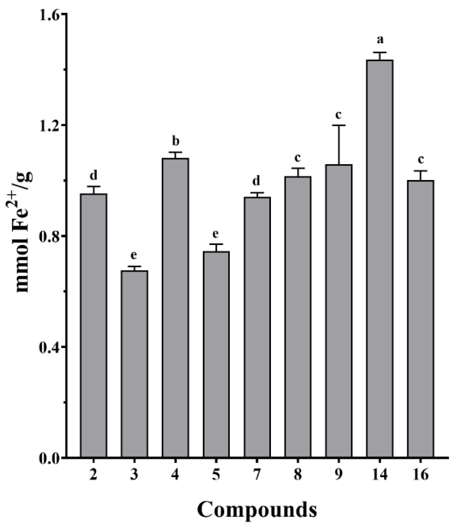
The isolated compounds from S. doederleinii were evaluated for their antioxidant activity by DPPH and FRAP assays. All the examined compounds exhibited radical scavenging abilities at different concentrations with the lowest and highest concentrations of 6.25 and 100 µM, respectively, as shown in Figure 6. Compound 14 expressed the best antioxidant activity among the tested compounds with an IC50 value of 89.3 ± 4.0 µM, while the positive control (Vitamin C) had an IC50 value of 20.3 ± 0.2 µM. The radical scavenging ability of the isolated compounds from S. doederleinii is attributed to the hydroxy groups in their structures, which donate a hydrogen atom to neutralize the free radicals, hence suppressing their oxidation potentials. The tested compounds expressed close free radical scavenging abilities even at the highest concentration of 100 µM, except compound 14 which had a higher value as compared to the rest. The FRAP assay results (Figure 7) also revealed that compound 14 exhibited the highest ferric reducing ability with a value of 1.4 ± 0.03 mM Fe2+/g, followed by compound 4 with a value of 1.1 ± 0.02 mmol Fe2+/g, which also exhibited the second highest DPPH radical scavenging rate at concentration of 100 µM. Vitamin C was used as the positive control on the FRAP assay and it exhibited an ion-reducing capacity with a value of 7.8 ± 1.2 mM Fe2+/g. The antioxidant activity of the isolated compounds from S. doederleinii has not been reported before, hence our work reports this for the first time. Flavonoids derived from plants have been reported to be strong antioxidants [56]. Bedir et al. [44] evaluated the antioxidant activity of flavonoids and four biflavonoids (Amentoflavone, Bilobetin, Ginkgetin, and Sciadopitysin). The flavonoids exhibited noble antioxidant activity. However, none of the four biflavonoids evaluated exhibited strong antioxidant activity. Another study by Orčić et al. [57] revealed low antioxidant activity of biflavonoids isolated from Hypericum perforatum species, whereas the monomer flavonoids exhibited strong antioxidant activities. Previous studies in the same species had reported low antioxidant activities of isolated biflavonoids. This is in support of our findings, whereby flavonoid compound 14 exhibited the strongest antioxidant activity compared to the rest of the tested compounds, which were mainly biflavonoids.
3.6. Antiproliferation Activity of Compounds Isolated from S. doederleinii
All the isolated compounds were evaluated for their antiproliferation activity against three human cancer cell lines: HT-29, HeLa, and A549 by the MTT method. All the compounds showed antiproliferation activity on the three tested cancer cell lines to different degrees. Interestingly, these compounds expressed some level of antiproliferation on cancer cell line A549, which could give an insight into its major traditional use for lung cancer treatment and management. Among the 16 compounds, three (8, 9, 16) expressed the best activity by inhibiting the rate of cell growth in a dose-dependent manner on the three cancer cell lines, and their IC50 values were shown in Table 4. Interestingly, the three were biflavonoids, which have continued to be of interest in the search for cancer drugs [28,58]. Compounds 8 and 16 exhibited noble activities on cancer cell line A549 as compared to their activities on the other cell lines. This affirmed the DCM fraction antiproliferative activity on cancer cell line A549, which exhibited the best activity compared to the other cell lines. Additionally, it supports the main use of this species, which is traditionally in the treatment and management of lung cancer.
3.7. Structure–Activity Relationship of S. doederleinii Phytochemicals
The structure–activity relationship study of our results was interesting, with all compounds exhibiting obvious cytotoxicity on the three cancer cell lines. The two amentoflavone derivatives (8 and 9) exhibited antiproliferation activity, with 8 having the best activity on the HeLa and A549 cancer cell lines and 9 having the best activity on the HT-29 cell line, as shown in Table 4. Compound 8 exhibited better activity than 9, this could be attributed to the OH at C-5,7 of ring A and C-4′ of ring B of the first flavonoid unit as compared to 9, which had OCH3 at C-7,4′. This confirms the importance of OH at C-5,7 of ring A and at C-3′,4′ of ring B [59,60,61]. When comparing the antiproliferation of the hinokiflavone derivatives, compound 16 exhibited more interesting activity than 15 with the best activity on the three human cancer cell lines among the tested compounds. The noble activity of 16 was enhanced by the methoxy group at C-7″ of ring A of the second flavonoid unit. This was in accordance with Du et al. [62], who established that methylation at ring A enhances the antiproliferative activity of flavones.
4. Conclusions
In this study, the TFC, antioxidant (DPPH and FRAP assays), and antiproliferative potentials of the ethanol extract and its fractions were evaluated. The DCM and EA fractions depicted good potency on the three bioassays. The phytochemical investigation was carried out to identify the phytochemicals responsible for its antioxidant and antiproliferative activities. This resulted in the isolation of 16 compounds, including two new compounds (1 and 3). The isolated compounds were then evaluated for their antioxidative and antiproliferative potentials. All the evaluated compounds exhibited some free radical scavenging ability. Compound 14 expressed the best antioxidant activity on the DPPH assay and the highest ferric reducing antioxidant ability on the FRAP assay. The antiproliferative activity was tested by MTT assay on three human cancer cell lines: HT-29, HeLa, and A549. Compound 16 (7″-methyl ether Tetrahydrohinokiflavone) exhibited the strongest activity by inhibiting the rate of cell growth in a dose-dependent manner on the three cancer cell lines. Compounds 8 and 16 exhibited noble antiproliferative activities on the A549 cancer cell line, hence they could be promising lung cancer drug candidates. The study has therefore supported the traditional use of S. doederleinii in cancer treatment and identified the bioactive chemical constituents responsible for its pharmacological properties. Additionally, the study has enriched the phytochemical constitution of S. doederleinii as well as its pharmacological profile. However, we strongly suggest more isolation work to expand the phytochemical profile of this species with new compounds of different classes as it has been reported in other species of the genus Selaginella.
Supplementary Materials
The following supporting information can be downloaded at: https://www.mdpi.com/article/10.3390/antiox11061189/s1. Figure S1. Isolation flow diagram of compounds 1–16, Figure S2. UPLC-QTOF-MS spectrum of compound 1, Figure S3. 1H NMR (600 MHz) spectrum of compound 1 in Methanol-d4, Figure S4. 13C NMR (150 MHz) spectrum of compound 1 in Methanol-d4, Figure S5. DEPT (150 MHz) spectrum of compound 1 in Methanol-d4, Figure S6. HSQC (600 MHz) spectrum of compound 1 in Methanol-d4, Figure S7. 1H-1H COSY (600 MHz) spectrum of compound 1 in Methanol-d4, Figure S8. HMBC (600 MHz) spectrum of compound 1 in Methanol-d4, Figure S9. NOESY (600 MHz) spectrum of compound 1 in Methanol-d4, Figure S10. UPLC-QTOF-MS spectrum of compound 3, Figure S11. 1H NMR (600 MHz) spectrum of compound 3 in Methanol-d4, Figure S12. 13C NMR (150 MHz) spectrum of compound 3 in Methanol-d4, Figure S13. DEPT (150 MHz) spectrum of compound 3 in Methanol-d4, Figure S14. HSQC (600 MHz) spectrum of compound 3 in Methanol-d4, Figure S15. 1H-1H COSY (600 MHz) spectrum of compound 3 in Methanol-d4, Figure S16. HMBC (600 MHz) spectrum of compound 3 in Methanol-d4, Figure S17. NOESY (600 MHz) spectrum of compound 3 in Methanol-d4.
Author Contributions
M.G. conceived of, designed, and supervised the study. F.W.M., Y.L., G.C. and Y.Z. performed the experiments, analyzed the data. F.W.M. wrote the original manuscript. Y.L. revised the manuscript and co-supervised the study. All authors have read and agreed to the published version of the manuscript.
Funding
This research was partly supported by the Natural Science Foundation of Hubei Province, grant number 2020CFB486, to Ye Liu.
Institutional Review Board Statement
Not applicable.
Informed Consent Statement
Not applicable.
Data Availability Statement
Not applicable.
Conflicts of Interest
The authors declare no conflict of interest.
References
- Siegel, R.L.; Miller, K.D.; Jemal, A. Cancer statistics, 2019. CA Cancer J. Clin. 2019, 69, 7–34. [Google Scholar] [CrossRef] [PubMed] [Green Version]
- Arnold, M.; Sierra, M.S.; Laversanne, M.; Soerjomataram, I.; Jemal, A.; Bray, F. Global patterns and trends in colorectal cancer incidence and mortality. Gut 2017, 66, 683. [Google Scholar] [CrossRef] [PubMed] [Green Version]
- Saunders, F.R.; Wallace, H.M. On the natural chemoprevention of cancer. Plant Physiol. Biochem. 2010, 48, 621–626. [Google Scholar] [CrossRef] [PubMed]
- Caddeo, C.; Gabriele, M.; Nácher, A.; Fernàndez-Busquets, X.; Valenti, D.; Maria Fadda, A.; Pucci, L.; Manconi, M. Resveratrol and artemisinin eudragit-coated liposomes: A strategy to tackle intestinal tumors. Int. J. Pharm. 2021, 592, 120083. [Google Scholar] [CrossRef]
- Attia, M.; Essa, E.A.; Zaki, R.M.; Elkordy, A.A. An Overview of the Antioxidant Effects of Ascorbic Acid and Alpha Lipoic Acid (in Liposomal Forms) as Adjuvant in Cancer Treatment. Antioxidants 2020, 9, 359. [Google Scholar] [CrossRef]
- George, S.; Abrahamse, H. Redox Potential of Antioxidants in Cancer Progression and Prevention. Antioxidants 2020, 9, 156. [Google Scholar] [CrossRef]
- Suraweera, L.T.; Rupasinghe, H.P.V.; Dellaire, G.; Xu, Z. Regulation of Nrf2/ARE Pathway by Dietary Flavonoids: A Friend or Foe for Cancer Management? Antioxidants 2020, 9, 973. [Google Scholar] [CrossRef]
- Liguori, I.; Russo, G.; Curcio, F.; Bulli, G.; Aran, L.; Della-Morte, D.; Gargiulo, G.; Testa, G.; Cacciatore, F.; Bonaduce, D. Oxidative stress, aging, and diseases. Clin. Interv. Aging 2018, 13, 757. [Google Scholar] [CrossRef] [Green Version]
- O’Neill, E.J.; Den Hartogh, D.J.; Azizi, K.; Tsiani, E. Anticancer Properties of Carnosol: A Summary of In Vitro and In Vivo Evidence. Antioxidants 2020, 9, 961. [Google Scholar] [CrossRef]
- Zhuang, X.-C.; Chen, G.-L.; Liu, Y.; Zhang, Y.-L.; Guo, M.-Q. New Lignanamides with Antioxidant and Anti-Inflammatory Activities Screened Out and Identified from Warburgia ugandensis Combining Affinity Ultrafiltration LC-MS with SOD and XOD Enzymes. Antioxidants 2021, 10, 370. [Google Scholar] [CrossRef]
- Ullah, R.; Khan, M.; Shah, S.A.; Saeed, K.; Kim, M.O. Natural Antioxidant Anthocyanins—A Hidden Therapeutic Candidate in Metabolic Disorders with Major Focus in Neurodegeneration. Nutrients 2019, 11, 1195. [Google Scholar] [CrossRef] [PubMed] [Green Version]
- Moosavi, M.A.; Haghi, A.; Rahmati, M.; Taniguchi, H.; Mocan, A.; Echeverría, J.; Gupta, V.K.; Tzvetkov, N.T.; Atanasov, A.G. Phytochemicals as potent modulators of autophagy for cancer therapy. Cancer Lett. 2018, 424, 46–69. [Google Scholar] [CrossRef] [PubMed]
- Tuorkey, M.J. Cancer Therapy with Phytochemicals: Present and Future Perspectives. Biomed. Environ. Sci. 2015, 28, 808–819. [Google Scholar] [CrossRef]
- Al-Ishaq, R.K.; Overy, A.J.; Büsselberg, D. Phytochemicals and Gastrointestinal Cancer: Cellular Mechanisms and Effects to Change Cancer Progression. Biomolecules 2020, 10, 105. [Google Scholar] [CrossRef] [Green Version]
- Si, H.; Liu, D. Dietary antiaging phytochemicals and mechanisms associated with prolonged survival. J. Nutr. Biochem. 2014, 25, 581–591. [Google Scholar] [CrossRef] [PubMed] [Green Version]
- Ma, X.K.; Li, X.F.; Zhang, J.Y.; Lei, J.; Li, W.W.; Wang, G. Analysis of the Volatile Components in Selaginella doederleinii by Headspace Solid Phase Microextraction-Gas Chromatography-Mass Spectrometry. Molecules 2020, 25, 115. [Google Scholar] [CrossRef] [Green Version]
- Li, S.; Wang, X.; Wang, G.; Shi, P.; Lin, S.; Xu, D.; Chen, B.; Liu, A.; Huang, L.; Lin, X.; et al. Ethyl Acetate Extract of Selaginella doederleinii Hieron Induces Cell Autophagic Death and Apoptosis in Colorectal Cancer via PI3K-Akt-mTOR and AMPKα-Signaling Pathways. Front. Pharmacol. 2020, 11, 565090. [Google Scholar] [CrossRef]
- Lee, N.Y.; Min, H.Y.; Lee, J.; Nam, J.W.; Lee, Y.J.; Han, A.R.; Wiryawan, A.; Suprapto, W.; Lee, S.K.; Seo, E.K. Identification of a New Cytotoxic Biflavanone from Selaginella doederleinii. Chem. Pharm. Bull. 2008, 56, 1360–1361. [Google Scholar] [CrossRef] [Green Version]
- Zou, Z.; Xu, P.; Zhang, G.; Cheng, F.; Chen, K.; Li, J.; Zhu, W.; Cao, D.; Xu, K.; Tan, G. Selagintriflavonoids with BACE1 inhibitory activity from the fern Selaginella doederleinii. Phytochemistry 2017, 134, 114–121. [Google Scholar] [CrossRef]
- Zhang, Y.; Yang, L.; Wang, L. Identification of Biflavones in Ethyl Acetate Fraction from Ethanol Extract of Selaginella doederleinii Hieron. Adv. Mater. Res. 2012, 550–553, 1862–1865. [Google Scholar] [CrossRef]
- Lin, R.C.; Skaltsounis, A.-L.; Seguin, E.; Tillequin, F.; Koch, M. Phenolic Constituents of Selaginella doederleinii. Planta Med. 1994, 60, 168–170. [Google Scholar] [CrossRef] [PubMed]
- Shim, S.-Y.; Lee, S.-g.; Lee, M. Biflavonoids Isolated from Selaginella tamariscina and Their Anti-Inflammatory Activities via ERK 1/2 Signaling. Molecules 2018, 23, 926. [Google Scholar] [CrossRef] [PubMed] [Green Version]
- Pan, K.-Y.; Lin, J.-L.; Chen, J.-S. Severe Reversible Bone Marrow Suppression Induced by Selaginella doederleinii. J. Toxicol. Clin. Toxicol. 2001, 39, 637–639. [Google Scholar] [CrossRef] [PubMed]
- Liu, H.; Peng, H.; Ji, Z.; Zhao, S.; Zhang, Y.; Wu, J.; Fan, J.; Liao, J. Reactive oxygen species-mediated mitochondrial dysfunction is involved in apoptosis in human nasopharyngeal carcinoma CNE cells induced by Selaginella doederleinii extract. J. Ethnopharmacol. 2011, 138, 184–191. [Google Scholar] [CrossRef] [PubMed]
- Li, S.; Zhao, M.; Li, Y.; Sui, Y.; Yao, H.; Huang, L.; Lin, X. Preparative Isolation of six Anti-Tumour Biflavonoids from Selaginella doederleinii Hieron by High-Speed Counter-Current Chromatography. Phytochem. Anal. 2014, 25, 127–133. [Google Scholar] [CrossRef]
- Chao, L.R.; Seguin, E.; Skaltsounis, A.L.; Tillequin, F.; Koch, M. Synthesis of the glycoalkaloids of Selaginella doederleinii and structure revision of one of them. J. Nat. Prod. 1990, 53, 882–893. [Google Scholar] [CrossRef]
- Zou, Z.X.; Tan, G.-S.; Zhang, G.-G.; Yu, X.; Xu, P.-S.; Xu, K.-P. New cytotoxic apigenin derivatives from Selaginella doederleinii. Chin. Chem. Lett. 2017, 28, 931–934. [Google Scholar] [CrossRef]
- Sui, Y.; Yao, H.; Li, S.; Jin, L.; Shi, P.; Li, Z.; Wang, G.; Lin, S.; Wu, Y.; Li, Y.; et al. Delicaflavone induces autophagic cell death in lung cancer via Akt/mTOR/p70S6K signaling pathway. J. Mol. Med. 2017, 95, 311–322. [Google Scholar] [CrossRef]
- Lin, L.C.; Kuo, Y.C.; Chou, C.J. Cytotoxic Biflavonoids from Selaginella delicatula. J. Nat. Prod. 2000, 63, 627–630. [Google Scholar] [CrossRef]
- Lian, R.; Li, J.; Ma, H.; Zhang, G.; Guo, X.; Li, X.; Yang, J. Effect of ethanol extract of Selaginella doederleinii Hieron on the proliferation of nasopharyngeal carcinoma CNE-1 and C666-1 cells. Afr. J. Tradit. Complement. Altern. Med. 2013, 10, 490–493. [Google Scholar] [CrossRef] [Green Version]
- Li, D.; Qian, Y.; Tian, Y.J.; Yuan, S.M.; Wei, W.; Wang, G. Optimization of Ionic Liquid-Assisted Extraction of Biflavonoids from Selaginella doederleinii and Evaluation of Its Antioxidant and Antitumor Activity. Molecules 2017, 22, 586. [Google Scholar] [CrossRef] [PubMed] [Green Version]
- Wang, G.; Yao, S.; Zhang, X.X.; Song, H. Rapid Screening and Structural Characterization of Antioxidants from the Extract of Selaginella doederleinii Hieron with DPPH-UPLC-Q-TOF/MS Method. Int. J. Anal. Chem. 2015, 2015, 849769. [Google Scholar] [CrossRef] [PubMed] [Green Version]
- Zou, Y.; Chang, S.K.; Gu, Y.; Qian, S.Y. Antioxidant activity and phenolic compositions of lentil (Lens culinaris var. Morton) extract and its fractions. J. Agric. Food Chem. 2011, 59, 2268–2276. [Google Scholar] [CrossRef] [Green Version]
- Ru, Q.M.; Wang, L.J.; Li, W.M.; Wang, J.L.; Ding, Y.T. In Vitro Antioxidant Properties of Flavonoids and Polysaccharides Extract from Tobacco (Nicotiana tabacum L.) Leaves. Molecules 2012, 17, 11281. [Google Scholar] [CrossRef] [PubMed] [Green Version]
- Liu, Y.; Zhang, Y.; Muema, F.W.; Kimutai, F.; Chen, G.; Guo, M. Phenolic Compounds from Carissa spinarum Are Characterized by Their Antioxidant, Anti-Inflammatory and Hepatoprotective Activities. Antioxidants 2021, 10, 652. [Google Scholar] [CrossRef] [PubMed]
- Mutungi, M.M.; Muema, F.W.; Kimutai, F.; Xu, Y.-B.; Zhang, H.; Chen, G.L.; Guo, M.Q. Antioxidant and Antiproliferative Potentials of Ficus glumosa and Its Bioactive Polyphenol Metabolites. Pharmaceuticals 2021, 14, 266. [Google Scholar] [CrossRef] [PubMed]
- Xu, Y.-B.; Chen, G.L.; Guo, M.Q. Antioxidant and Anti-Inflammatory Activities of the Crude Extracts of Moringa oleifera from Kenya and Their Correlations with Flavonoids. Antioxidants 2019, 8, 296. [Google Scholar] [CrossRef] [Green Version]
- Hemlata; Meena, P.R.; Singh, A.P.; Tejavath, K.K. Biosynthesis of Silver Nanoparticles Using Cucumis prophetarum Aqueous Leaf Extract and Their Antibacterial and Antiproliferative Activity Against Cancer Cell Lines. ACS Omega 2020, 5, 5520–5528. [Google Scholar] [CrossRef] [Green Version]
- Sivaraman, A.; Johnson, M.; Parimelazhagan, T.; Irudayaraj, V. Evaluation of antioxidant potential of ethanolic extracts of selected species of Selaginella. NIScPR Online Period. Repos. 2013, 4, 238–244. [Google Scholar]
- Siddhuraju, P.; Becker, K. Antioxidant properties of various solvent extracts of total phenolic constituents from three different agroclimatic origins of drumstick tree (Moringa oleifera Lam.) leaves. J. Agric. Food Chem. 2003, 51, 2144–2155. [Google Scholar] [CrossRef]
- Song, G.; Yao, S.; Cheng, L.; Luo, Y.F.; Song, H. Antioxidant and anticancer effection of the volatile oil from various habitats of Selaginella doederleinii Hieron. Technol. Health Care 2015, 23 (Suppl. S1), S21–S27. [Google Scholar]
- Wang, J.Z.; Li, J.; Zhao, P.; Ma, W.T.; Feng, X.H.; Chen, K.L. Antitumor Activities of Ethyl Acetate Extracts from Selaginella doederleinii Hieron In Vitro and In Vivo and Its Possible Mechanism. Evid. Based. Complement. Alternat. Med. 2015, 2015, 865714. [Google Scholar] [CrossRef] [PubMed] [Green Version]
- Baker, W.; Finch, A.; Ollis, W.; Robinson, K. The structures of the naturally occurring biflavonyls. J. Chem. Soc. (Resumed) 1963, 208, 1477–1490. [Google Scholar] [CrossRef]
- Bedir, E.; Tatli, I.I.; Khan, R.A.; Zhao, J.; Takamatsu, S.; Walker, L.A.; Goldman, P.; Khan, I.A. Biologically Active Secondary Metabolites from Ginkgo biloba. J. Agric. Food Chem. 2002, 50, 3150–3155. [Google Scholar] [CrossRef] [PubMed]
- He, C.W.; Wei, J.H.; Zeng, L.Y.; Deng, J.G. Triterpenoids and Flavonoids from Cassava Leaves. Chem. Nat. Compd. 2020, 56, 331–333. [Google Scholar] [CrossRef]
- Das, B.; Mahender, G.; Koteswara Rao, Y.; Prabhakar, A.; Jagadeesh, B. Biflavonoids from Cycas beddomei. Chem. Pharm. Bull. 2005, 53, 135–136. [Google Scholar] [CrossRef] [Green Version]
- Chen, J.J.; Duh, C.Y.; Chen, J.F. New cytotoxic biflavonoids from Selaginella delicatula. Planta Med. 2005, 71, 659–665. [Google Scholar] [CrossRef]
- Skopp, G.; Schwenker, G. Biflavonoide aus Schinus terebinthifolius Raddi (Anacardiaceae)/Biflavonoids from Schinus terebinthifolius Raddi (Anacardiaceae). Z. Naturforsch. B. 1986, 41, 1479–1482. [Google Scholar] [CrossRef]
- Zheng, J.X.; Wang, N.L.; Liu, H.W.; Chen, H.F.; Li, M.M.; Wu, L.Y.; Fan, M.; Yao, X.S. Four new biflavonoids from Selaginella uncinata and their anti-anoxic effect. J. Asian Nat. Prod. Res. 2008, 10, 945–952. [Google Scholar] [CrossRef]
- Tan, C.X.; Schrader, K.K.; Khan, I.A.; Rimando, A.M. Activities of Wogonin Analogs and Other Flavones against Flavobacterium columnare. Chem. Biodivers. 2015, 12, 259–272. [Google Scholar] [CrossRef]
- Yao, H.; Yuan, Z.; Wei, G.; Chen, C.; Duan, J.; Li, Y.; Wang, Y.; Zhang, C.; Liu, Y. Thevetiaflavone from Wikstroemia indica ameliorates PC12 cells injury induced by OGD/R via improving ROS-mediated mitochondrial dysfunction. Mol. Med. Rep. 2017, 16, 9197–9202. [Google Scholar] [CrossRef] [PubMed] [Green Version]
- Boukaabache, R.; Boubekri, N.; Boumaza, O.; Mekkiou, R.; Seghiri, R.; Sarri, D.; Zama, D.; Benayache, F.; Benayache, S. Phytochemical study of ethyl acetate extract and antioxidant activity of Genista quadriflora Munby (Fabaceae). Der. Pharm. Lett. 2013, 5, 56–59. [Google Scholar]
- Das, B.; Mahender, G.; Rao, Y.K.; Thirupathi, P. A new biflavonoid from Cycas beddomei. Indian J. Chem. 2006, 450, 1933–1935. [Google Scholar]
- Fan, X.; Xu, J.; Lin, X.; Chen, K. Study on biflavonoids from Selaginella uncinata (Desv.) Spring. Chin. Pharm. J. 2009, 44, 15–19. [Google Scholar]
- Setyawan, A.D. Natural products from genus Selaginella (Selaginellaceae). Nusant. Biosci. 2011, 3, 44–58. [Google Scholar] [CrossRef]
- Karak, P. Biological activities of flavonoids: An overview. Int. J. Pharm. Sci. Res. 2019, 10, 1567–1574. [Google Scholar]
- Orčić, D.Z.; Mimica-Dukić, N.M.; Francišković, M.M.; Petrović, S.S.; Jovin, E.Đ. Antioxidant activity relationship of phenolic compounds in Hypericum perforatum L. Chem. Cent. J. 2011, 5, 1–8. [Google Scholar] [CrossRef] [Green Version]
- Goossens, J.-F.; Goossens, L.; Bailly, C. Hinokiflavone and Related C–O–C-Type Biflavonoids as Anti-cancer Compounds: Properties and Mechanism of Action. Nat. Prod. Bioprospect. 2021, 11, 365–377. [Google Scholar] [CrossRef]
- Kothandan, G.; Gadhe, C.G.; Madhavan, T.; Choi, C.H.; Cho, S.J. Docking and 3D-QSAR (quantitative structure activity relationship) studies of flavones, the potent inhibitors of p-glycoprotein targeting the nucleotide binding domain. Eur. J. Med. Chem. 2011, 46, 4078–4088. [Google Scholar] [CrossRef]
- Dao, P.T.A.; Le Quan, T.; Mai, N.T.T. Antioxidant constituents from the stem of Tetrastigma erusbescense Planch (Vitaceae). Nat. Prod. Sci. 2014, 20, 22–28. [Google Scholar]
- López-Posadas, R.; Ballester, I.; Abadía-Molina, A.C.; Suárez, M.D.; Zarzuelo, A.; Martínez-Augustin, O.; Sánchez de Medina, F. Effect of flavonoids on rat splenocytes, a structure–activity relationship study. Biochem. Pharmacol. 2008, 76, 495–506. [Google Scholar] [CrossRef] [PubMed]
- Du, Q.; Chen, H. The methoxyflavones in Citrus reticulata Blanco cv. ponkan and their antiproliferative activity against cancer cells. Food Chem. 2010, 119, 567–572. [Google Scholar]
Figure 1.
The total flavonoids content (TFC) of the ethanol extract and its fractions of S. doederleinii expressed rutin equivalents (RE) of dry weight sample. EE, ethanol extract; PE, petroleum ether; DCM, dichloromethane; EA, ethyl acetate; n-BuOH, n-butanol. All the data were expressed as mean ± standard deviation (n = 3). The letters (a–e) denote that the means are significantly different at a level of p < 0.05 (n = 3) by one-way ANOVA DMRT.
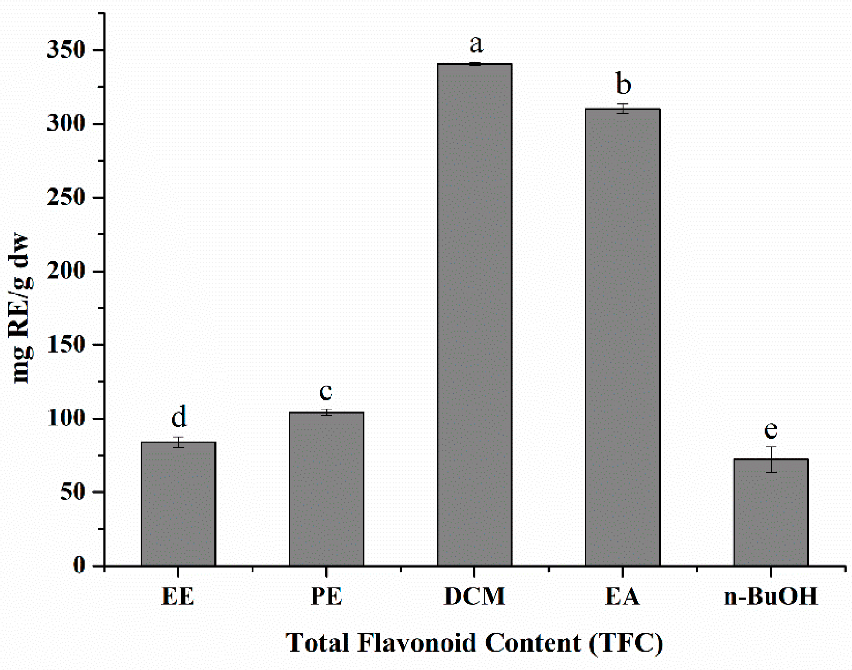
Figure 2.
The radical scavenging percentage of ethanol extract and its PE (petroleum ether) fraction. DCM (dichloromethane), EA (ethyl acetate), and n-BuOH (n-butanol) of S. doederleinii by DPPH assay.
Figure 2.
The radical scavenging percentage of ethanol extract and its PE (petroleum ether) fraction. DCM (dichloromethane), EA (ethyl acetate), and n-BuOH (n-butanol) of S. doederleinii by DPPH assay.
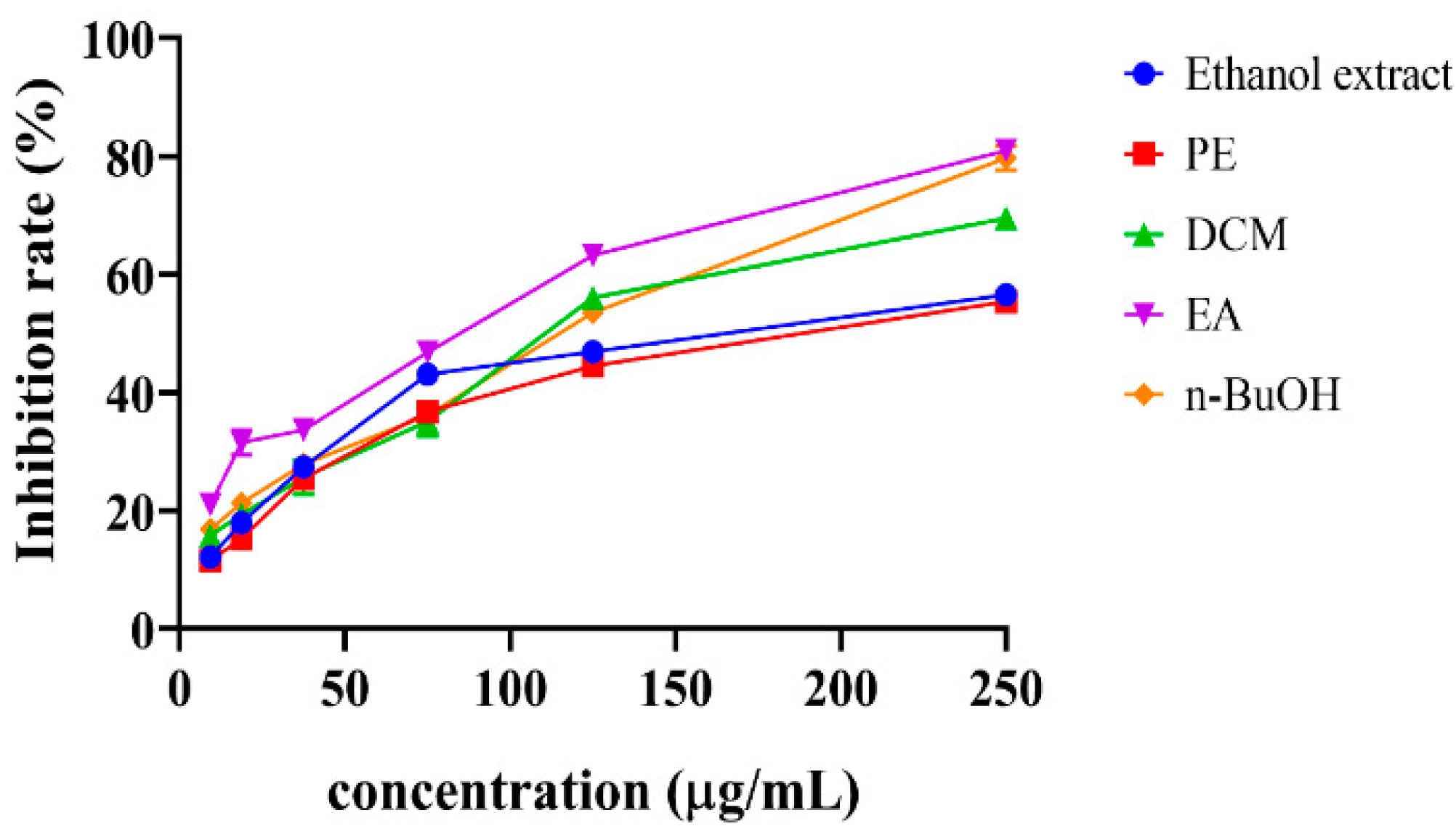
Figure 3.
Antiproliferative activity in inhibition rate (%) at different concentrations of DCM (dichloromethane) and EA (ethyl acetate) fractions, respectively, against cancer cell lines HT-29, Hela, and A549 by the MTT assay. The cell growth inhibition rate of the ethanol extract, petroleum ether, and n-butanol could be expressed at a higher concentration than 200 µg/mL. The solvent’s inhibition rate was near zero value on all the cell lines. The data were expressed as mean ± SD (n = 3).

Figure 4.
Chemical structures of isolated compounds (1–16) from DCM fraction of S. doederleinii.

Figure 5.
Main 1H-1H COSY and HMBC correlations of compounds 1, 3, 4 and 5.

Figure 6.
Free radical scavenging rates of isolated compounds from S. doederleinii by DPPH assay. The positive control used was Vitamin C (VC). Letters (a–f) indicate that the values are significantly different at a level of p < 0.05 (n = 3) by one-way ANOVA DMRT.
Figure 6.
Free radical scavenging rates of isolated compounds from S. doederleinii by DPPH assay. The positive control used was Vitamin C (VC). Letters (a–f) indicate that the values are significantly different at a level of p < 0.05 (n = 3) by one-way ANOVA DMRT.

Figure 7.
Antioxidant activities of isolated compounds from S. doederleinii evaluated by FRAP assay. The data were expressed as means ± SD (n = 3). The mean values denoted with letters (a–e) are significantly different at a level of p < 0.05 (n = 3) by one-way ANOVA DMRT.
Figure 7.
Antioxidant activities of isolated compounds from S. doederleinii evaluated by FRAP assay. The data were expressed as means ± SD (n = 3). The mean values denoted with letters (a–e) are significantly different at a level of p < 0.05 (n = 3) by one-way ANOVA DMRT.

Table 1.
Antioxidant activities of ethanol extract of S. doederleinii and its PE, DCM, EA, and n-BuOH fractions, and positive controls of Vitamin C and BHT evaluated by DPPH and FRAP assays.
Table 1.
Antioxidant activities of ethanol extract of S. doederleinii and its PE, DCM, EA, and n-BuOH fractions, and positive controls of Vitamin C and BHT evaluated by DPPH and FRAP assays.
| Sample | DPPH | FRAP |
|---|---|---|
| IC50 (µg/mL) | mmol Fe2+/g | |
| Ethanol extract | 187.5 ± 1.3 a | 1.1 ± 0.0 c |
| PE | 176.5 ± 0.8 b | 0.9 ± 0.1 c |
| DCM | 110.6 ± 1.3 d | 2.6 ± 0.1 b |
| EA | 82.1 ± 1.1 e | 1.7 ± 0.0 bc |
| n-BuOH | 115.2 ± 1.4 c | 1.6 ± 0.0 bc |
| Vitamin C | 5.8 ± 1.9 f | 7.8 ± 1.2 a |
| BHT | 5.9 ± 1.6 f | Nt |
Data were expressed as means ± standard deviation (n = 3). The mean values denoted by letters (a–f) are significantly different at level p < 0.05 by one-way ANOVA DMRT. Nt denote, not tested.
Table 2.
IC50 values of DCM and EA fractions on HT-29, HeLa, and A549.
| Fraction | IC50 (µg/mL) | ||
|---|---|---|---|
| HT-29 | HeLa | A549 | |
| DCM | 145.4 ± 3.0 | 92.5 ± 0.6 | 55.9 ± 12.6 |
| EA | 55.6 ± 1.3 | 69.2 ± 1.3 | 112.7 ± 6.7 |
Data expressed as means ± standard deviation (n = 3).
Table 3.
1H NMR and 13C NMR data of compounds 1, 3–5.
| 1 | 3 | 4 | 5 | |||||
|---|---|---|---|---|---|---|---|---|
| H/C | δH (J in Hz) | δC | δH (J in Hz) | δC | δH (J in Hz) | δC | δH (J in Hz) | δC |
| 2 | 164.6 | 165.7 | 163.5 | 163.4 | ||||
| 3 | 6.68 1H, s | 103.8 | 6.68 1H, s | 103.9 | 6.67 1H, s | 103.9 | 6.71 1H, s | 103.9 |
| 4 | 184.5 | 184.3 | 179.4 | 179.4 | ||||
| 5 | 163.1 | 162.5 | 164.6 | |||||
| 6 | 6.60 1H, s | 96.4 | 6.40 1H, s | 100.1 | 6.38 1H, s | 99.9 | 6.63 1H, s | 96.4 |
| 7 | 163.3 | 166.7 | 164.4 | 164.4 | ||||
| 8 | 105.6 | 105.4 | 104.6 | 105.7 | ||||
| 9 | 155.5 | 156.5 | 156.8 | 155.5 | ||||
| 10 | 103.8 | 104.3 | 104.9 | 106.4 | ||||
| 1′ | 124.3 | 124.6 | 124.7 | 124.3 | ||||
| 2′/6′ | 7.56 2H, d, J = 8.9 Hz | 129.1 | 7.64 2H, d, J = 8.9 Hz | 129.1 | 7.54 2H, d, J = 8.9 Hz | 129.0 | 7.56 2H, d, J = 8.9 Hz | 129.0 |
| 3′/5′ | 6.93 2H, d, J = 8.9 Hz | 115.5 | 6.94 2H, d, J = 8.9 Hz | 115.5 | 6.93 2H, d, J = 8.9 Hz | 115.5 | 6.94 2H, d, J = 8.9 Hz | 115.5 |
| 4′ | 164.4 | 164.3 | 164.4 | 165.0 | ||||
| 1″ | 122.0 | 119.7 | 122.6 | 122.5 | ||||
| 2″ | 163.1 | 163.3 | 163.3 | 163.3 | ||||
| 3″ | 7.20 1H, d, J = 8.7 Hz | 111.4 | 7.08 1H, d, J = 8.5 Hz | 116.7 | 7.24 1H, d, J = 8.8 Hz | 111.8 | 7.28 1H, d, J = 8.8 Hz | 111.7 |
| 4″ | 8.13 1H, dd, J = 8.7, 2.2 Hz | 132.7 | 8.01 1H, dd, J = 8.4, 2.3 Hz | 130.9 | 8.16 1H, dd, J = 8.7, 2.4 Hz | 131.9 | 8.17 1H, dd, J = 8.7, 2.3 Hz | 131.1 |
| 5″ | 126.2 | 130.1 | 130.9 | 130.9 | ||||
| 6″ | 7.93 1H, d, J = 2.2 Hz | 135.5 | 7.99 1H, d, J = 2.2 Hz | 135.7 | 7.99 1H, d, J = 2.3 Hz | 134.8 | 7.96 1H, d, J = 2.3 Hz | 134.6 |
| 7″ | 165.9 | 199.6 | 196.5 | 199.5 | ||||
| 8″ | 2.57 3H, s | 26.9 | 2.57 3H, s | 26.9 | 2.57 3H, s | 28.1 | ||
| OMe-7 | 3.82 3H, s | 56.0 | 3.84 3H, s | 56.9 | ||||
| OMe-4′ | 3.86 3H, s | 56.8 | 3.84 3H, s | 56.0 | 3.82 3H, s | 56.5 | 3.87 3H, s | 56.5 |
| OMe-2″ | 3.77 3H, s | 56.3 | 3.82 3H, s | 56.0 | 3.82 3H, S | 56.0 | ||
600 MHz 1H-NMR and 150 MHz 13C-NMR data recorded in Methanol-d4.
Table 4.
IC50 values of compounds 8, 9, and 16 on HT-29, HeLa, and A549.
| Compound No. | IC50 (µM) | ||
|---|---|---|---|
| HT-29 | HeLa | A549 | |
| 8 | 56.9 ± 3.4 | 43.5 ± 4.2 | 24.3 ± 1.2 |
| 9 | 44.7 ± 3.2 | >100 | >100 |
| 16 | 27.9 ± 1.0 | 35.5 ± 4.2 | 20.7 ± 3.4 |
Publisher’s Note: MDPI stays neutral with regard to jurisdictional claims in published maps and institutional affiliations. |
© 2022 by the authors. Licensee MDPI, Basel, Switzerland. This article is an open access article distributed under the terms and conditions of the Creative Commons Attribution (CC BY) license (https://creativecommons.org/licenses/by/4.0/).
Share and Cite
MDPI and ACS Style
Muema, F.W.; Liu, Y.; Zhang, Y.; Chen, G.; Guo, M. Flavonoids from Selaginella doederleinii Hieron and Their Antioxidant and Antiproliferative Activities. Antioxidants 2022, 11, 1189. https://doi.org/10.3390/antiox11061189
AMA Style
Muema FW, Liu Y, Zhang Y, Chen G, Guo M. Flavonoids from Selaginella doederleinii Hieron and Their Antioxidant and Antiproliferative Activities. Antioxidants. 2022; 11(6):1189. https://doi.org/10.3390/antiox11061189
Chicago/Turabian StyleMuema, Felix Wambua, Ye Liu, Yongli Zhang, Guilin Chen, and Mingquan Guo. 2022. "Flavonoids from Selaginella doederleinii Hieron and Their Antioxidant and Antiproliferative Activities" Antioxidants 11, no. 6: 1189. https://doi.org/10.3390/antiox11061189
Note that from the first issue of 2016, this journal uses article numbers instead of page numbers. See further details here.