Article

Complete Genome Sequence, Molecular Characterization and Phylogenetic Relationships of a Temminck’s Stint Calicivirus: Evidence for a New Genus within Caliciviridae Family

1
Federal State Budgetary Institution “Centre for Strategic Planning and Management of Biomedical Health Risks” of the Federal Medical Biological Agency, 119121 Moscow, Russia
2
Center of Life Sciences, Skolkovo Institute of Science and Technology, 143026 Moscow, Russia
3
Faculty of Biology, Lomonosov Moscow State University, 119991 Moscow, Russia
*
Author to whom correspondence should be addressed.
Microorganisms 2022, 10(8), 1540; https://doi.org/10.3390/microorganisms10081540
Submission received: 28 June 2022 / Revised: 26 July 2022 / Accepted: 26 July 2022 / Published: 29 July 2022
(This article belongs to the Special Issue Viral Metagenomic Analysis in Animals)

Abstract

:
Caliciviridae is a family of viral pathogens that naturally infects vertebrates, including humans, and causes a range of highly contagious infectious diseases. Caliciviruses are not well studied because of the lack of a universal approach to their cultivation; however, the development of molecular genetics and bioinformatics methods can shed light on their genetic architecture and evolutionary relationships. Here, we present and characterize the complete genome sequence of calicivirus isolated from a sandpiper—Temminck’s stint (Calidris temminckii), preliminarily named Temminck’s stint calicivirus (TsCV). Its genome is a linear, non-segmented, single-stranded (+sense) RNA with genome organization typical of avian caliciviruses. Comparative studies have shown significant divergence of the nucleotide sequence of the TsCV genome, as well as the amino acid sequence of the major capsid protein from all publicly available genomic and protein sequences, with the highest genome sequence similarity to unclassified Ruddy turnstone calicivirus A (43.68%) and the lowest pairwise divergence of the major capsid protein with unclassified goose calicivirus (57.44%). Phylogenetic analysis, as well as a comparative analysis of the homologous proteins, showed evidence of another separate genus within the Caliciviridae family—previously proposed, but not yet accepted by International Committee on Taxonomy of Viruses (ICTV)—the Sanovirus genus, which combines seven previously unclassified genomic sequences of avian caliciviruses, including the newly discovered TsCV, which we propose to consider as a separate species.

1. Introduction

Caliciviruses are small non-enveloped pathogens with a single-stranded RNA genome varying from 6.4 to 8.5 kb in length belonging to the Caliciviridae family [1]. These viruses are known to infect mammals, birds and fish [2,3]. According to the current International Committee on Taxonomy of Viruses (ICTV) report, 11 genera are currently accepted within the Caliciviridae family—Lagovirus, Norovirus, Nebovirus, Recovirus, Sapovirus, Valovirus, Vesivirus, Bavovirus, Nacovirus, Minovirus and Salovirus [4], with each genus including one to two species [5]. The taxonomic classification of Caliciviridae is based on the protein sequence of the major capsid protein (VP1) with isolates with less than 60% sequence identity being assigned to different genera [4]. However, a number of calicivirus isolates currently remain unclassified and several new genera are proposed, including Sanovirus [6] and Secalivirus [7].
The genomic RNA of caliciviruses is organized in one of two ways. In the genomes of representatives of Norovirus, Recovirus and Vesivirus genera, three open reading frames (ORFs) are present. ORF1 of murine norovirus encodes a polyprotein, which is cleaved into six to seven nonstructural proteins (NS): N-term (NS1/2), NTPase (helicase, NS3), 3A-like (NS4), VPg (virion genome-linked protein, NS5), viral protease (NS6) and RNA-dependent RNA polymerase (RdRp, NS7) [8]. Similarities with homologous proteins of other positive-sense single-stranded RNA viruses were used to reveal the functions of some nonstructural proteins of caliciviruses (NTPase, VPg, protease and RdRp) [9]. The calicivirus NTPase participates in viral replication, unwinds dsRNA intermediates, remodels structured RNA and forms vesicular structures for replication [10,11]; the caliciviral VPg is used as primer for the replication of a viral genome in host cells [12]; proteolytic cleavage of the viral polyprotein is performed by the calicivirus protease [13]; and the RdRp replicates the viral genome [14]. A major capsid protein (VP1) is encoded by ORF2 and ORF3 encodes a minor capsid protein (VP2). In murine norovirus, an additional ORF, ORF4, was detected encoding virulence factor 1 (VF1), lying within ORF2 with 1 nucleotide shift [15]. In genera Lagovirus, Nacovirus, Nebovirus, Sapovirus and Valovirus, on the other hand, only two ORFs are present, with VP1 and the nonstructural polyprotein being encoded together by ORF1 and VP2 being encoded by ORF2. The 3′ end of the RNA genome is polyadenylated and the 5′ end is linked to VPg [9].
Cup-shaped depressions located on the capsid surface of caliciviruses are considered to be a unique morphological feature of the group [16]. The capsid of a calicivirus consists of 180 copies of VP1 in three different conformers (A, B and C). A short N-terminal arm, a shell domain and a protruding domain form a mature capsid protein. VP2 is also integrated into the virion, but the copy number is comparatively lower [17].
Birds are known hosts for caliciviruses from genera Bavovirus, Nacovirus and Norovirus. Bird-infecting caliciviruses are pathogens of known importance, since they are able to infect poultry, including chicken [18], turkey [19] and geese [20]. Additionally, birds are already known to be carriers for pathogens that are able to infect humans, such as West Nile virus (WNV) [21,22,23], Japanese encephalitis virus [24] and several subtypes of avian influenza virus [25,26,27]. At least some caliciviruses are suggested to be able to cross the species barrier [28,29], which makes migrating birds a reservoir with potential epidemiological importance. In this study, we have sequenced, assembled and characterized a complete genome of previously undescribed Temminck’s stint calicivirus (TsCV) isolated from Temminck’s stint. The amino acid sequence of the TsCV major capsid protein, which is used for taxonomic classification of the Caliciviridae family, is more than 60% diverged from any other classified caliciviruses, suggesting that the newly identified virus does not belong to any of the ICTV-accepted genera, and shows the highest similarity (57.4%) to currently unclassified goose calicivirus (NCBI accession number KY399947.1), which was previously described as a founding member of the proposed Sanovirus genus [6].

2. Materials and Methods

2.1. Sampling

The sample under the study was collected in 2017 on the banks of the Yenisei River in Krasnoyarsk Region (Russia, Siberia) [30] and belongs to the Temminck’s stint (Calidris temminckii), a small-sized shorebird of the Sandpipers family (Scolopacidae). The biological sample was collected without direct contact with animals; no invasive interventions on animals were carried out. The birds have been observed and identified by qualified zoologists at close distance from a camouflaged hideout. Bird droppings were collected immediately after discharge, taking only the surface part of the fecal pellet to avoid contamination.
To ensure the preservation of the nucleic acids of viral pathogens, fecal samples were placed in sterile tubes containing the transport medium (reagent for transportation and storage of clinical material, Amplisens, Moscow, Russia). After transportation to the laboratory, the sample was stored in a low-temperature refrigerator.

2.2. Sample Preparation and Sequencing

For the extraction of nucleic acids, an Allprep DNA/RNA mini kit (Qiagen, Hilden, Germany) was used. All manipulations were carried out in accordance with the manufacturer’s instructions. Preliminary screening for avian viral pathogens was carried out as described earlier [30].
Library preparation for high-throughput sequencing was performed with a NEBNext Ultra II RNA Library Prep Kit (New England Biolabs, Ipswich, MA, USA) following the manufacturer’s recommendations for partially degraded samples. No additional steps involving depletion and any kind of enrichment were used. Sequencing with MiSeq Reagent Kit v2 (500-cycles) (Illumina, San Diego, CA, USA) on the Illumina MiSeq platform (Illumina, San Diego, CA, USA) resulted in 1.276 M paired-end reads per the sample under the study.

2.3. Assembly and Genome Annotation

SPAdes software v.3.15.3 [31] (CAB SPbU, St. Petersburg, Russia) was used for de novo metagenomic assembly. The main script with standard parameters was used, except for the activation of the “careful” option. The resulting contigs were used for taxonomic classification by nucleotide and translated protein sequences using the BLAST algorithm [32] with Nr/Nt and the NCBI Taxonomy databases [33]. The taxonomic classification of the host was further confirmed by the analysis of contigs related to eukaryotes (Table A1). Contigs attributed to the Caliciviridae family were used to obtain draft whole-genome assembly using the SeqMan NGen program (DNASTAR, Madison, WI, USA).
Additionally, the original raw reads were mapped to the draft whole-genome assembly to perform errors correction. BWA v.0.7.17 [34] was used for used for mapping and Samtools package v.1.10 [35] for operations with sam/bam files. Assembly check and correction was performed using Tablet program v.1.19.09.03 [36]. The quality and integrity of the 3′ end of the assembly was assessed manually.
An NCBI open reading frame finder [37] was used to annotate the open reading frames (ORFs). The following search parameters were used: minimal ORF length 150 amino acids, genetic code 1 and start codon “ATG only”. Protein-coding genes were identified by analyzing homologous protein sequences using the BLAST algorithm [32] with translated nucleotide search. The domain enhanced look-up time accelerated BLAST (DELTA-BLAST) algorithm [38] was used to detect highly distant protein homologues in the absence of significant hits in the standard blast search. Visualization of the annotated sequence and search for characteristic conserved protein motifs were carried out using the SnapGen Viewer software [39] (Dotmatics, Boston, MA, USA).

2.4. Comparative Analysis

The annotated genome assemblies used in the comparative analyses were retrieved from the GenBank database [40] [date of access: 15 May 2022].
The pairwise alignments of whole-genome sequences, and amino acid sequences of annotated major capsid proteins of representative genomes of each Caliciviridae genus (Table A2), unclassified Caliciviridae representatives (Table A3) and TsCV were constructed using MAFFT software [41] for every possible pair of genomes and protein sequences. Pairwise identity for each nucleotide alignment was calculated using DistanceCalculator from Bio.Phylo.TreeConstruction module of BioPython [42] and ‘identity’ model for calculation of nucleotide divergence. Evolutionary divergence between VP1 amino acid sequences was estimated using MEGA11 software [43] with a frequencies model with a gamma distribution of variation including invariant sites, as described in [4].

2.5. Phylogenetic Analysis

To build a phylogenetic tree of representatives of the Caliciviridae family, including TsCV, we used the amino acid sequences of annotated major capsid proteins of the above set of genomes (Table A2 and Table A3). The multiple sequences alignment was performed using MAFFT [41]. The maximum likelihood unrooted tree was generated using RAxML-NG v.1.0.2 according to the recommendations of the ICTV [4] with the only improvement being the choice of an evolutionary model, which was determined using PartitionFinder v.2.1.1 [44] under the corrected Akaike (AICc) and the Bayesian (BIC) information criteria. LG + I + G + F was determined as the best-fitting model. Partial and duplicate sequences (YP_009666353.1, AFH89835.1, YP_009028574.1, AAB60927.1, QXO14962.1, UNY48346.1 and QXO14970.1) have been removed from further analysis. Bootstrapping converged after 650 replicates.
To build a high-resolution phylogenetic tree of TsCV and its closest relatives, we used the nucleotide sequences of the complete genomes of TsCV and a set of the most closely related members of the Caliciviridae family according to the criterion of percent sequence identity (Table A3). This list includes representatives of the genera Bavovirus and Nacovirus, as well as unclassified caliciviruses. The multiple sequences alignment was performed using MAFFT [45]. To eliminate poorly aligned and diverged regions, Gblocks v.0.91b [46] was used with the default parameters. The analysis of possible recombination events was performed using the GARD program implemented in the HyPhy software v.2.5.40 [47]. The maximum likelihood unrooted tree was generated using RAxML-NG v.1.0.2 [48] with GTR + I + G as the most parameter-rich model [49,50]. Bootstrapping converged after 300 replicates. The trees were visualized and rooted in midpoint using iTOL v.6 [51].

2.6. Species Demarcation

We used three approaches that propose de novo species partitions to confirm the species status of the TsCV virus as described previously in [52]: the GMYC [53], bPTP [54] and ASAP [55] methods. Calculations have been carried out for the ICTV set of representative genomes of the Caliciviridae family (Table A2), supplemented with genome sequences of unclassified caliciviruses that are most closely related to TsCV (Table A3). We used the single-threshold version of the GMYC method, since the multiple-threshold version tends to overestimate the number of species partitions [53]. The ultrametric timetree as input tree for GMYC analysis was obtained by applying the RelTime method [56,57] implemented in the MEGA-11 software v.11.0.11 [58] using the appropriate evolutionary model. bPTP analysis was run with default parameters using 500,000 MCMC generations. A matrix of patristic distances as input matrix for ASAP analysis was obtained using the cophenetic.phylo function implemented in the R package ape [59].

2.7. Protein 3D Structure Prediction

To predict 3D structures of TsCV proteins we used a machine learning approach, AlphaFold2, which is able to predict protein structures with an accuracy close to experimental [60]. To build a multiple alignment, we searched for homologues in the following databases: Uniref90 [61], Mgnify [62], BFD [63], UniClust30 [64] and pdb70 [65]. We obtained five relaxed models and five unrelaxed models for each protein ranging by per-residue confidence score (pLDDT). For each protein, the model with the best pLDDT score was chosen for subsequent analysis. Visualization for all individual proteins and structures was performed using UCSF Chimera [66]. Comparison of 3D structures was carried out using the “match maker” function of the UCSF Chimera [66].

3. Results

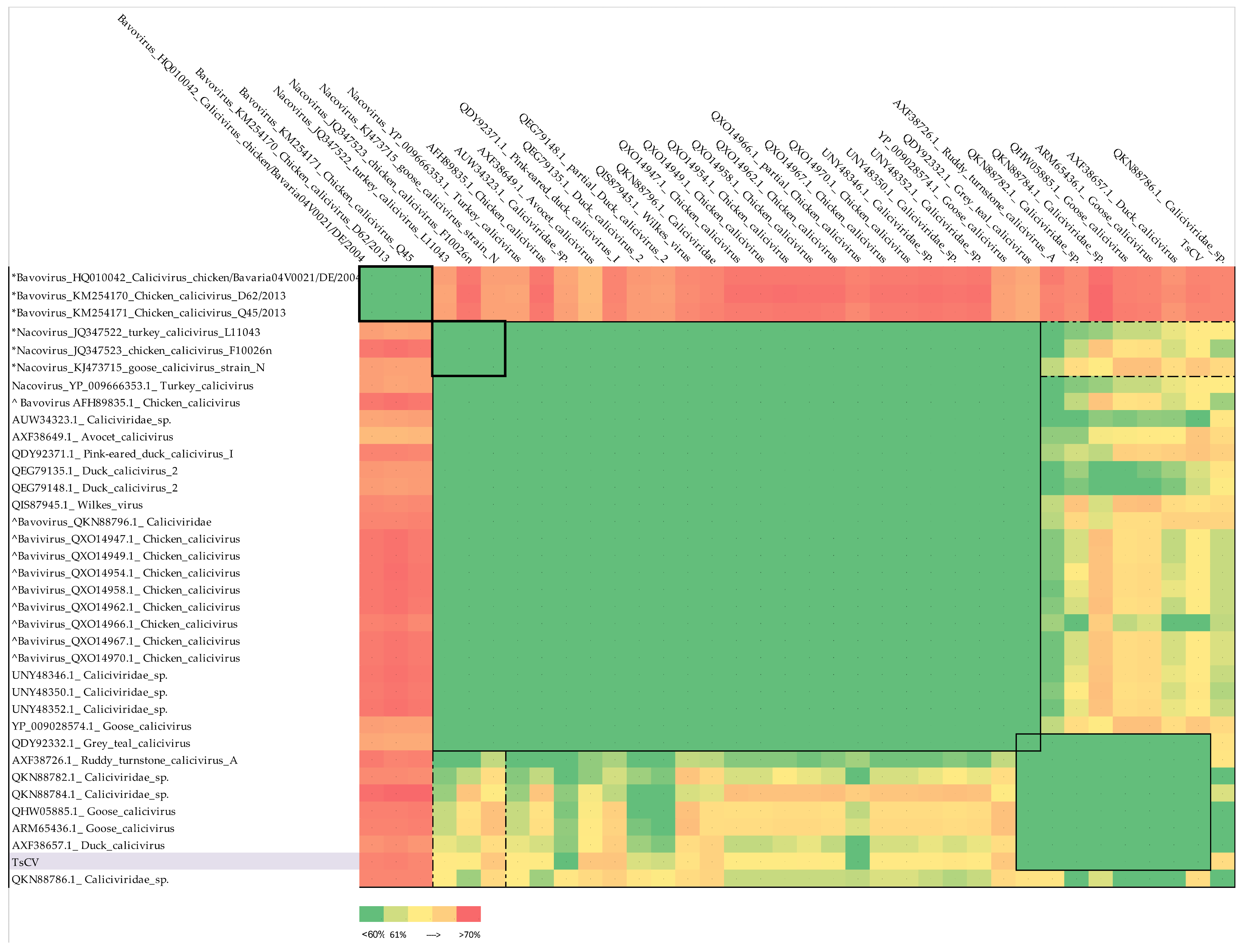
3.1. Annotation of TsCV Genome and Comparative Analysis

The genome of Temminck’s stint calicivirus (TsCV) is a linear, non-segmented, single-stranded positive-sense RNA, comprised of 8575 bases with an average G + C content of 51.73% (Figure 1).
The TsCV genome showed significant divergence from all publicly available genomic sequences with the highest similarity to unclassified Ruddy turnstone calicivirus A (MH453861.1, 43.68%) [67] (Table 1). The G + C proportion is also most similar to the nucleotide composition of the Ruddy turnstone calicivirus A genome. Open reading frames (ORFs) prediction showed genome structure typical of avian caliciviruses—TsCV coding regions are organized into two ORFs: ORF1 of 6849 bases and short ORF2 of 711 bases separated by 1 nucleotide frameshift (Figure 1). However, the relative position of open reading frames differs from its closest relatives—in the TsCv genome, the first and second open reading frames do not overlap, while for the genomes of its closest relatives the overlap is from 18 to 74 nucleotides, according to their annotation (Table 1). In addition, the length of the nucleotide sequence of the ORF1 is noticeably shorter, whereas the non-transcribed regions and the ORF2 sequence show the average lengths.
The ORF1 translation product was identified as polyprotein by analysis of homologous sequences. The protein sequence encoded by ORF2 was identified as VP2 protein using the DELTA-BLAST algorithm and the NCBI’s Conserved Domain Database (the only match was VP2 protein of grey teal calicivirus, QDY92333.1 [68]). The calculated molecular weight of the polyprotein and VP2 were 249.5 and 25.5 kDa, respectively.
ORF1 encoded an immature polyprotein of 2282 aa, which contained characteristic protein motifs conserved in caliciviruses: NTpase/helicase motifs 526GPPGIGKT533 and 603KRKLFTSKLILATTN617; VPg motif 992DEYDTW997; protease motif 1169GDCGLP1174; RdRp motifs 1379KDELL1383, 1453DYSKWDST1460, 1556YGDD1559 and 1603FLKR1606; and VP1 (major capsid protein) motifs 1859PPG1861 and 1944FCLLKEP1950 (Figure 2).
The prediction of cleavage sites was based on the alignment of the amino acid sequence of the polyprotein of TsCV and goose calicivirus (KY399947, [6])—the one fully annotated sequence of the closest relatives to date, caliciviral 3C-like protease cleavage sites preferences [69] and average weights of mature proteins. The cleavage sites of the polyprotein were predicted to be: E370/G, Q811/N, Q971/G, E1047/G and E1736/S. Based on the indicated cleavage sites, the molecular weights of mature proteins were predicted to be 40.7 kDa for Nterm protein (370 aa), 48.5 kDa for NTPase (441 aa), 18 kDa for NS3 protein (160 aa), 8.3 kDa for VPg protein (76 aa), 75.8 kDa for Pro-Pol (689 aa) and 58.2 kDa for major capsid protein (546 aa) (Figure 2).

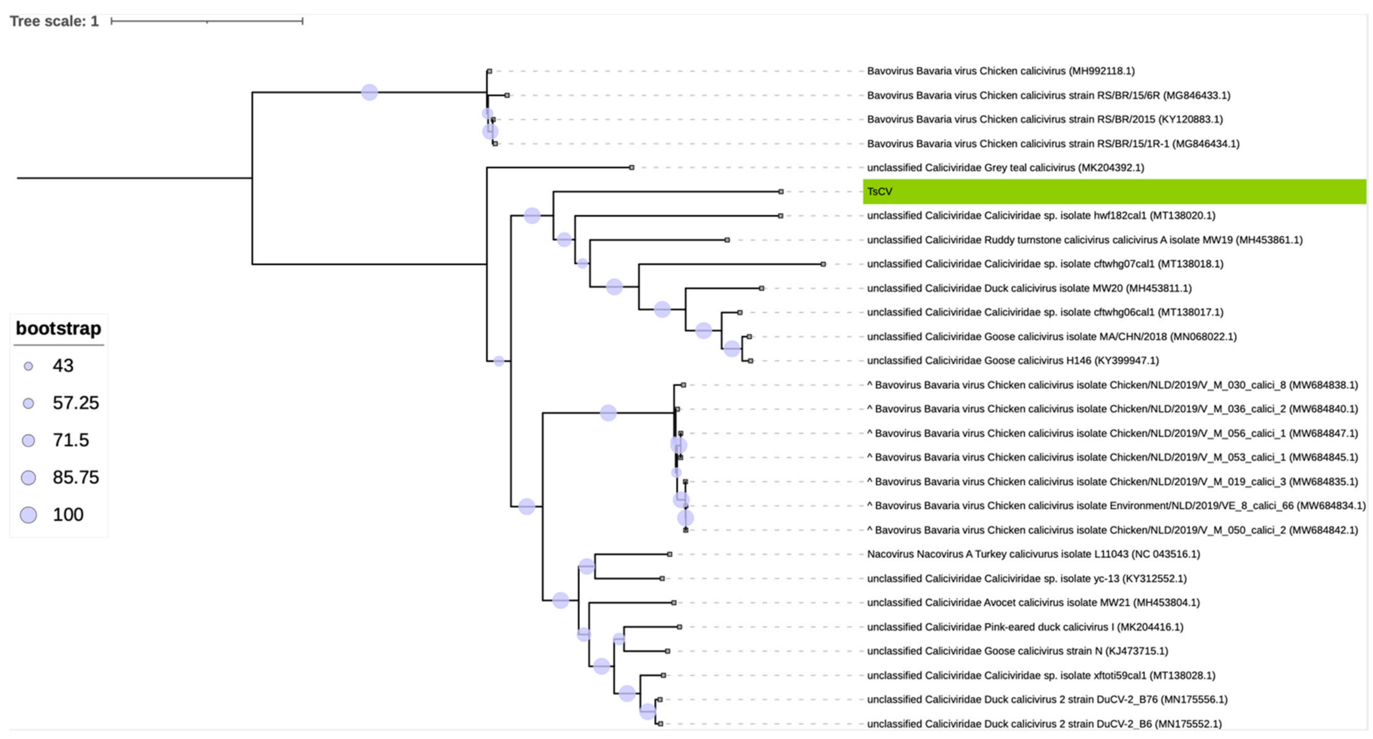
3.2. Taxonomic Classification of TsCV by ICTV Criteria

According to the International Committee on Taxonomy of Viruses (ICTV), in the Caliciviridae family, the amino acid sequence of the major capsid protein (VP1) is used for taxonomic classification wherein the criterion for species demarcation is the divergence of the VP1 amino acid sequence of more than 60% [4].
We calculated the divergence of the VP1 amino acid sequence of TsCV and the VP1 sequences of representatives of each of the accepted genera. A set of representative sequences provided by ICTV was used for calculations (Table A2). Thus, according to the accepted criterion, TsCV cannot be assigned to any of the accepted genera of the Caliciviridae family, since all the given divergence values, with their standard deviations considered, are more than 60% (Table 2). The VP1 sequence of the virus is closest to the representatives of the Nacovirus genus with an average value of 63%. The next in order of increasing degree of divergence is the Bavovirus genus (71%). Both of these genera include avian caliciviruses.
To establish the taxonomic relationship of TsCV with currently known caliciviruses with publicly available major capsid protein sequences, we performed a search for homologous proteins using the BLASTp algorithm and nr database [date of access: 15 May 2022]. List of top BLAST hits is shown in Table A3. This set consisted of unclassified caliciviruses, as well as representatives of the genera Nacovirus and Bavovirus according to the specified taxonomy. From each polyprotein sequence, a VP1 protein region was isolated based on sequence annotation and/or alignment with annotated members of the Nacovirus and Bavovirus genera (listed in the Table A2). Then, identical sequences were filtered out and representative sequences of Bavovirus and Nacovirus genera were added to the analyzed set. After that, the matrix of pairwise divergence of VP1 sequences was calculated, which is presented as a heat map (Figure 3).
Sequences belonging to unclassified caliciviruses are divided into two main clusters: a major one, which also includes representative sequences of Nacovirus genus, and a minor one, which consists entirely of unclassified caliciviruses, but includes the characterized goose calicivirus (KY399947.1, [6]). With this paper, the authors carried out a comparative and phylogenetic study showing that the discovered calicivirus did not belong to any of the accepted genera and proposed a new genus Sanovirus. Our results also support this assumption. Based on the given divergence values, considering the accepted criterion, it can be concluded that:
1.
Goose calicivirus, (ARM65436.1 and QHW05885.1), duck calicivirus (AXF38657.1) and Caliciviridae sp. with accession numbers QKN88782.1, QKN88786.1 and QKN88784.1, as well as TsCV, cannot be assigned to the Nacovirus genus and can be combined into one separate genus with a member of the proposed Sanovirus genus included (the spread of divergence values within the proposed genus is 49.8 ± 7.8%, the divergence with members of the Nacovirus genus is 62.7 ± 1.7%).
2.
Ruddy turnstone calicivirus A (AXF38726.1) has borderline divergence values from representatives of the genus Nacovirus (60.3 ± 0.8%) but is much closer to the proposed genus Sanovirus (values of pairwise divergence with goose calicivirus 53.3% and with putative members 56.1 ± 2.5%).
3.
The inclusion of ruddy turnstone calicivirus A (AXF38726.1) virus in the proposed genus Sanovirus does not violate the demarcation criterion (the spread of divergence values within the proposed genus is 52.1 ± 7.2% and the divergence with members of the Nacovirus genus is 62.3 ± 1.8%).
4.
Grey teal calicivirus (QDY92332.1) cannot be classified according to the accepted criterion, since the divergence values of its VP1 amino acid sequence are less than 60%, both in comparison with representative sequences of Nacovirus and with putative members of the proposed Sanovirus genus.
5.
Chicken caliciviruses accession numbers QXO14947, QXO14949, QXO14954, QXO14958, QXO14962, QXO14966, QXO14967, QXO14970, AFH89835.1 and Caliciviridae sp. QKN88796 appear to be misclassified to the genus Bavovirus and should be moved to the genus Nacovirus.

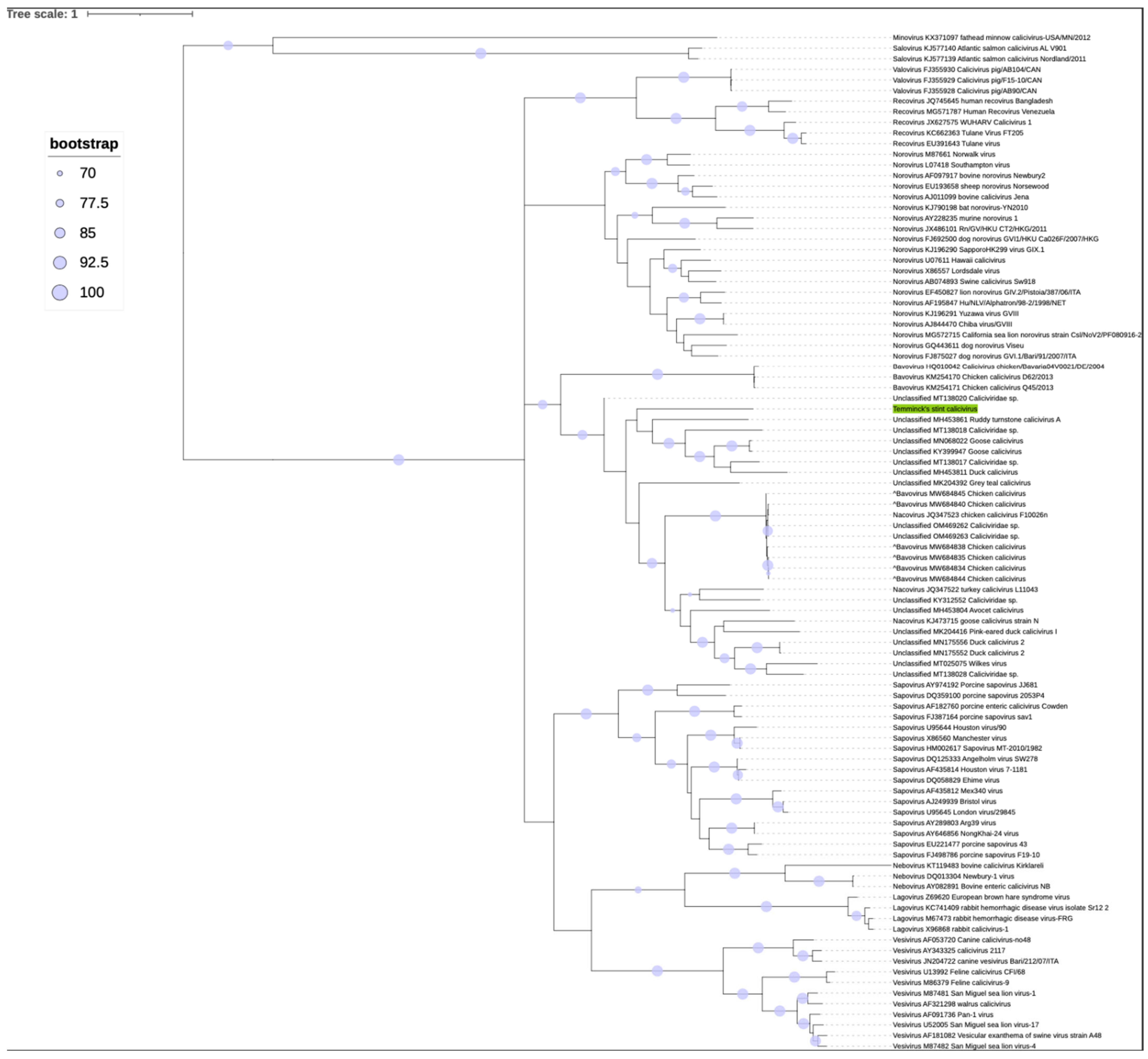
3.3. Phylogenetic Analysis

A phylogenetic analysis was performed to determine the evolutionary relationship between TsCV and other Caliciviridae members. The tree was built on the basis of the amino acid sequences of major capsid proteins in accordance with the ICTV recommendations [4], since this sequence is the gold standard for identifying the Caliciviridae family.
The topology of external nodes of the obtained phylogenetic tree (Figure 4) was strongly supported by bootstrap values. The topology of the tree was consistent with the ICTV phylogeny that is traditionally used to characterize the Caliciviridae family [4]. Genome TsCV was located within the clade containing many unclassified members of the family, as well as members of the genera Bavovirus and Nacovirus. Since all of the characterized members of the clade have been isolated from birds, the clade appears to represent a group of related bird-infecting caliciviruses.
Since the topology in the clade of interest was not supported by high bootstrap values (the bootstrap value of the TsCV branch was 58), which can often be expected when constructing a phylogenetic tree of highly divergent sequences, we constructed a more accurate phylogenetic tree with a higher resolution on the basis of the whole genome nucleotide sequences of the TsCV and its closest relatives (Figure 5). The tree, regardless of the slight differences, is mostly consistent with the topology based on the amino acid sequences of the major capsid protein. Strong bootstrap support for TsCV clustering with other proposed Sanovirus sequences supports our hypothesis.

3.4. Species Demarcation

Various single-locus approaches based on the amino-acid sequence of the major capsid protein have been used to distinguish species. To create species partitions using paired patristic genetic distances, we employed the assemble species by automatic partitioning (ASAP) approach. The partition with the best ASAP score was selected. As a result of applying this method, the studied set of 96 Caliciviridae representatives was partitioned into 69 groups corresponding to different species. In addition, we applied the bPTP web interface, which uses the phylogenetic species concept to delimit species. Using both maximum likelihood and Bayesian approaches, 73 and 68 species partitions were identified, respectively. In both cases, the TsCV formed an independent operational taxonomic unit. Finally, using the GMYC method, 73 species groups with a single-threshold approach were identified (Table A4 contains complete data for the family Caliciviridae).
The result shows that the TsCV genome is not partitioned with other genomes when using any of the listed models. Thus, we have shown the TsCV does not belong to a previously sequenced species of the Caliciviridae family. We propose assigning the TsCV virus to a new, previously undescribed species, preliminarily named Temminck’s stint calicivirus.
All three methods we used unanimously attributed the following groups of caliciviruses to common species:
1.
Chicken calicivirus Q45/2013 (KM254171) and chicken calicivirus D62/2013 (KM254170) belonging the genus Bavovirus;
2.
Bovine enteric calicivirus NB (AY082891) and Newbury-1 virus (DQ013304) belonging the genus Nebovirus;
3.
Chiba virus/GVIII (AJ844470) and Yuzawa virus GVIII (KJ196291) belonging the genus Norovirus;
4.
Calicivirus pig/AB104/CAN (FJ355930), calicivirus_pig/AB90/CAN (FJ355928) and calicivirus_pig/F15-10/CAN (FJ355929) belonging the genus Valovirus;
5.
Sapovirus Angelholm virus SW278 (DQ125333), Ehime_virus (DQ058829) and Houston virus 7-1181 (AF435814) belonging the genus Sapovirus.
6.
NongKhai-24 virus (AY646856) and Arg39 virus (AY289803) belonging the genus Sapovirus;
7.
Sapovirus MT-2010/1982 (HM002617), sapovirus U65427 and Manchester virus (X86560) belonging the genus Sapovirus;
8.
London_virus/29845 (U95645) and Bristol_virus (AJ249939) belonging the genus Sapovirus;
9.
Rabbit_calicivirus-1 (X96868) and rabbit hemorrhagic disease virus-FRG (M67473) belonging the genus Lagovirus;
10.
Unclassified duck calicivirus 2 MN175552 and MN175556;
11.
Unclassified goose calicivirus KY399947 and MN068022;
12.
Chicken calicivirus F10026n (JQ347523, Nacovirus according to the ICTV Caliciviridae report [4]), unclassified Caliciviridae_sp. OM469263 and OM469262 and six chicken caliciviruses strains (MW684845, MW684838, MW684835, MW684844, MW684834 and MW684840) presumably misclassified to the genus Bavovirus.
In addition, according to the species delimitation criteria used in this study, the following pairs of viruses should also be considered as the one species, since their major capsid protein sequences were exactly identical:
1.
Goose calicivirus strain N (KJ473715) belonging the genus Nacovirus according to the ICTV Caliciviridae report [4] and ucclassified goose calicivirus (NC_024078);
2.
Caliciviridae sp. OM469263.1 and OM469260.

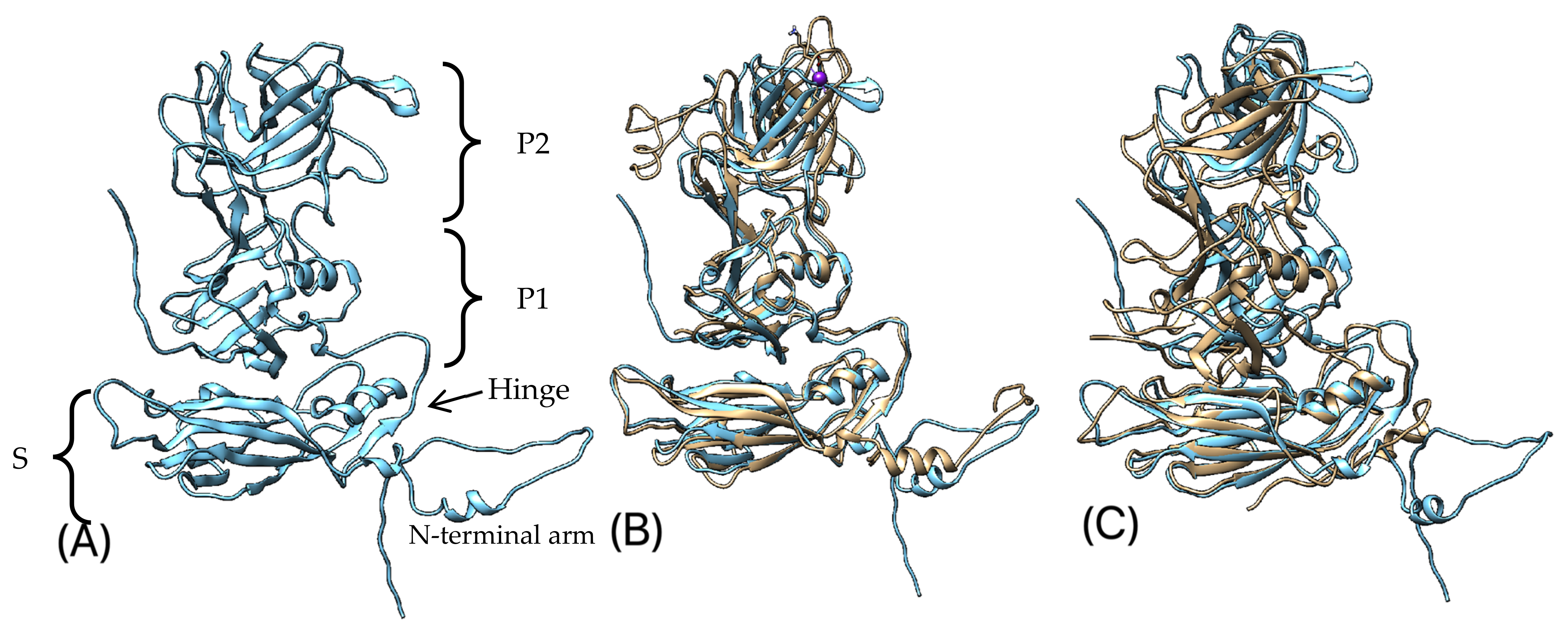
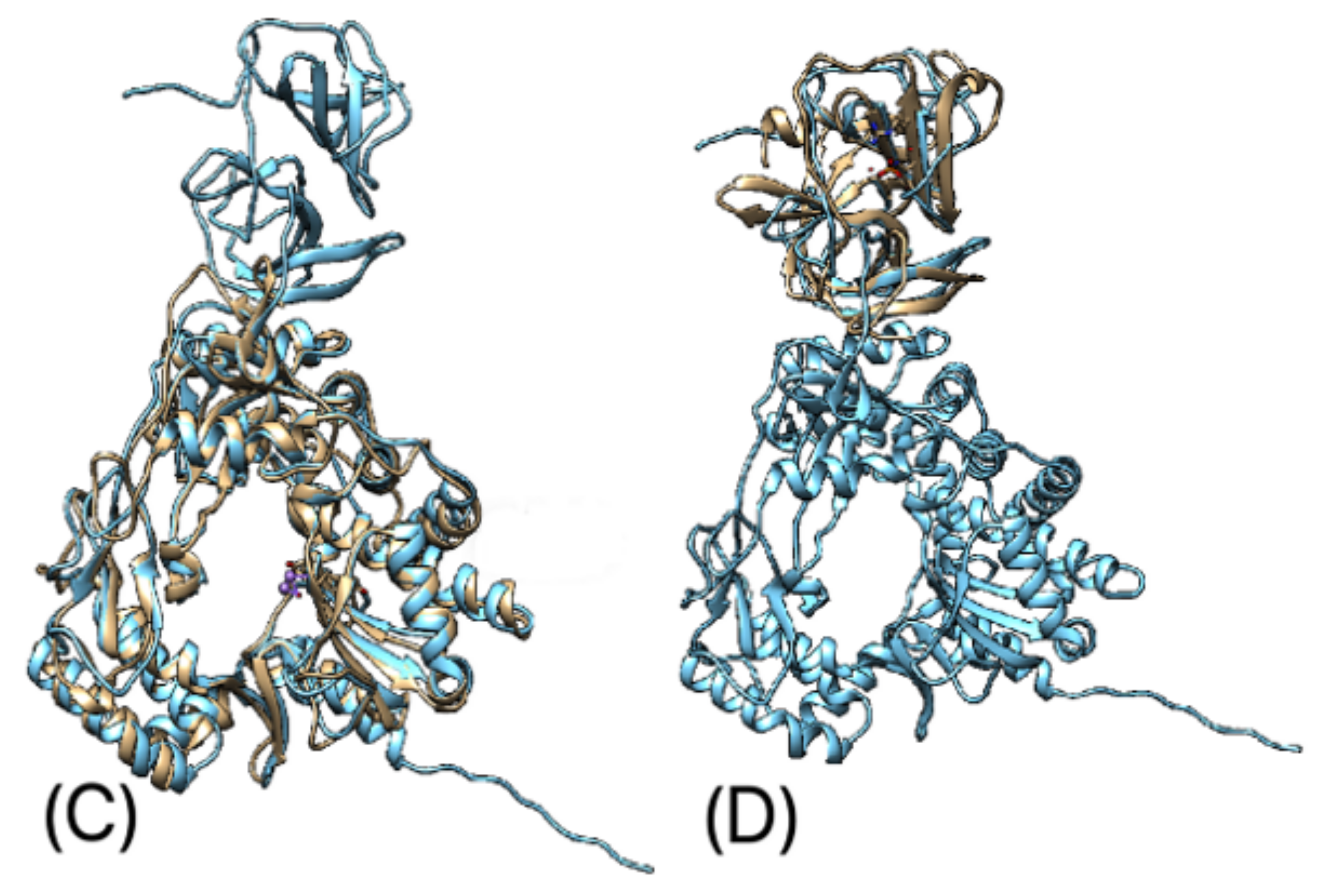
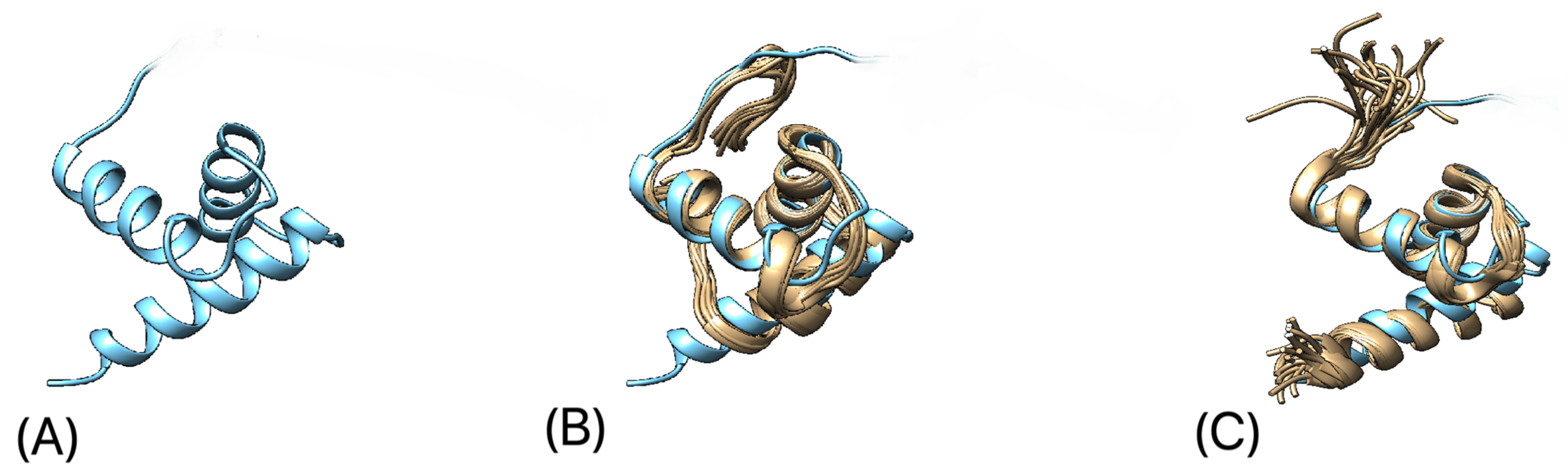
3.5. Protein 3D-Structure Prediction

We predicted the 3D structure of the following TsCV proteins: the VP1 protein, the proteinase–polymerase precursor protein (Pro-Pol) and the core domain of the VPg protein. All predictions were made using the machine learning approach AlphaFold 2 [60] based on the primary sequence of each protein. Of the five models generated for each protein, the one with the highest per-residue confidence score (pLDDT) was selected (Figure 6A, Figure 7A, Figure 8A).
The predicted VP1 protein has a structure typical of the major capsid protein of caliciviruses and consists of short N-terminal arm and two main domains, shell (S) and protruding (P), linked by a flexible hinge (Figure 6A). P domains divided into two subdomains, P1 and P2. S domain of TsCV VP1 has a classical for viral capsids structure of eight-stranded anti-parallel β sandwich with two well-defined α-helices and shows high structural similarity to other caliciviruses (on the example of known crystal structures of VP1 of feline calicivirus [17] and Norwalk virus [70]). The P1 domain, which shows a high correspondence of the spatial arrangement of structural elements with the feline calicivirus, differs from it by the presence of an additional short alpha helix and two extra β strands. The P2 domain, which includes the host-specific receptor binding site and major immunodominant epitopes [17], folds into β-barrel-like structure, but in comparison with Norwalk virus has two extra C-terminal β-strands.
Figure 6. (A) Predicted 3D structure of VP1 protein of TsCV; (B)—predicted 3D structure of VP1 protein of TsCV (blue) aligned with VP1 protein of feline calicivirus strain F9, chain A (yellow) [17], a purple sphere represents the potassium ion—a ligand included in the crystal structure of the VP1 protein of feline calicivirus; (C)—predicted 3D structure of VP1 protein of TsCV (blue) aligned with VP1 protein of Norwalk virus (yellow) [70].
Microorganisms 10 01540 g006
The predicted structure of the Pro-Pol complex contains proteinase and polymerase pro-domains (Figure 7A). Domain I of TsCV structurally resembles Norwalk virus [71], but misses two α-helices: one at the C-terminus end and in the proximal part of the domain. Domain II folds into a β-barrel-like structure, similar to Norwalk virus, but misses a β-strand and a short α-helix in the proximal part of the domain. The polymerase pro-domain shows high structural similarity to that of both rabbit hemorrhagic disease virus (RHDV) [72] and Norwalk virus [73], typical for three-dimensional structures of most other polynucleotide polymerases. The N-terminal region of TsCV contains two additional short β-strands compared to Norwalk virus (or one, compared to RHDV) and misses a short α-helix present in RHDV. Several additional short β-strands compared to both RHDV and Norwalk virus are also found in the fingers domain. An additional α-helix in the C-terminal part of the thumb domain was predicted in the TsCV polymerase pro-domain.
Figure 7. (A) Predicted 3D structure of Pro-Pol precursor protein of TsCV (domains of both pro-domains are written in italic); (B) predicted 3D structure of Pro-Pol precursor protein (blue) aligned with RdRp of Norwalk virus (yellow) [73]; (C) predicted 3D structure of Pro-Pol precursor protein (blue) aligned with RdRp of rabbit hemorrhagic disease virus (yellow) [72]; (D) predicted 3D structure of Pro-Pol precursor protein (blue) aligned with protease of Norwalk virus (yellow) [71].
Microorganisms 10 01540 g007aMicroorganisms 10 01540 g007b
Figure 8. (A) Predicted 3D structure of VPg protein core domain of TsCV; (B) predicted 3D structure of VPg protein core domain of TsCV (blue) aligned with VPg protein core domain of feline calicivirus (yellow) [74]; (C) predicted 3D structure of VPg protein core domain of TsCV (blue) aligned with VPg protein core domain of porcine sapovirus (yellow) [75].
Microorganisms 10 01540 g008
The core domain of the predicted 3D structure of VPg protein adopts a helical structure with N-terminus and C-terminus regions at two separate ends of the domain. The structure of the TsCV VPg core domain is highly similar to feline calicivirus [74] and porcine sapovirus [75] VPg.
For the VP2 protein of TsCV, we were unable to obtain a 3D structure with acceptable pLDDT scores (the highest pLDDT score was 43.09); however, according to all the models obtained, the core part of the VP2 protein of TsCV consists of three consecutive alpha-helices—one long and two short. The predicted order of the secondary structures was in complete agreement with the structure of the VP2 protein of the feline calicivirus obtained with cryo-electron microscopy [17].

4. Discussion

With the development of metagenomics, the inability to obtain a culture of microorganisms has ceased to be a problem that limits the ability to characterize the species diversity of an ecological niche and study the genomic features of its representatives. Here, we identified and characterized the complete genome of a novel Temminck’s stint calicivirus (TsCV), isolated from the wild bird Calidris temminckii captured in Russia, Siberia. Using metagenomic data, we were able to obtain the complete genome sequence of TsCV, annotate its CDS, describe its proteins and model their 3D structure, and carry out taxonomic classification and phylogenetic study.
We showed that the structure of the TsCV genome corresponds to that of the accepted genera Lagovirus, Nacovirus, Nebovirus, Sapovirus and Valovirus, for which the coding part is organized into two open reading frames encoding the polyprotein and VP1. The TsCV polyprotein has all the expected proteins, arranged in an order conservative among all caliciviruses [76]. However, TsCV is genetically distant from all known caliciviruses and, in addition, has a different relative arrangement of open reading frames, indicating that this virus apparently does not use a termination-re-initiation mechanism during VP2 translation [77,78], as suggested for all caliciviruses [4].
The spatial similarity of the predicted 3D structures of TsCV proteins and the crystal structures of their homologues shows that machine-learning approaches can be successfully applied to model the caliciviral proteins and additionally confirms the CDS annotation. The ability to obtain the 3D structure of the caliciviral VP1 protein is a task of particular importance, since in caliciviruses, capsid-related functions, such as antigenicity and host specificity, are predominantly determined by its primary sequence and spatial configuration [79].
Metagenomic sequencing has provided researchers with a multitude of genomes, many of which require de novo classification. Development and improvement of bioinformatics methods and robust classification of metagenomic sequences allows a significant expansion of the formal taxonomy of viruses in the way of future studies of virus diversity [80,81]. However, it is obvious that by using this approach, most of the recommended species and genus classification criteria are inaccessible to the researcher. However, on the other hand, most of the traditional species-defining traits [82] have not been fully characterized for most caliciviruses since all of them require viral cultures [1]. Therefore, species-delimitation methods based mainly on genomic information are gaining popularity. Such methods include ANI (average nucleotide identity) and ANI-like approaches based on pairwise distances between genome nucleotide sequences [83], as well as single-locus distances methods. In the case of caliciviruses, a recognized criterion for the delimitation of genera is the percentage identity of the amino acid sequence of the major capsid protein [4], while there is no single criterion for the division of species at all. Thus, such methods, although widely used, exhibit disadvantages, among which is the need to a priori establish a threshold for taxonomic delimitation, which in some cases cannot be correctly established. The taxonomic classification of TsCV according to the criteria established by the international committee does not allow it to be placed in one of the accepted genera of the Caliciviridae family. Then we applied the ICTV genus delimitation criterion to the unclassified viruses belonging to the Caliciviridae family and showed that TsCV can be assigned to the proposed Sanovirus genus [6] together with goose calicivirus, (KY399947 [6] and MN068022), duck calicivirus (MH453811, [67]) and Caliciviridae sp. with accession numbers MT138017, MT138020 and MT138018.
The general definition developed by the Caliciviridae Study Group (CSG) for a caliciviral species was as follows: “A calicivirus species will be defined as a cluster of viruses that constitutes a major phylogenetic branch within a genus and is also distinguishable from other branch(es) by one or more of the following biologic properties: natural host range, natural cell and tissue tropism, and antigenicity” [83]. As can be seen from the obtained phylogenetic trees (Figure 4 and Figure 5), the clade containing TsCV, which we propose to consider as a separate genus, is characterized by long branches (except for goose caliciviruses KY399947 [6] and MN068022). According to this criterion, there is no reason to believe that the TsCV virus forms a species together with other closely related published caliciviruses.
We tested this assumption using several other single-locus methods for species delimitation. In this study, to classify species, we used GMYC and bPTP coalescent-based methods that combine population genetic and phylogenetic theory to provide an objective means for the delimitation of evolutionarily significant units of diversity [53,54]. In addition to the methodologies mentioned above, the ASAP method was applied in this research. Compared to GMYC and PTP, ASAP utilizes a phenetic approach where similar sequences are clustered in the same group/species [55]. Since the tools use different approaches, combining them improves the accuracy of the analysis. The most reliable can be considered species partitions, confirmed by several different methods [45]. The approaches we apply have the benefit of proposing de novo species divisions and requiring no a priori-defined intraspecific genetic distances. All the methods we used classified the virus TsCV as a separate novel species. All the methods also confirmed that the remaining members of this proposed genus also belong to separate species, except for goose caliciviruses KY399947 [6] and MN068022, which appear to be strains of the same species. In addition, these methods have revealed groups of caliciviruses that are very closely related and could potentially be considered as strains of the same species.
Despite the ICTV approach being traditional and widely used, we applied an additional method to obtain a more accurate phylogenetic tree with a higher resolution. For this, we applied the approach described in [52] with the only difference being that we used whole genome sequences to construct an alignment. Since the genome of the TsCV is extremely divergent from all other known representatives of the family, we used sequences belonging only to the closest relatives of the TsCV virus. This allowed us to obtain a reliable phylogenetic tree containing the representatives of Bavovirus and Nacovirus genera, as well as unclassified caliciviruses, among which we localized the new TsCV virus. The tree demonstrates some minor differences in topology, e.g., the localization of branches MK204392.1 and MT138020.1. It should be noted that the nucleotide-based topologies are based on more phylogenetic information than amino acid-based topologies. In addition, it was shown that the use of alignment-editing methods allows the obtaining of a more correct topology, although sometimes with less robust supports [46].
Several viral species known to generate major disease burdens in people and animals, such as influenza viruses, West Nile virus, and Newcastle disease virus, have natural reservoirs in birds. Migratory birds, as a result, play a crucial role in the development and spread of dangerous viruses [58]. Extensive metagenomic investigations have significantly increased our understanding of the viromes of various ecosystems in recent years, including the identification of new viruses in domestic and wild bird species [84]. Caliciviruses have previously been detected in migratory birds such as ruddy turnstones (Arenaria interpres) [18]. To our knowledge, Temminck’s stint has not previously been described as a host for caliciviruses.
Temminck’s stint breeds in the north of Eurasia, mainly from Scandinavia to the east to Chukotka, Anadyr and Kamchatka, with more than 93% of the population occurring in Russia [85,86]. A typical migratory bird, it winters in the tropical climates of southern Europe, Africa and South and Southeast Asia. Infections transmitted by migrating birds potentially have the ability to travel long distances. Therefore, the Temminck’s stint can be a source of viral spreading in the Russia and other countries. Migratory bird virome characterization can help monitor potential infectious disease outbreaks in poultry and other animals, including humans.

Author Contributions

Conceptualization, A.M. and M.D.; methodology, A.M. and M.D.; software, A.M.; validation, A.M. and M.D.; formal analysis, A.M. and M.D.; investigation, A.S.; resources, V.B. and I.A.; data curation, A.M.; writing—original draft preparation, A.M. and M.D.; writing—review and editing, G.S.; visualization, A.M. and M.D.; supervision, G.S.; project administration, G.S.; funding acquisition, G.S. All authors have read and agreed to the published version of the manuscript.

Funding

This research received no external funding.

Institutional Review Board Statement

Ethical review and approval were waived for this study due to the biological sample was collected without direct contact with animals; no invasive interventions on animals were carried out.

Informed Consent Statement

Not applicable.

Data Availability Statement

Temminck’s stint calicivirus complete genome and annotation available in the GenBank database, accession number ON815296.

Acknowledgments

The authors express their gratitude to Kamil Khafizov for the opportunity to work with archival material and for the supervision and administrative support during the collection of specimens and pre-screening study.

Conflicts of Interest

The authors declare no conflict of interest.

Appendix A

Table A1. List of contigs assigned to eukaryotes. Statistics are given for contigs with a length of more than 600 nucleotides and taxonomic groups with a total length of contigs of more than 2000 nucleotides.
TaxaNumber of ContigsTotal Length of ContigsGroup
Scolopacidae/Calidris2119,912Bird
Turdidae/Erithacus22730
Sylviidae/Curruca32430
Amphipleuraceae/Halamphora33460Algae
Bacillariaceae/Cylindrotheca32757
Stauroneidaceae/Stauroneis32574
Danionidae/Danio67526Fish
Serranidae/Epinephelus43906
Batrachoididae/Thalassophryne22494
Apogonidae/Sphaeramia32442
Bovichtidae/Cottoperca22420
Echeneidae/Echeneis22005
Acanthosomatidae/Elasmucha55581Insect
Chironomidae/Chironomus44822
Apidae/Apis22064
Ostreidae/Crassostrea56755Mollusc
Eimeriidae/Eimeria77273Parasite
Rhabditidae/Caenorhabditis23541
Cornaceae/Cornus87911Plant
Malvaceae/Gossypium44437
Solanaceae/Solanum44272
Poaceae/Panicum22716
Brassicaceae/Brassica32585
Fabaceae/Cercis12406
Fabaceae/Cicer12348
Hominidae/Homo44027Human
Cercopithecidae/Macaca12868Primate
Amoebidiaceae/Amoebidium43011Protozoa
Protaspidae/Cryothecomonas12136
Octodontidae/Octodon12964Rodent
Sciuridae/Sciurus22732
Vespertilionidae/Eptesicus12138Bat
Table A2. List of protein sequences used for comparative and phylogenetic analysis. A set of representative sequences accepted by ICTV.
GenusSequence NameGenBank Accession
Bavoviruscalicivirus chicken/Bavaria04V0021/DE/2004HQ010042
Bavoviruschicken calicivirus D62/2013KM254170
Bavoviruschicken calicivirus Q45/2013KM254171
Lagovirusrabbit hemorrhagic disease virus isolate Sr12 2KC741409
Lagovirusrabbit hemorrhagic disease virus-FRGM67473
Lagovirusrabbit calicivirus-1X96868
LagovirusEuropean brown hare syndrome virusZ69620
Minovirusfathead minnow calicivirus-USA/MN/2012KX371097
Nacovirusturkey calicivirus L11043JQ347522
Nacoviruschicken calicivirus F10026nJQ347523
Nacovirusgoose calicivirus strain NKJ473715
NebovirusNewbury-1 virusDQ013304
Nebovirusbovine enteric calicivirus NBAY082891
Nebovirusbovine calicivirus KirklareliKT119483
Noroviruslion norovirus GIV.2/Pistoia/387/06/ITAEF450827
Norovirusswine calicivirus Sw918AB074893
Norovirusbovine norovirus Newbury2AF097917
NorovirusHu/NLV/Alphatron/98-2/1998/NETAF195847
Norovirusbovine calicivirus JenaAJ011099
NorovirusChiba virus/GVIIIAJ844470
Norovirusmurine norovirus 1AY228235
Norovirussheep norovirus NorsewoodEU193658
Norovirusdog norovirus GVI1/HKU Ca026F/2007/HKGFJ692500
Norovirusdog norovirus GVI.1/Bari/91/2007/ITAFJ875027
Norovirusdog norovirus ViseuGQ443611
NorovirusRn/GV/HKU CT2/HKG/2011JX486101
NorovirusYuzawa virus GVIIIKJ196291
Norovirusbat norovirus-YN2010KJ790198
NorovirusSouthampton virusL07418
NorovirusNorwalk virusM87661
NorovirusCalifornia sea lion norovirus strain Csl/NoV2/PF080916-2MG572715
NorovirusHawaii calicivirusU07611
NorovirusSapporoHK299 virus GIX.1KJ196290
NorovirusLordsdale virusX86557
RecovirusTulane virusEU391643
Recovirushuman recovirus BangladeshJQ745645
Recovirushuman recovirus VenezuelaMG571787
RecovirusWUHARV Calicivirus 1JX627575
RecovirusTulane virus FT205KC662363
SalovirusAtlantic salmon calicivirus Nordland/2011KJ577139
SalovirusAtlantic salmon calicivirus AL V901KJ577140
Sapovirusporcine enteric calicivirus CowdenAF182760
SapovirusMex340 virusAF435812
SapovirusHouston virus 7-1181AF435814
SapovirusArg39 virusAY289803
SapovirusNongKhai-24 virusAY646856
Sapovirusporcine sapovirus JJ681AY974192
SapovirusEhime virusDQ058829
SapovirusAngelholm virus SW278DQ125333
Sapovirusporcine sapovirus 2053P4DQ359100
Sapovirusporcine sapovirus 43EU221477
Sapovirusporcine sapovirus sav1FJ387164
Sapovirusporcine sapovirus F19-10FJ498786
SapovirusSapovirus MT-2010/1982HM002617
SapovirusSapporo virusU65427
SapovirusHouston virus/90U95644
SapovirusManchester virusX86560
SapovirusBristol virusAJ249939
SapovirusLondon virus/29845U95645
Valoviruscalicivirus pig/AB90/CANFJ355928
Valoviruscalicivirus pig/F15-10/CANFJ355929
Valoviruscalicivirus pig/AB104/CANFJ355930
Vesiviruscanine calicivirus-no48AF053720
VesivirusPan-1 virusAF091736
Vesivirusvesicular exanthema of swine virus strain A48AF181082
Vesiviruswalrus calicivirusAF321298
Vesiviruscalicivirus 2117AY343325
Vesiviruscanine vesivirus Bari/212/07/ITAJN204722
VesivirusFeline calicivirus-9M86379
VesivirusSan Miguel sea lion virus-1M87481
VesivirusSan Miguel sea lion virus-4M87482
Vesivirusfeline calicivirus CFI/68U13992
VesivirusSan Miguel sea lion virus-17U52005
Table A3. List of top BLAST hits obtained by analyzing the VP1 amino acid sequence of TsCV using the BLASTp search algorithm and the nr database of the NCBI.
GenBank AccessionSequence Name
QKN88784.1Caliciviridae sp.
QHW05885.1Goose calicivirus
AXF38657.1Duck calicivirus
ARM65436.1Goose calicivirus
QKN88782.1Caliciviridae sp.
AXF38726.1Ruddy turnstone calicivirus A
AUW34323.1Caliciviridae sp.
QKN88786.1Caliciviridae sp.
QDY92332.1Grey teal calicivirus
QKN88796.1Caliciviridae sp.
YP_9666353.1Turkey calicivirus
QIS87945.1Wilkes virus
AFH89835.1Chicken calicivirus
QEG79135.1Duck calicivirus 2
QEG79148.1Duck calicivirus 2
QDY92371.1Pink-eared duck calicivirus I
QXO14949.1Chicken calicivirus
QXO14947.1Chicken calicivirus
QXO14962.1Chicken calicivirus
QXO14958.1Chicken calicivirus
UNY48352.1Caliciviridae sp.
UNY48346.1Caliciviridae sp.
QXO14954.1Chicken calicivirus
UNY48350.1Caliciviridae sp.
QXO14967.1Chicken calicivirus
YP_9028574.1Goose calicivirus
AXF38649.1Avocet calicivirus
QXO14966.1Chicken calicivirus
QXO14970.1Chicken calicivirus
Table A4. Species delimitation schemes were obtained using the ASAP, PTP and GMYC approaches. The values in the cells correspond to the number of representatives per taxonomic unit.
VirusASAPbPTP MLbPTP BayesianGMYC (Single Threshold)
Minovirus_KX371097_fathead_minnow_calicivirus-USA/MN/2012111outgroup
Salovirus_KJ577139_Atlantic_salmon_calicivirus_Nordland/2011111outgroup
Salovirus_KJ577140_Atlantic_salmon_calicivirus_AL_V901111outgroup
Bavovirus_HQ010042_Calicivirus_chicken/Bavaria04V0021/DE/20043332
Bavovirus_KM254171_Chicken_calicivirus_Q45/2013
Bavovirus_KM254170_Chicken_calicivirus_D62/20131
Temminck’s stint calicivirus1111
Unclassified_MT138020_Caliciviridae_sp.1111
Nebovirus_KT119483_bovine_calicivirus_Kirklareli1111
Nebovirus_AY082891_Bovine_enteric_calicivirus_NB2222
Nebovirus_DQ013304_Newbury-1_virus
^Bavovirus_MW684845_Chicken_calicivirus9999
^Bavovirus_MW684838_Chicken_calicivirus
^Bavovirus_MW684835_Chicken_calicivirus
^Bavovirus_MW684844_partial_Chicken_calicivirus
^Bavovirus_MW684834_Chicken_calicivirus
^Bavovirus_MW684840_Chicken_calicivirus
Nacovirus_JQ347523_chicken_calicivirus_F10026n
Unclassified_OM469263_Caliciviridae_sp.
Unclassified_OM469262_Caliciviridae_sp.
Unclassified_MK204392_Grey_teal_calicivirus1111
Unclassified_MH453861_Ruddy_turnstone_calicivirus_A1111
Valovirus_FJ355930_Calicivirus_pig/AB104/CAN3333
Valovirus_FJ355928_Calicivirus_pig/AB90/CAN
Valovirus_FJ355929_Calicivirus_pig/F15-10/CAN
Unclassified_MH453804_Avocet_calicivirus1111
Unclassified_MT138018_Caliciviridae_sp.1111
Unclassified_MK204416_Pink-eared_duck_calicivirus_I1111
Nacovirus_KJ473715_goose_calicivirus_strain_N1111
Sapovirus_DQ125333_Angelholm_virus_SW2783333
Sapovirus_DQ058829_Ehime_virus
Sapovirus_AF435814_Houston_virus_7-1181
Norovirus_KJ790198_bat_norovirus-YN20101111
Norovirus_FJ692500_dog_norovirus_GVI1/HKU_Ca026F/2007/HKG1111
Unclassified_KY312552_Caliciviridae_sp.1111
Nacovirus_JQ347522_turkey_calicivirus_L110431111
Unclassified_MH453811_Duck_calicivirus1111
Unclassified_MT138017_Caliciviridae_sp.1111
Unclassified_KY399947_Goose_calicivirus2222
Unclassified_MN068022_Goose_calicivirus
Norovirus_MG572715_California_sea_lion_norovirus_strain_Csl/NoV2/PF080916-21111
Norovirus_AJ844470_Chiba_virus/GVIII2222
Norovirus_KJ196291_Yuzawa_virus_GVIII
Sapovirus_DQ359100_porcine_sapovirus_2053P41111
Sapovirus_AY974192_Porcine_sapovirus_JJ6811111
Unclassified_MT138028_Caliciviridae1111
Unclassified_MT025075_Wilkes_virus1111
Unclassified_MN175552_Duck_calicivirus_22222
Unclassified_MN175556_partial_Duck_calicivirus_2
Sapovirus_AY646856_NongKhai-24_virus2222
Sapovirus_AY289803_Arg39_virus
Norovirus_KJ196290_SapporoHK299_virus_GIX.11111
Norovirus_FJ875027_dog_norovirus_GVI.1/Bari/91/2007/ITA1111
Norovirus_GQ443611_dog_norovirus_Viseu1111
Norovirus_U07611_Hawaii_calicivirus1111
Norovirus_JX486101_Rn/GV/HKU_CT2/HKG/20111111
Norovirus_AY228235_murine_norovirus_11111
Norovirus_AB074893_Swine_calicivirus_Sw9181111
Norovirus_X86557_Lordsdale_virus1111
Norovirus_AF195847_Hu/NLV/Alphatron/98-2/1998/NET1111
Norovirus_EF450827_lion_norovirus_GIV.2/Pistoia/387/06/ITA1111
Norovirus_L07418_Southampton_virus1111
Norovirus_M87661_Norwalk_virus1111
Vesivirus_AF053720_Canine_calicivirus-no481111
Recovirus_MG571787_Human_Recovirus_Venezuela1111
Recovirus_JQ745645_human_recovirus_Bangladesh1111
Norovirus_AF097917_bovine_norovirus_Newbury21111
Norovirus_AJ011099_bovine_calicivirus_Jena1111
Norovirus_EU193658_sheep_norovirus_Norsewood1111
Sapovirus_U95644_Houston_virus/901111
Sapovirus_HM002617_Sapovirus_MT-2010/19822222
Sapovirus_X86560_Manchester_virus
Vesivirus_AF091736_Pan-1_virus1141
Vesivirus_U52005_San_Miguel_sea_lion_virus-17111
Vesivirus_M87482_San_Miguel_sea_lion_virus-4211
Vesivirus_AF181082_Vesicular_exanthema_of_swine_virus_strain_A4811
Lagovirus_Z69620_European_brown_hare_syndrome_virus1111
Lagovirus_KC741409_rabbit_hemorrhagic_disease_virus_isolate_Sr12_23331
Lagovirus_X96868_rabbit_calicivirus-12
Lagovirus_M67473_rabbit_hemorrhagic_disease_virus-FRG
Sapovirus_FJ498786_porcine_sapovirus_F19-101111
Sapovirus_EU221477_porcine_sapovirus_431111
Recovirus_JX627575_WUHARV_Calicivirus_11111
Recovirus_EU391643_Tulane_virus2221
Recovirus_KC662363_Tulane_Virus_FT2051
Vesivirus_JN204722_canine_vesivirus_Bari/212/07/ITA2111
Vesivirus_AY343325_calicivirus_2117111
Vesivirus_AF321298_walrus_calicivirus1111
Vesivirus_M87481_San_Miguel_sea_lion_virus-11111
Sapovirus_AF435812_Mex340_virus1111
Sapovirus_U95645_London_virus/298452222
Sapovirus_AJ249939_Bristol_virus
Sapovirus_FJ387164_porcine_sapovirus_sav12121
Sapovirus_AF182760_porcine_enteric_calicivirus_Cowden11
Vesivirus_M86379_Feline_calicivirus-92121
Vesivirus_U13992_Feline_calicivirus_CFI/6811
TOTAL:68726772

References

  1. Green, K.Y.; Ando, T.; Balayan, M.S.; Berke, T.; Clarke, I.N.; Estes, M.K.; Matson, D.O.; Nakata, S.; Neill, J.D.; Studdert, M.J.; et al. Taxonomy of the Caliciviruses. J. Infect. Dis. 2000, 181, S322–S330. [Google Scholar] [CrossRef] [PubMed] [Green Version]
  2. L’Homme, Y.; Sansregret, R.; Plante-Fortier, É.; Lamontagne, A.-M.; Ouardani, M.; Lacroix, G.; Simard, C. Genomic Characterization of Swine Caliciviruses Representing a New Genus of Caliciviridae. Virus Genes 2009, 39, 66–75. [Google Scholar] [CrossRef] [PubMed]
  3. Ghosh, S.; Malik, Y.S.; Kobayashi, N. Animal Caliciviruses. In Animal-Origin Viral Zoonoses; Livestock Diseases and Management; Malik, Y.S., Singh, R.K., Dhama, K., Eds.; Springer: Singapore, 2020; pp. 81–109. ISBN 9789811526503. [Google Scholar]
  4. Vinjé, J.; Estes, M.K.; Esteves, P.; Green, K.Y.; Katayama, K.; Knowles, N.J.; L’Homme, Y.; Martella, V.; Vennema, H.; White, P.A.; et al. ICTV Virus Taxonomy Profile: Caliciviridae. J. Gen. Virol. 2019, 100, 1469–1470. [Google Scholar] [CrossRef] [PubMed]
  5. Desselberger, U. Caliciviridae Other Than Noroviruses. Viruses 2019, 11, 286. [Google Scholar] [CrossRef] [Green Version]
  6. Wang, F.; Wang, M.; Dong, Y.; Zhang, B.; Zhang, D. Genetic Characterization of a Novel Calicivirus from a Goose. Arch. Virol. 2017, 162, 2115–2118. [Google Scholar] [CrossRef] [PubMed]
  7. Ng, T.F.F.; Marine, R.; Wang, C.; Simmonds, P.; Kapusinszky, B.; Bodhidatta, L.; Oderinde, B.S.; Wommack, K.E.; Delwart, E. High Variety of Known and New RNA and DNA Viruses of Diverse Origins in Untreated Sewage. J. Virol. 2012, 86, 12161–12175. [Google Scholar] [CrossRef] [PubMed] [Green Version]
  8. Royall, E.; Locker, N. Translational Control during Calicivirus Infection. Viruses 2016, 8, 104. [Google Scholar] [CrossRef] [Green Version]
  9. Smertina, E.; Hall, R.N.; Urakova, N.; Strive, T.; Frese, M. Calicivirus Non-Structural Proteins: Potential Functions in Replication and Host Cell Manipulation. Front. Microbiol. 2021, 12, 712710. [Google Scholar] [CrossRef]
  10. Li, T.-F.; Hosmillo, M.; Schwanke, H.; Shu, T.; Wang, Z.; Yin, L.; Curry, S.; Goodfellow, I.G.; Zhou, X. Human Norovirus NS3 Has RNA Helicase and Chaperoning Activities. J. Virol. 2018, 92, e01606-17. [Google Scholar] [CrossRef] [Green Version]
  11. Cotton, B.T.; Hyde, J.L.; Sarvestani, S.T.; Sosnovtsev, S.V.; Green, K.Y.; White, P.A.; Mackenzie, J.M. The Norovirus NS3 Protein Is a Dynamic Lipid- and Microtubule-Associated Protein Involved in Viral RNA Replication. J. Virol. 2017, 91, e02138-16. [Google Scholar] [CrossRef] [Green Version]
  12. Goodfellow, I. The Genome-Linked Protein VPg of Vertebrate Viruses—A Multifaceted Protein. Curr. Opin. Virol. 2011, 1, 355–362. [Google Scholar] [CrossRef] [PubMed] [Green Version]
  13. Boniotti, B.; Wirblich, C.; Sibilia, M.; Meyers, G.; Thiel, H.J.; Rossi, C. Identification and Characterization of a 3C-like Protease from Rabbit Hemorrhagic Disease Virus, a Calicivirus. J. Virol. 1994, 68, 6487–6495. [Google Scholar] [CrossRef] [PubMed] [Green Version]
  14. Mahar, J.E.; Bok, K.; Green, K.Y.; Kirkwood, C.D. The Importance of Intergenic Recombination in Norovirus GII.3 Evolution. J. Virol. 2013, 87, 3687–3698. [Google Scholar] [CrossRef] [PubMed] [Green Version]
  15. Thackray, L.B.; Wobus, C.E.; Chachu, K.A.; Liu, B.; Alegre, E.R.; Henderson, K.S.; Kelley, S.T.; Virgin, H.W. Murine Noroviruses Comprising a Single Genogroup Exhibit Biological Diversity despite Limited Sequence Divergence. J. Virol. 2007, 81, 10460–10473. [Google Scholar] [CrossRef] [Green Version]
  16. Schaffer, F.L.; Bachrach, H.L.; Brown, F.; Gillespie, J.H.; Burroughs, N.; Madin, S.H.; Madeley, R.; Povey, C.; Scott, F.; Smith, A.W.; et al. Caliciviridae. Intervirology 1980, 14, 1–6. [Google Scholar] [CrossRef]
  17. Conley, M.J.; McElwee, M.; Azmi, L.; Gabrielsen, M.; Byron, O.; Goodfellow, I.G.; Bhella, D. Calicivirus VP2 Forms a Portal-like Assembly Following Receptor Engagement. Nature 2019, 565, 377–381. [Google Scholar] [CrossRef] [Green Version]
  18. De Souza, W.M.; Fumagalli, M.J.; de Araujo, J.; Ometto, T.; Modha, S.; Thomazelli, L.M.; Durigon, E.L.; Murcia, P.R.; Figueiredo, L.T.M. Discovery of Novel Astrovirus and Calicivirus Identified in Ruddy Turnstones in Brazil. Sci. Rep. 2019, 9, 5556. [Google Scholar] [CrossRef] [Green Version]
  19. Wolf, S.; Reetz, J.; Hoffmann, K.; Gründel, A.; Schwarz, B.-A.; Hänel, I.; Otto, P.H. Discovery and Genetic Characterization of Novel Caliciviruses in German and Dutch Poultry. Arch. Virol. 2012, 157, 1499–1507. [Google Scholar] [CrossRef] [PubMed]
  20. Liao, Q.; Wang, X.; Wang, D.; Zhang, D. Complete Genome Sequence of a Novel Calicivirus from a Goose. Arch. Virol. 2014, 159, 2529–2531. [Google Scholar] [CrossRef]
  21. Ahlers, L.R.H.; Goodman, A.G. The Immune Responses of the Animal Hosts of West Nile Virus: A Comparison of Insects, Birds, and Mammals. Front. Cell. Infect. Microbiol. 2018, 8, 96. [Google Scholar] [CrossRef] [Green Version]
  22. Luo, H.; Wang, T. Recent Advances in Understanding West Nile Virus Host Immunity and Viral Pathogenesis. F1000Research 2018, 7, 338. [Google Scholar] [CrossRef] [PubMed] [Green Version]
  23. Wheeler, S.S.; Taff, C.C.; Reisen, W.K.; Townsend, A.K. Mosquito Blood-Feeding Patterns and Nesting Behavior of American Crows, an Amplifying Host of West Nile Virus. Parasit. Vectors 2021, 14, 331. [Google Scholar] [CrossRef] [PubMed]
  24. Mulvey, P.; Duong, V.; Boyer, S.; Burgess, G.; Williams, D.T.; Dussart, P.; Horwood, P.F. The Ecology and Evolution of Japanese Encephalitis Virus. Pathogens 2021, 10, 1534. [Google Scholar] [CrossRef]
  25. Horman, W.S.J.; Nguyen, T.H.O.; Kedzierska, K.; Bean, A.G.D.; Layton, D.S. The Drivers of Pathology in Zoonotic Avian Influenza: The Interplay Between Host and Pathogen. Front. Immunol. 2018, 9, 1812. [Google Scholar] [CrossRef] [PubMed] [Green Version]
  26. Blagodatski, A.; Trutneva, K.; Glazova, O.; Mityaeva, O.; Shevkova, L.; Kegeles, E.; Onyanov, N.; Fede, K.; Maznina, A.; Khavina, E.; et al. Avian Influenza in Wild Birds and Poultry: Dissemination Pathways, Monitoring Methods, and Virus Ecology. Pathogens 2021, 10, 630. [Google Scholar] [CrossRef]
  27. Liu, W.J.; Wu, Y.; Bi, Y.; Shi, W.; Wang, D.; Shi, Y.; Gao, G.F. Emerging HxNy Influenza A Viruses. Cold Spring Harb. Perspect. Med. 2022, 12, a038406. [Google Scholar] [CrossRef]
  28. Kocher, J.F.; Lindesmith, L.C.; Debbink, K.; Beall, A.; Mallory, M.L.; Yount, B.L.; Graham, R.L.; Huynh, J.; Gates, J.E.; Donaldson, E.F.; et al. Bat Caliciviruses and Human Noroviruses Are Antigenically Similar and Have Overlapping Histo-Blood Group Antigen Binding Profiles. mBio 2018, 9, e00869-18. [Google Scholar] [CrossRef] [PubMed] [Green Version]
  29. Berry, E.S.; Skilling, D.E.; Barlough, J.E.; Vedros, N.A.; Gage, L.J.; Smith, A.W. New Marine Calicivirus Serotype Infective for Swine. Am. J. Vet. Res. 1990, 51, 1184–1187. [Google Scholar]
  30. Ayginin, A.A.; Pimkina, E.V.; Matsvay, A.D.; Speranskaya, A.S.; Safonova, M.V.; Blinova, E.A.; Artyushin, I.V.; Dedkov, V.G.; Shipulin, G.A.; Khafizov, K. The Study of Viral RNA Diversity in Bird Samples Using De Novo Designed Multiplex Genus-Specific Primer Panels. Adv. Virol. 2018, 2018, 3248285. [Google Scholar] [CrossRef]
  31. Bankevich, A.; Nurk, S.; Antipov, D.; Gurevich, A.A.; Dvorkin, M.; Kulikov, A.S.; Lesin, V.M.; Nikolenko, S.I.; Pham, S.; Prjibelski, A.D.; et al. SPAdes: A New Genome Assembly Algorithm and Its Applications to Single-Cell Sequencing. J. Comput. Biol. 2012, 19, 455–477. [Google Scholar] [CrossRef] [Green Version]
  32. McGinnis, S.; Madden, T.L. BLAST: At the Core of a Powerful and Diverse Set of Sequence Analysis Tools. Nucleic Acids Res. 2004, 32, W20–W25. [Google Scholar] [CrossRef] [PubMed]
  33. Federhen, S. The NCBI Taxonomy Database. Nucleic Acids Res. 2012, 40, D136–D143. [Google Scholar] [CrossRef] [PubMed] [Green Version]
  34. Li, H.; Durbin, R. Fast and Accurate Short Read Alignment with Burrows-Wheeler Transform. Bioinforma. Oxf. Engl. 2009, 25, 1754–1760. [Google Scholar] [CrossRef] [Green Version]
  35. Li, H.; Handsaker, B.; Wysoker, A.; Fennell, T.; Ruan, J.; Homer, N.; Marth, G.; Abecasis, G.; Durbin, R. 1000 Genome Project Data Processing Subgroup The Sequence Alignment/Map Format and SAMtools. Bioinform. Oxf. Engl. 2009, 25, 2078–2079. [Google Scholar] [CrossRef] [PubMed] [Green Version]
  36. Milne, I.; Stephen, G.; Bayer, M.; Cock, P.J.A.; Pritchard, L.; Cardle, L.; Shaw, P.D.; Marshall, D. Using Tablet for Visual Exploration of Second-Generation Sequencing Data. Brief. Bioinform. 2013, 14, 193–202. [Google Scholar] [CrossRef] [PubMed]
  37. NCBI Software Tool to Search for Open Reading Frames (ORFs) in the DNA Sequence Open Reading Frame Finder (RRID:SCR_016643). Available online: https://www.ncbi.nlm.nih.gov/orffinder (accessed on 1 May 2022).
  38. Boratyn, G.M.; Schäffer, A.A.; Agarwala, R.; Altschul, S.F.; Lipman, D.J.; Madden, T.L. Domain Enhanced Lookup Time Accelerated BLAST. Biol. Direct 2012, 7, 12. [Google Scholar] [CrossRef] [Green Version]
  39. SnapGen Viewer Software by Dotmatics (from Insightful Science; Available at Snapgene.Com); Insightful Science: San Diego, CA, USA, 2022.
  40. Clark, K.; Karsch-Mizrachi, I.; Lipman, D.J.; Ostell, J.; Sayers, E.W. GenBank. Nucleic Acids Res. 2016, 44, D67–D72. [Google Scholar] [CrossRef] [PubMed] [Green Version]
  41. Katoh, K. MAFFT: A Novel Method for Rapid Multiple Sequence Alignment Based on Fast Fourier Transform. Nucleic Acids Res. 2002, 30, 3059–3066. [Google Scholar] [CrossRef] [PubMed] [Green Version]
  42. Cock, P.J.A.; Antao, T.; Chang, J.T.; Chapman, B.A.; Cox, C.J.; Dalke, A.; Friedberg, I.; Hamelryck, T.; Kauff, F.; Wilczynski, B.; et al. Biopython: Freely Available Python Tools for Computational Molecular Biology and Bioinformatics. Bioinform. Oxf. Engl. 2009, 25, 1422–1423. [Google Scholar] [CrossRef]
  43. Tamura, K.; Stecher, G.; Kumar, S. MEGA11: Molecular Evolutionary Genetics Analysis Version 11. Mol. Biol. Evol. 2021, 38, 3022–3027. [Google Scholar] [CrossRef]
  44. Lanfear, R.; Frandsen, P.B.; Wright, A.M.; Senfeld, T.; Calcott, B. PartitionFinder 2: New Methods for Selecting Partitioned Models of Evolution for Molecular and Morphological Phylogenetic Analyses. Mol. Biol. Evol. 2017, 34, 772–773. [Google Scholar] [CrossRef] [PubMed] [Green Version]
  45. Ducasse, J.; Ung, V.; Lecointre, G.; Miralles, A. LIMES: A Tool for Comparing Species Partition. Bioinformatics 2020, 36, 2282–2283. [Google Scholar] [CrossRef] [PubMed]
  46. Castresana, J. Selection of Conserved Blocks from Multiple Alignments for Their Use in Phylogenetic Analysis. Mol. Biol. Evol. 2000, 17, 540–552. [Google Scholar] [CrossRef] [PubMed] [Green Version]
  47. Kosakovsky Pond, S.L.; Poon, A.F.Y.; Velazquez, R.; Weaver, S.; Hepler, N.L.; Murrell, B.; Shank, S.D.; Magalis, B.R.; Bouvier, D.; Nekrutenko, A.; et al. HyPhy 2.5—A Customizable Platform for Evolutionary Hypothesis Testing Using Phylogenies. Mol. Biol. Evol. 2020, 37, 295–299. [Google Scholar] [CrossRef] [PubMed]
  48. Kozlov, A.M.; Darriba, D.; Flouri, T.; Morel, B.; Stamatakis, A. RAxML-NG: A Fast, Scalable and User-Friendly Tool for Maximum Likelihood Phylogenetic Inference. Bioinformatics 2019, 35, 4453–4455. [Google Scholar] [CrossRef] [PubMed] [Green Version]
  49. Abadi, S.; Azouri, D.; Pupko, T.; Mayrose, I. Model Selection May Not Be a Mandatory Step for Phylogeny Reconstruction. Nat. Commun. 2019, 10, 934. [Google Scholar] [CrossRef] [PubMed] [Green Version]
  50. Spielman, S.J. Relative Model Fit Does Not Predict Topological Accuracy in Single-Gene Protein Phylogenetics. Mol. Biol. Evol. 2020, 37, 2110–2123. [Google Scholar] [CrossRef] [PubMed]
  51. Letunic, I.; Bork, P. Interactive Tree Of Life (ITOL) v5: An Online Tool for Phylogenetic Tree Display and Annotation. Nucleic Acids Res. 2021, 49, W293–W296. [Google Scholar] [CrossRef]
  52. Matsvay, A.; Dyachkova, M.; Mikhaylov, I.; Kiselev, D.; Say, A.; Burskaia, V.; Artyushin, I.; Khafizov, K.; Shipulin, G. Complete Genome Sequence, Molecular Characterization and Phylogenetic Relationships of a Novel Tern Atadenovirus. Microorganisms 2021, 10, 31. [Google Scholar] [CrossRef]
  53. Fujisawa, T.; Barraclough, T.G. Delimiting Species Using Single-Locus Data and the Generalized Mixed Yule Coalescent Approach: A Revised Method and Evaluation on Simulated Data Sets. Syst. Biol. 2013, 62, 707–724. [Google Scholar] [CrossRef] [Green Version]
  54. Zhang, J.; Kapli, P.; Pavlidis, P.; Stamatakis, A. A General Species Delimitation Method with Applications to Phylogenetic Placements. Bioinformatics 2013, 29, 2869–2876. [Google Scholar] [CrossRef] [PubMed] [Green Version]
  55. Puillandre, N.; Brouillet, S.; Achaz, G. ASAP: Assemble Species by Automatic Partitioning. Mol. Ecol. Resour. 2021, 21, 609–620. [Google Scholar] [CrossRef] [PubMed]
  56. Tamura, K.; Tao, Q.; Kumar, S. Theoretical Foundation of the RelTime Method for Estimating Divergence Times from Variable Evolutionary Rates. Mol. Biol. Evol. 2018, 35, 1770–1782. [Google Scholar] [CrossRef] [PubMed]
  57. Tamura, K.; Battistuzzi, F.U.; Billing-Ross, P.; Murillo, O.; Filipski, A.; Kumar, S. Estimating Divergence Times in Large Molecular Phylogenies. Proc. Natl. Acad. Sci. USA 2012, 109, 19333–19338. [Google Scholar] [CrossRef] [PubMed] [Green Version]
  58. Chan, J.F.-W.; To, K.K.-W.; Chen, H.; Yuen, K.-Y. Cross-Species Transmission and Emergence of Novel Viruses from Birds. Curr. Opin. Virol. 2015, 10, 63–69. [Google Scholar] [CrossRef]
  59. Paradis, E.; Claude, J.; Strimmer, K. APE: Analyses of Phylogenetics and Evolution in R Language. Bioinformatics 2004, 20, 289–290. [Google Scholar] [CrossRef] [Green Version]
  60. Jumper, J.; Evans, R.; Pritzel, A.; Green, T.; Figurnov, M.; Ronneberger, O.; Tunyasuvunakool, K.; Bates, R.; Žídek, A.; Potapenko, A.; et al. Highly Accurate Protein Structure Prediction with AlphaFold. Nature 2021, 596, 583–589. [Google Scholar] [CrossRef]
  61. Suzek, B.E.; Wang, Y.; Huang, H.; McGarvey, P.B.; Wu, C.H.; UniProt Consortium. UniRef Clusters: A Comprehensive and Scalable Alternative for Improving Sequence Similarity Searches. Bioinformatics 2015, 31, 926–932. [Google Scholar] [CrossRef] [Green Version]
  62. Mitchell, A.L.; Almeida, A.; Beracochea, M.; Boland, M.; Burgin, J.; Cochrane, G.; Crusoe, M.R.; Kale, V.; Potter, S.C.; Richardson, L.J.; et al. MGnify: The Microbiome Analysis Resource in 2020. Nucleic Acids Res. 2020, 48, D570–D578. [Google Scholar] [CrossRef]
  63. Steinegger, M.; Söding, J. Clustering Huge Protein Sequence Sets in Linear Time. Nat. Commun. 2018, 9, 2542. [Google Scholar] [CrossRef] [Green Version]
  64. Mirdita, M.; von den Driesch, L.; Galiez, C.; Martin, M.J.; Söding, J.; Steinegger, M. Uniclust Databases of Clustered and Deeply Annotated Protein Sequences and Alignments. Nucleic Acids Res. 2017, 45, D170–D176. [Google Scholar] [CrossRef] [PubMed]
  65. Berman, H.M. The Protein Data Bank. Nucleic Acids Res. 2000, 28, 235–242. [Google Scholar] [CrossRef] [PubMed] [Green Version]
  66. Pettersen, E.F.; Goddard, T.D.; Huang, C.C.; Couch, G.S.; Greenblatt, D.M.; Meng, E.C.; Ferrin, T.E. UCSF Chimera? A Visualization System for Exploratory Research and Analysis. J. Comput. Chem. 2004, 25, 1605–1612. [Google Scholar] [CrossRef] [PubMed] [Green Version]
  67. Wille, M.; Eden, J.; Shi, M.; Klaassen, M.; Hurt, A.C.; Holmes, E.C. Virus–Virus Interactions and Host Ecology Are Associated with RNA Virome Structure in Wild Birds. Mol. Ecol. 2018, 27, 5263–5278. [Google Scholar] [CrossRef] [PubMed]
  68. Wille, M.; Shi, M.; Klaassen, M.; Hurt, A.C.; Holmes, E.C. Virome Heterogeneity and Connectivity in Waterfowl and Shorebird Communities. ISME J. 2019, 13, 2603–2616. [Google Scholar] [CrossRef] [PubMed] [Green Version]
  69. Sosnovtsev, S.V.; Belliot, G.; Chang, K.-O.; Prikhodko, V.G.; Thackray, L.B.; Wobus, C.E.; Karst, S.M.; Virgin, H.W.; Green, K.Y. Cleavage Map and Proteolytic Processing of the Murine Norovirus Nonstructural Polyprotein in Infected Cells. J. Virol. 2006, 80, 7816–7831. [Google Scholar] [CrossRef] [Green Version]
  70. Prasad, B.V.V.; Hardy, M.E.; Dokland, T.; Bella, J.; Rossmann, M.G.; Estes, M.K. X-ray Crystallographic Structure of the Norwalk Virus Capsid. Science 1999, 286, 287–290. [Google Scholar] [CrossRef] [PubMed] [Green Version]
  71. Zeitler, C.E.; Estes, M.K.; Venkataram Prasad, B.V. X-ray Crystallographic Structure of the Norwalk Virus Protease at 1.5-Å Resolution. J. Virol. 2006, 80, 5050–5058. [Google Scholar] [CrossRef] [PubMed] [Green Version]
  72. Ng, K.K.S.; Cherney, M.M.; Vázquez, A.L.; Machın, Á.; Alonso, J.M.M.; Parra, F.; James, M.N.G. Crystal Structures of Active and Inactive Conformations of a Caliciviral RNA-Dependent RNA Polymerase. J. Biol. Chem. 2002, 277, 1381–1387. [Google Scholar] [CrossRef] [Green Version]
  73. Ng, K.K.-S.; Pendás-Franco, N.; Rojo, J.; Boga, J.A.; Machín, À.; Alonso, J.M.M.; Parra, F. Crystal Structure of Norwalk Virus Polymerase Reveals the Carboxyl Terminus in the Active Site Cleft. J. Biol. Chem. 2004, 279, 16638–16645. [Google Scholar] [CrossRef] [Green Version]
  74. Leen, E.N.; Kwok, K.Y.R.; Birtley, J.R.; Simpson, P.J.; Subba-Reddy, C.V.; Chaudhry, Y.; Sosnovtsev, S.V.; Green, K.Y.; Prater, S.N.; Tong, M.; et al. Structures of the Compact Helical Core Domains of Feline Calicivirus and Murine Norovirus VPg Proteins. J. Virol. 2013, 87, 5318–5330. [Google Scholar] [CrossRef] [PubMed] [Green Version]
  75. Hwang, H.-J.; Min, H.J.; Yun, H.; Pelton, J.G.; Wemmer, D.E.; Cho, K.-O.; Kim, J.-S.; Lee, C.W. Solution Structure of the Porcine Sapovirus VPg Core Reveals a Stable Three-Helical Bundle with a Conserved Surface Patch. Biochem. Biophys. Res. Commun. 2015, 459, 610–616. [Google Scholar] [CrossRef] [PubMed]
  76. Lambden, P.R.; Clarke, I.N. Genome Organization in the Caliciviridae. Trends Microbiol. 1995, 3, 261–265. [Google Scholar] [CrossRef]
  77. Powell, M.L.; Brown, T.D.K.; Brierley, I. Translational Termination–Re-Initiation in Viral Systems. Biochem. Soc. Trans. 2008, 36, 717–722. [Google Scholar] [CrossRef] [Green Version]
  78. Napthine, S.; Lever, R.A.; Powell, M.L.; Jackson, R.J.; Brown, T.D.K.; Brierley, I. Expression of the VP2 Protein of Murine Norovirus by a Translation Termination-Reinitiation Strategy. PLoS ONE 2009, 4, e8390. [Google Scholar] [CrossRef] [Green Version]
  79. Chen, R.; Neill, J.D.; Estes, M.K.; Prasad, B.V.V. X-ray Structure of a Native Calicivirus: Structural Insights into Antigenic Diversity and Host Specificity. Proc. Natl. Acad. Sci. USA 2006, 103, 8048–8053. [Google Scholar] [CrossRef] [PubMed] [Green Version]
  80. Simmonds, P.; Adams, M.J.; Benkő, M.; Breitbart, M.; Brister, J.R.; Carstens, E.B.; Davison, A.J.; Delwart, E.; Gorbalenya, A.E.; Harrach, B.; et al. Virus Taxonomy in the Age of Metagenomics. Nat. Rev. Microbiol. 2017, 15, 161–168. [Google Scholar] [CrossRef]
  81. de Vries, J.J.C.; Brown, J.R.; Couto, N.; Beer, M.; Le Mercier, P.; Sidorov, I.; Papa, A.; Fischer, N.; Oude Munnink, B.B.; Rodriquez, C.; et al. Recommendations for the Introduction of Metagenomic Next-Generation Sequencing in Clinical Virology, Part II: Bioinformatic Analysis and Reporting. J. Clin. Virol. 2021, 138, 104812. [Google Scholar] [CrossRef]
  82. van Regenmortel, M.H.V.; Bishop, D.H.L.; Fauquet, C.M.; Mayo, M.A.; Maniloff, J.; Calisher, C.H. Guidelines to the Demarcation of Virus Species. Arch. Virol. 1997, 142, 1505–1518. [Google Scholar] [CrossRef]
  83. Konstantinidis, K.T.; Tiedje, J.M. Genomic Insights That Advance the Species Definition for Prokaryotes. Proc. Natl. Acad. Sci. USA 2005, 102, 2567–2572. [Google Scholar] [CrossRef] [Green Version]
  84. Bodewes, R. Novel Viruses in Birds: Flying through the Roof or Is a Cage Needed? Vet. J. 2018, 233, 55–62. [Google Scholar] [CrossRef] [PubMed]
  85. Zockler, C. Patterns in Biodiversity in Arctic Birds. WCMC Biodiversity Bulletin; WCMC: Cambridge, UK, 1998; Volume 3, pp. 1–16. [Google Scholar]
  86. Stepanian, L.S.; Pavlov, D.S. Conspectus of the Ornithological Fauna of Russia and Adjacent Territories (within the Borders of the USSR as a Historic Region); IKTS Akademkniga: Moscow, Russia, 2003; ISBN 978-5-94628-093-8. [Google Scholar]
Figure 1. Schematic view of Temminck’s stint calicivirus (TsCV) genome. Open reading frames (ORFs) are indicated as gray arrows indicating the direction of transcription; the regions of the genes encoding the indicated protein sequences are marked in pink. A color scale from blue (minimum value) to red (maximum value) indicates G + C content.
Microorganisms 10 01540 g001
Figure 2. Schematic view of predicted cleavage map of TsCV polyprotein. Red lines indicated predicted cleavage sites; regions of mature proteins are delimited by gray rectangles; callouts list identified protein motifs that are conserved for caliciviruses.
Microorganisms 10 01540 g002
Figure 3. The pairwise divergence of the amino acid sequence of VP1, calculated for a set of sequences, including TsCV, representative sequences of the genera Nacovirus and Bavovirus (marked with *) and closest homologues of the TsCV VP1, presented as a heat map. The bright green color shows values of evolutionary distances less than 60%, which is the accepted criterion for genus demarcation. The color scale, from pale green to red, shows an increase in pairwise divergence from 60 to 73, the maximum value for the set under the study. Bold lines delimit areas of the map containing values for representative sequences of accepted genera. Thin lines separate clusters of values that match the criterion for a separate genus. The dotted line indicates the values used for comparison with the Nacovirus genus. The (^) denotes sequences presumably misclassified into the genus Bavovirus.
Microorganisms 10 01540 g003
Figure 4. Phylogenetic tree constructed using the amino acid sequences of the VP1 protein of representative sequences of all accepted genera of the Caliciviridae family, TsCV (highlighted in green) and its closest relatives, found by analysis of VP1 protein homologues. The scale bar corresponds to the expected mean number of nucleotide substitutions per site. The support value of the TsCV branch is 58 (not shown). The (^) denotes sequences presumably misclassified into the genus Bavovirus; see Section 3.2.
Microorganisms 10 01540 g004
Figure 5. Phylogenetic tree constructed using the nucleotide sequences of the whole genome of representative sequences of all accepted genera of the Caliciviridae family, TsCV (highlighted in green) and its closest relatives, found by analysis of VP1 protein homologues. The scale bar corresponds to the expected mean number of nucleotide substitutions per site. The tree subdivides the genus Bavovirus into two non-closely related clades, resulting in the Bavovirus group not being monophyletic. The (^) denotes sequences presumably misclassified into the genus Bavovirus; see Section 3.2.
Microorganisms 10 01540 g005
Table 1. Comparison the nucleotide sequence and structure of TeAdV-1 genome with the genomes of the closest relatives.
TsCVRuddy turnstone calicivirus A [67] (MH453861.1)Duck Calicivirus [67] (MH453811.1)Goose
Calicivirus (MN068022.1)
Goose
Calicivirus [6] (KY399947.1)
Caliciviridae sp. (MT138017.1)Caliciviridae sp. (MT138020.1)
%GC51.73%51.77%50.71%48.66%49.13%47.93%50.78%
Genome nucleotide identity to TsCV 43.68%40.33%42.39%41.82%42.64%42.37%
VP1 protein divergence with TsCV 59.70%58.42%57.44%57.44%59.69%59.18%
3′ UTR length816 nt521 nt901 nt15 nt18 nt225 nt731 nt
ORF1 length6849 nt7221 nt7827 nt7254 nt7254 nt7827 nt7173 nt
Distance between ORF1 and ORF21 nt−17 nt−74 nt−8 nt−8 nt−8 nt−10 nt
ORF2 length711 nt621 nt765 nt855 nt855 nt852 nt957 nt
5′ URT length198 nt452 nt289 nt323 nt330 nt99 nt92 nt
Genome length8575 nt8798 nt9780 nt8439 nt8449 nt8995 nt8943 nt
Table 2. Pairwise divergence by the VP1 protein sequence of ICTV representatives for Caliciviridae family and TsCV. The numbers indicate the average values of the specified parameter and its standard deviation within the compared taxonomic groups. The standard deviation calculation method is not applicable to the Minovirus genus, since only one sequence is available.
BavovirusLagovirusMinovirusNacovirusNebovirusNorovirusRecovirusSalovirusSapovirusValovirusVesivirus
TsCV70.8 ± 0.3%78.4 ± 0.3%87.763.1 ± 1.7%78.1 ± 0.3%82.0 ± 1.4%83.7 ± 0.2%87.4 ± 0.9%71.2 ± 1.2%82.7 ± 0.3%75.1 ± 1.1%
Publisher’s Note: MDPI stays neutral with regard to jurisdictional claims in published maps and institutional affiliations.

