Article

The StkSR Two-Component System Influences Colistin Resistance in Acinetobacter baumannii

by
Sarah K. Giles
1,*,
Uwe H. Stroeher
1,2,
Bhavya Papudeshi
1,
Robert A. Edwards
1,
Jessica AP. Carlson-Jones
1,
Michael Roach
1 and
Melissa H. Brown
1,*
1
College of Science and Engineering, Flinders University, Adelaide, SA 5042, Australia
2
Research and Development, Neutrog Australia Pty Ltd., Kanmantoo, SA 5252, Australia
*
Authors to whom correspondence should be addressed.
Microorganisms 2022, 10(5), 985; https://doi.org/10.3390/microorganisms10050985
Submission received: 28 February 2022 / Revised: 13 April 2022 / Accepted: 22 April 2022 / Published: 8 May 2022
(This article belongs to the Section Antimicrobial Agents and Resistance)

Abstract

:
Acinetobacter baumannii is an opportunistic human pathogen responsible for numerous severe nosocomial infections. Genome analysis on the A. baumannii clinical isolate 04117201 revealed the presence of 13 two-component signal transduction systems (TCS). Of these, we examined the putative TCS named here as StkSR. The stkR response regulator was deleted via homologous recombination and its progeny, ΔstkR, was phenotypically characterized. Antibiogram analyses of ΔstkR cells revealed a two-fold increase in resistance to the clinically relevant polymyxins, colistin and polymyxin B, compared to wildtype. PAGE-separation of silver stained purified lipooligosaccharide isolated from ΔstkR and wildtype cells ruled out the complete loss of lipooligosaccharide as the mechanism of colistin resistance identified for ΔstkR. Hydrophobicity analysis identified a phenotypical change of the bacterial cells when exposed to colistin. Transcriptional profiling revealed a significant up-regulation of the pmrCAB operon in ΔstkR compared to the parent, associating these two TCS and colistin resistance. These results reveal that there are multiple levels of regulation affecting colistin resistance; the suggested ‘cross-talk’ between the StkSR and PmrAB two-component systems highlights the complexity of these systems.

1. Introduction

Acinetobacter baumannii is a Gram-negative nosocomial opportunistic pathogen that causes severe infections worldwide and is at the top of the World Health Organization’s (WHO) list of priority pathogens for the R&D of new antibiotics [1]. Pandemic outbreaks of A. baumannii are increasing in incidence and severity due to its multidrug and pandrug-resistant nature [2,3,4,5]. Patients presenting with these infections commonly rely on “last resort” antibiotics such as the polymyxin class of drugs. Polymyxin E (colistin) and polymyxin B are cationic peptides composed of a cyclic heptapeptide covalently attached to a fatty acyl chain [6,7]. Colistin is bactericidal with positively charged ɑ,ɣ-diamonobutyric acid residues which interact by attaching and displacing the magnesium and calcium divalent cations of the negatively charged phosphate groups of lipid A. Lipid A stabilizes the lipooligosaccharide or lipopolysaccharide component of the bacterial membrane [8,9,10]. Displacement of these cations increases the permeability of the outer membrane causing uptake into the periplasm and perturbation and surface corrugation of the outer and inner membranes. This can result in the collapse of the outer membrane and leakage of the cellular content, a phenomenon known as blebbing [11,12].
For A. baumannii and other pathogenic bacteria, the ability to sense and respond to external and internal signals is critical for survival in the environment as well as within the host. Two-component signal transduction systems (TCS) aid in the regulation of pathogenesis and thus are implicated in the circumvention of antibiotic treatments [5,8,13,14,15]. Regulation of these systems typically involves a membrane-bound histidine kinase and its cognate response regulator, however, ‘cross-talk’ between histidine kinases and non-cognate response regulators also occurs [16,17]. When a signal is received by a histidine kinase, the histidine kinase protein forms a dimer and is autophosphorylated at a conserved histidine residue. The phosphate molecule activates its cognate response regulator protein by being transferred to the conserved aspartate residue in the N-terminal region [5,18,19]. Activation of the response regulator typically produces a conformational change in the C-terminal or variable region of the response regulator protein [18], and in the case of DNA-binding response regulators, this allows for stimulation or repression of target genes thereby affecting cellular processes [19,20].
TCS are associated with alterations in antibiogram profiles; for example, in A. baumannii the PmrAB system is linked to increased polymyxin resistance, where mutations in pmrA and/or pmrB result in up-regulation of the pmrCAB operon leading to the modification of lipid A by the addition of phosphoethanolamine (pEtN) to either 4′ or 1′ phosphate lipid A [5,21,22,23]. Other two-component systems implicated in antibiotic resistance include the AdeRS system which regulates an efflux pump which significantly contributes to antibiotic resistance of aminoglycosides and tigecycline in A. baumannii [5,24]. Additionally, in other bacteria like Klebsiella pneumoniae, TCS cascades have been identified in the PhoPQ and the PmrAB systems involved in regulating colistin resistance via the pmrCAB operon [25,26].
Two main mechanisms for colistin resistance have been previously identified in A. baumannii strains. One is the complete loss of the lipid A portion of the lipooligosaccharide, which correlates with mutations in lpxA, lpxC, lpxD, lpsB, lpsD, and vacJ genes which encode acyltransferases essential to lipid A biosynthesis associated with permeability defects [8,9,27,28]. The second is the modification of lipooligosaccharide by the addition of pEtN or 4-amino-4-deoxy-l-arabinose to the phosphate groups of lipid A [2,8]. Two distinct pathways for this modification have been identified; the first requires pmrC encoded in an operon with the PmrAB two-component system [10,27,28,29,30,31]. The second is correlated to the insertion of an IS element (ISAba125) in an H-NS-family transcriptional regulator, increasing expression of eptA which encodes a second pEtN transferase [32]. The increase in pEtN reduces the overall membrane electronegativity, thereby reducing its affinity to polymyxins [23]. Here we identified a novel regulator that influences the expression of pmrCAB elucidating that multiple layers of regulation can be employed by A. baumannii to confer colistin resistance.

2. Materials and Methods

2.1. Bacterial Strains and Media

The clinical A. baumannii bacterial strains used in this study have been described previously [33] and were cultured as previously described [34]. For maintenance, conjugation, cloning, and replicating plasmids, Escherichia coli DH5α, SM10, or JM109 cells were used [35,36,37]. The A. baumannii 04117201 isolate belongs to the International Clone type II lineage and was obtained from a tracheal aspirate sample at Flinders Private Hospital, SA, Australia.

2.2. Eukaryotic Cell Adherence Assay

Adherence of A. baumannii to human type 2 A549 pneumocytes was investigated as previously described [34]. The data collected for the adherence assays were obtained from at least three independent experiments and represent data points from each experiment in quadruplicate wells.

2.3. Minimum Inhibitory Concentration and Disk Diffusion Susceptibility

Minimum inhibitory concentrations (MIC) were determined using standard methods as previously described [38]. In brief, 96-well microtiter trays containing a 2-fold dilution series of the compound of interest were prepared and cultures added 1:1 in Mueller-Hinton broth (MH) (Oxoid, ThermoFisher, Adelaide, SA, Australia) before incubating overnight at 37 °C with shaking (~50 rpm). Bacterial growth was examined by absorbance at OD600 using a Multiskan EX (Adelab Scientific, Adelaide, SA, Australia). The standard agar disc diffusion technique was performed on MH 1% agar with bacterial strains diluted to the McFarland 0.5 standard. Results of the Kirby–Bauer disc diffusion were evaluated according to the Clinical and Laboratory Standards Institute guidelines [39,40].

2.4. Deletion Replacement Mutant Construction by Homologous Recombination

Construction of A. baumannii 04117201 stkR-inactivated derivatives was undertaken by homologous recombination as described previously [24] with the following modification. DNA fragments corresponding to 1.2 kb upstream and 1 kb downstream of stkR were PCR amplified using VELOCITYTM DNA polymerase (Bioline, Sydney, NSW, Australia) using specific oligonucleotides designed in PRIMER Biosoft NetPrimer (www.premiersoft.com, accessed on 22 September 2015) (Table S1). Amplified PCR products of the appropriate size were cloned into the suicide vector pEX18Tc via XbaI, BamHI, and SacI restriction sites and transformed into E. coli DH5α cells. Following sequence confirmation, pEX18Tc clones were electroporated into freshly prepared electrocompetent A. baumannii 04117201 bacterial cells and homologuous recombination was undertaken as previously described [41]. Two ΔstkR derivatives were generated, and the phenotypic analysis of one is presented here.

2.5. Cell Surface Hydrophobicity Tests

Cell surface hydrophobicity was examined as described previously [34,41]. The OD600 of the cell suspension was determined before (ODinitial) and after (ODfinal) the addition of xylene. The hydrophobicity index was calculated as (ODinitial − ODfinal/ODinitial) × 100. Quantitative data were collected from at least three experiments from three different days.

2.6. Transmission Electron Microscopy

Transmission electron microscopy (TEM) was undertaken as previously described [42] with the following modifications. A. baumannii cells grown to log phase were treated for 1 h at 37 °C with shaking in the presence of colistin at 0, 3, or 12 µg/mL. Washed and fixed samples were dehydrated in an ethanol series and embedded in 1 mL EMbed 812/Araldite 502 resin (Emgrid, Adelaide, SA, Australia). Samples were cut with a diamond knife (Diatome, Philadelphia, PA, USA) and microtome (Leica EM UC6 Ultramicrotome) (Leica, Sydney, NSW, Australia) and examined using a TEM (FEI Tecnai G2 Spirit, New York, NY, USA). A magnification of 60,000× was used for bacterial cell analysis, whereas for determination of blebs per cell 30 TEM slides (~600 cells) at a magnification of 11,500× was used. A total of 30 independent fields, including a minimum of 10 cells per field of view, spanning three different slides, were used to calculate blebs/cell.

2.7. SDS-PAGE and Lipooligosaccharide Silver Staining

SDS-PAGE and lipooligosaccharide silver staining was undertaken as previously described [43,44]. In short, Proteinase K treated samples were resolved on an SDS-PAGE gel. Fixation was achieved using ethanol and glacial acetic acid and followed by oxidization using ethanol and glacial acetic acid and periodic acid. The gel was washed with MilliQ and stained with NaOH, NH4OH and AgNO3.

2.8. RNA Isolation

RNA was extracted as previously described [34] with the following modifications. Cells were grown in MH broth (Oxoid, ThermoFisher, Adelaide, SA, Australia) to an OD600 of 0.6 and treated with colistin at 3 μg/mL for 1 h. Harvested bacterial cells were lysed and TRizol and chloroform treated. Phases were separated by centrifugation and RNA extracted using the RNA Mini Kit II (Bioline, Australia) following the manufacturer’s recommendations before DNaseI treatment.

2.9. Quantitative Reverse Transcription PCR

The iScript reverse-transcriptase cDNA synthesis kit (Bio-Rad, Sydney, NSW, Australia) was used to synthesize cDNA following the manufacturer’s recommendations. Quantitative reverse transcription PCR was performed using iTaqTM Universal SYBR® green supermix (Bio-Rad, Sydney, NSW, Australia) in conjunction with a Rotor-Gene Q (Qiagen, Melbourne, VIC, Australia) with oligonucleotides designed in PRIMER Biosoft NetPrimer (www.premiersoft.com, accessed on 10 July 2016) (Table S1). A typical quantitative reverse transcription PCR run was 2 min at 95 °C followed by 40 cycles of 10 s at 95 °C, 15 s at 60 °C, and 20 s at 72 °C [45]. Transcriptional differences were calculated using the 2−ΔΔCT method [46] and the 16S rRNA (A1S_r01) and GAPDH (A1S_2501) transcription levels were used as a reference.

2.10. DNA Extraction

Bacterial cultures of A. baumannii 04117201 and ΔstkR were grown in MH broth to an OD600 of 0.6. DNA purification was conducted using a spin column purification kit (NucleoSpin tissue, Macherey-Nagel, Düren, Germany) following manufacturer’s instructions. Library preparation for sequencing on a MinION using a Flongle Flow cell and adaptor (Oxford Nanopour, Düren, UK) was undertaken following the SQK-RBK004 protocol as per the manufacturer’s instructions.

2.11. Library Quality Control and Bioinformatics

Base calling was achieved using guppy-basecaller converting fast5 files to fastq format, along with demultiplexing and adapter trimming. Filtlong v0.2.0 (https://github.com/rrwick/Filtlong, accessed on 15 January 2021) was used to remove reads less than 1000 bp and 5% of the lowest quality reads. Post quality control (QC), Nanopore reads were de novo assembled using Unicycler v0.4.8 (https://github.com/rrwick/Unicycler, accessed on 20 January 2021) [47] producing a complete chromosome for the 04117201 wild-type (WT) strain and the ΔstkR mutant strain. To correct the indels in the assembly, Pilon v1.23 [48] was run to polish the complete assemblies with RNAseq Illumina reads. To confirm the quality of the assembly improved, the pre pilon assembly and post pilon assembly was run through ideel (https://github.com/mw55309/ideel, accessed on 27 January 2021). Ideel calculates the ratio of the protein sequences predicted in the assemblies compared to the top protein hits they aligned against in the Uniprot database. Finally, Bandage was used to visualize the completed assembly [49]. To identify the variants between strains, reads from the 04117201 WT strain and two ΔstkR mutant strains (Section 2.4) were aligned against the complete 04117201 WT strain assembly using Bowtie2 v2.3.5 [50]. The mapped reads were piled up against the reference 04117201 WT strain using SAMtools v1.9 [51]. VCFools v0.1.17 [52] was used to identify the SNPs observed in the alignment. To identify which genes the SNPs were found in, the assembled genomes were annotated using Prokka v1.14.6 [53] and validated using progressive Mauve (v2) and Artemis.

2.12. Statistical Analysis

The Graphpad Prism 7.0 (La Jolla, CA, USA) statistical package was used to analyze the results where appropriate. Assessments of skewness, kurtosis, and the Shapiro–Wilk normality test [54] were undertaken on all measures, and non-parametric tests were applied as needed.

2.13. Accession Numbers

Sanger sequencing of the DNA sequence of the two-component signal transduction system StkRS of A. baumannii 04117201 has been deposited in Genbank ID 2135660.

3. Results

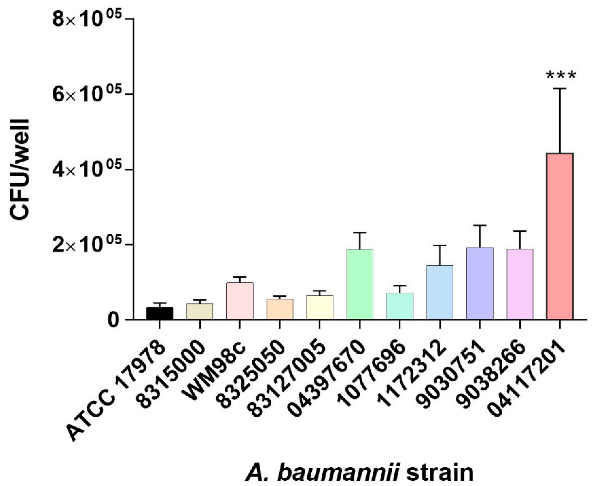
3.1. Selection of A. baumannii Isolate

The commonly used reference strain A. baumannii ATCC 17978 is not highly virulent in our hands, as seen in Figure 1 and Table 1, therefore we sought to identify a potentially virulent A. baumannii isolate. Within our collection, 11 clinical isolates were screened for two important clinical virulence-related phenotypes: cell adherence and antibiotic resistance [55]. Incubation with human A549 pneumocytes revealed the A. baumannii isolate 04117201 to be significantly more adherent than the reference ATCC 17978 strain (p < 0.001; Figure 1), and more adherent than the other clinical strains tested (Figure 1).
Antimicrobial resistance screening of the 11 clinical isolates identified the 04117201 strain as more resistant to many of the compounds tested compared to the reference ATCC 17978 strain. However, it is sensitive to erythromycin (Table 1), allowing the use of this antibiotic as a selection marker for mutant construction. As previously published, we have shown that the A. baumannii 04117201 cells can form a well-established biofilm, which is an important virulence factor [56].

3.2. Construction of an A. baumannii 04117201ΔstkR Derivative

The genome of the WT A. baumannii 04117201 strain was sequenced by Sanger sequencing at the Welcome Trust Sanger Institute and assembled using Velvet v1.2.03. Visual inspection of the sequence using RAST v2.0 identified 13 TCS response regulator proteins in the genome. A comparison of the 13 TCS identified in the clinical 04117201 strain was undertaken against those present in the A. baumannii SDF and ATCC 17978 strains identifying five response regulators proteins AdeR, BarA, StkR and two orphan response regulators NasT and RsbU not present in the SDF avirulent strain (Table 2). The TCS not present in A. baumannii SDF we named as StkSR ‘sticky’ due to the observed increased adherence to eukaryotic cells. The StkSR system identified in the clinical 04117201 strain encompasses a membrane bound hybrid histidine kinase and a cytosolic response regulator. The StkR response regulator has a conserved C-terminal helix-turn-helix motif placing it in the LuxR family. To assess the role of the StkSR system in the clinical 04117201 strain, two independent A. baumannii ΔstkR mutants were constructed. One ΔstkR mutant was selected and its growth kinetics compared to the WT in different media representing different types of environment conditions including, MH (laboratory), minimal M9 medium (desiccation on surfaces and ventilator tubing), lung medium (pneumonia) [57], and whole sheep blood (septicemia) (Figure S1). Growth of ΔstkR was comparable to WT in MH, M9 and lung medium. However, bacterial growth was significantly reduced in whole sheep’s blood. Both the WT and ΔstkR strains showed a 5-log decrease in colony forming units (CFU) in the first hour post exposure to whole sheep’s blood, after which the CFU of both strains did not change for 2 h. The ΔstkR mutant strain showed limited recovery and reached 104 CFU by the 4-h time point, whereas the WT did not reach a 104 CFU for another hour; both the WT and ΔstkR strains recovered to 106 CFU by 7 h. These data indicate that the deletion of the response regulator stkR does not result in a general decrease in bacterial cell fitness when grown in MH, M9 or lung media; however, there is a marked increase in bacteria cell CFU recovery of the ΔstkR mutant strain compared to the WT strain after exposure to whole sheep’s blood.

3.3. Assessment of the A. baumannii ΔstkR Mutant Strain Antibiogram

As TCS are known for regulating a variety of genes involved in antibiotic resistance, the ΔstkR and WT strains were assessed for their ability to resist a variety of antimicrobial compounds by MIC assays (Table 3). Five of the 16 compounds tested had altered antibiograms between the WT and ΔstkR strains. There was at least a two-fold increase in resistance to colistin and polymyxin B (both cationic antimicrobial compounds) for ΔstkR. Chloramphenicol and rifampicin showed a two-fold increase of the ΔstkR mutant compared to the WT strain and kanamycin resistance increased past the MIC threshold of detection in the ΔstkR mutant compared to the WT strain (Table 3).

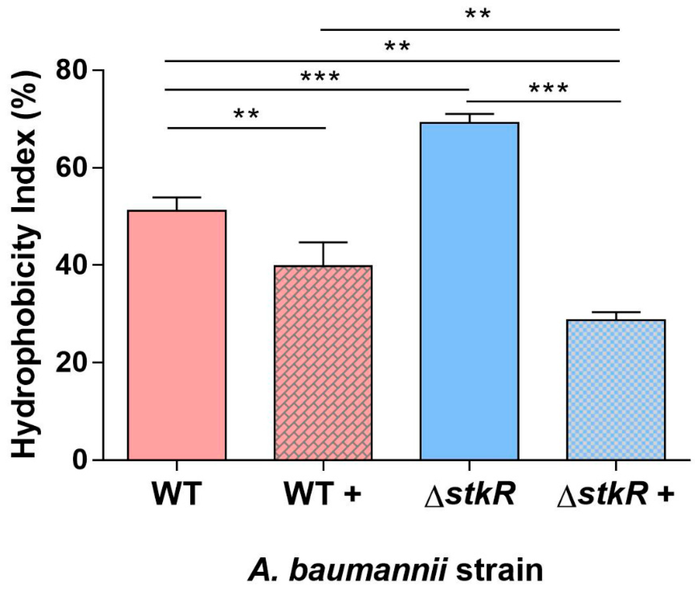
3.4. Examination of Cell Surface Hydrophobicity of the A. baumannii ΔstkR Mutant Strain When Exposed to Sub-MIC Levels of Colistin

Colistin is a cationic antimicrobial compound that acts by attaching and displacing the magnesium and calcium cations and thereby destabilizing the lipooligosaccharide in A. baumannii [69]. The hydrophobicity of the cell surface influences the affinity of colistin to the membrane [8,70]. Therefore, the cell surface hydrophobicity of the ΔstkR mutant strain was compared with the WT by its affinity to xylene with and without exposure to sub-MIC levels of colistin [41,71]. Under no colistin stress the ΔstkR mutant strain showed a higher cell surface hydrophobicity compared with the WT strain (p < 0.001; Figure 2), however, under sub-MIC levels of colistin, the ΔstkR mutant strain showed a significant decrease in cell surface hydrophobicity compared with the WT strain (p < 0.01; Figure 2). These results demonstrate the ability of the StkR response regulator to alter the bacterial cells surface hydrophobicity in response to external stressors.

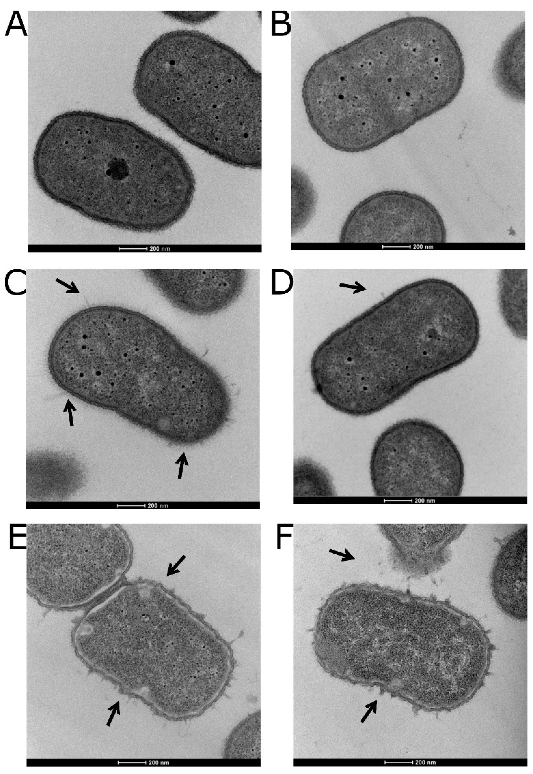
3.5. Visualizing the Effects of Colistin Stress on the Bacterial Cell Envelope

As the mode of action of colistin involves the disruption of the cell surface lipopolysaccharide [72], changes to the bacterial cell surface were assessed by TEM. Neither the WT nor ΔstkR mutant strain showed disruption of the bacterial cell outer membrane without colistin treatment (Figure 3A,B). However, after exposure to a sub-MIC level of colistin (3 µg/mL) (Figure 3C,D) significantly more protruding events per bacterial cell (blebs/cell) were observed in the WT strain (2.5 ± 0.3 blebs/cell) compared to the ΔstkR strain (0.7 ± 0.05 blebs/cell; p < 0.0001) [23,73]. Treatment of the WT and ΔstkR mutant strains with 12 µg/mL of colistin resulted in both strains exhibiting extensive membrane perturbation leading to a breakdown of the bacterial cell wall with partially disintegrated cells (Figure 3E,F).

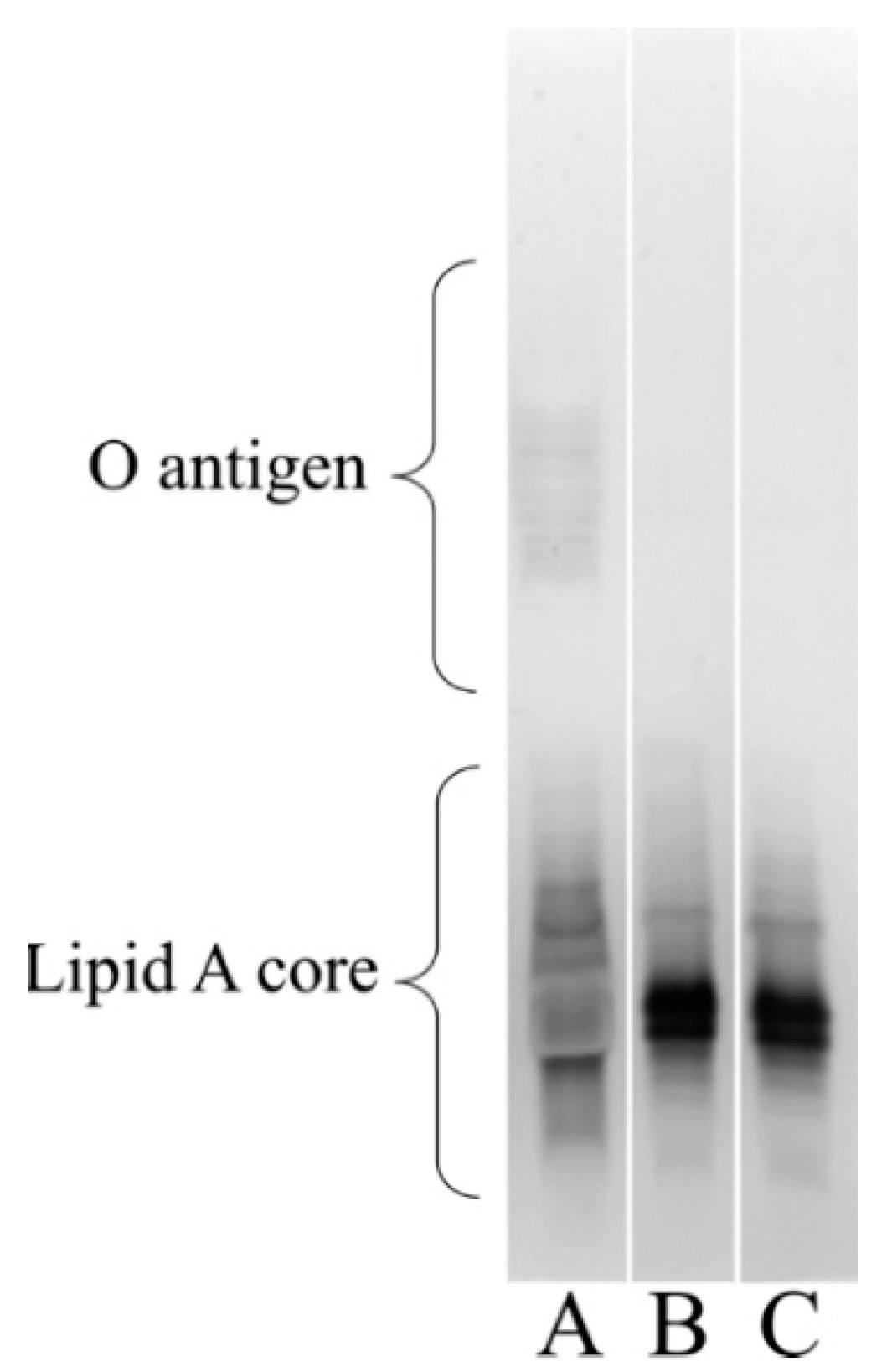
3.6. Visualizing the Lipooligosaccharide Composition of the A. baumannii ΔstkR Mutant and WT Strain

A. baumannii acquires resistance to colistin by two known mechanisms: one is the complete loss of the lipid A portion of lipooligosaccharide [70,74] and the other is through the modification of lipid A [75]. To see if disruption of stkR influences lipid A production, silver staining of the bacterial cell lipooligosaccharide was performed. The WT and ΔstkR bacterial strains were grown under normal conditions and their lipid A profiles assessed. Colistin resistance cannot be attributed to the loss of lipid A as both the WT and ΔstkR cells were visually observed to be identical (Figure 4).

3.7. Transcriptional Profiling of the A. baumannii ΔstkR Mutant and WT Strain Treated with Sub-MIC Concentrations of Colistin

The PmrAB TCS regulates pmrC, whose product is responsible for lipid A modification by the addition of pEtN leading to an increase in colistin resistance [74,75]. Quantitative reverse transcription polymerase chain reaction (qRT-PCR) of the pmrA, pmrB and pmrC genes identified an increase in transcription of these genes in the ΔstkR mutant strain when exposed to the sub-MIC of colistin, by 6.63-fold (p < 0.02), 8.22-fold (p < 0.0001) and 17.63-fold (p < 0.0003), respectively, compared to the WT strain. Increased transcription of pmrCAB can be related to point mutations in PmrAB itself [72]. A sequencing analysis was therefore undertaken on the ΔstkR mutant strain to identify if any mutations in the pmrAB genes were present. Sanger sequencing analysis of the 3.6 kb region encompassing the pmrCAB operon identified no point mutants in the ΔstkR mutant strain compared to the
WT (data not shown). This finding suggests that deletion of the stkR gene influences the pmrCAB operon and is likely a new mechanism influencing colistin resistance.

3.8. Comparative Genomes

To determine whether any other mutations may be causing the phenotypes exhibited by the ΔstkR mutant strain, the WT A. baumannii strain 04117201 was resequenced and the ΔstkR mutant strain sequenced using a Nanopore MinION. Two independent ΔstkR mutant strains were compared with the WT strain, which identified 14 single nucleotide polymorphisms (SNPs) present in both of the ΔstkR strains assessed (Table 4). Out of the 14 SNPs identified, seven are in non-coding regions of the chromosome; two are in repeat regions including a hypothetical protein; two are synonymous and three are non-synonymous. The first of the non-synonymous SNPs is at position 1150782 within a gene annotated as an aldehyde dehydrogenase. This produces an amino acid change from a stop codon to a glutamine, therefore the open reading frame now encodes a fusion of two aldehyde dehydrogenase transcribed together and this mRNA is unlikely to form an active protein. Further analysis of the WT 04117201 identified six copies of aldehyde dehydrogenase in the chromosome. Aldehyde dehydrogenase is implicated in a broad range of metabolic pathways; however, as it is commonly found in multiple copies in the genome, this inactive gene is likely to have minimal, if any, impact on cellular regulation [76]. The second non-synonymous change is at position 3132365 and is annotated as the phenazine biosynthesis protein (PhzF), a secondary metabolite known to be produced in Pseudomonas spp. which correlates to the colour exhibited by the species [77]. Although there is a change from a nonpolar (phenylalanine) to a polar amino acid (serine), this change is close to the C-terminal end of the protein and is unlikely to affect the secondary structure and function. To date, this gene has not been implicated in antibiotic resistance. The final non-synonymous change is at position 3445432, encoding an alcyl-CoA dehydrogenase, a class of enzymes that are associated with oxidation in the mitochondria and not involved in cellular regulation. “The non-coding regions harboring SNPs were also investigated. All were between 26 to 47 bps from the stop or start codon of any potential coding regions, predicting that these mutations would not influence gene transcription. The SNP at position 538323 results in a glutamine to a glycine change in the region annotated as a repeat hypothetical protein. This region is also upstream from an annotated tail protein and downstream of a major capsid protein suggesting that this region is potential a prophage. The other hypothetical protein containing a SNP is at position 3201037 placed downstream of a polymerase sigma factor RpoN and upstream of the outer membrane phospholipid-binding lipoprotein MlaA. No genetic factors have been described that indicate a link between these proteins”.

4. Discussion

Two-component systems have gained considerable attention due to their role in modulating a plethora of virulence mechanisms and multidrug resistance. In A. baumannii these systems include: BfmRS, which is involved in fitness, capsule production and biofilm formation; AdeRS, which regulates an efflux pump associated with antibiotic resistance; GacSA, which is involved with regulating the phenyl acetic acid catabolic pathway as well as attenuated virulence in a mouse infection model; the BaeSR system, which modulates efflux pumps and has been implicated in cross talk with the AdeRS systems, and the PmrAB system, which is involved in colistin resistance [4,5,26,66,78]. In this study the diverse nature of these systems was explored through the uncharacterized TCS, StkSR. We chose to investigate the role of this system in the clinical isolate 0411201 because of its potential involvement in influencing virulence (Figure 1) (Table 1) and its ability to form strong biofilms [56].
Comparative bioinformatics analysis of the genes encoding potential response regulator proteins in the WT 04117201 A. baumannii strain identified five TCS that are absent from the avirulent A. baumannii strain SDF and therefore are hypothesized to affect virulence potential (Table 2). These systems included two orphan response regulators, NasT and RsbU, and two previously characterized response regulators, AdeR, which is associated with efflux pump regulation, and BarA, coordinating the control of type IV pili [24,66]. The novel hybrid TCS named here as StkSR was chosen for further phenotypic investigation.
A knockout mutant was constructed by replacing the stkR gene with an erythromycin resistance cassette giving rise to the ΔstkR mutant strain. This produced a significant increase in expression of the pmrC, pmrA and pmrB genes in the ΔstkR mutant after sub-MIC exposure to colistin, identifying the stkR gene as a potential repressor of the genes pmrA and pmrB either directly or indirectly. Thus, this result has identified a potential regulatory relationship between the StkSR and PmrAB TCS.
The increased spread of multidrug- and pan-resistant strains of A. baumannii is of global concern. As carbapenem resistance increases, clinicians are more reliant on colistin as a ‘last resort’ antibiotic. As a consequence of its use, colistin resistant strains are continuing to be prevalent, which is resulting in an increase in mortality rates [3,74]. Inactivation of the stkR response regulator in A. baumannii strain 04117201 resulted in a 2-fold increase in resistance to the polymyxins, colistin and polymyxin B (Table 3). Colistin requires unmodified lipooligosaccharide or lipopolysaccharide to be present to exert its antibacterial properties; to date two mechanisms of resistance to colistin have been described in A. baumannii, the complete loss of the lipooligosaccharide portion of the outer membrane and modification of phosphate groups of the lipid A by the addition of pEtN or 4-amino-4-deoxy-l-arabinose, both of which reduce the overall membrane electronegativity [2,8,73,79]. Silver staining of purified lipooligosaccharide from the ΔstkR mutant and its WT parent identified the presence of lipooligosaccharide and a similar lipid A composition. Therefore, unlike Henry et al. (2012), the increase in colistin resistance could not be attributed to the loss of lipid A or the gross modification of lipid A under normal conditions [80,81]. However, bacterial cells which have a reduced cell surface hydrophobicity also show an increase in colistin resistance [82]. Therefore, cell surface hydrophobicity was assessed in both ΔstkR and WT cells. This revealed a significant reduction in the cell surface hydrophobicity of the ΔstkR derivative compared to the WT when in the presence of colistin (Figure 2). This suggests that modification of the bacterial cell is decreasing the electronegative of the cell and thereby decreasing the cell surface hydrophobicity. This modification could be a result of the addition of pEtN to lipid A, correlating the altered expression of the pmrCAB operon with cell surface hydrophobicity. When there is no colistin stress on the ΔstkR and WT bacterial cells there is an increased cell surface hydrophobicity in ΔstkR compared to the WT parent, we suggest that modification of the cell surface is only occurring when the WT and ΔstkR mutant strains are stressed with sub-MIC concentrations of colistin, showing that the bacterial cell is actively responding to the antibiotic as suggested previously by Boinett et al. 2019 [28].
To visualize the physical alteration of the cell membrane when exposed to colistin, TEM analysis identified varying levels of membrane perforation, from partially disintegrated cells to complete lysis of the WT and ΔstkR mutant strains. As the cell surface hydrophobicity of the bacterial cell changes, so does the electronegativity and therefore the ability of colistin to attach and displace the cationic cations Ca+ and Mg+ causing blebbing events on the cell surface. Under sub-MIC treatment with colistin, the WT strain displayed a significant increase in membrane perturbation/protrusion known as blebbing events compared to the ΔstkR mutant strain (Figure 3C,D). These results are supported by other studies investigating the effect of colistin on the membrane showing envelope disruption and outer membrane collapse [23,78].
The increased substitution of lipid A by pEtN through transcription of pmrC, eptA a pmrC homologue, or the plasmid mediated mcr-1 gene, results in increased colistin resistance [31,35,36,83]. Plasmids carrying MCR-1, a pEtN transferase enzyme that contributes to colistin resistance, have been identified in E. coli, K. pneumoniae and the Acinetobacter species [79]. Genome analysis did not identify this plasmid in the A. baumannii strain 04117201 nor the ΔstkR derivative (data not shown). The pmrC homologue eptA has been associated with an IS element (ISAba125) insertion into a H-NS regulator gene altering transcription of eptA. However, no IS element was found within H-NS identified in the ΔstkR mutant strain, therefore we discounted these mechanisms as possible means of colistin resistance [83,84,85,86,87,88]. Similarly, an alternate means of colistin resistance is the complete loss of lipopolysaccharide/lipooligosaccharide, achieved by insertion elements affecting the transcription of the lpxA genes [89]. Whole genome sequencing confirmed that this mutation was not present and there were no other mutations likely to cause the phenotypes observed.
The pmrCAB system was examined by qRT-PCR, as it plays a major role in conferring colistin resistance by the modification of lipid A with pEtN. The PmrAB TCS can be regulated by other two-component systems as seen in Salmonella enterica and K. pneumoniae. In these cases, the PhoPQ system leads to the activation of a small polypeptide known as pmrD, which binds to pmrA and stabilizes it in its phosphorylated state leading to an increase in transcription of pmrC [30]. Even though a pmrD homologue was not identified in A. baumannii 04117201, the cross-regulation of pmrCAB is not limited to interactions with the PhoPQ system but can be further complicated by other TCS (e.g., preAB in S. enterica) [90]. This PreAB TCS is like the qseBC system found in E. coli and appears to be able to influence the expression of pmrCAB as well, suggesting that the pmrCAB system could be regulated by several TCS.
We showed that the levels of pmrA, pmrB, and pmrC were significantly increased in ΔstkR compared to the WT when exposed to colistin. Additionally, we demonstrated that the PmrAB TCS is a likely contributor to colistin resistance and that the StkSR two-component system is a probable influence on the regulation of the PmrAB system. Taken collectively, these data could explain the modified colistin resistance seen when stkR is inactivated [90,91]. Lastly, an increase in colistin resistance through the PmrAB TCS is reported to lead to a decrease in fitness [30,92]. However, we have yet to identify any decrease in fitness of the ΔstkR mutant.

5. Conclusions

This is the first study to describe the novel TCS StkSR identified in an A. baumannii strain isolated from an Australian hospital. We have demonstrated that the observed increase in colistin resistance seen for the ΔstkR derivative of this isolate is not due to the loss of lipid A but correlates to the increased transcription of the pmrCAB operon which is linked to the modification of lipid A. This modification decreases the overall membrane electronegativity and therefore decreases the cells affinity to the colistin compound. Colistin represents a “last resort” antibiotic used for the treatment of severe A. baumannii infections, and understanding the resistance mechanisms to this clinically important compound is vital for its continued application.

Supplementary Materials

The following are available online at https://www.mdpi.com/article/10.3390/microorganisms10050985/s1, Figure S1: Growth analysis of A. baumannii 04117201 and ΔstkR mutant strains, Table S1: Oligonucleotides used in this study.

Author Contributions

Conceptualization, S.K.G., U.H.S. and M.H.B.; methodology, S.K.G., J.A.C.-J. and U.H.S.; software, B.P., M.R. and R.A.E.; validation, S.K.G., U.H.S., R.A.E. and M.H.B.; formal analysis, S.K.G., B.P.; investigation, S.K.G., U.H.S. and B.P.; resources, S.K.G. and M.H.B.; data curation, S.K.G., B.P., M.R. and R.A.E.; writing—original draft preparation, S.K.G., U.H.S., B.P. and M.H.B.; writing—review and editing, S.K.G., M.R., J.A.C.-J., R.A.E. and M.H.B.; visualization, S.K.G., U.H.S. and B.P.; supervision, R.A.E. and M.H.B.; project administration, S.K.G. and M.H.B.; funding acquisition, S.K.G., U.H.S. and M.H.B. All authors have read and agreed to the published version of the manuscript.

Funding

This research was funded by National Health and Medical Research Council, Australia, grant number 535053 to M.H.B., S.K.G. was the recipient of a Flinders University Research Scholarship.

Institutional Review Board Statement

Not applicable.

Informed Consent Statement

Not applicable.

Acknowledgments

We thank Ruth Hall from the University of Sydney for providing the initial sequence of A. baumannii strain 04117201, Sophie Leterme at Flinders University, for her contribution to the statistical analysis and David Ogunniyi Institute at the University of South Australia, for providing the A549 human pneumocytes cell line. Thank you to Felise Adams and Paul Radford for their critical editing of the paper.

Conflicts of Interest

The authors declare that they have no conflicts of interest.

References

  1. WHO. Essential Medicines and Health Products: Global Priority List of Antibiotic-Resistant Bacteria to Guide Researcher, Discovery, and Development of New Antibiotics. 2017. Available online: www.who.int/medicines/publications/global-priority-list-antibiotic-resistant-bacteria/en/ (accessed on 27 February 2017).
  2. Sabnis, A.; Hagart, K.L.; Klockner, A.; Becce, M.; Evans, L.E.; Furniss, R.C.D.; Mavridou, D.A.; Murphy, R.; Stevens, M.M.; Davies, J.C.; et al. Colistin kills bacteria by targeting lipopolysaccharide in the cytoplasmic membrane. elife 2021, 10, e65836. [Google Scholar] [CrossRef] [PubMed]
  3. Erdem, F.; Abulaila, A.; Aktas, Z.; Oncul, O. In vitro evaluation of double carbapenem and colistin combinations against OXA-48, NDM carbapenemase-producing colistin-resistant Klebsiella pneumoniae strains. Antimicrob. Resis. Infect. Control 2020, 9, 70–79. [Google Scholar] [CrossRef] [PubMed]
  4. Allen, J.L.; Tomlinson, B.R.; Casella, L.G.; Shaw, L.N. Regulatory networks important for survival of Acinetobacter baumannii within the host. Curr. Opin. Microbiol. 2020, 55, 74–80. [Google Scholar] [CrossRef] [PubMed]
  5. De Silva, P.M.; Kumar, A. Signal transduction proteins in Acinetobacter baumannii: Role in antibiotic resistance, virulence, and potential as drug targets. Front. Microbiol. 2019, 10, 1–12. [Google Scholar] [CrossRef] [PubMed] [Green Version]
  6. Leung, L.M.; McElheny, C.L.; Gardner, F.M.; Chandler, C.E.; Bowler, S.L.; Mettus, R.T.; Spychala, C.N.; Fowler, E.L.; Opene, B.N.A.; Myers, R.A.; et al. A prospective study of Acinetobacter baumannii complex isolates and colistin susceptibility monitoring by mass spectrometry of microbial membrane glycolipids. J. Clin. Microbiol. 2019, 57, e01100-18. [Google Scholar] [CrossRef] [PubMed] [Green Version]
  7. Trimble, M.J.; Mlynarcik, P.; Kolar, M.; Hancock, R.E. Polymyxin: Alternative mechanisms of action and resistance. Cold Spring Harbor Perspect. Med. 2016, 6, 1–12. [Google Scholar] [CrossRef] [Green Version]
  8. Kyriakidis, I.; Vasileiou, E.; Pana, Z.D.; Tragiannidis, A. Acinetobacter baumannii antibiotic resistance mechanisms. Pathogens 2021, 10, 373. [Google Scholar] [CrossRef]
  9. Da Silva, G.J.; Domingues, S. Interplay between colistin resistance, virulence and fitness in Acinetobacter baumannii. Antibiotics 2017, 6, 28. [Google Scholar] [CrossRef] [Green Version]
  10. Bakthavatchalam, Y.D.; Pragasam, A.K.; Biswas, I.; Veeraraghavan, B. Polymyxin susceptibility testing, interpretative breakpoints and resistance mechanisms: An update. J. Global Antimicrob. Res. 2018, 12, 124–136. [Google Scholar] [CrossRef]
  11. Falagas, M.E.; Rafailidis, P.I.; Ioannidou, E.; Alexiou, V.G.; Matthaiou, D.K.; Karageorgopoulos, D.E.; Kapaskelis, A.; Nikita, D.; Michalopoulos, A. Colistin therapy for microbiologically documented multidrug-resistant Gram-negative bacterial infections: A retrospective cohort study of 258 patients. Internat. J. Antimicrob. Agents 2010, 35, 194–199. [Google Scholar] [CrossRef] [Green Version]
  12. Hancock, R.E. Peptide antibiotics. Lancet 1997, 349, 418–422. [Google Scholar] [CrossRef]
  13. Fiester, S.E.; Nwugo, C.C.; Penwell, W.F.; Neary, J.M.; Beckett, A.C.; Arivett, B.A.; Schmidt, R.E.; Geiger, S.C.; Connerly, P.L.; Menke, S.M.; et al. Role of the carboxy terminus of secA in iron acquisition, protein translocation, and virulence of the bacterial pathogen Acinetobacter Baumannii. Infect. Immun. 2015, 83, 1354–1365. [Google Scholar] [CrossRef] [Green Version]
  14. Srinivasan, V.B.; Vaidyanathan, V.; Rajamohan, G. AbuO, a TolC-like outer membrane protein of Acinetobacter baumannii, is involved in antimicrobial and oxidative stress resistance. Antimicrob. Agents Chemother. 2015, 59, 1236–1245. [Google Scholar] [CrossRef] [PubMed] [Green Version]
  15. Gaddy, J.A.; Tomaras, A.P.; Actis, L.A. The Acinetobacter baumannii 19606 OmpA protein plays a role in biofilm formation on abiotic surfaces and in the interaction of this pathogen with eukaryotic cells. Infect. Immun. 2009, 77, 3150–3160. [Google Scholar] [CrossRef] [PubMed] [Green Version]
  16. Howell, A.; Dubrac, S.; Noone, D.; Varughese, K.I.; Devine, K. Interactions between the YycFG and PhoPR two-component systems in Bacillus subtilis: The PhoR kinase phosphorylates the non-cognate YycF response regulator upon phosphate limitation. Molec. Microbiol. 2006, 59, 1199–1215. [Google Scholar] [CrossRef] [PubMed]
  17. Rowland, M.A.; Deeds, E.J. Crosstalk and the evolution of specificity in two-component signaling. Proc. Natl. Acad. Sci. USA 2014, 111, 5550–5555. [Google Scholar] [CrossRef] [PubMed] [Green Version]
  18. Capra, E.J.; Laub, M.T. Evolution of two-component signal transduction systems. Ann. Rev. Microbiol. 2012, 66, 325–347. [Google Scholar] [CrossRef] [PubMed] [Green Version]
  19. Rajput, A.; Seif, Y.; Choudhary, K.S.; Dalldorf, C.; Poudel, S.; Monk, J.M.; Palsson, B.O. Pangenome analytics reveal two-component systems as conserved targets in ESKAPEE pathogens. mSystems 2021, 6, e00981-20. [Google Scholar] [CrossRef]
  20. Cerqueira, G.M.; Kostoulias, X.; Khoo, C.; Aibinu, I.; Qu, Y.; Traven, A.; Peleg, A.Y. A global virulence regulator in Acinetobacter baumannii and its control of the phenylacetic acid catabolic pathway. J. Infect. Dis. 2014, 210, 46–55. [Google Scholar] [CrossRef] [Green Version]
  21. Quesada, A.; Porrero, M.C.; Tellez, S.; Palomo, G.; Garcia, M.; Dominguez, L. Polymorphism of genes encoding PmrAB in colistin-resistant strains of Escherichia coli and Salmonella enterica isolated from poultry and swine. J. Antimicrob. Chemother. 2015, 70, 71–74. [Google Scholar] [CrossRef] [Green Version]
  22. Park, Y.K.; Choi, J.Y.; Shin, D.; Ko, K.S. Correlation between overexpression and amino acid substitution of the PmrAB locus and colistin resistance in Acinetobacter Baumannii. Int. J. Antimicrob. Agents 2011, 37, 525–530. [Google Scholar] [CrossRef] [PubMed]
  23. Girardello, R.; Visconde, M.; Cayo, R.; Figueiredo, R.C.; Mori, M.A.; Lincopan, N.; Gales, A.C. Diversity of polymyxin resistance mechanisms among Acinetobacter baumannii clinical isolates. Diagnost.Microbiol. Infect. Dis. 2017, 87, 37–44. [Google Scholar] [CrossRef] [PubMed]
  24. Adams, F.G.; Stroeher, U.H.; Hassan, K.A.; Marri, S.; Brown, M.H. Resistance to pentamidine is mediated by AdeAB, regulated by AdeRS, and influenced by growth conditions in Acinetobacter baumannii ATCC 17978. PLoS ONE 2018, 13, e0197412. [Google Scholar] [CrossRef] [PubMed] [Green Version]
  25. Richmond, G.E.; Evans, L.P.; Anderson, M.J.; Wand, M.E.; Bonney, L.C.; Ivens, A.; Chua, K.L.; Webber, M.A.; Sutton, J.M.; Peterson, M.L.; et al. The Acinetobacter baumannii two-component system AdeRS regulates genes required for multidrug efflux, biofilm formation, and virulence in a strain-specific manner. mBio 2016, 7, e00430-16. [Google Scholar] [CrossRef] [Green Version]
  26. Cheng, H.Y.; Chen, Y.F.; Peng, H.L. Molecular characterization of the PhoPQ-PmrD-PmrAB mediated pathway regulating polymyxin B resistance in Klebsiella pneumoniae CG43. J. Biomed. Sci. 2010, 17, 60–76. [Google Scholar] [CrossRef] [Green Version]
  27. Jaidane, N.; Naas, T.; Mansour, W.; Radhia, B.B.; Jerbi, S.; Boujaafar, N.; Bouallegue, O.; Bonnin, R.A. Genomic analysis of in vivo acquired resistance to colistin and rifampicin in Acinetobacter Baumannii. Internat. J. Antimicrob. Agents 2018, 51, 266–269. [Google Scholar] [CrossRef]
  28. Boinett, C.J.; Cain, A.K.; Hawkey, J.; Do Hoang, N.T.; Khanh, N.N.T.; Thanh, D.P.; Dordel, J.; Campbell, J.I.; Lan, N.P.H.; Mayho, M.; et al. Clinical and laboratory-induced colistin-resistance mechanisms in Acinetobacter Baumannii. Micro. Genom. 2019, 5, 1–10. [Google Scholar] [CrossRef]
  29. Beceiro, A.; Llobet, E.; Aranda, J.; Bengoechea, J.A.; Doumith, M.; Hornsey, M.; Dhanji, H.; Chart, H.; Bou, G.; Livermore, D.D.; et al. Phosphoethanolamine modification of lipid A in colistin-resistant variants of Acinetobacter baumannii mediated by the pmrAB two-component regulatory system. Antimicrob. Agents Chemother. 2011, 55, 3370–3379. [Google Scholar] [CrossRef] [Green Version]
  30. Jones, C.L.; Singh, S.S.; Alamneh, Y.; Casella, L.G.; Ernst, R.K.; Lesho, E.P.; Waterman, P.E.; Zurawski, D.V. In vivo fitness adaptations of colistin-resistant Acinetobacter baumannii isolates to oxidative stress. Antimicrob. Agents Chemother. 2017, 61, e00598-16. [Google Scholar] [CrossRef] [Green Version]
  31. Trebosc, V.; Gartenmann, S.; Totzl, M.; Lucchini, V.; Schellhorn, B.; Pieren, M.; Lociuro, S.; Gitzinger, M.; Tigges, M.; Bumann, D.; et al. Dissecting colistin resistance mechanisms in extensively drug-resistant Acinetobacter baumannii clinical isolates. mBio 2019, 10, e01083-19. [Google Scholar] [CrossRef] [Green Version]
  32. Deveson Lucas, D.; Crane, B.; Wright, A.; Han, M.L.; Moffatt, J.; Bulach, D.; Gladman, S.L.; Powell, D.; Aranda, J.; Seemann, T.; et al. Emergence of high-level colistin resistance in an Acinetobacter baumannii clinical isolate mediated by inactivation of the global regulator H-NS. Antimicrob. Agents Chemother. 2018, 62, e02442-16. [Google Scholar] [CrossRef] [PubMed] [Green Version]
  33. Eijkelkamp, B.A.; Hassan, K.A.; Paulsen, I.T.; Brown, M.H. Investigation of the human pathogen Acinetobacter baumannii under iron limiting conditions. BMC Genom. 2011, 12, 126–140. [Google Scholar] [CrossRef] [PubMed] [Green Version]
  34. Giles, S.K.; Stroeher, U.H.; Eijkelkamp, B.A.; Brown, M.H. Identification of genes essential for pellicle formation in Acinetobacter Baumannii. BMC Microbiol. 2015, 15, 116–129. [Google Scholar] [CrossRef] [Green Version]
  35. Hanahan, D. Studies on transformation of Escherichia coli with plasmids. J. Molec. Biol. 1983, 166, 557–580. [Google Scholar] [CrossRef]
  36. Simon, R.; Priefer, U.; Puhler, A. A broad host range mobilization system for in vivo genetic-engineering: Transposon mutagenesis in Gram-negative bacteria. Biotechnol. Appl. Microbiol. 1983, 1, 784–791. [Google Scholar] [CrossRef]
  37. Yanisch-Perron, C.; Vieira, J.; Messing, J. Improved M13 phage cloning vectors and host strains: Nucleotide sequences of the M13mp18 and pUC19 vectors. Gene 1985, 33, 103–119. [Google Scholar] [CrossRef]
  38. Wiegand, I.; Hilpert, K.; Hancock, R.E. Agar and broth dilution methods to determine the minimal inhibitory concentration (MIC) of antimicrobial substances. Nat. Protoc. 2008, 3, 163–175. [Google Scholar] [CrossRef]
  39. CLSI. Performance Standards for Antimicrobial Disk Susceptibility Testing, Seventeenth Information Supplement; Clinical and Laboratory Standards Institute: Wayne, PA, USA, 2007; Volume 27, p. M100-S17. [Google Scholar]
  40. CLSI. Clinical Laboratory Standards Institute evaluation protocols. MLO-Med. Lab. Obs. 2006, 38, 40–41. [Google Scholar]
  41. Eijkelkamp, B.A.; Stroeher, U.H.; Hassan, K.A.; Elbourne, L.D.; Paulsen, I.T.; Brown, M.H. H-NS plays a role in expression of Acinetobacter baumannii virulence features. Infect. Immun. 2013, 81, 2574–2583. [Google Scholar] [CrossRef] [Green Version]
  42. Gui, S.; Li, R.; Feng, Y.; Wang, S. Transmission electron microscopic morphological study and flow cytometric viability assessment of Acinetobacter baumannii susceptible to Musca domestica cecropin. Sci. World J. 2014, 2014, 1–6. [Google Scholar]
  43. Tsai, C.M.; Frasch, C.E. A sensitive silver stain for detecting lipopolysaccharides in polyacrylamide gels. Anal. Biochem. 1982, 119, 115–119. [Google Scholar] [CrossRef]
  44. Fomsgaard, A.; Freudenberg, M.A.; Galanos, C. Modification of the silver staining technique to detect lipopolysaccharide in polyacrylamide gels. J. Clin. Microbiol. 1990, 28, 2627–2631. [Google Scholar] [CrossRef] [PubMed] [Green Version]
  45. Brazma, A.; Vilo, J. Gene expression data analysis. Microbes Infect. 2001, 3, 823–829. [Google Scholar] [CrossRef]
  46. Livak, K.J.; Schmittgen, T.D. Analysis of Relative Gene Expression Data Using Real-Time Quantitative PCR and the 2−ΔΔCT Method. Methods 2001, 25, 402–408. [Google Scholar] [CrossRef] [PubMed]
  47. Wick, R.R.; Judd, L.M.; Gorrie, C.L.; Holt, K.E. Unicycler: Resolving bacterial genome assemblies from short and long sequencing reads. PLoS Comp. Biol. 2017, 13, e1005595. [Google Scholar] [CrossRef] [PubMed] [Green Version]
  48. Walker, B.J.; Abeel, T.; Shea, T.; Priest, M.; Abouelliel, A.; Sakthikumar, S.; Cuomo, C.A.; Zeng, Q.; Wortman, J.; Young, S.K.; et al. Pilon: An integrated tool for comprehensive microbial variant detection and genome assembly improvement. PLoS ONE 2014, 9, e112963. [Google Scholar] [CrossRef] [PubMed]
  49. Wick, R.R.; Schultz, M.B.; Zobel, J.; Holt, K.E. Bandage: Interactive visualization of de novo genome assemblies. Bioinformatics 2015, 31, 3350–3352. [Google Scholar] [CrossRef] [Green Version]
  50. Langmead, B.; Salzberg, S.L. Fast gapped-read alignment with Bowtie 2. Nat. Methods 2012, 9, 357–359. [Google Scholar] [CrossRef] [Green Version]
  51. Li, H.; Handsaker, B.; Wysoker, A.; Fennell, T.; Ruan, J.; Homer, N.; Marth, G.; Abecasis, G.; Durbin, R. 1000 Genome Project Data Processing Subgroup The sequence alignment/map format and SAMtools. Bioinformatics 2009, 25, 2078–2079. [Google Scholar] [CrossRef] [Green Version]
  52. Danecek, P.; Auton, A.; Abecasis, G.; Albers, C.A.; Banks, E.; DePristo, M.A.; Handsaker, R.E.; Lunter, G.; Marth, G.T.; Sherry, S.T.; et al. The variant call format and VCFtools. Bioinformatics 2011, 27, 2156–2158. [Google Scholar] [CrossRef]
  53. Seemann, T. Prokka: Rapid prokaryotic genome annotation. Bioinformatics 2014, 30, 2068–2069. [Google Scholar] [CrossRef]
  54. Samuels, M.L.; Witmer, J.A.; Schaffner, A.A. Statistics for the Life Sciences, 4th ed.; Pearson Education: Boston, MA, USA, 2012. [Google Scholar]
  55. Brossard, K.A.; Campagnari, A.A. The Acinetobacter baumannii biofilm-associated protein plays a role in adherence to human epithelial cells. Infect. Immun. 2012, 80, 228–233. [Google Scholar] [CrossRef] [Green Version]
  56. Eijkelkamp, B.A.; Stroeher, U.H.; Hassan, K.A.; Papadimitrious, M.S.; Paulsen, I.T.; Brown, M.H. Adherence and motility characteristics of clinical Acinetobacter baumannii isolates. FEMS Microbiol. Lett. 2011, 323, 44–51. [Google Scholar] [CrossRef] [PubMed] [Green Version]
  57. Palmer, K.L.; Aye, L.M.; Whiteley, M. Nutritional cues control Pseudomonas aeruginosa multicellular behavior in cystic fibrosis sputum. J. Bacteriol. 2007, 189, 8079–8087. [Google Scholar] [CrossRef] [PubMed] [Green Version]
  58. Marchand, I.; Damier-Piolle, L.; Courvalin, P.; Lambert, T. Expression of the RND-type efflux pump AdeABC in Acinetobacter baumannii is regulated by the AdeRS two-component system. Antimicrob. Agents Chemother. 2004, 48, 3298–3304. [Google Scholar] [CrossRef] [PubMed] [Green Version]
  59. Adams, M.D.; Goglin, K.; Molyneaux, N.; Hujer, K.M.; Lavender, H.; Jamison, J.J.; MacDonald, I.J.; Martin, K.M.; Russo, T.; Campagnari, A.A.; et al. Comparative genome sequence analysis of multidrug-resistant Acinetobacter Baumannii. J. Bacteriol. 2008, 190, 8053–8064. [Google Scholar] [CrossRef] [PubMed] [Green Version]
  60. Lin, M.F.; Lin, Y.Y.; Lan, C.Y. The role of the two-component system BaeSR in disposing chemicals through regulating transporter systems in Acinetobacter baumannii. PLoS ONE 2015, 10, e0132843. [Google Scholar] [CrossRef] [PubMed]
  61. Heermann, R.; Jung, K. The complexity of the ‘simple’ two-component system KdpD/KdpE in Escherichia coli. FEMS Microbiol. Lett. 2010, 304, 97–106. [Google Scholar] [CrossRef] [Green Version]
  62. Bhagirath, A.Y.; Li, Y.; Patidar, R.; Yerex, K.; Ma, X.; Kumar, A.; Duan, K. Two component regulatory systems and antibiotic resistance in Gram-negative pathogens. Internat. J. Molec. Sci. 2019, 20, 1781. [Google Scholar] [CrossRef] [Green Version]
  63. Li, W.; Xue, M.; Yu, L.; Qi, K.; Ni, J.; Chen, X.; Deng, R.; Shang, F.; Xue, T. QseBC is involved in the biofilm formation and antibiotic resistance in Escherichia coli isolated from bovine mastitis. Peer J. 2020, 8, e8833. [Google Scholar] [CrossRef] [Green Version]
  64. Meng, X.; Ahator, S.D.; Zhang, L.H. Molecular mechanisms of phosphate stress activation of Pseudomonas aeruginosa quorum sensing systems. mSphere 2020, 5, e00119-20. [Google Scholar] [CrossRef] [PubMed] [Green Version]
  65. Tomaras, A.P.; Flagler, M.J.; Dorsey, C.W.; Gaddy, J.A.; Actis, L.A. Characterization of a two-component regulatory system from Acinetobacter baumannii that controls biofilm formation and cellular morphology. Microbiology 2008, 154, 3398–3409. [Google Scholar] [CrossRef] [PubMed] [Green Version]
  66. Xu, K.; Shen, D.; Yang, N.; Chou, S.H.; Gomelsky, M.; Qian, G. Coordinated control of the type IV pili and c-di-GMP-dependent antifungal antibiotic production in Lysobacter by the response regulator PilR. Molec. Plant Pathol. 2021, 22, 602–617. [Google Scholar] [CrossRef] [PubMed]
  67. Leech, A.J.; Sprinkle, A.; Wood, L.; Wozniak, D.J.; Ohman, D.E. The NtrC family regulator AlgB, which controls alginate biosynthesis in mucoid Pseudomonas aeruginosa, binds directly to the algD promoter. J. Bacteriol. 2008, 190, 581–589. [Google Scholar] [CrossRef] [PubMed] [Green Version]
  68. Delumeau, O.; Dutta, S.; Brigulla, M.; Kuhnke, G.; Hardwick, S.W.; Volker, U.; Yudkin, M.D.; Lewis, R.J. Functional and structural characterization of RsbU, a stress signaling protein phosphatase 2C. J. Biol. Chem. 2004, 279, 40927–40937. [Google Scholar] [CrossRef] [Green Version]
  69. Yoon, E.J.; Kim, H.S.; Woo, H.; Choi, Y.J.; Won, D.; Choi, J.R.; Kim, Y.A.; Jeong, S.H. Trajectory of genetic alterations associated with colistin resistance in Acinetobacter baumannii during an in-hospital outbreak of infection. J. Antimicrob. Chemother. 2021, 77, 69–73. [Google Scholar] [CrossRef]
  70. Moffatt, J.H.; Harper, M.; Harrison, P.; Hale, J.D.; Vinogradov, E.; Seemann, T.; Henry, R.; Crane, B.; St Michael, F.; Cox, A.D.; et al. Colistin resistance in Acinetobacter baumannii is mediated by complete loss of lipopolysaccharide production. Antimicrob. Agents Chemother. 2010, 54, 4971–4977. [Google Scholar] [CrossRef] [Green Version]
  71. Rosenberg, E.; Gottlieb, A.; Rosenberg, M. Inhibition of bacterial adherence to hydrocarbons and epithelial cells by emulsan. Infect. Immun. 1983, 39, 1024–1028. [Google Scholar] [CrossRef] [Green Version]
  72. Sun, B.; Liu, J.; Jiang, Y.; Shao, L.; Yang, S.; Chen, D. New mutations involved in colistin resistance in Acinetobacter Baumannii. Msphere 2020, 5, e00895-19. [Google Scholar] [CrossRef] [Green Version]
  73. Alves, C.S.; Melo, M.N.; Franquelim, H.G.; Ferre, R.; Planas, M.; Feliu, L.; Bardaji, E.; Kowalczyk, W.; Andreu, D.; Santos, N.C.; et al. Escherichia coli cell surface perturbation and disruption induced by antimicrobial peptides BP100 and pepR. J. Biol. Chem. 2010, 285, 27536–27544. [Google Scholar] [CrossRef] [Green Version]
  74. Vrancianu, C.O.; Gheorghe, I.; Czobor, I.B.; Chifiriuc, M.C. Antibiotic resistance profiles, molecular mechanisms and innovative treatment strategies of Acinetobacter Baumannii. Microorganisms 2020, 8, 935. [Google Scholar] [CrossRef] [PubMed]
  75. Arroyo, L.A.; Herrera, C.M.; Fernandez, L.; Hankins, J.V.; Trent, M.S.; Hancock, R.E. The pmrCAB operon mediates polymyxin resistance in Acinetobacter baumannii ATCC 17978 and clinical isolates through phosphoethanolamine modification of Lipid A. Antimicrob. Agents Chemother. 2011, 55, 3743–3751. [Google Scholar] [CrossRef] [PubMed] [Green Version]
  76. Riveros-Rosas, H.; Julian-Sanchez, A.; Moreno-Hagelsieb, G.; Munoz-Clares, R.A. Aldehyde dehydrogenase diversity in bacteria of the Pseudomonas genus. Chem.-Biologic. Interac. 2019, 304, 83–87. [Google Scholar] [CrossRef]
  77. Price-Whelan, A.; Dietrich, L.E.; Newman, D.K. Rethinking ‘secondary’ metabolism: Physiological roles for phenazine antibiotics. Nat. Chem. Biol 2006, 2, 71–78. [Google Scholar] [CrossRef] [PubMed]
  78. Mohamed, Y.F.; Abou-Shleib, H.M.; Khalil, A.M.; El-Guink, N.M.; El-Nakeeb, M.A. Membrane permeabilization of colistin toward pan-drug resistant Gram-negative isolates. Brazil. J. Microbiol. 2016, 47, 381–388. [Google Scholar] [CrossRef] [PubMed] [Green Version]
  79. Hameed, F.; Khan, M.A.; Muhammad, H.; Sarwar, T.; Bilal, H.; Rehman, T.U. Plasmid-mediated mcr-1 gene in Acinetobacter baumannii and Pseudomonas aeruginosa: First report from Pakistan. Rev. Soc. Bras. Med. Tropic. 2019, 52, e20190237. [Google Scholar] [CrossRef] [PubMed] [Green Version]
  80. Henry, R.; Vithanage, N.; Harrison, P.; Seemann, T.; Coutts, S.; Moffatt, J.H.; Nation, R.L.; Li, J.; Harper, M.; Adler, B.; et al. Colistin-resistant, lipopolysaccharide-deficient Acinetobacter baumannii responds to lipopolysaccharide loss through increased expression of genes involved in the synthesis and transport of lipoproteins, phospholipids, and poly-beta-1,6-N-acetylglucosamine. Antimicrob. Agents Chemother. 2012, 56, 59–69. [Google Scholar] [CrossRef] [PubMed] [Green Version]
  81. Velkov, T.; Thompson, P.E.; Nation, R.L.; Li, J. Structure activity relationships of polymyxin antibiotics. J. Med. Chem. 2010, 53, 1898–1916. [Google Scholar] [CrossRef] [Green Version]
  82. Soon, R.L.; Li, J.; Boyce, J.D.; Harper, M.; Adler, B.; Larson, I.; Nation, R.L. Cell surface hydrophobicity of colistin-susceptible vs resistant Acinetobacter baumannii determined by contact angles: Methodological considerations and implications. J. Appl. Microbiol. 2012, 113, 940–951. [Google Scholar] [CrossRef] [Green Version]
  83. Coetzee, J.; Corcoran, C.; Prentice, E.; Moodley, M.; Mendelson, M.; Poirel, L.; Nordmann, P.; Brink, A.J. Emergence of plasmid-mediated colistin resistance (MCR-1) among Escherichia coli isolated from South African patients. S. Afr. Med. J. 2016, 106, 449–450. [Google Scholar] [CrossRef]
  84. Liu, Y.Y.; Wang, Y.; Walsh, T.R.; Yi, L.X.; Zhang, R.; Spencer, J.; Doi, Y.; Tian, G.; Dong, B.; Huang, H.; et al. Emergence of plasmid-mediated colistin resistance mechanism MCR-1 in animals and human beings in China: A microbiological and molecular biological study. Lancet Infect. Dis. 2016, 16, 161–168. [Google Scholar] [CrossRef]
  85. Stokes, J.M.; MacNair, C.R.; Ilyas, B.; French, S.; Cote, J.P.; Bouwman, C.; Farha, M.A.; Sieron, A.O.; Whitfield, C.; Coombes, B.K.; et al. Pentamidine sensitizes Gram-negative pathogens to antibiotics and overcomes acquired colistin resistance. Nat. Microbiol. 2017, 2, 1–21. [Google Scholar] [CrossRef] [PubMed] [Green Version]
  86. Elnahriry, S.S.; Khalifa, H.O.; Soliman, A.M.; Ahmed, A.M.; Hussein, A.M.; Shimamoto, T.; Shimamoto, T. Emergence of plasmid-mediated colistin resistance gene mcr-1 in a clinical Escherichia coli isolate from Egypt. Antimicrob. Agents Chemother. 2016, 60, 3249–3250. [Google Scholar] [CrossRef] [PubMed] [Green Version]
  87. Yu, C.Y.; Ang, G.Y.; Chin, P.S.; Ngeow, Y.F.; Yin, W.F.; Chan, K.G. Emergence of mcr-1-mediated colistin resistance in Escherichia coli in Malaysia. Internat. J. Antimicrob. Agents 2016, 47, 504–505. [Google Scholar] [CrossRef]
  88. Vijayakumar, S.; Kanthan, K.; Veeraraghavan, B. Whole-genome shotgun sequences of seven colistin-resistant Acinetobacter baumannii isolates from bacteraemia. J. Antimicrob. Resis. 2018, 12, 155–156. [Google Scholar] [CrossRef]
  89. Moffatt, J.H.; Harper, M.; Adler, B.; Nation, R.L.; Li, J.; Boyce, J.D. Insertion sequence ISAba11 is involved in colistin resistance and loss of lipopolysaccharide in Acinetobacter Baumannii. Antimicrob. Agents Chemother. 2011, 55, 3022–3024. [Google Scholar] [CrossRef] [Green Version]
  90. Adams, M.D.; Nickel, G.C.; Bajaksouzian, S.; Lavender, H.; Murthy, A.R.; Jacobs, M.R.; Bonomo, R.A. Resistance to colistin in Acinetobacter baumannii associated with mutations in the PmrAB two-component system. Antimicrob. Agents Chemother. 2009, 53, 3628–3634. [Google Scholar] [CrossRef] [Green Version]
  91. Qureshi, Z.A.; Hittle, L.E.; O’Hara, J.A.; Rivera, J.A.; Syed, A.; Shields, R.K.; Pasculle, A.W.; Ernst, R.K.; Doi, Y. Colistin-resistant Acinetobacter baumannii: Beyond carbapenem resistance. Clin. Infect. Dis. 2015, 60, 1295–1303. [Google Scholar] [CrossRef] [Green Version]
  92. Lopez-Rojas, R.; Dominguez-Herrera, J.; McConnell, M.J.; Docobo-Perez, F.; Smani, Y.; Fernandez-Reyes, M.; Rivas, L.; Pachon, J. Impaired virulence and in vivo fitness of colistin-resistant Acinetobacter Baumannii. J. Infect. Dis. 2011, 203, 545–548. [Google Scholar] [CrossRef] [Green Version]
Figure 1. Cell adherence capacity of A. baumannii clinical strains to A549 pneumocytes. The CFU/well values of 11 A. baumannii strains were examined after exposure to A549 cells. Bars indicate the standard error of three separate experiments. Assessments of skewness, kurtosis and the Shapiro–Wilk normality test were undertaken. *** represents a p-value of < 0.001 as determined by an analysis of variance test, between ATCC 17978 and the clinical isolate.
Microorganisms 10 00985 g001
Figure 2. Cell surface hydrophobicity of A. baumannii 04117201 and ΔstkR mutant. Cell surface hydrophobicity was assessed by the cells’ affiliation to xylene. A. baumannii strain 04117201 (WT) is without colistin exposure, WT + is with exposure to 3 μg/mL of colistin. A. baumannii 04117201 ΔstkR mutant is without colistin exposure, ΔstkR+ is with exposure to 3 μg/mL of colistin. Bars represent the standard deviation of three separate experiments. A Kruskal–Wallis test was performed to compare the means of the WT and ΔstkR mutant with and without colistin exposure. *** represents a p-value < 0.001 and ** represents a p-value of <0.01 as established by an analysis of variance test.
Microorganisms 10 00985 g002
Figure 3. Membrane perturbation of A. baumannii 04117201 (WT) and ΔstkR mutant after colistin stress. WT and ΔsktR mutant strains were subjected to sub-MIC and MIC levels of colistin (3 μg/mL and 12 μg/mL) and cross-sections were examined by TEM (60,000× magnification); 200 nm scale is shown in white. Images are of (A) WT, (B) ΔsktR, (C) WT after exposure to 3 μg/mL colistin, (D) ΔstkR after exposure to 3 μg/mL of colistin, (E) WT after exposure to 12 μg/mL colistin, and (F) ΔstkR after exposure to 12 μg/mL colistin; arrows show membrane perturbation and/or leakage of cellular contents.
Microorganisms 10 00985 g003
Figure 4. Lipooligosaccharide analysis of the 04117201 (WT) and ΔstkR mutant. The lipid A and O-antigen components of the strains’ membrane was resolved on a 15% polyacrylamide gel and silver stained. (A) control lipopolysaccharide from Pseudomonas aeruginosa PA01 showing the presence of O-antigen; (B) WT, and (C) ΔstkR. Brackets identify either the lipid A core or O-antigen regions.
Microorganisms 10 00985 g004
Table 1. Antimicrobial resistance profiles of Acinetobacter baumannii clinical strains.
A. baumannii Strains
Compound abATCC 17978041172011172312WM98 c083150000439767090307511077697903826608325050083217005
KAN cRRRSSRRSRSR
ERYSSSSRSSISIR
TETSRRIRIRIRRR
AMP250>500>500250>500>500>500>500>500>500>500
CIP3.2530303.25>24060606060>240>240
CST488168832321644
TEL0.931.871.871.873.753.751.871.873.753.753.75
GEN3250050016125250125125250125125
PMB81616168163232888
RIF8888>64444444
H2O2 d125250250500250125250250250125250
a KAN, kanamycin; ERY, erythromycin; TET, tetracycline; AMP, ampicillin; CIP, ciprofloxacin; CST, colistin; TEL, tellurite; GEN, gentamicin; PMB, polymyxin B; RIF, rifampicin; H2O2, hydrogen peroxide. b (µg/mL). c R, Resistant; I, Intermediate; S, Susceptible; determined by Kirby Bauer against the CLSI standards. d (nM).
Table 2. Response regulators identified in the clinical Acinetobacter baumannii 04117201.
Peg # in 04117201Location in 04117201A1S + # in ATCC 17978Location in ATCC 17978Present in SDFResponse Regulator aDescription
25 18192–18935A1S_17532038890–2044193NAdeRTCS, known as AdeRS, involved in antibiotic resistance and AdeABC efflux pump [58]
95 40498–39824A1S_27513193938- 3189264YPmrATCS, known as PmrAB involved in lipid A modification [59]
226 180539–179853A1S_28833334601–3333044YBaeRTCS known as BaeRS involved in chemical transport and regulation+ of AdeABC and AdeIJK pumps [60]
519 97888–98604A1S_21372497340–2492622YKdpETCS, known as KdpED involved in potassium transport [61]
838 118775–122230A1S_13941636507–1641464NStkRTCS known as GerE (renamed here as StkSR)
1104 21883–21119A1S_32293720752–3725517YOmpRTCS, EnvZ-OmpR, involved in osmotic stress [62]
1270 84347–85009A1S_22882655031–2650369YQseBTCS, known as QseBC involved in biofilm formation [63]
1649 134448–135158A1S_33753881425–3886134YPhoBTCS, known as PhoRB involved in phosphate stress and quorum sensing [64]
1825 90896–91486A1S_20062319938–2324522NNasTOrphan response regulator known as NasT; no histidine kinase identifiable up or down stream
1962 98859–99575A1S_0748887026–892617YRstATCS, known as BfmR involved in biofilm formation [65]
2189 37589–36954A1S_0233259526–264163NPilRTCS, known as PilR involved in Type 4 fimbriae expression [66]
2213 62365–63105A1S_0261284993–289733YAlgBTCS, known as AlgBZ involved in alginate biosynthesis [67]
2465 1290–2258A1S_0621670111–674210NRsbUOrphan response regulator known as RsbU; no histidine kinase identifiable up or down stream [68]
a Annotated by Rapid Annotation using subsystem technology (RAST) https://rast.nmpdr.org/ (accessed on 15 March 2015). # Locus tag number.
Table 3. Antimicrobial resistance profile of the Acinetobacter baumannii 04117201 and ΔstkR mutant.
Acinetobacter baumannii Strain
CompoundWT (µg/mL)ΔstkR (µg/mL)Antibiotic Family Group
Novobiocin3131Aminocoumaria
Amikacin1010Aminoglycoside
Gentamicin500500Aminoglycoside
Kanamycin3000>3000Aminoglycoside
Streptomycin>300>300Aminoglycoside
Chloramphenicol;55–10Amphenicol
Rifampicin44–8Ansamycins
Ampicillin>500>500Beta-lactam
Ciprofloxacin3030Carboxy fluoroquinoline
Chlorhexidine7.5–1515Chlorobenzenes
Triclosan0.15–0.650.31–0.65Diphenyl ethers
Tellurite1.871.87Metal
Triton X1006464Nonionic surfactant
Pentamidine125125Phenol ether
Colistin816Polymyxin
Polymyxin1632Polymyxin
Nalidixic acid12501250Quinolone
Table 4. Single nucleotide polymorphisms regions identified in the ΔstkR mutant compared to the WT strain.
SNP Position aCodon in WTCodon in ΔstkRAmino Acid in WTAmino Acid in ΔstkRAnnotation of Region b
176528 ----Non-coding region
538323 GAGGGGGluGlyHypothetical protein (Repeat region)
1001055 ----Non-coding region
1058011 ----Non-coding region
1098529 ----Non-coding region
1150782 TAACAAStopGlnAldehyde dehydrogenase
1590889 GCTGCCAlaAlaHypothetical protein
2730447 TTTTTCPhePheHypothetical protein
2796261 ----Non-coding region
3132365 TTCTCCPheSerPhenazine biosynthesis protein (PhzF)
3201037 CCCTCCProSerRepeat region
3361708 ----Non-coding region
3370031 ----Non-coding region
3445432 AAAGAALysGluAlcyl-CoA Dehydrogenase
a SNP positions identified with SAMtools v1.9 and VCFools v0.11.17. b Annotation of region undertaken using Prokka.
Publisher’s Note: MDPI stays neutral with regard to jurisdictional claims in published maps and institutional affiliations.

