Article

Micromechanisms of Deformation and Fracture in Porous L-PBF 316L Stainless Steel at Different Strain Rates

1
Institute of Physics of Metals, Ural Branch of Russian Academy of Sciences, 620049 Ekaterinburg, Russia
2
The Department of Engineering and Physics, Karlstad University, SE-65188 Karlstad, Sweden
3
Institute of New Materials and Technologies, Ural Federal University, 620002 Ekaterinburg, Russia
*
Author to whom correspondence should be addressed.
Metals 2021, 11(11), 1870; https://doi.org/10.3390/met11111870
Submission received: 12 October 2021 / Revised: 9 November 2021 / Accepted: 11 November 2021 / Published: 21 November 2021

Abstract

:
The process of an unstable plastic flow associated with the strain rate sensitivity of mechanical properties was studied in porous 316L austenitic steel samples manufactured by laser powder bed fusion (L-PBF). Different micromechanisms of deformation and fracture of porous samples dependent on strain rate were found. It was found that despite the porosity, the specimens showed high strength, which increased with the loading rate. Porosity led to lower ductility of the studied specimens, in comparison with literature data for low porous 316L L-PBF samples and resulted in de-localization of plastic deformation. With an increase in strain rate, nucleation of new pores was less pronounced, so that at the highest strain rate of 8 × 10⁻³ s−1, only pore coalescence was observed as the dominating microscopic mechanism of ductile fracture.

1. Introduction

The interest in obtaining digital metal products using additive technologies is very high. The as-built microstructure of L-PBF (laser powder bed fusion) stainless steel is usually described as a cellular columnar structure. At solidification, colonies of crystallographically coherent cells grow in the same direction and form colonies separated by high-angle boundaries [1,2]. Additionally, oxide nanoparticles and the segregation of elements at cell boundaries have been reported [3]. The strengthening effect of the oxide particles in as-built 316 steel was proposed, but an absence of the nanoparticle strengthening effect after annealing makes this hypothesis arguable. Formation of segregation and specific dislocation structures at cell boundaries have also been reported [2,4,5,6,7]. The dislocation structures observed in as-built L-PBF seem to be quite stable, and they have recovered and annihilated only after annealing at 800–950 °C. Mobility of these dislocations is not high as the as-built 316L steel, which has higher strength and lower ductility characteristics in a comparison with the conventional bulk material [8,9]. Additionally, under high strain rates, twinning can be activated in L-PBF 316-L samples [10].
A technological feature of L-PBF manufacturing is commonly associated with the formation of specific for L-PBF micro defects such as lack-of-fusion pores, interlayer defects, spattering, keyhole pores [3,10]. These defects of course influence the life span of the component, and quantification of the deteriorating effect of each type of defect is crucial for/in applications. Porosity is commonly considered a critical defect negatively influencing mechanical characteristics. The main effect is a decrease of the load-bearing cross-section; additionally, sharp pore edges introduce possible stress concentration points. Nevertheless, the influence of porosity on micromechanisms of fracture initiation is not fully understood. Whether it is the growth of existing pores or the nucleation and coalescence of micro pores under strain is still an open question.
An unstable plastic flow of metals and alloys is one of the serious fundamental problems associated with damage of structural metallic materials. It is associated with many physical effects; among them is the Portevin le Chatelier effect, which is explained by the occurrence of propagative localized bands [11,12] and strain rate sensitivity of mechanical properties [13]. In structural materials, these effects can manifest themselves both with negative and positive sides. For example, steel products show increased fragility in a certain temperature range (blue-brittleness), which is a decrease in the ductility of steel with a simultaneous increase in strength observed upon deformation at the 200–300 °C temperatures that causes a blue discoloration of the steel sample [14]. Unlike traditional austenitic steels, in which, with an increase in the deformation rate, an increase in strength and a decrease in plasticity occur, in samples of the same austenitic steels manufactured by laser 3D printing, an increase in both strength and plasticity with an increasing strain rate has been observed [15]. The micromechanisms behind this behaviour are still unclear.
Porosity in materials produced by additive manufacturing (AM) is commonly considered a critical defect. At the same time, the impact of deformation and porosity in the fracture micromechanisms at different strain rates has not been investigated thoroughly. Therefore, intentionally porous specimens were used for the study. On the other hand, in selected biomedical applications, the materials porosity is desirable to improve required properties of the implants. This investigation reveals micromechanisms of fracture observed in L-PBF 316L stainless steel with a density of 92% under different strain rates. Focus was paid to the investigations of the deformation mechanisms and final elongation to fracture, along with the investigation of a contribution of porosity into the fracture mechanisms at different strain rates.

2. Materials and Methods

Porous samples of 316L steel were manufactured by the Renishaw AM 400 (Renishaw Inc., Wotton-under-Edge, UK) metal laser printer equipped with a 400 W laser. Spherical gas-atomized standard ASTM F3184 (316L steel) powder provided by Renishaw was used for the study.
To intentionally manufacture porous specimen, laser power of 150 W, point distance of 60 µm, hatch distance of 110 µm, exposure time of 80 µs the manufacturing parameters recommended by the equipment manufacturer were used. The laser power was 25% lower than the recommended by the manufacturer laser power of 200 W. To prevent oxidation, the manufacturing was performed under Ar atmosphere. The islands/chess-board strategy with the bi-directional/zig-zag scan path, scan length of 5 mm, and 23° rotation between layers was used [16]. The measured density by the hydrostatic underwater weighing method was 92%. The measured density by the Archimedes’ method, which is commonly used for AM materials and is based on a calculation of the specimen density from the difference of the weight of the specimen in air and distilled water, was 92%. The tested specimens were manufactured horizontally so that the build direction was perpendicular to the stress application direction of the tensile specimens. Tensile cylinder specimens of the standard size of 6.5 mm in length and 0.5 mm in diameter were machined and polished before tests.
The deformation of the samples was carried out with a mechanical testing machine Instron by tensile at room temperature and different rates: 3 × 10⁻⁴ s−1, 1 × 10⁻³ s−1, 8 × 10⁻³ s−1. Nine samples were tested, three at each strain rate. The dimensions of the samples for mechanical tests were selected by GOST 1497-84 (ISO 6892-84) requirements. The microstructure and crystallographic texture of the studied samples were studied with a ZEISS Cross Beam AURIGA scanning electron microscope (SEM) (Carl Zeiss NTS, Oberkochen, Germany) equipped with an EBSD HKL Inca spectrometer used an Oxford Instruments Channel 5 analysing system. EDS elemental analysis was conducted on the polished sample near the fracture surface (1 mm from the edge); the statistical calculations for the medium values were used. TEM study was carried out with a Tecnai G2-30 Twin transmission electron microscope (FEI, Hillsboro, OR, USA). X-ray diffraction phase analysis was carried out with a DRON-3 X-ray diffractometer (Bourevestnik, JSC, St. Petersburg, Russia) using Cu Kα radiation.

3. Results and Discussion
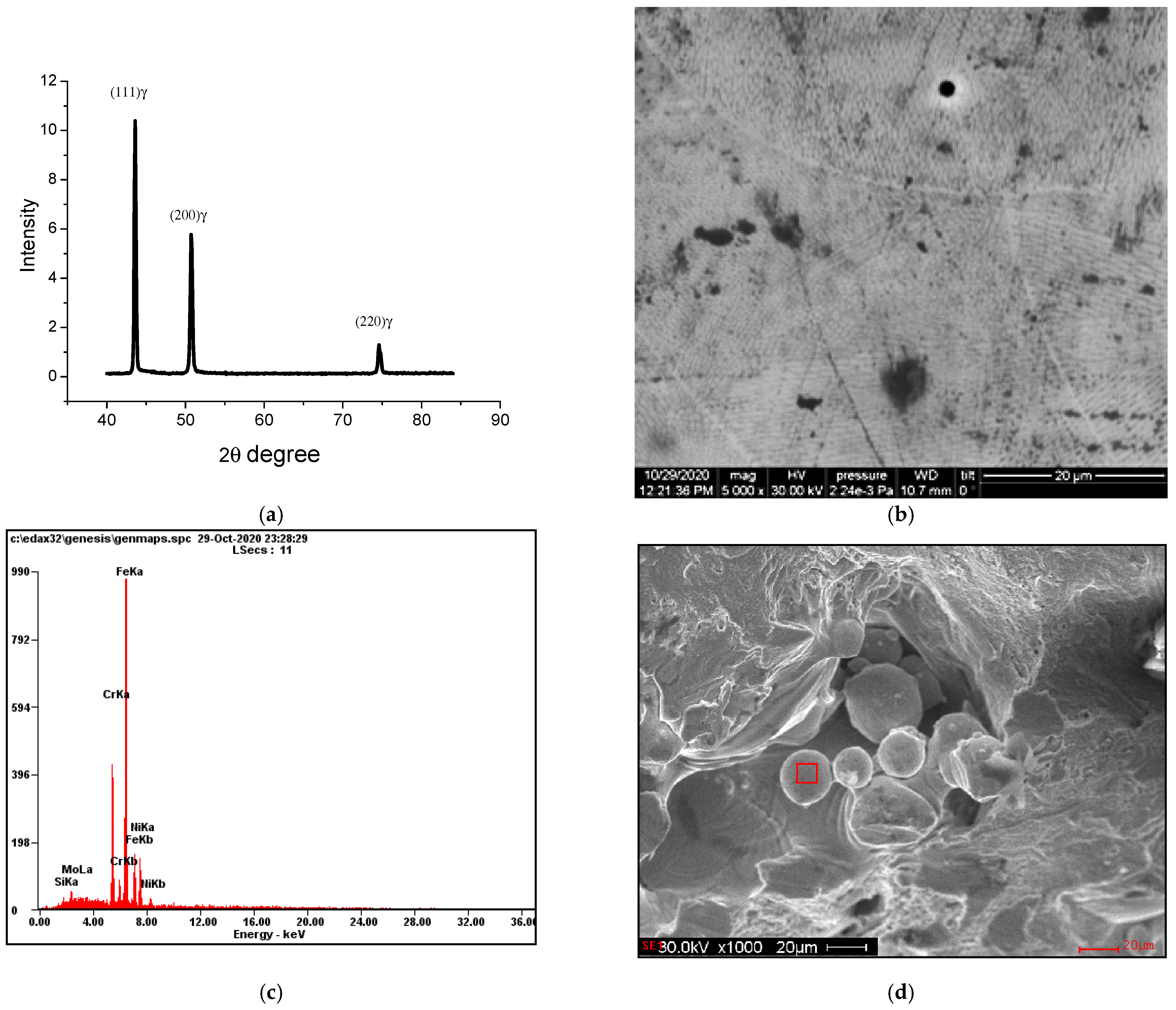
3.1. As-Built State

X-ray diffraction analysis showed that as-built samples were in an austenitic state of face-centred cubic (FCC) γ-phase with the parameter of the crystal lattice a = 0.3601 nm (Figure 1a). The distribution of the intensity of the diffraction lines corresponds to the standard for FCC phases. This may indicate the absence of the <100> cubic texture typical for steel samples obtained by the L-PBF method [16].
Figure 1 illustrates microstructure (Figure 1b) and porosity (Figure 1c,d) in the as-built state of the 316L sample; some colonies of different sizes are visible in Figure 1b as well. Colonies in AM material are elongated, and their visual size often depends on the colony growth direction and the polished surface orientation. The longitudinal cross-section is similar to an elongated grain, transverse such as a small round grain. Two types of pores were found in the structure of as-built samples, namely small, rounded gas pores, and large, irregular technological lack of fusion pores. Technical pores were filled with the unmelted powder particles; the pore size ranged from 30 µm to 200 µm (Figure 1d). Results of EDX analysis of the unmelted power inside the pore (area is pointed with a square in Figure 1d) are presented in Table 1.
Results of the TEM study of the as-built sample are presented in Figure 2. The structure of the austenitic FCC phase with the high density of the dislocation and FCC twins was observed in the as-built sample (Figure 2). The twins were very thin and sparsely located (Figure 2c,d).
The L-PBF process can be considered as a nonequilibrium metallurgical process, in which nonequilibrium phases or nonequilibrium structural states can form due to rapid solidification, fast cooling, and thermal cycling. FCC twinning was observed previously in an as-built 316L steel sample in [3] where it was explained by the thermal stresses that arose during rapid solidification in the L-PBF process. In our case, transmission electron microscope studies did not find any areas of the martensite (ε-phase, α′-phase) or ferrite phases in the structure of the as-built sample.

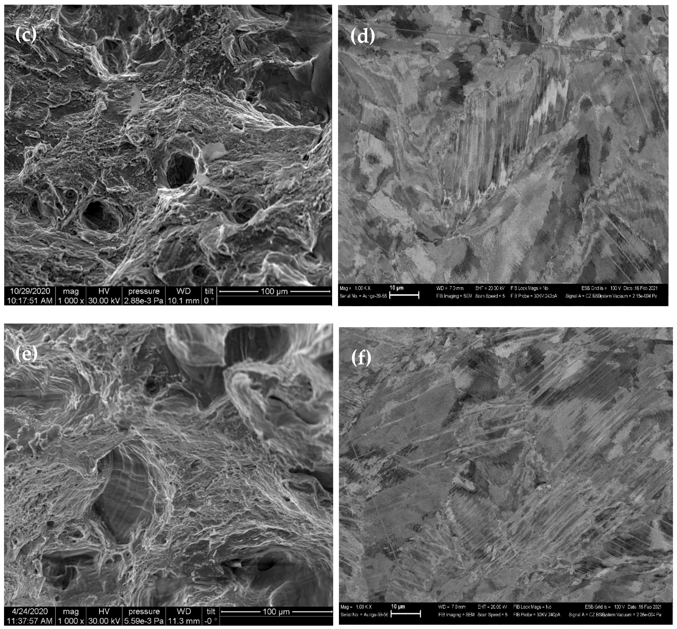
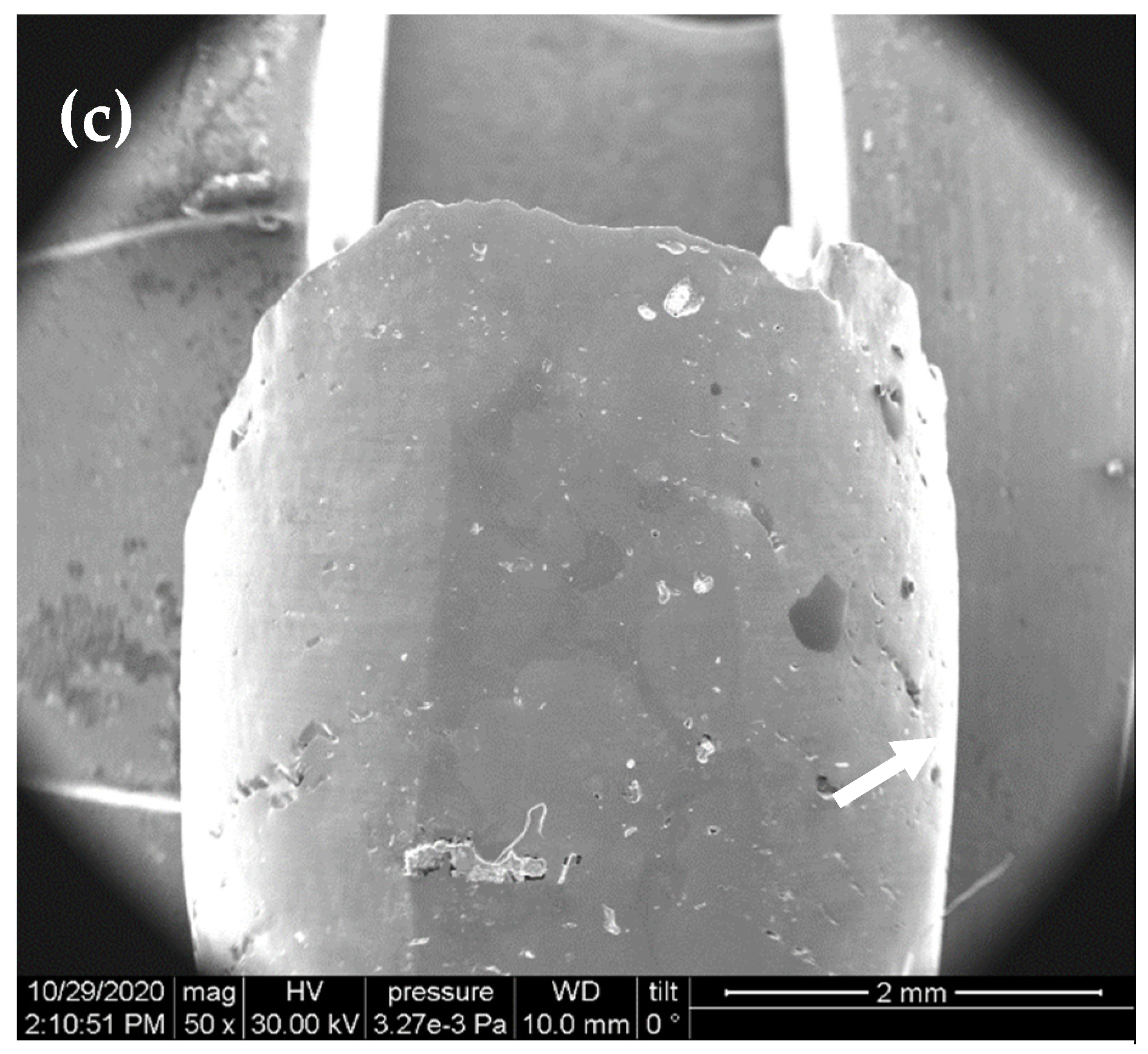
3.2. Analysis of the Structure and Mechanical Properties of the Deformed Samples

Figure 3 and Table 2 show the results of the mechanical tests of the studied LPBF samples. As it can be seen, with an increase in the strain rate, there is a simultaneous increase in both the strength and plasticity of the samples. LPBF 316L stainless steel sample in as-built conditions usually demonstrates higher than conventional steel strength characteristics. Strength characteristics of AM austenitic stainless steels vary in the range 300–600 MPa and 350–760 MPa for YS and UTS, respectively [17,18]. As it was shown in [19] the high strength and ductility might be produced in LPBF 316L samples by enhancing the TWIP, twinning induced plasticity effect that occurred due to the formation of twins and dislocation structures. The presence of the martensite phase could also lead to the high yield strength (transition induced plasticity, TRIP effect) [20]. In conventional 316L steel, the formation of α′-martensite phase was observed at strain rate 10−3 s−1 under tension for the true strain of 50% [20] and at cold rolling [21].
The strength properties measured in the present investigation are in the standard for convention austenitic steel range but do not approach the highest values, as we suggest, due to porosity in the specimens. An increase in strain rate commonly results in higher strength characteristics measured under tension experimentally. Additionally, a significant decrease in plastic properties was found in all studied samples in comparison with cold rolled sheet and plate from conventional 316 steel, where elongation is accepted as 40% (EN 10088-2 standard). Similar low plasticity (2%) was observed in bulk convention austenitic 316L steel samples with nano-twinned austenitic grains (~30 nm), while the tensile strength was found 1400 MPa [20]. In our study, the as-built sample showed the cellular structure; the average cell size was about 100 nm, therefore, the low plasticity was probably associated with the high porosity of the specimens and low mobility of dislocations in cell walls.
Table 2 shows the average values of the characteristics obtained.
Analysis of the microstructure after deformation showed that fracture occurs along with technological pores, the sharp irregular edges of which served as stress concentrators (Figure 4 and Figure 5). The presence of a large number of technological pores affected the nature of the fracture of the porous L-PBF sample 316L. The mixed brittle-ductile fracture may be observed in L-PBF 316L sample after deformation at 3 × 10⁻⁴ s−1 (Figure 4a). The brittle fracture area with the ledge (or step) is visible in Figure 4a (is pointed with arrow). With the increase in strain rate, ductile fracture along the pores was observed, which is unexpected for a material with high porosity (Figure 4c,e). Twins were revealed by SEM in all deformed samples near the edge of fracture (Figure 4b,d,f). Twinning was more pronounced in specimens deformed with a higher strain rate (Figure 4f). Investigated L-PBF specimens have a specific cellular structure. Commonly, segregation of elements and increased dislocation density are observed at cell walls. Dislocations in these cell structures are less mobile, and under high deformation rate cannot glide easily. Because of that, twinning, as the other mechanism of plastic deformation is activated. The higher the strain rate used, the higher contribution of twinning is observed (Figure 4). This resulted in higher elongation at fracture in the L-PBF 316L specimens tested with a higher strain rate (Table 2).
In metals with an FCC lattice, the easy dislocation gliding occurs in {111}<110> system. The close-packed (111) plane is also the plane of FCC twinning: (111)[11-2] or (11-1)[11-2]. FCC metals deformed by tension, drawing or extrusion show multicomponent texture <111> + <100>, and as the energy of stacking faults decreases, the <100> texture component intensity increases. This is because of a decrease in the effect of the transverse slip of the coiled components and the development of deformation twins. Compression of FCC metals produces a simple texture <110> but with a significant scatter [20].
During the L-PBF process, growth of columnar crystals during crystallization occurs in the direction of heat removal. In this case, the axis of the axial texture of cubic crystals coincides with the direction of crystal growth <100> [22]. In [23], it was shown that the L-PBF sample of 316L steel prepared with a laser scan perpendicular to the gas flow direction had a weak {110}<001> Goss texture. By varying the operating modes of the 3D laser printer, it was shown that two types of texture <110> or <100>, as well as textureless samples (random orientation) could be obtained in the L-PBF 316L steel sample [19,24,25].
In this work, we studied samples manufactured horizontally in a 3D printer chamber. All structural studies were carried out for the upper plane of the sample, which was cut perpendicular to the building direction and parallel to the tension direction. As can be seen from Figure 6, the initial as-built state may be rather related to the textureless samples. The increase in the strain rate led to a relative attenuation of texture components such as (011)[1-11] and an increase in the components of Goss texture (011)[100]. The presence of a highly diffuse but pronounced multicomponent axial crystallographic texture (011)[100] + (011)[1-11] was observed only in the deformed with a strain rate of 8 × 10⁻³ s−1. According to the literature, a change in texture, as well as additional hardening, is associated with changes in the deformation mechanism [19].

4. Conclusions

The process of an unstable plastic flow associated with the strain rate sensitivity of mechanical properties was studied in porous 316L austenitic steel samples manufactured by laser powder bed fusion (L-PBF). Presence of pores and specific cellular microstructure of LPBF 316L steel governed the deformation process and fracture mechanisms under tension at different strain rates. The following conclusions can be drawn:
  • Despite the porosity, the specimens showed high strength, the value of which increased with the loading rate. Porosity decreased the effective cross-section of the specimen, which led to lower ductility of the specimens. Additionally, porosity resulted in the de-localization of plastic deformation so that necking was not observed in all tested specimens at all strain rates.
  • With an increase in strain rate, nucleation of new pores was less pronounced, at the highest strain rate of 8 × 10⁻³ s−1, only pore coalescence was observed as the dominating microscopic mechanism of ductile fracture.
  • With an increase in strain rate, FCC (111)[11-2] or (11-1)[11-2] twinning was more developed, probably due to relatively low mobility of dislocations associated with cellular dislocation structures in L-PBF material. It resulted in an increased elongation at fracture measured in specimens tested at 8 × 10⁻³ s−1.
Commonly, microstructure of additively manufactured (AM) materials is not the same as in conventional bulk materials. Additionally, some specific defects such as gas porosity and lack of fusion may be found in AM materials. These features influence mechanical characteristics and performance of the material, and therefore, establishing the conditions of existence of instability of plastic flow is necessary for successful 3D metal product industrial applications.

Author Contributions

N.K.: Conceptualization, Methodology, Investigation, Writing original manuscript. P.K.: Conceptualization, Draft revision, writing assistance. M.Å.: Manufacturing of the L-PBF samples. Y.K.: Investigation, writing assistance. D.D.: Investigation. I.E.: Investigation. M.K.: Investigation. O.K.: Investigations. All authors have read and agreed to the published version of the manuscript.

Funding

This work was founded by the Swedish Agency for Economic and Regional Growth, grant #20201144 and Russian State program of Institute of Metal Physics “Additivity” № 121102900049-1.

Institutional Review Board Statement

Not applicable.

Informed Consent Statement

Not applicable.

Data Availability Statement

Data are available by request.

Conflicts of Interest

The authors declare no conflict of interest. The funders had no role in the design of the study; in the collection, analyses, or interpretation of data; in the writing of the manuscript, or in the decision to publish the results.

References

  1. Andreatta, F.; Lanzutti, A.; Vaglio, E.; Totis, G.; Sortino, M.; Fedrizzi, L. Corrosion behavior of 316L stainless steel manufactured by selective laser melting. Mater. Corros. 2019, 70, 1633–1645. [Google Scholar] [CrossRef]
  2. Zhong, Y.; Liu, L.; Wikman, S.; Cui, D.; Shen, Z. Intragranular cellular segregation network structure strengthening 316L stainless steel prepared by selective laser melting. J. Nucl. Mater. 2016, 470, 170. [Google Scholar] [CrossRef]
  3. Tucho, W.M.; Lysne, V.H.; Austbø, H.; Sjolyst-Kverneland, A.; Hansen, V. Investigation of effects of process parameters on microstructure and hardness of SLM manufactured SS316L. J. Alloys Compd. 2018, 740, 910. [Google Scholar] [CrossRef]
  4. Qiu, C.; Kindi, M.A.; Aladawi, A.S.; Al Hatmi, I. A comprehensive study on microstructure and tensile behaviour of a selectively laser melted stainless steel. Sci. Rep. 2018, 8, 7785. [Google Scholar] [CrossRef] [PubMed]
  5. Puichaud, A.-H.; Flament, C.; Chniouel, A.; Lomello, F.; Rouesne, E.; Giroux, P.-F.; Maskrot, H.; Schuster, F.; Béchade, J.-L. Microstructure and mechanical properties relationship of additively manufactured 316L stainless steel by selective laser melting. EPJ Nucl. Sci. Technol. 2019, 5, 23. [Google Scholar] [CrossRef]
  6. Krakhmalev, P.; Fredriksson, G.; Svensson, K.; Yadroitsev, I. Microstructure, solidification texture, and thermal stability of 316 L stainless steel manufactured by laser powder bed fusion. Metals 2018, 8, 643. [Google Scholar] [CrossRef] [Green Version]
  7. Saeidi, K.; Gao, X.; Lofaj, F.; Kvetková, L.; Shen, Z.J. Transformation of austenite to duplex austenite-ferrite assembly in annealed stainless steel 316L consolidated by laser melting. J. Alloys Compd. 2015, 633, 463–469. [Google Scholar] [CrossRef]
  8. Prashanth, K.G.; Eckert, J. Formation of metastable cellular microstructures in selective laser melted alloys. J. Alloys Compd. 2017, 707, 27–34. [Google Scholar] [CrossRef]
  9. Bertsch, K.M.; Meric de Bellefon, G.; Kuehl, B.; Thoma, D.J. Origin of dislocation structures in an additively manufactured austenitic stainless steel 316L. Acta Mater. 2020, 199, 19–33. [Google Scholar] [CrossRef]
  10. Phama, M.S.; Dovgyya, B.; Hooper, P.A. Twinning induced plasticity in austenitic stainless steel 316L made by additive manufacturing. Mater. Sci. Eng. A 2017, 704, 102–111. Available online: https://www.sciencedirect.com/science/article/abs/pii/S0921509317309899. (accessed on 1 October 2021). [CrossRef]
  11. Lee, S.-Y.; Takushima, C.; Hamada, J.-I.; Nakada, N. Macroscopic and microscopic characterizations of Portevin-Le Chatelier effect in austenitic stainless steel using high-temperature digital image correlation analysis. Acta Mater. 2021, 205, 116560. [Google Scholar] [CrossRef]
  12. Yilmaz, A. The Portevin–Le Chatelier effect: A review of experimental findings. Sci. Technol. Adv. Mater. 2011, 12, 063001. [Google Scholar] [CrossRef] [PubMed]
  13. Davidov, D.; Kazantseva, N.; Ezhov, I.; Fefelov, A.; Merkushev, A.; Afanasyev, S. Effect of dynamical aging in SLM Ti-6Al-4V alloys. Int. Sci. J. Mach. Technol. Mater. 2018, 12, 434–435. [Google Scholar]
  14. Chernov, M.; Kardashev, B.K.; Moroz, K.A. Low-temperature embrittlement and fracture of metals with different crystal lattices—Dislocation mechanisms. Nucl. Mater. Energy 2016, 9, 496–501. [Google Scholar] [CrossRef] [Green Version]
  15. Kamiura, T.; Takahashi, S. Study on effect of strain rate on elongation in advanced high strength steel. Procedia Eng. 2017, 207, 1988–1993. [Google Scholar] [CrossRef]
  16. Jia, H.; Sun, H.; Wang, H.; Wu, Y. Scanning strategy in selective laser melting (SLM): A review. Int. J. Adv. Manuf. Technol. 2021, 113, 1–23. [Google Scholar] [CrossRef]
  17. Bajaj, P.; Hariharan, A.; Kini, A.; Kürnsteiner, P.; Raabe, D.; Jägle, E.A. Steels in additive manufacturing: A review of their microstructure and properties. Mater. Sci. Eng. A 2020, 772, 138633. [Google Scholar] [CrossRef]
  18. DebRoy, T.; Wei, H.L.; Zuback, J.S.; Mukherjee, T.; Elmer, J.W.; Milewski, J.O.; Beese, A.M.; Wilson-Heid, A.; De, A.; Zhang, W. Additive manufacturing of metallic components—Process, structure and properties. Prog. Mater. Sci. 2018, 92, 112–224. [Google Scholar] [CrossRef]
  19. Sun, Z.; Tan, X.; Tor, S.B.; Chua, C.K. Simultaneously enhanced strength and ductility for 3D-printed stainless steel 316L by selective laser melting. NPG Asia Mater 2018, 10, 127–136. [Google Scholar] [CrossRef]
  20. El-Tahawy, M.; Jenei, P.; Kolonits, T.; Han, G.; Park, H.; Choe, H.; Gubicza, J. Different Evolutions of the Microstructure, Texture, and Mechanical Performance During Tension and Compression of 316L Stainless Steel. Met. Mater. Trans. A 2020, 51, 3447–3460. [Google Scholar] [CrossRef]
  21. Yan, F.K.; Liu, G.Z.; Tao, N.R.; Lu, K. Strength and ductility of 316L austenitic stainless steel strengthened by nano-scale twin bundles. Acta Mater. 2012, 60, 1059–1071. [Google Scholar] [CrossRef]
  22. Lobanov, M.L.; Yurovskikh, A.S.; Kardonina, N.I.; Rusakov, G.M. Methods for Texture Study in Materials; Ural Publishing House University: Ekaterinburg, Russia, 2014; p. 115. ISBN 978-5-7996-1107-1. [Google Scholar]
  23. Andreau, O.; Koutiri, I.; Peyre, P.; Penot, J.-D. Texture control of 316L parts by modulation of the melt pool morphology in selective laser melting. J. Mater. Process. Technol. 2019, 264, 21–31. [Google Scholar] [CrossRef] [Green Version]
  24. Donik, C.; Kraner, J.; Paulin, I.; Godec, M. Influence of the Energy Density for Selective Laser Melting on the Microstructure and Mechanical Properties of Stainless Steel. Metals 2020, 10, 919. [Google Scholar] [CrossRef]
  25. Ishimoto, T.; Wu, S.; Ito, Y.; Sun, S.-H.; Amano, H.; Nakano, T. Crystallographic Orientation Control of 316L Austenitic Stainless Steel via Selective Laser Melting. ISIJ Int. 2020, 60, 8. [Google Scholar] [CrossRef] [Green Version]
Figure 1. As-built state of 316L sample: (a) the X-ray diffractogram; (b) the microstructure and porosity; (c,d) SEM image.
Metals 11 01870 g001
Figure 2. Microstructure of the as-build 316L sample, TEM: (a) dislocations, the bright-field image; (b) SAED pattern to (a), zone axis [110]γ; (c) twins; the dark-field image taken with twin reflex, twin plane (1−1); (d) SAED pattern to (c), zone axis [110]m||[−1−10]t.
Metals 11 01870 g002
Figure 3. Tensile engineering stress–strain curves of the studied L-PBF samples, the deformation speed: 1— 3 × 10 4   s 1 ; 2—1 × 10 3   s 1 ; 3—8 × 10 3   s 1 .
Metals 11 01870 g003
Figure 4. The fracture surfaces and sub-fracture microstructures of the L-PBF 316L steel samples deformed with the different strain rates, (a,c,e) SEM images; (b,d,f) Electron backscattered SEM images: (a,b) 3 × 10 4   s 1 ; (c,d) 1 × 10 3   s 1 ; (e,f) 8 × 10 3   s 1 .
Metals 11 01870 g004aMetals 11 01870 g004b
Figure 5. The outer surface of the studied L-PBF samples near the fracture zone after deformation with the different strain rates, SEM images: (a) 3 × 10 4   s 1 ; (b) 1 ×   10 3   s 1 ; (c) 8 ×   10 3   s 1 .
Metals 11 01870 g005aMetals 11 01870 g005b
Figure 6. The pole figures with {111} and {100} zones obtained in the studied samples after deformation with the different strain rates, EBSD analysis: 1— 3 × 10 4   s 1 ; 2—1 ×   10 3   s 1 ; 3—8 ×   10 3   s 1 .
Metals 11 01870 g006
Table 1. Results of the Energy Dispercive X-ray Spectroscopy (EDS), wt.%.
FeNiCrMoSi
Bal.13.4216.772.401.23
Table 2. Mechanical properties of L-PBF 316L steel tested at different strain rates.
Strain Rate, s−1YS, MPaUTS, MPaδ, %
3 × 10 4 5355201.7
1 ×   10 3 5545265.0
8 ×   10 3 5935625.4
Publisher’s Note: MDPI stays neutral with regard to jurisdictional claims in published maps and institutional affiliations.

