Article

Influence of Sulfobacillus thermosulfidooxidans on Initial Attachment and Pyrite Leaching by Thermoacidophilic Archaeon Acidianus sp. DSM 29099

Biofilm Centre, Aquatische Biotechnologie, Universität Duisburg—Essen, Universitätsstraße 5, 45141 Essen, Germany
*
Author to whom correspondence should be addressed.
Minerals 2016, 6(3), 76; https://doi.org/10.3390/min6030076
Submission received: 29 April 2016 / Revised: 6 July 2016 / Accepted: 13 July 2016 / Published: 21 July 2016
(This article belongs to the Special Issue Biotechnologies and Mining)

Abstract

:
At the industrial scale, bioleaching of metal sulfides includes two main technologies, tank leaching and heap leaching. Fluctuations in temperature caused by the exothermic reactions in a heap have a pronounced effect on the growth of microbes and composition of mixed microbial populations. Currently, little is known on the influence of pre-colonized mesophiles or moderate thermophiles on the attachment and bioleaching efficiency by thermophiles. The objective of this study was to investigate the interspecies interactions of the moderate thermophile Sulfobacillus thermosulfidooxidans DSM 9293T and the thermophile Acidianus sp. DSM 29099 during initial attachment to and dissolution of pyrite. Our results showed that: (1) Acidianus sp. DSM 29099 interacted with S. thermosulfidooxidansT during initial attachment in mixed cultures. In particular, cell attachment was improved in mixed cultures compared to pure cultures alone; however, no improvement of pyrite leaching in mixed cultures compared with pure cultures was observed; (2) active or inactivated cells of S. thermosulfidooxidansT on pyrite inhibited or showed no influence on the initial attachment of Acidianus sp. DSM 29099, respectively, but both promoted its leaching efficiency; (3) S. thermosulfidooxidansT exudates did not enhance the initial attachment of Acidianus sp. DSM 29099 to pyrite, but greatly facilitated its pyrite dissolution efficiency. Our study provides insights into cell-cell interactions between moderate thermophiles and thermophiles and is helpful for understanding of the microbial interactions in a heap leaching environment.

Graphical Abstract

1. Introduction

Bioleaching is applied at an industrial scale (biomining) for extraction of valuable metals from mineral concentrates and low-grade ores [1,2], for instance copper, zinc, cobalt, and so on [3,4]. Bioleaching can occur in nature and accelerate acidity of ground water and cause serious environmental problems, like acid mine/rock drainage (AMD/ARD) [5,6]. Microorganisms involved in the dissolution of metal sulfides are mainly extremely acidophilic archaea and bacteria, which are able to oxidize reduced inorganic sulfur compounds (RISCs) and/or iron(II) ions [7,8].
Sulfobacillus sp., Gram-positive bacteria, are moderately thermophilic acidophiles that can utilize a broad range of energy substrates, including iron(II) ions, RISCs, and mineral sulfides in environments with high metal concentrations [9,10]. Since spore formation enables Sulfobacillus to survive long periods of time under unfavorable, or even deleterious, growth conditions, these bacteria are often found in heap leaching operations and acidic geothermal springs [10]. Acidianus sp. are facultatively anaerobic thermophilic archaea capable of growth at high temperature and at low pH with iron(II) ions, RISCs, metal sulfides, or complex organic compounds [11]. These thermophiles can be used to bioprocess recalcitrant minerals, e.g., chalcopyrite [12].
At the industrial scale, the main technologies used for metal recovery include dumps, heaps, in situ and in stope leaching, vat leaching, and stirred tank leaching [3]. Mesophilic, moderately thermophilic, and thermophilic microbes are involved in heap leaching [13]. The dominant microorganisms in both tank and heap leaching environments are Sulfobacillus, Acidithiobacillus caldus, Ferroplasma spp, Leptospirillum ferriphilum, and thermophiles Sulfolobus sp. and Acidianus sp. [13,14,15,16]. Fluctuations in temperature can have a pronounced effect on the growth of individual microbes and also on the composition of mixed microbial populations. Mesophilic and moderately thermophilic microbes are present in heaps at the beginning of heap leaching. Reactions involved in the biological oxidation of mineral sulphides (exothermic reactions) result in heat generation and the increased temperature provides favourable conditions for the growth of thermophilic microorganisms [13,17]. Mineral sulphide oxidation is compromised at elevated temperatures, unless an appropriate thermophilic consortium is present in the process [13].
Microbial attachment to and biofilm formation on mineral sulphides are of great importance for mineral bioleaching [18]. During the succession of microbial communities caused by temperature fluctuations, attached populations may subsequently change. It has been shown that the presence of L. ferriphilum in the biofilm can affect the initial attachment of S. thermosulfidooxidans to pyrite [19]. Additionally, the presence of an active biofilm of iron oxidizers may influence the subsequent cell attachment of other species [20]. It was shown that At. thiooxidans cells showed a 40% increased attachment to pyrite pre-colonized by cells of At. ferrooxidans or L. ferrooxidans. In addition, the attachment of At. thiooxidans was more rapid if the pyrite was already colonized by L. ferrooxidans, but not by At. ferrooxidans [21].
This study focused on how pyrite pre-colonized by moderately thermophilic microorganisms affected the attachment, colonization and pyrite bioleaching efficiency of thermophiles. In this study, we have chosen the moderate thermophile S. thermosulfidooxidans DSM 9293T and a thermophile Acidianus sp. DSM 29099 to study their interspecies interactions with respect to the pyrite-leaching process. The effect of pyrite pre-colonized with cells of S. thermosulfidooxidansT on initial attachment and bioleaching by Acidianus sp. DSM 29099 were evaluated in a simulated heap leaching environment.

2. Experimental Section

2.1. Strains and Media

Acidianus sp. DSM 29099 (GeneBank accession number of 16S rRNA: KJ921703) was isolated from one of the geothermal hot springs at Caviahue–Copahue, Neuquen, Argentina [22]. S. thermosulfidooxidans DSM 9293T was purchased from DSMZ (Deutsche Sammlung von Mikroorganismen und Zellkulturen, Braunschweig, Germany). The cultures were cultivated in Mackintosh (MAC) medium [23] with an initial pH of 1.7. Cultures were grown on pyrite (10% w/v) with yeast extract (0.2 g/L). Cells of Acidianus sp. DSM 29099 and S. thermosulfidooxidansT were cultivated at 65 and 45 °C, respectively, with shaking at 120 rpm.

2.2. Pyrite Preparation

Pyrite grains with a diameter of 200–500 μm were selected after grinding and sieving of pyrite cubes (origin Navajun, Spain). These were used for the attachment and leaching experiments. Pyrite slices with a size of approx. 1 cm × 1 cm × 2 mm were cut from pyrite cubes and were used for visualization of biofilms. Both slices and grains were washed with boiling 6 M HCl for 30 min, and rinsed with deionized water until a neutral pH. Afterwards, pyrite samples were washed three times with acetone. These treatments were done in order to remove impurities such as iron ions and sulphur compounds on pyrite surface. After cleaning, pyrite was dried at 80 °C for 12 h and sterilized at 120 °C in a nitrogen atmosphere for 24 h [24].

2.3. Iron and pH Determination

Iron ions were determined according to German standard method for the examination of water, wastewater, and sludge (DIN 38406-1). The determination was performed in 96-well-plate and scanned at 492 nm with a spectrophotometer equipped with a microplate reader (Infinite®200 PRO, TECAN, Männedorf, Switzerland). The iron standard solutions used for calibration curve were prepared in the range of 0.8–6 mg/L.
For iron(II) ion determination, each sample was mixed with 2 mL of master solution consisting of ammonium acetate solution (16 g/L in glacial acetic acid) and (1,10)-phenanthrolinium chloride monohydrate (0.2 g/L). The mixtures were incubated for 15 min in the dark. After the measurement, hydroxylammonium chloride (4 g/L) was added into the samples for total iron determination. After 15 min incubation in the dark, samples were measured again at 492 nm. Iron(III) ion concentration can be determined by subtracting the one of iron(II) ions from the one of total iron ions.
A WTW (pH 537) pH meter with a Mettler Toledo electrode (InLab® 422 semimicro pH electrode; Mettler Toledo GmbH, Urdorf, Switzerland) was used for pH determination. The pH meter was calibrated before measurements by using pH calibration solutions (Roth, Germany).

2.4. Preparation of Pyrite Pre-Colonized by S. thermosulfidooxidansT or of S. thermosulfidooxidansT Exudates

Cells of S. thermosulfidooxidansT with an initial cell number of 5 × 10⁸ cells/mL were inoculated in 100 mL Erlenmeyer flasks containing 10% (w/v) pyrite grains and 50 mL MAC medium. The cultures were incubated at 45 °C with shaking at 120 rpm for seven days. Pyrite grains colonized with active or inactivated biofilm cells of S. thermosulfidooxidansT were prepared according to the previous report [21]. Briefly, pyrite grains colonized with active cells were obtained by washing the grains twice with 50 mL sterile MAC medium. To prepare pyrite grains colonized by inactive cells, pyrite grains attached with cells were subsequently incubated at 80–100 °C for 2 h.
The efficiency of this inactivation procedure was evaluated. Briefly, the inactive cells and active cells were both inoculated in the fresh MAC medium at 45 °C for three days. Cell growth from both inactive cells and active cells was compared.
The culture supernatants were filtered into a new sterilized 100 mL Erlenmeyer flask through a filter with the pore-size of 0.2 μm. The exudates were determined to be contained 0.5 g/L iron(III) ions. Thus, supplementation with 0.5 g/L iron(III) ions was tested to evaluate their influence on cell initial attachment and leaching efficiency by Acidianus sp. DSM 29099.

2.5. Initial Attachment

Initial attachment tests were carried out in 100 mL Erlenmeyer flasks containing 50 mL MAC medium, 0.2 g/L yeast extract, and 10% (w/v) pyrite or pyrite pre-colonized by cells of S. thermosulfidooxidansT. Cell attachment of Acidianus sp. DSM 29099 to clean pyrite in the presence of S. thermosulfidooxidansT exudates was also tested.
Surface area may be a limiting factor for cell attachment. Based on surface area of a pyrite grain ( = 4 π r 2 ) and a cell ( = 2 π rh + 2 π r 2 ) used in the experiments, 10% of pyrite was used for the attachment experiments. This provided sufficient surface area for cell attachment (Supplementary Figure S1). Briefly, cells were cultivated at 65 °C (pure culture of Acidianus sp. DSM 29099) or 45 °C (pure culture of Acidianus sp. DSM 29099, S. thermosulfidooxidansT and mixed cultures) and shaking at 120 rpm. Cell numbers were monitored by direct counting using a Thoma chamber within the first 6 h. Control experiments without adding pyrite were also included. Each experiment was performed in triplicate. The initial cell number was 1 × 10⁸ cells/mL for pure cultures. For mixed cultures, the initial total cell number was 1 × 10⁸ cells/mL (Mix 1) or 2 × 10⁸ cells/mL (Mix 2) with equal cell numbers for each species.
The attached cells were calculated by subtracting the number of planktonic cells from the initial total cell number. The theoretical attachment rate of mixed cultures (At) was calculated by averaging the sum of the attachment rate of each pure culture [25]. The formula is as follows:
At = (Aa+AS.t)/2
where At = theoretical attachment rate of mixed cultures, a = Acidianus sp. DSM 29099, and S.t = S. thermosulfidooxidansT.

2.6. Leaching Experiments

Leaching experiments were carried out in 100 mL Erlenmeyer flasks containing 50 mL MAC medium, 0.2 g/L yeast extract, and 10% (w/v) of pyrite or pyrite pre-colonized by cells of S. thermosulfidooxidansT. Leaching activity of Acidianus sp. DSM 29099 with clean pyrite in the presence of S. thermosulfidooxidansT exudates was also tested. Briefly, leaching experiments were carried out for three weeks. In addition, pyrite leaching experiments in MAC medium without adding cells of S. thermosulfidooxidansT as an abiotic control were included. To determine the bioleaching efficiency, 1 mL of leach solutions were withdrawn from each flask at specific intervals and used for determining planktonic cell number, pH, and total iron concentration. The increase of total iron was used to estimate the pyrite dissolution efficiency. Each experiment was performed in triplicate.

2.7. Visualization of Biofilm Formation by Confocal Laser Scanning Microscopy

For visualization of cells and biofilms on pyrite, samples were stained and observed by confocal laser scanning microscopy (CLSM) according to the previous report [26]. Briefly, pyrite samples colonized with cells were rinsed by 1 mL deionized, particle-free water three times. Nucleic acid staining was done by covering the samples with 6 μM SYTO 9 (Invitrogen, Carlsbad, CA, USA) for 15 min. After staining, a washing step was carefully performed by using 1 mL particle-free deionized water to remove the unbound stain. Pyrite grains were fixed on diagnostic microscope slides (Gerhard Menzel, Braunschweig, Germany, GmbH) by using an anti-fading agent (Citifluor, Ltd., London, UK, AF2) and covered by coverslips. All staining was performed in the dark. Examination of stained samples was performed by CLSM using a laser scanning module (LSM 510 Carl Zeiss®, Jena, Germany) coupled to an inverted Axiovert100MBP microscope (Zeiss®, Hallbergmoos, Germany). The laser excitation wavelength (Argon laser) 488 nm was used to detect fluorophore signals. Surface topography and texture of the pyrite surface were recorded by using the CLSM in reflection mode. Fluorescence images were analysed by an extended version of the software ImageJ (NIH, Bethesda, MD, USA) [27].

3. Results and Discussion

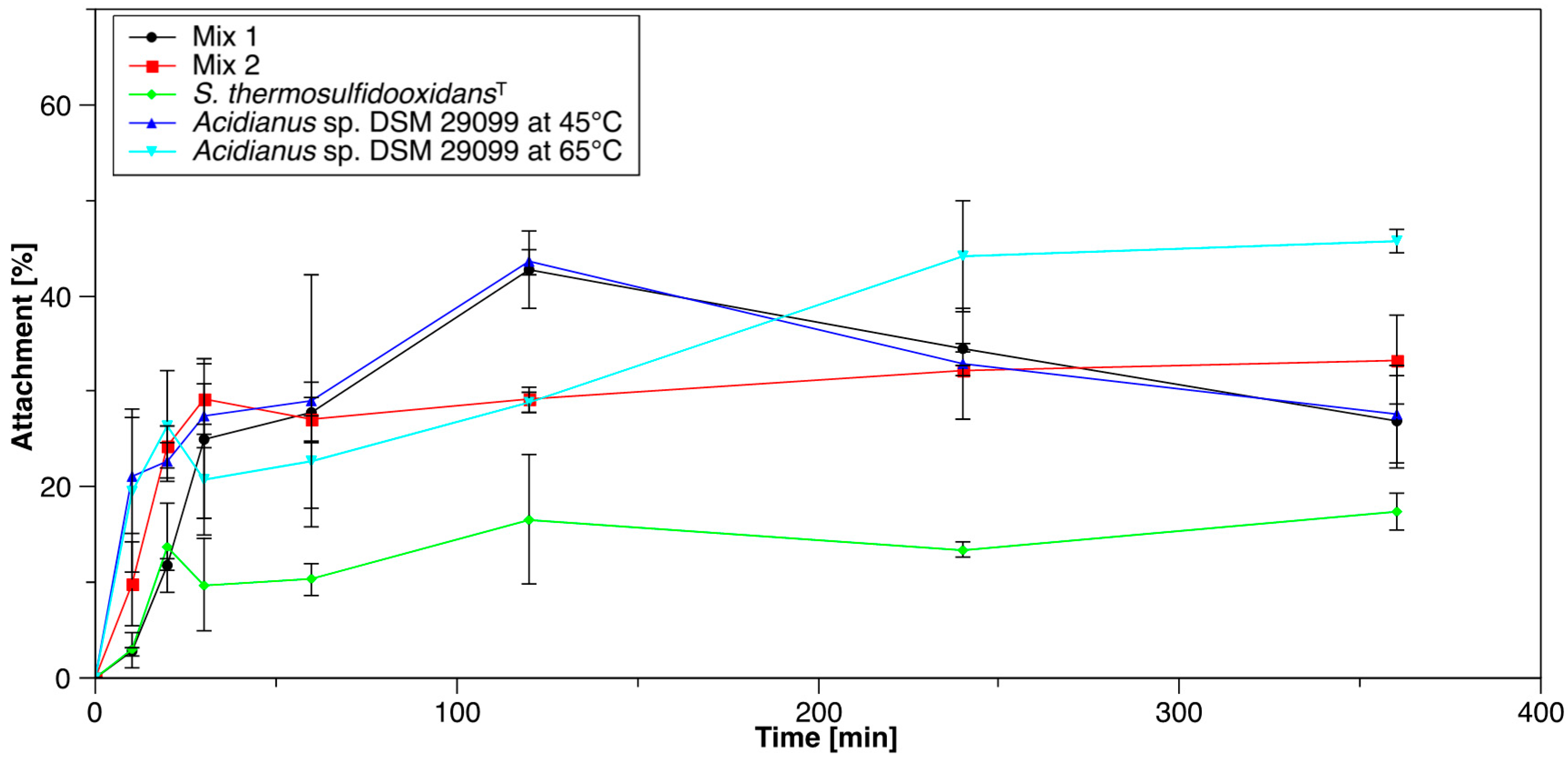
3.1. Initial Attachment of Cells to Pyrite

3.1.1. Pure and Mixed Cultures

Pure cultures of S. thermosulfidooxidansT exhibited the lowest attachment to pyrite grains, with 17% of total cells attached to pyrite after 6 h incubation (Figure 1), which coincides with previous reports [19,28]. In case of Acidianus sp. DSM 29099, cells showed the highest attachment, with 46% of total cells attached to pyrite when cultivated at 65 °C. Both species did not show a clear tendency to attach to the glass wall of Erlenmeyer flasks (Supplementary Figure S2). In a previous study, it was found that up to 60% and 35% of cells adhered to pyrite, and chalcopyrite surfaces within the first 2 h [29]. In case of Acidianus copahuensis 29038, maximum initial cell attachment to pyrite and chalcopyrite were 43% and 69%, respectively [30,31]. These data suggest that cell attachment strongly depends on the nature of the species and the pre-cultivation conditions [18,32]. In addition, it has been reported that even within the same species, different strains have a different affinity for the same mineral [33].
Attachment of Acidianus sp. DSM 29099 at 45 °C was around 28% (Figure 1). It was 65% less than when cultivated at 65 °C (its optimal growth temperature). Obviously, the cultivation temperature influences the initial attachment of cells of Acidianus sp. DSM 29099. This is in agreement with the previous report that attachment and biofilm formation by Metallosphaera hakonensis showed the highest attachment at the optimal growth temperature and less cells attached to minerals at reduced temperature [34].
The attachment of mixed cultures was compared to each pure culture with the theoretical value (for the pure culture): (23% = (28% + 17%)/2). In this test, 27% of the cells in sample Mix 1 (initial total cell number: 1 × 10⁸ cell/mL) and 33% of cells in sample Mix 2 (initial total cell number: 2 × 10⁸ cell/mL) were detected to attach to pyrite, respectively (Figure 1). Both attachment rates of mixed cultures showed an increase compared to the theoretical attachment of pure cultures. This suggests that cells of Acidianus sp. DSM 29099 and S. thermosulfidooxidansT may have some interactions influencing the attachment. Although no quorum-sensing molecules from acidophilic archaea have been detected, specific interactions like cell-cell communication in the form of quorum sensing between the two species may exist. Cells in Mix 2 showed a considerably increased attachment (24%) if compared to Mix 1, indicating that initial cell density could influence the initial attachment in mixed cultures. The attachment dependency on initial cell number has been reported for Acidithiobacillus ferrooxidans to refractory gold concentrate [27]. It might be that cell-cell interactions between Acidianus sp. DSM 29099 and S. thermosulfidooxidans are cell density-dependent. In the case of Leptospirillum ferrooxidans DSM 2391 and Acidithiobacillus caldus DSM 8584, a mixed culture of the two species showed 37% increased attachment ratio to pyrite compared to the theoretical attachment value for pure cultures [25].

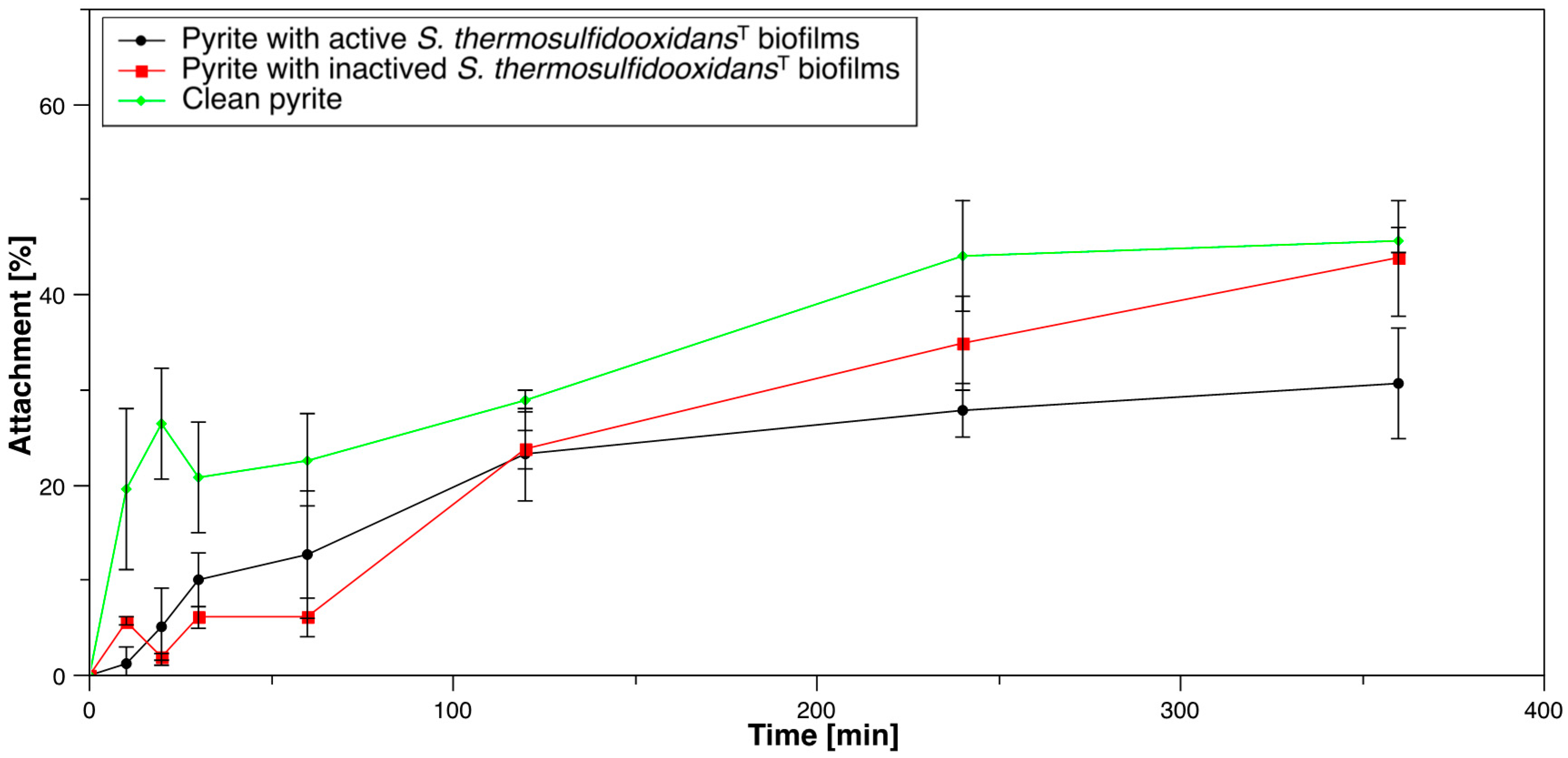
3.1.2. Acidianus sp. DSM 29099 to Pyrite Pre-Colonized with S. thermosulfidooxidansT

The efficiency of the inactivation procedure was confirmed due to the fact that no cell growth was observed in the fresh MAC medium with inactive cells on pyrite after three days incubation. By contrast, with active cells on pyrite cell numbers reached 3 × 10⁸ cells/L (not shown). Attached cells of Acidianus sp. DSM 29099 increased over time within 6 h under all test conditions (Figure 2). A notable increase of the attached cells to clean pyrite was noticed and the attachment reached 28% within the first 20 min. By comparison, only 5% of inoculum attached to the pyrite pre-colonized by active cells of S. thermosulfidooxidansT and almost no cells attached to the pyrite colonized with dead S. thermosulfidooxidansT cells within the first 20 min. There is no notable difference between the attachment of Acidianus sp. DSM 29099 to pre-colonized pyrite with active cells or inactive cells during the early stages of the experiments. They do not diverge until about 2 h of incubation. After 6 h, more than 46% of the total cells attached to the clean pyrite, while approximately 44% and 31% of the total cells attached to the pyrite covered with active and inactivated S. thermosulfidooxidansT cells, respectively. The presence of active and inactive cells of S. thermosulfidooxidansT on the surface of pyrite grains both inhibited the attachment of Acidianus sp. DSM 29099 at the beginning of the experiment. The pre-colonization by cells of S. thermosulfidooxidansT may change the surface properties (e.g., hydrophobicity) in a way which is inhibitory to cell attachment of cells of Acidianus sp. DSM 29099. Another assumption is that the sites favourable for cell attachment have been occupied by S. thermosulfidooxidansT cells, resulting in a reduced number of active sites available for cells of Acidianus sp. DSM 29099. In addition, the release of some antagonistic compounds by S. thermosulfidooxidansT cells may be possible.

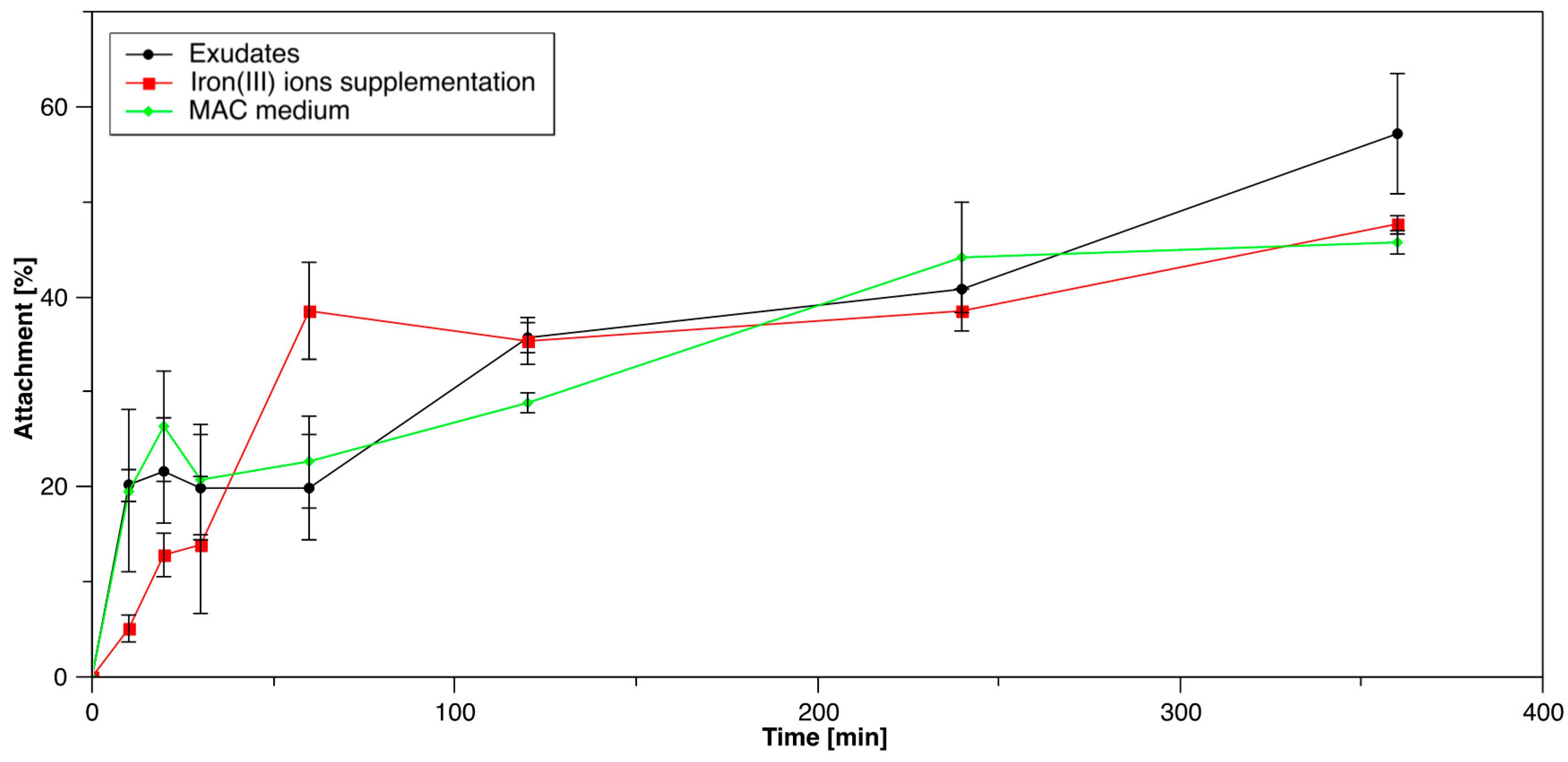
3.1.3. Influence of S. thermosulfidooxidansT Exudates on Initial Attachment of Acidianus sp. DSM 29099

After 6 h incubation, 46% and 48% of total cells attached to pyrite with and without 0.5 g/L iron(III) ion supplementation, respectively (Figure 3). Thus, it seems that iron(III) ions had no significant effect on initial cell attachment of Acidianus sp. DSM 29099. This is, however, in contrast to the previous study indicating that attachment of Acidithiobacillus was promoted by addition of 1 mM iron(III) ions, especially when cells were pre-grown on pyrite [35]. When incubated with S. thermosulfidooxidansT exudates, 57% of the cells of Acidianus sp. DSM 29099 attached to pyrite within 6 h (Figure 3). However, there was no significant difference before 4 h incubation. Thus, S. thermosulfidooxidansT exudates did not show clear stimulation on the initial attachment of Acidianus sp. DSM 29099. The organic compounds in the S. thermosulfidooxidansT exudates, probably humic acids, proteins, and polysaccharides [36], may be relevant to the cell attachment of Acidianus sp. DSM 29099. It has been reported that surfaces immersed in an aquatic environment adsorb dissolved organic matter [37,38]. This is called the conditioning film. The EPS from acidophiles grown on minerals can be partly considered as surface active compounds [39]. These can create a conditioning film at the interface and thus affect the cellular attachment [40]. Such compounds have already been detected on surfaces of cells of Acidianus, Sulfolobus metallicus, Ferroplasma acidiphilum, and Acidithiobacillus sp. and they may be involved in the initial stages of adhesion to mineral or sulphur surfaces [22,41,42]. This conditioning film formed by S. thermosulfidooxidansT exudates may have an effect on the cellular attachment to surfaces [43,44].

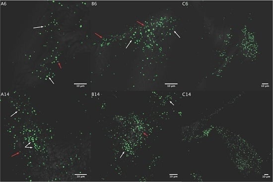
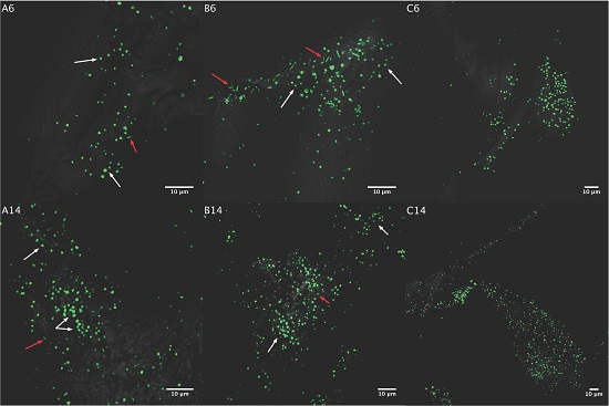
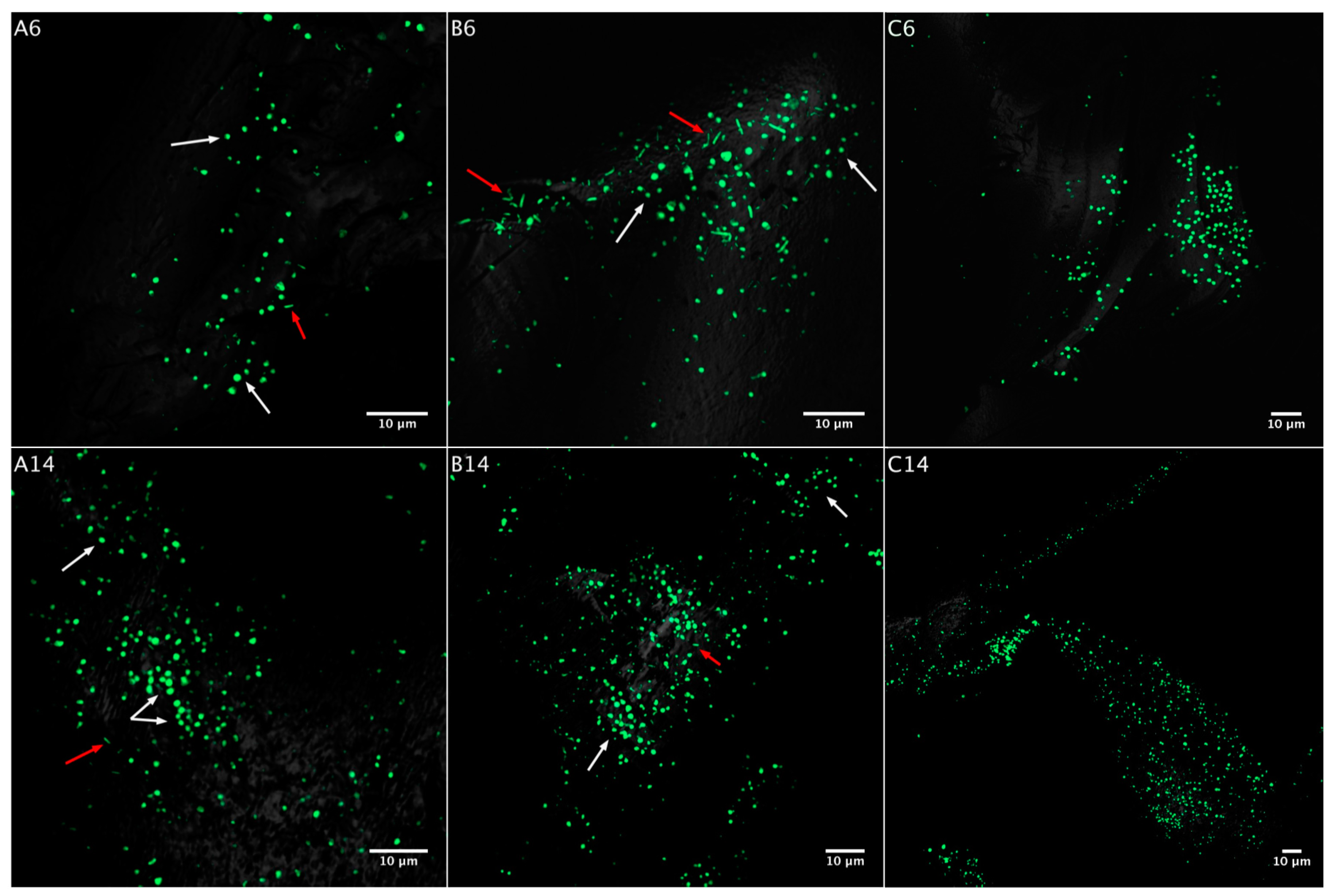
3.2. Biofilm Formation

3.2.1. Acidianus sp. DSM 29099 on Pyrite Pre-Colonized by S. thermosulfidooxidansT

We first observed the morphology of biofilm cells on pyrite. Cells of Acidianus sp. DSM 29099 show a spherical appearance, but S. thermosulfidooxidansT cells are rod-shaped. Thus, the two strains in mixed cultures are distinguishable by cell morphology (Supplementary Figure S3). Biofilm formation by S. thermosulfidooxidansT cells on pyrite decreased after seven days of incubation [45]. The biofilm formation of Acidianus sp. DSM 29099 on pyrite pre-colonized with active/inactivated S. thermosulfidooxidansT biofilms is shown in Figure 4. Biofilm cells of Acidianus sp. DSM 29099 increased over time on all kind of pyrite grains. On day 6, biofilm cells of Acidianus sp. DSM 29099 were found on clean pyrite as well as on pyrite pre-colonized by cells of S. thermosulfidooxidansT. Pre-colonized S. thermosulfidooxidansT cells were also observed (Figure 4, upper row). In addition, pyrite surfaces showed more S. thermosulfidooxidansT cells after inactivation treatment than the surfaces without treatment. It could be that active cells of S. thermosulfidooxidansT detached from the pyrite surface because of the unfavourable conditions (65 °C). On the pyrite surface with inactivated S. thermosulfidooxidansT cells, Acidianus sp. DSM 29099 and pre-established S. thermosulfidooxidansT cells showed comparable amounts on day 6. Due to the constant increase of attached Acidianus sp. DSM 29099 cells, the proportion of S. thermosulfidooxidansT cells was reduced on pyrite (Figure 4). However, there were no notable differences in the biofilm formation by Acidianus sp. DSM 29099 on the grains as a result of the pyrite pre-treatment.
S. thermosulfidooxidansT cells were detected on pyrite grains, which were pre-colonized either by active or inactivated cells during the whole test. However, in both cases no growth of cells of S. thermosulfidooxidansT were observed (not shown). Cells of Acidianus sp. DSM 29099 were heterogeneously distributed over the pyrite surface. No physical contact between the two species was observed (Figure 4).

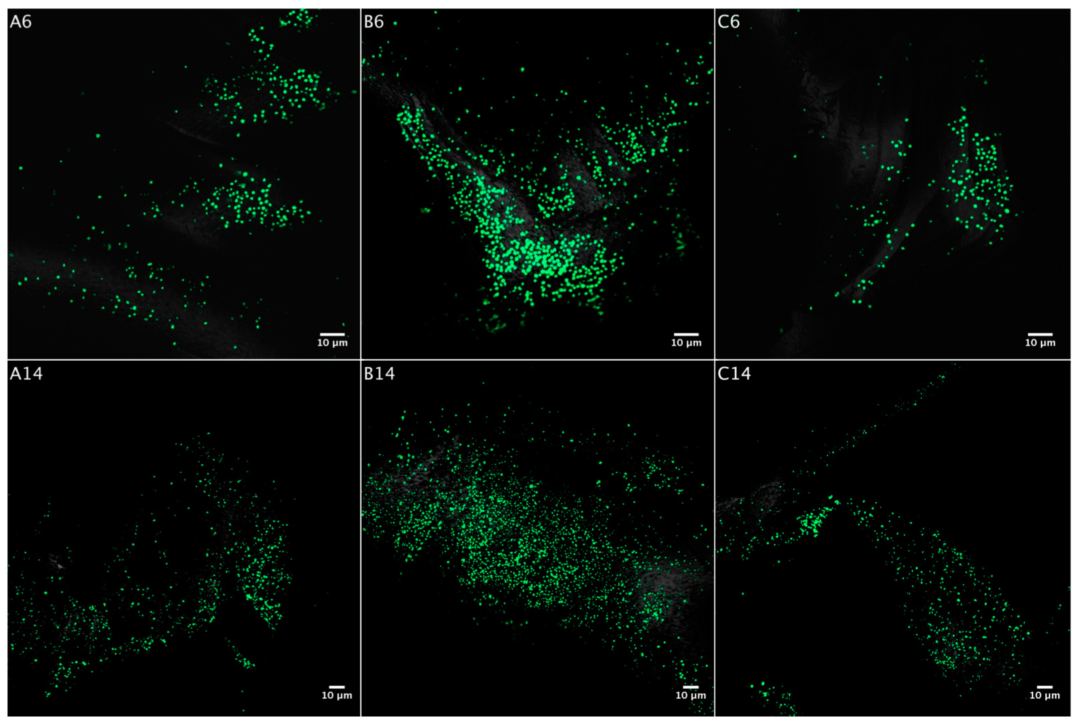
3.2.2. Influence of S. thermosulfidooxidansT Exudates on Biofilm Formation by Acidianus sp. DSM 29099

Cells of Acidianus sp. DSM 29099 grown with iron(III) ion supplementation were often found on pyrite grains on day 3. In contrast, cells were rarely observed on pyrite grains, when grown in S. thermosulfidooxidansT exudates (Supplementary Table S1). Attached cells on pyrite grains increased in all tested conditions over time (Figure 5). On day 6, biofilms were much denser with additional iron(III) ions than in the other growth conditions. This phenomenon was also observed on day 14 (Supplementary Table S1). Thus it seems that biofilm formation by cells of Acidianus sp. DSM 29099 is stimulated by additional iron(III) ions. Also, S. thermosulfidooxidansT exudates seem to promote its biofilm development (Figure 5). It has been reported that addition of iron(III) ions promotes A. ferrooxidans attachment on pyrite [32,46]. Although addition of iron(III) ions showed no significant effects on cell initial attachment of Acidianus sp. DSM 29099 (Figure 3), biofilm cells of Acidianus sp. DSM 29099 on pyrite surfaces were greatly increased. Thus, iron(III) ions did not enhance the cell attachment at early stage (Figure 3) but may be relevant in the persistence of attachment and biofilm maintenance (Figure 4).

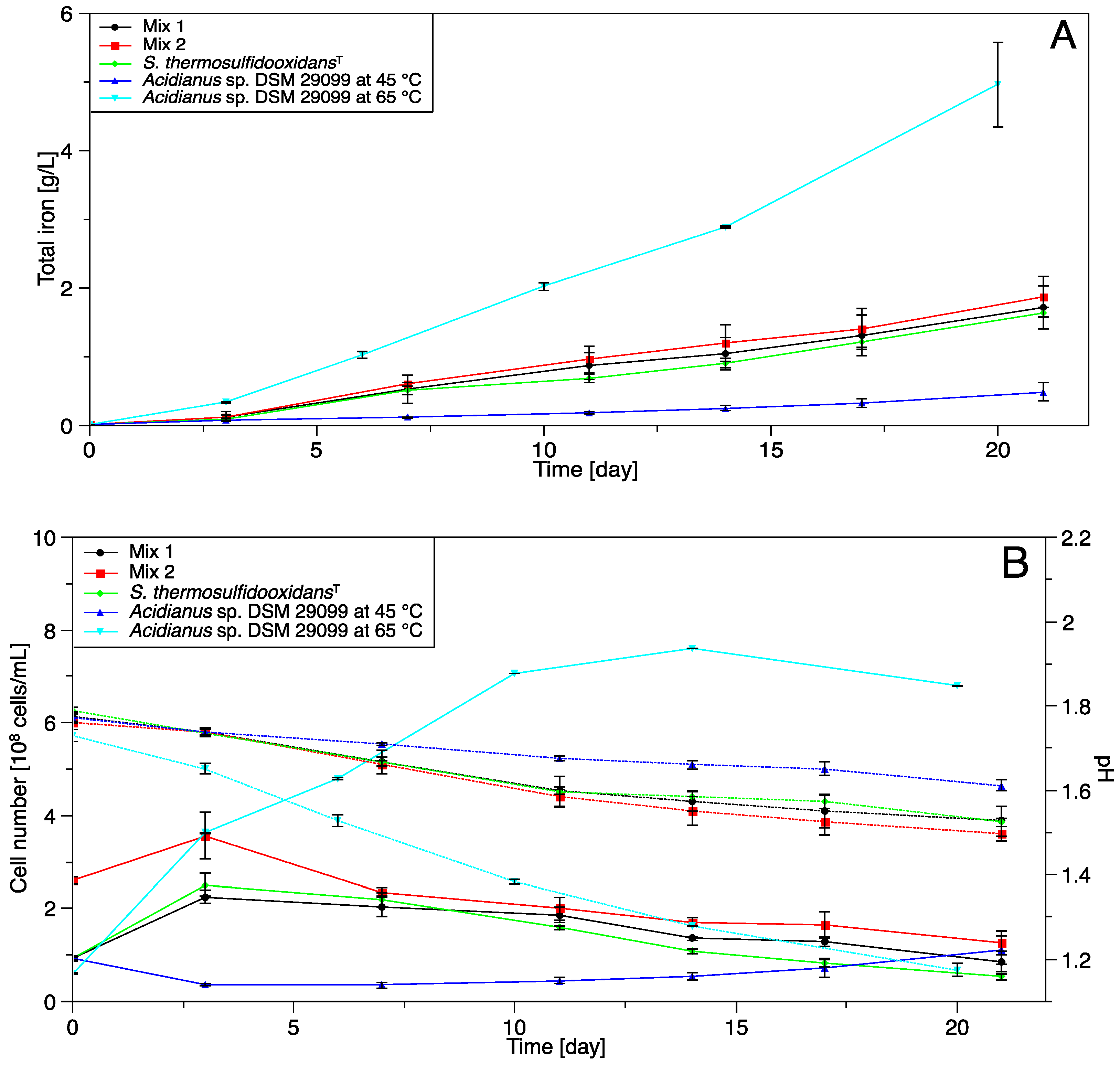
3.3. Pyrite Leaching

3.3.1. Pure and Mixed Cultures

Pure culture of S. thermosulfidooxidansT showed a low leaching efficiency (Figure 6A), which is in agreement with the previous reports that pyrite oxidation by Sulfobacillus strains was slow [47,48]. Acidianus sp. DSM 29099 showed the highest pyrite leaching efficiency at 65 °C according to the highest concentration of total iron (5.0 g/L), the highe st cell number (6.8 × 10⁸ cells/mL) and pH changes (0.56). However, at 45 °C cell number or pH did not change notably (Figure 6B) and no significant leaching was noticed (Figure 6A). Pyrite leaching at 45 °C in both mixed cultures (Mix 1 and 2) was similar with that in the pure culture of S. thermosulfidooxidansT. In abiotic controls at 65 °C around 190 mg/L total iron was detected, which was much higher than for the assay at 45 °C (30 mg/L, Supplementary Figure S4). In any case, abiotic leaching at 65 °C was negligible compared to bioleaching by Acidianus sp. DSM 29099. This agrees with the fact that pyrite can be oxidized by iron(III) ions but not proton [8]. A formation of flocs was observed for Acidianus sp. DSM 29099 in pure and mixed cultures with cells of S. thermosulfidooxidansT if cultivated at 45 °C (Supplementary Figure S5).

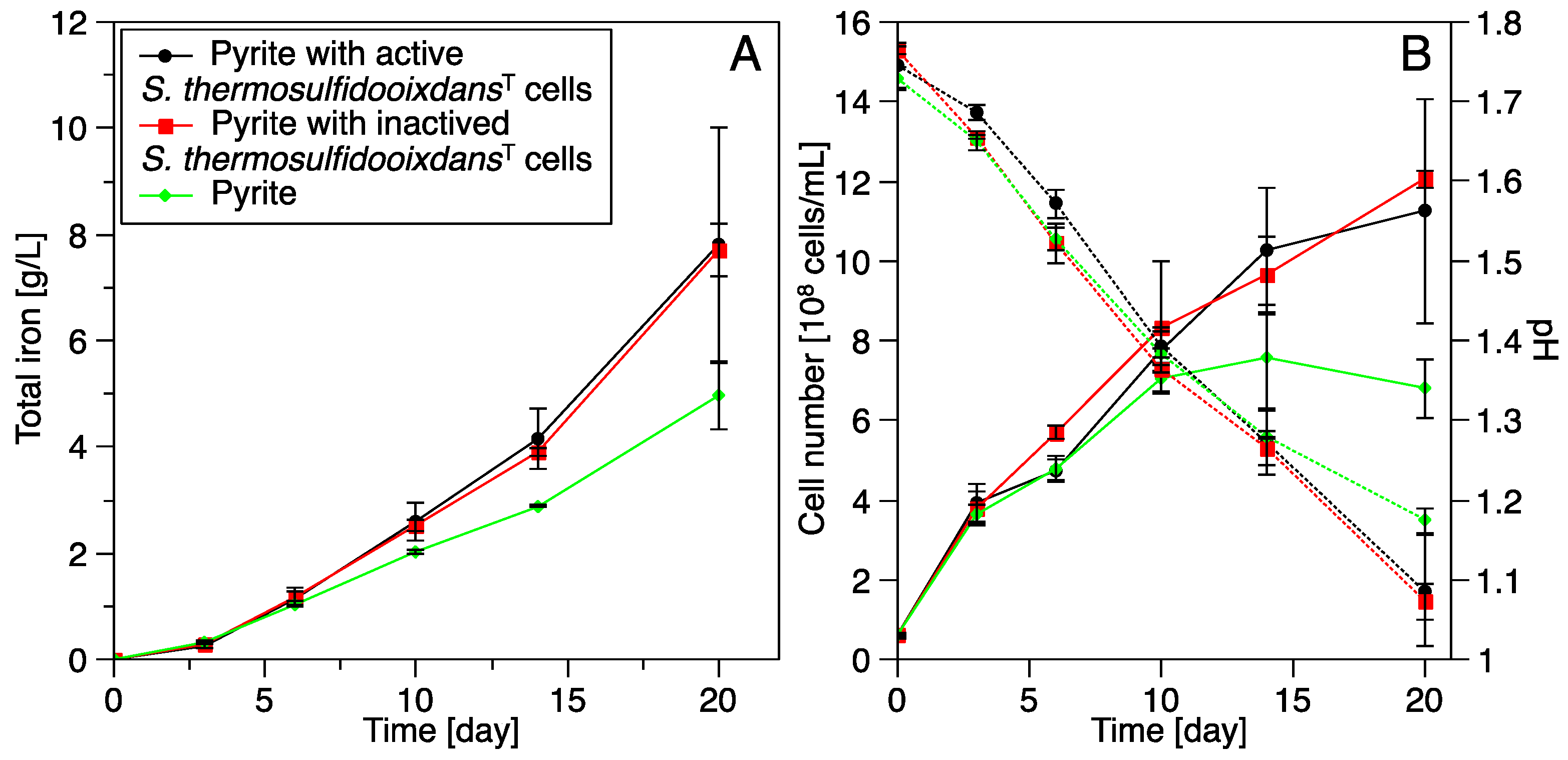
3.3.2. Bioleaching by Acidianus sp. DSM 29099 with Pyrite Pre-Colonized by S. thermosulfidooxidansT

The cell number of Acidianus sp. DSM 29099 grown on clean pyrite increased from 1 × 10⁸ to 8 × 10⁸ cells/mL after 14 days (Figure 7B). Then it slightly decreased to 7 × 10⁸ cells/mL until the end of the incubation. When grown on pyrite pre-colonized with active or inactivated cells of S. thermosulfidooxidansT, the numbers of planktonic cells kept increasing and reached 11 × 108 and 12 × 108 cells/mL, respectively (Figure 7B). Meanwhile, the decline of pH in all tested cultures from around 1.7 to 1.1 was correlated with the increased cell numbers of Acidianus sp. DSM 29099. The drop of pH in Acidianus sp. DSM 29099 cultures grown on pyrite pre-colonized by both active and inactivated cells of S. thermosulfidooxidansT (0.7 ΔpH) was slightly higher than the one for clean pyrite (0.6) (Figure 7B). Total iron concentrations in all samples were continuously increasing during the experiments. The highest total dissolved iron concentration (~7.8 g/L) was detected in the culture with pyrite pre-colonized by both active and inactivated cells of S. thermosulfidooxidansT (Figure 7A). Bioleaching by Acidianus sp. DSM 29099 with pyrite pre-colonized by either active or inactivated S. thermosulfidooxidansT cells was ~55% higher than that with clean pyrite. This clearly shows that pyrite leaching by Acidianus sp. DSM 29099 was enhanced by pre-established biofilms of S. thermosulfidooxidansT. However, no significant difference in leaching efficiency of Acidianus sp. DSM 29099 were detected with clean pyrite or pyrite pre-colonized by S. thermosulfidooxidansT cells before 10 days of incubation. Although EPS are playing a key role in bioleaching, killed cells of A. ferrooxidans with EPS caused no significant pyrite dissolution [46]. We observed that both, the active biofilm cells of S. thermosulfidooxidansT on pyrite and the inactivated S. thermosulfidooxidansT cells, showed no pyrite oxidation activity at 65 °C (not shown). The yeast extract was most probably exhausted in the medium after 10 days’ incubation and EPS of S. thermosulfidooxidansT may have facilitated bioleaching by Acidianus sp. DSM 29099. As mentioned above, the main components of S. thermosulfidooxidansT EPS, such as humic acid, proteins, and polysaccharides [36] can be utilized by Acidianus sp. DSM 29099. Considering the reduced initial cellular attachment of Acidianus sp. DSM 29099 to pre-colonized pyrite by active cells of S. thermosulfidooxidansT (Figure 2), it seems that the initial attachment to pyrite is not always correlated with pyrite dissolution.

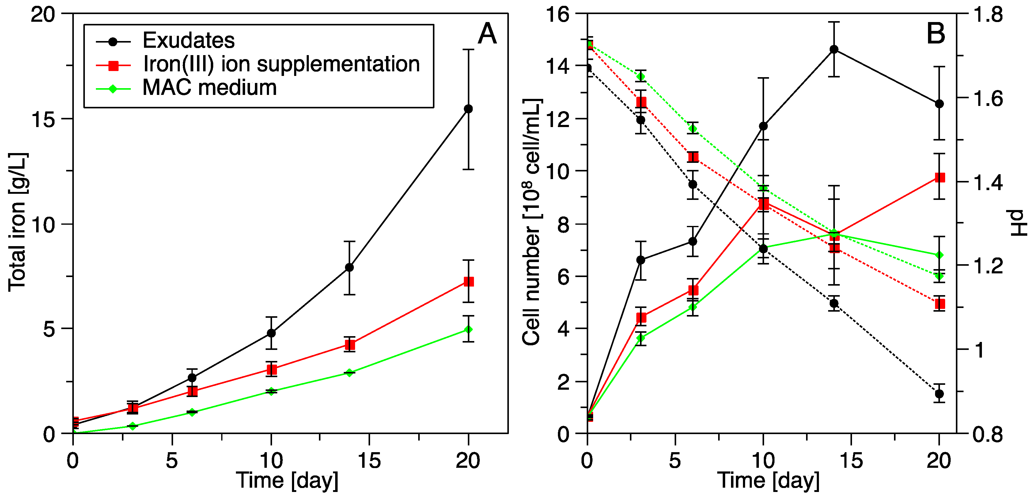
3.3.3. Influence of S. thermosulfidooxidansT Exudates on Pyrite Leaching by Acidianus sp. DSM 29099

Cell numbers of Acidianus sp. DSM 29099 under all growth conditions increased gradually in the first 10 days (Figure 8). The maximum was reached in the culture with no supplementations (MAC medium) and in the one with iron(III) ion supplementation on day 10. However, cells in the culture with exudates of S. thermosulfidooxidansT kept increasing and reached 1.5 × 10⁹ cells/mL after 14 days, slightly decreasing to 1.3 × 10⁸ cells/mL at the end of the experiments. Obviously, cell growth of Acidianus sp. DSM 29099 was promoted by S. thermosulfidooxidansT exudates. The highest pH drop was noticed in the culture with S. thermosulfidooxidansT exudates. The lowest pH change was found in the culture with no supplementation (MAC medium). The pH decrease was obviously correlated with cell growth and pyrite oxidation activity.
The highest total iron of 15.4 g/L until the end of the study, was measured in the culture with S. thermosulfidooxidansT exudates. In the culture with supplementation of iron(III) ions, 7.3 g/L of total iron was detected. By contrast, only around 5 g/L of total iron was released in the culture with no supplementation (Figure 8). These data clearly show that exudates of S. thermosulfidooxidansT or supplementation of iron(III) ions significantly enhance pyrite dissolution by Acidianus sp. DSM 29099 cells if compared to the ones grown in MAC medium with no supplementations. Due to metabolic versatility of Acidianus [49], exudates of S. thermosulfidooxidansT could be used as energy source and promote cell growth. In addition, it is possible that certain molecules in S. thermosulfidooxidansT exudates may influence pyrite leaching by Acidianus sp. DSM 29099. However, this needs further investigations.
It has been shown that cell attachment to ores as well as biofilm formation is correlated with pyrite leaching by acidophiles [18,50]. A recent study shows that biofilm populations of At. ferrooxidansT are responsible for pyrite dissolution at the early stage (first 4–5 days). However, planktonic cells are contributing substantially later on in the bioleaching process [35]. Thus, it is not surprising that the initial attachment of Acidianus sp. DSM 29099 was not enhanced (Figure 3) or even inhibited (Figure 2) but pyrite leaching was promoted (Figure 7 and Figure 8).

4. Summary and Conclusions

We studied the interspecies interactions of S. thermosulfidooxidansT and Acidianus sp. DSM 29099 during pyrite leaching. Pre-colonized biofilms of S. thermosulfidooxidansT promoted pyrite leaching by Acidianus sp. DSM 29099, while the initial attachment of Acidanus sp. DSM 29099 was not positively influenced. S. thermosulfidooxidansT exudates did not enhance the initial attachment but significantly facilitated the pyrite leaching by Acidianus sp. DSM 29099. Our data suggest the presence of interactions between S. thermosulfidooxidansT and Acidianus sp. DSM 29099 during pyrite dissolution. Further investigations on the interaction mechanisms between these acidophilic bacteria and archaea may allow to design and optimize microbial consortia to be used in bio-heap leaching applications.

Supplementary Materials

The following are available online at www.mdpi.com/2075-163X/6/3/76/s1.

Acknowledgments

Thomas R. Neu (Department of River Ecology, Helmholtz Centre for Environmental Research–UFZ, Magdeburg, Germany) is acknowledged for assistance in Confocal Laser Scanning Microscopy imaging. Mario Vera (Schools of Engineering, Biological Sciences and Medicine; School of Engineering, Pontificia Universidad Católica de Chile, Santiago, Chile) is acknowledged for helpful discussions. We gratefully thank the three anonymous reviewers for their critical and constructive suggestions.

Author Contributions

Jing Liu performed the laboratory tasks and carried out the experiments. Ruiyong Zhang and Wolfgang Sand conceived and designed the experiments. Qian Li performed some CLSM analyses. Jing Liu wrote the first draft of the manuscript. Ruiyong Zhang and Wolfgang Sand assisted in preparing the manuscript, all authors read and approved the final version.

Conflicts of Interest

The authors declare no conflict of interest.

References

  1. Brierley, C.L.; Brierley, J.A. Progress in bioleaching: Part B: Applications of microbial processes by the minerals industries. Appl. Microbiol. Biotechnol. 2013, 97, 7543–7552. [Google Scholar] [CrossRef] [PubMed]
  2. Johnson, D.B. Biomining—Biotechnologies for extracting and recovering metals from ores and waste materials. Curr. Opin. Biotechnol. 2014, 30, 24–31. [Google Scholar] [CrossRef] [PubMed]
  3. Watling, H. Review of biohydrometallurgical metals extraction from polymetallic mineral resources. Minerals 2014, 5, 1–60. [Google Scholar] [CrossRef]
  4. Gentina, J.C.; Acevedo, F. Copper bioleaching in Chile. Minerals 2016, 6, 23. [Google Scholar] [CrossRef]
  5. Johnson, D.B.; Hallberg, K.B. Acid mine drainage remediation options: A review. Sci. Total Environ. 2005, 338, 3–14. [Google Scholar] [CrossRef] [PubMed]
  6. Sand, W.; Jozsa, P.-G.; Kovacs, Z.-M.; Sasaran, N.; Schippers, A. Long-term evaluation of acid rock drainage mitigation measures in large lysimeters. J. Geochem. Explor. 2007, 92, 205–211. [Google Scholar] [CrossRef]
  7. Schippers, A.; Breuker, A.; Blazejak, A.; Bosecker, K.; Kock, D.; Wright, T. The biogeochemistry and microbiology of sulfidic mine waste and bioleaching dumps and heaps, and novel Fe(II)-oxidizing bacteria. Hydrometallurgy 2010, 104, 342–350. [Google Scholar] [CrossRef]
  8. Vera, M.; Schippers, A.; Sand, W. Progress in bioleaching: Fundamentals and mechanisms of bacterial metal sulfide oxidation—Part A. Appl. Microbiol. Biotechnol. 2013, 97, 7529–7541. [Google Scholar] [CrossRef] [PubMed]
  9. Da Costa, M.S.; Rainey, F.A.; Albuquerque, L. Sulfobacillus. In Bergey's Manual of Systematics of Archaea and Bacteria; Whitman, W.B., Ed.; John Wiley & Sons, Ltd: Hoboken, NJ, USA, 2015; pp. 1–6. [Google Scholar]
  10. Watling, H.R.; Perrot, F.A.; Shiers, D.W. Comparison of selected characteristics of Sulfobacillus species and review of their occurrence in acidic and bioleaching environments. Hydrometallurgy 2008, 93, 57–65. [Google Scholar] [CrossRef]
  11. Wheaton, G.; Counts, J.; Mukherjee, A.; Kruh, J.; Kelly, R. The confluence of heavy metal biooxidation and heavy metal resistance: Implications for bioleaching by extreme thermoacidophiles. Minerals 2015, 5, 397–451. [Google Scholar] [CrossRef]
  12. Norris, P.R.; Burton, N.P.; Clark, D.A. Mineral sulfide concentrate leaching in high temperature bioreactors. Miner. Eng. 2013, 48, 10–19. [Google Scholar] [CrossRef]
  13. Franzmann, P.D.; Hawkes, R.B.; Haddad, C.M.; Plumb, J.J. Mining with microbes. Microbiol. Aust. 2007, 28, 124–126. [Google Scholar]
  14. Plumb, J.J.; Hawkes, R.B.; Franzmann, P.D. The microbiology of moderately thermophilic and transiently thermophilic ore heaps. In Biomining; Springer: Berlin, Germany, 2007; pp. 217–235. [Google Scholar]
  15. Rawlings, D.E. Heavy metal mining using microbes. Annu. Rev. Microbiol. 2002, 56, 65–91. [Google Scholar] [CrossRef] [PubMed]
  16. Norris, P.R.; Johnson, D.B. Acidophilic microorganisms. In Extremophiles: Microbial Life in Extreme Environments; Horikoshi, K., Grant, W.D., Eds.; Wiley-Liss: New York, NY, USA; 1998. [Google Scholar]
  17. Dixon, D.G. Analysis of heat conservation during copper sulphide heap leaching. Hydrometallurgy 2000, 58, 27–41. [Google Scholar] [CrossRef]
  18. Zhang, R.; Bellenberg, S.; Neu, T.R.; Sand, W.; Vera, M. The biofilm lifestyle of acidophilic metal/sulfur-oxidizing microorganisms. In Biotechnology of Extremophiles: Advances and Challenges; Rampelotto, H.P., Ed.; Springer International Publishing: Cham, Germany, 2016; pp. 177–213. [Google Scholar]
  19. Li, Q.; Zhang, R.; Krok, B.A.; Vera, M.; Sand, W. Biofilm formation of Sulfobacillus thermosulfidooxidans on pyrite in the presence of Leptospirillum ferriphilum. Adv. Mater. Res. 2015, 1130, 141–144. [Google Scholar] [CrossRef]
  20. Noël, N.; Florian, B.; Sand, W. AFM & EFM study on attachment of acidophilic leaching organisms. Hydrometallurgy 2010, 104, 370–375. [Google Scholar]
  21. Bellenberg, S.; Diaz, M.; Noel, N.; Sand, W.; Poetsch, A.; Guiliani, N.; Vera, M. Biofilm formation, communication and interactions of leaching bacteria during colonization of pyrite and sulfur surfaces. Res. Microbiol. 2014, 165, 773–781. [Google Scholar] [CrossRef] [PubMed]
  22. Zhang, R.; Neu, T.; Bellenberg, S.; Kuhlicke, U.; Sand, W.; Vera, M. Use of lectins to in situ visualize glycoconjugates of extracellular polymeric substances in acidophilic archaeal biofilms. Microb. Biotechnol. 2015, 8, 448–461. [Google Scholar] [CrossRef] [PubMed]
  23. Mackintosh, M. Nitrogen fixation by Thiobacillus ferrooxidans. J. Gen. Microbiol. 1978, 105, 215–218. [Google Scholar] [CrossRef]
  24. Schippers, A.; Jozsa, P.; Sand, W. Sulfur chemistry in bacterial leaching of pyrite. Appl. Environ. Microbiol. 1996, 62, 3424–3431. [Google Scholar] [PubMed]
  25. Florian, B.; Noël, N.; Thyssen, C.; Felschau, I.; Sand, W. Some quantitative data on bacterial attachment to pyrite. Miner. Eng. 2011, 24, 1132–1138. [Google Scholar] [CrossRef]
  26. Zhang, R.; Bellenberg, S.; Castro, L.; Neu, T.R.; Sand, W.; Vera, M. Colonization and biofilm formation of the extremely acidophilic archaeon Ferroplasma acidiphilum. Hydrometallurgy 2014, 150, 245–252. [Google Scholar] [CrossRef]
  27. González, R.; Gentina, J.; Acevedo, F. Attachment behaviour of Thiobacillus ferrooxidans cells to refractory gold concentrate particles. Biotechnol. Lett. 1999, 21, 715–718. [Google Scholar] [CrossRef]
  28. Maulani, N. Interaction of the Acidophilic Archaeon Ferroplasma acidiphilum with Leaching Bacteria during Initial Biofilm Formation on Pyrite. Master’s Thesis, Universität Duisburg-Essen, Essen, Germany, 2015. [Google Scholar]
  29. Zhang, R.; Zhang, Y.; Neu, T.R.; Li, Q.; Bellenberg, S.; Sand, W.; Vera, M. Initial attachment and biofilm formation of a novel crenarchaeote on mineral sulfides. Adv. Mater. Res. 2015, 1130, 127–130. [Google Scholar] [CrossRef]
  30. Castro, C.; Vera, M.; Donati, E.; Sand, W. Visualization of attachment and colonization of pyrite surfaces by a novel species of Acidianus. Adv. Mater. Res. 2013, 825, 70–73. [Google Scholar] [CrossRef]
  31. Castro, C.; Donati, E. Effects of different energy sources on cell adhesion and bioleaching of a chalcopyrite concentrate by extremophilic archaeon Acidianus copahuensis. Hydrometallurgy 2016, 162, 49–56. [Google Scholar] [CrossRef]
  32. Gehrke, T.; Hallmann, R.; Kinzler, K.; Sand, W. The EPS of Acidithiobacillus ferrooxidans—A model for structure-function relationships of attached bacteria and their physiology. Water Sci. Technol. 2001, 43, 159–167. [Google Scholar] [PubMed]
  33. Afzal Ghauri, M.; Okibe, N.; Barrie Johnson, D. Attachment of acidophilic bacteria to solid surfaces: The significance of species and strain variations. Hydrometallurgy 2007, 85, 72–80. [Google Scholar] [CrossRef]
  34. Africa, C.-J.; van Hille, R.P.; Sand, W.; Harrison, S.T.L. Investigation and in situ visualisation of interfacial interactions of thermophilic microorganisms with metal-sulphides in a simulated heap environment. Miner. Eng. 2013, 48, 100–107. [Google Scholar] [CrossRef]
  35. Bellenberg, S.; Barthen, R.; Boretska, M.; Zhang, R.; Sand, W.; Vera, M. Manipulation of pyrite colonization and leaching by iron-oxidizing Acidithiobacillus species. Appl. Microbiol. Biotechnol. 2015, 99, 1435–1449. [Google Scholar] [CrossRef] [PubMed]
  36. Aguirre Morales, M. Extracellular Polymeric Substances (EPS) Production in Sulfobacillus thermosulfidooxidans and Its Relevance on Attachment to Metal Sulfides. Master’s Thesis, Universidad Nacional de Colombia, Bogota, Colombia, 2013. [Google Scholar]
  37. Baier, R. Influence of the Initial Surface Condition of Materials on Bioadhesion. In Proceedings of the Third International Congress on Marine Corrosion and Fouling, Gaithersburg, MD, USA, 2–6 October 1973; pp. 633–639.
  38. Bhosle, N.B.; Garg, A.; Fernandes, L.; Citon, P. Dynamics of amino acids in the conditioning film developed on glass panels immersed in the surface seawaters of Dona Paula Bay. Biofouling 2005, 21, 99–107. [Google Scholar] [CrossRef] [PubMed]
  39. Sand, W.; Gehrke, T. Analysis and function of the EPS from the strong acidophile Thiobacillus ferrooxidans. In Microbial Extracellular Polymeric Substances; Springer: Berlin, Germany, 1999; pp. 127–141. [Google Scholar]
  40. Neu, T.R. Significance of bacterial surface-active compounds in interaction of bacteria with interfaces. Microbiol. Rev. 1996, 60, 151. [Google Scholar] [PubMed]
  41. Starkey, R.; Jones, G.; Frederick, L. Effects of medium agitation and wetting agents on oxidation of sulphur by Thiobacillus thiooxidans. J. Gen. Microbiol. 1956, 15, 329–334. [Google Scholar] [CrossRef] [PubMed]
  42. Zhang, R.; Neu, T.R.; Zhang, Y.; Bellenberg, S.; Kuhlicke, U.; Li, Q.; Sand, W.; Vera, M. Visualization and analysis of EPS glycoconjugates of the thermoacidophilic archaeon Sulfolobus metallicus. Appl. Microbiol. Biotechnol. 2015, 99, 7343–7356. [Google Scholar] [CrossRef] [PubMed]
  43. Jain, A.; Bhosle, N.B. Biochemical composition of the marine conditioning film: Implications for bacterial adhesion. Biofouling 2009, 25, 13–19. [Google Scholar] [CrossRef] [PubMed]
  44. De Kerchove, A.J.; Elimelech, M. Impact of alginate conditioning film on deposition kinetics of motile and nonmotile Pseudomonas aeruginosa strains. Appl. Environ. Microbiol. 2007, 73, 5227–5234. [Google Scholar] [CrossRef] [PubMed]
  45. Li, Q.; Sand, W.; Zhang, R. Enhancement of biofilm formation on pyrite by Sulfobacillus thermosulfidooxidans. Minerals 2016, 6, 71. [Google Scholar] [CrossRef]
  46. Gehrke, T.; Telegdi, J.; Thierry, D.; Sand, W. Importance of extracellular polymeric substances from Thiobacillus ferrooxidans for bioleaching. Appl. Environ. Microbiol. 1998, 64, 2743–2747. [Google Scholar] [PubMed]
  47. Norris, P.; Owen, J. Mineral sulphide oxidation by enrichment cultures of novel thermoacidophilic bacteria. FEMS Microbiol. Rev. 1993, 11, 51–56. [Google Scholar] [CrossRef]
  48. Norris, P. Factors affecting bacterial mineral oxidation: The example of carbon dioxide in the context of bacterial diversity. In Proceedings of the International Biohydrometallurgical Symposium, Jakson Hole, WY, USA, 13–18 August 1989; pp. 3–14.
  49. Giaveno, M.A.; Urbieta, M.S.; Ulloa, J.R.; Toril, E.G.; Donati, E.R. Physiologic versatility and growth flexibility as the main characteristics of a novel thermoacidophilic Acidianus strain isolated from Copahue geothermal area in Argentina. Microb. Ecol. 2013, 65, 336–346. [Google Scholar] [CrossRef] [PubMed]
  50. Vardanyan, A.; Stepanyan, S.; Vardanyan, N.; Markosyan, L.; Sand, W.; Vera, M.; Zhang, R. Study and assessment of microbial communities in natural and commercial bioleaching systems. Miner. Eng. 2015, 81, 167–172. [Google Scholar] [CrossRef]
Figure 1. Initial attachment of cells of Acidianus sp. DSM 29099 and S. thermosulfidooxidansT to pyrite in pure and mixed cultures. The initial cell number was 1 × 10⁸ cells/mL for each pure cultureand Mix 1, and 2 × 10⁸ cells/mL for Mix 2, respectively. Cells were cultivated in 50 mL MAC medium (pH 1.7) with 0.2 g/L yeast extract containing 10% pyrite grains (200–500 μm) at 45 °C and 120 rpm.
Minerals 06 00076 g001
Figure 2. Initial attachment of cells of Acidianus sp. DSM 29099 to pyrite pre-colonized by cells of S. thermosulfidooxidansT. Cells of Acidianus sp. DSM 29099 (1 × 10⁸ cells/mL) were incubated in 50 mL MAC medium (pH 1.7) containing 0.2 g/L yeast extract and 10% (w/v) pyrite grains (200–500 μm) at 65 °C and 120 rpm.
Minerals 06 00076 g002
Figure 3. Influence of S. thermosulfidooxidansT exudates on the initial attachment of cells of Acidianus sp. DSM 29099 to pyrite. Cells of Acidianus sp. DSM 29099 with an initial cell number of 1 × 10⁸ cells/mL were incubated at 65 °C and 120 rpm.
Minerals 06 00076 g003
Figure 4. Biofilm formation of Acidianus sp. DSM 29099 on pyrite pre-colonized by S. thermosulfidooxidansT cells. Cells of Acidianus sp. DSM 29099 with an initial cell number of 1 × 10⁸ cells/mL were incubated at 65 °C and 120 rpm. A: Biofilms on pyrite pre-colonized by active S. thermosulfidooxidansT. B: Biofilms on pyrite pre-colonized by inactivated S. thermosulfidooxidansT. C: Biofilms on clean pyrite. Numbers behind the letters: days of incubation. Red arrows indicate S. thermosulfidooxidansT cells and white arrows indicate Acidianus sp. DSM 29099 cells. All samples were stained by SYTO 9.
Minerals 06 00076 g004
Figure 5. Influence of S. thermosulfidooxidansT exudates on biofilms formation of Acidianus sp. DSM 29099 on pyrite. Cells of Acidianus sp. DSM 29099 with an initial cell number of 1 × 10⁸ cells/mL were incubated at 65 °C and 120 rpm. A: Biofilms on pyrite in the presence of S. thermosulfidooxidansT exudates. B: Biofilms on pyrite in the culture with additional iron(III) ions (0.5 g/L). C: Biofilms formation with no additions. Numbers behind the letters: days of incubation. All samples were stained by SYTO 9.
Minerals 06 00076 g005
Figure 6. Pyrite leaching by cells of Acidianus sp. DSM 29099 and S. thermosulfidooxidansT in pure and mixed cultures. (A) total iron concentrations; and (B) planktonic cell numbers (solid lines) and pH changes (dot lines).
Minerals 06 00076 g006
Figure 7. Influence of pre-established biofilms of S. thermosulfidooxidansT on pyrite leaching by Acidianus sp. DSM 29099. (A) Total iron concentration; and (B) planktonic cell (solid lines) and pH changes (dot lines). Cells of Acidianus sp. DSM 29099 with an initial cell number of 1 × 10⁸ cells/mL were incubated with 10% (w/v) pyrite grains (200–500 μm) and cultivated at 65 °C and 120 rpm.
Minerals 06 00076 g007
Figure 8. Influence of S. thermosulfidooxidansT exudates on pyrite leaching by Acidianus sp. DSM 29099. (A) total iron concentration; and (B) planktonic cell (solid lines) and pH change (dot lines). Cells of Acidianus sp. DSM 29099 with an initial cell number of 1 × 10⁸ cells/mL were incubated with 10% (w/v) pyrite grains (200–500 μm) and cultivated at 65 °C and 120 rpm.
Minerals 06 00076 g008

