Article

Immunotherapy of B-Cell Lymphoma with an Engineered Bispecific Antibody Targeting CD19 and CD5

1
Department of Translational Immunology, German Cancer Research Center and National Center for Tumor Diseases, 69120 Heidelberg, Germany
2
Department of Tumor Genetics and Immunogenetics, Max-Delbrück-Center of Molecular Medicine, 13125 Berlin, Germany
3
Molecular Immunotherapy, Max-Delbrück Center of Molecular Medicine, 13125 Berlin, Germany
4
Department of Hematology, Oncology and Tumor Immunology, Charité Universitätsmedizin, 13353 Berlin, Germany
5
Department Peptide Arrays and Antibody Libraries, Karlsruhe Institute of Technology (KIT), 76344 Eggenstein-Leopoldshafen, Germany
*
Author to whom correspondence should be addressed.
Antibodies 2013, 2(2), 338-352; https://doi.org/10.3390/antib2020338
Submission received: 26 March 2013 / Revised: 22 April 2013 / Accepted: 22 April 2013 / Published: 14 May 2013
(This article belongs to the Special Issue Bispecific Antibodies for Dual Targeting Strategies)

Abstract

:
Using genetic engineering a humanized Fab fragment with specificity for CD19 was fused to a disulfide-stabilized single-chain antibody (dsFv) recognizing CD5. This format should show reduced immunogenicity and improved tissue penetration. The specificity of bsAb FabCD19xdsFvCD5 binding to target cells was verified by flow cytometry on B and T lymphoma cell lines. Binding affinities of both arms were compared with the bivalent parental antibodies against CD19 and CD5 by binding competition assay. Redirected lysis of B lymphoma cells by preactivated PBMC from healthy donors was demonstrated in a chromium-release assay. A clear dose-response relationship could be established in the range from 1 ng/mL to 10 μg/mL bsAb. To evaluate the in vivo efficacy of bsAb FabCD19xdsFvCD5, NOD/SCID mice were intravenously injected with luciferase transfected Raji lymphoma cells together with pre-activated PBMC. Mice received five injections of therapeutic bsAb or control antibodies. While in the control groups all mice died within 40 to 50 days, 40% of bsAb treated animals survived longer than 60 days.

1. Introduction

Non-Hodgkin’s lymphoma (NHL) is the most common lymphoma with an estimated 70,130 new cases and 18,940 deaths in 2012 as registered in the United States [1]. Monoclonal antibody therapy of NHL has proven to be rather successful. Most attempts were focused on the B cell-restricted CD20 antigen against which the prototype antibody rituximab and three additional antibody therapeutics have been approved so far [2,3]. However, the common view that CD20 represents an ideal lymphoma target deserves more careful consideration with regard to the clinical outcome of those therapies. In particular, the weak expression of CD20 in most cases of B-chronic lymphocytic leukemia (B-CLL) and its absence in childhood and acute lymphoblastic leukemia (B-ALL) has limited the use of CD20 antibodies. Another point of concern is the development of resistance to anti-CD20 therapy caused by a variety of mechanisms as for instance CD20 antigen down regulation, decreased expression of pro apoptotic genes of the Bcl-2 family and polymorphism of Fc-gamma receptors [4]. Bispecific antibodies (bsAb) represent a new class of therapeutics that holds big promise for the treatment of NHL. They are man-made molecules, which carry two different antigen binding sites. They are excellent activators of the immune system and have been generated against different combinations of tumor antigens and trigger molecules on effector cells [5].
As an alternative and attractive target for lymphoma therapy, we have chosen CD19 that shows the broadest expression among the B cell-restricted antigens. CD19 expression starts from the early pre-B stage and lasts throughout B cell differentiation up to mature B cells before it is lost during terminal plasma cell differentiation. The pattern of CD19 expression is recapitulated in B cell malignancies from indolent to aggressive entities including B-CLL and B-ALL. Structurally, CD19 is a type I transmembrane glycoprotein of the immunoglobulin Ig superfamily with a molecular mass of 95 kDa [6]. It represents the dominant signaling component within the multimolecular cell surface signal-transduction complex, which includes CD21, CD81, and CD225. The cytoplasmic domain of CD19 contains nine conserved tyrosine residues essential for the recruitment of regulatory molecules to the cell surface when phosphorylated after B cell receptor and/or CD19 ligation [7].
Most bsAb constructs have used the TCR-associated CD3 molecule for redirecting of effector T cells. Depending on the epitope recognized, some bsAb do not require further costimulation in order to induce a vigorous T cell activation. Although this strengthens the potency of therapeutic bsAb, at the same time it enhances the risk of adverse side effects caused by the release of lymphokines and cytokines. In the worst case, a global T cell activation can cause a cytokine-release syndrome (“cytokine storm”), which represents a life-threatening adverse event. Based on an incidence in 2006 following the application of a superagonistic anti-CD28 antibody in six human healthy volunteers with the development of unexpected severe adverse events including multiorgan failure [8] the German Paul-Ehrlich-Institut (regulatory authority for the approval of mAb) has classified bsAb as high-risk drugs [9]. In order to minimize the risk of excessive T cell activation by bsAb we employed CD5 as the T cell targeting moiety. CD5 is a 67 kDa type I transmembrane glycoprotein, which belongs to the highly conserved scavenger-receptor cysteine-rich superfamily. It is exposed on all mature T cells and a subset of mature B cells. Functionally, CD5 behaves as a negative regulator of TCR and BCR signaling [10].
The advance in genetic engineering has facilitated the creation of second- and third-generation bispecific molecules of different sizes and binding strengths. These recombinant bispecific constructs paved the way for a revival of bsAb and renewed the interest in this novel type of modified antibodies [11,12,13,14,15,16,17]. In this study, we have investigated the in vitro and in vivo properties of the newly established bsAb FabCD19xdsFvCD5. Most importantly, this recombinant bsAb shows efficacy in a NOD/SCID xenotransplantation model of Raji lymphoma comparable to that of a quadroma-derived bsAb of the specificity CD19xCD3. To the best of our knowledge, this is the first description and preclinical evaluation of a bispecific antibody of the FabxdsFv format simultaneously targeting CD19 and CD5.

2. Materials and Methods

2.1. Cell Lines and Hybridomas

The CD19 expressing Burkitt’s cell line Raji und the CD3-positive T acute lymphoblastic leukemia line Jurkat were obtained from the American Type Culture Collection (Manassas, VA, USA). The luciferase-expressing Raji cell line was prepared by lentiviral transduction [18]. Hybridoma cell lines specific for CD19 (BCE19), CD5 (T5.16), and EpCAM (HEA125), were raised in our laboratory [19,20,21]. Chimerization of hybridoma lines resulting in human constant antibody domains of IgG1 isotype was achieved by homologous recombination of immunoglobulin genes [22]. Generation and purification of quadroma-derived bsAb CD19xCD3 was previously described [23,24]. Cells were cultivated in RPMI 1640 medium (Sigma-Aldrich, Taufkirchen, Germany) supplemented with 10% heat-inactivated FCS (Biochrom, Berlin, Germany), 2 mM L-glutamine and 1 mM sodium pyruvate.

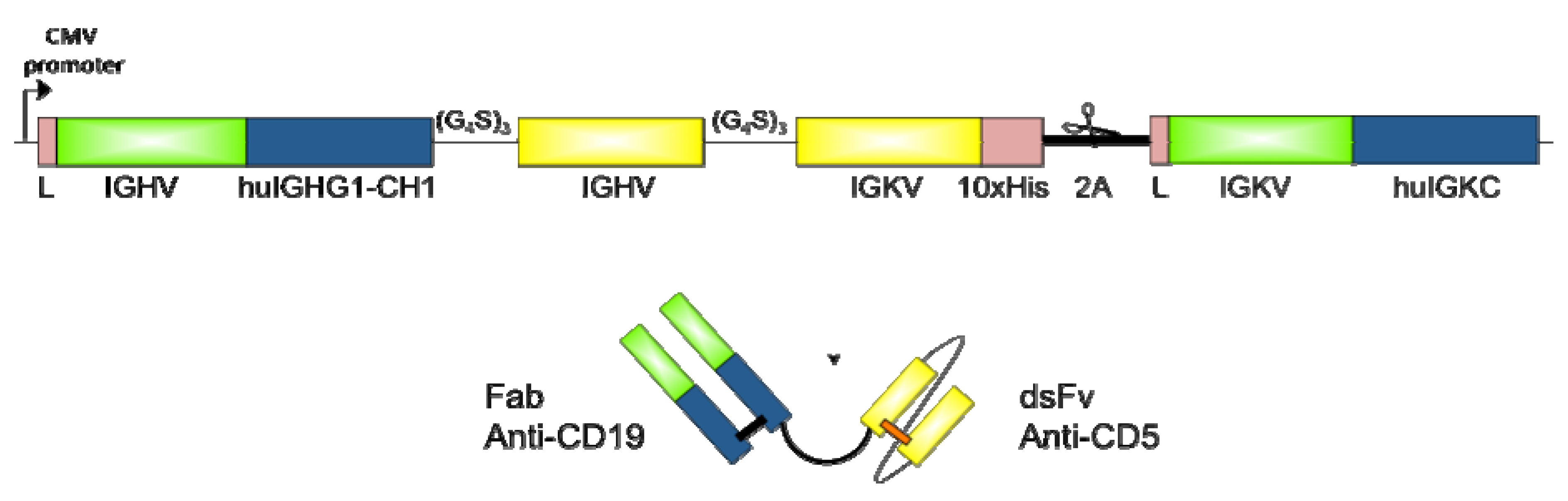
2.2. Design and Cloning of bsAb FabCD19xdsFvCD5

In brief, the bispecific construct should contain the Fab fragment of the chimerized anti CD19 antibody chiBCE19, which is covalently linked to a disulphide-stabilized Fv antibody derived from the anti-CD5 hybridoma T5.16. For this, expression plasmids were designed as outlined in Figure 1. After computational design of the coding region, DNA was synthesized by GeneArt AG (Regensburg, Germany), except for the Fab arm sequences. These were PCR-amplified from cDNA of chiBCE19 hybridoma and cloned into the entry plasmid via the respective restriction sites.

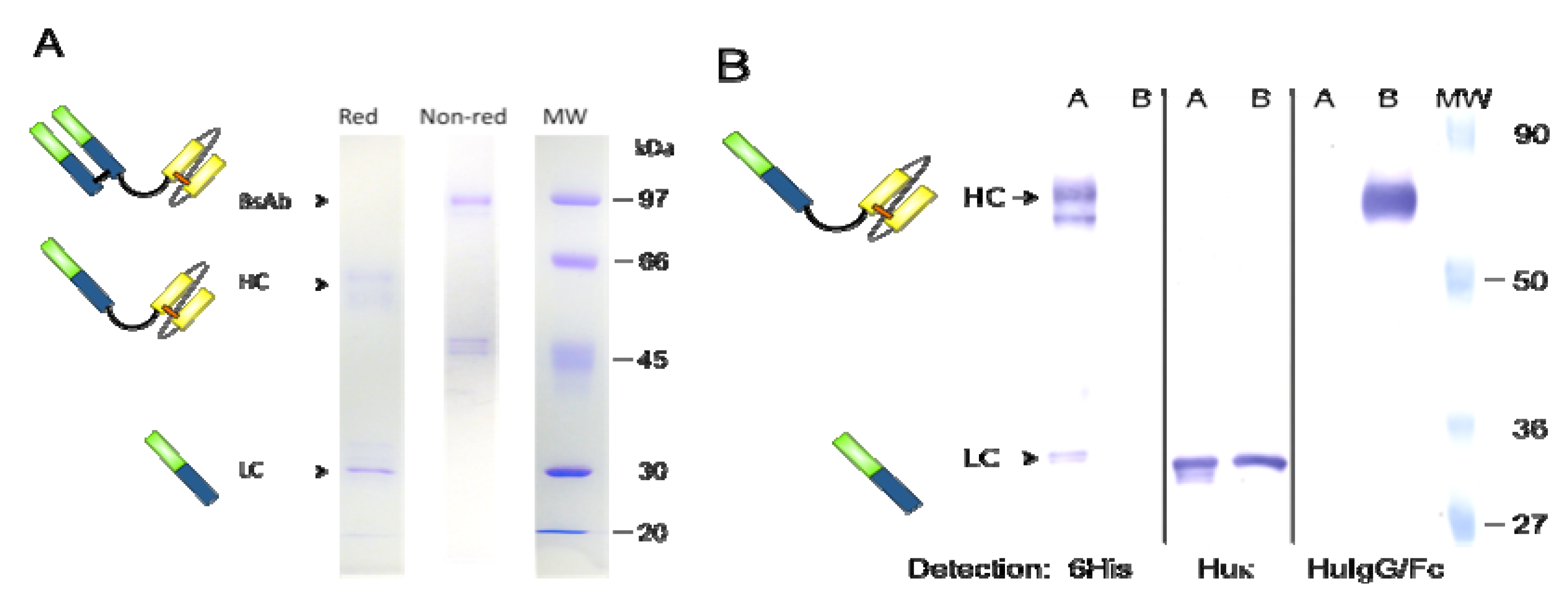
2.3. Expression and Purification of bsAb FabCD19xdsFvCD5

HEK293T cells were transfected with the plasmid using Lipofectamine (Life Technologies Darmstadt, Germany) according to the manufacturer’s instruction. For positive selection of stably transduced cells, a ble gene expressing plasmid (pBudCE4.1, Life Technologies) mediating zeocine resistance was co-transfected and lipofected cells were cultured in presence of zeocine (700 µg/mL) starting from day four after transfection. After two weeks of selection, growing colonies were picked and diluted to single cell clones which were then analyzed by ELISA and flow cytometry for bsAb secretion. High antibody secreting clones were identified and produced in a Miniperm bioreactor (Vivascience, Goettingen, Germany). Purification of bsAb was carried out by affinity-chromatography over a Sepharose CL-4B column (GE Healthcare, Freiburg, Germany) to which goat anti-human IgG antibodies (own preparation) were coupled. Purity of the eluted material was assessed by sodium dodecylsulfate polyacrylamide gel electrophoresis (SDS-PAGE).
Figure 1. Schematic representation of genetic and protein organization of bsAb. Expression plasmids for bsAbs contain one open reading frame with both heavy and light chain genes one after the other, divided by the 2A sequence coding for an autocatalytical peptide. Expression is driven by the CMV promoter. Both chains are preceded by the endogenous leader sequences of the tumor targeting Abs. Heavy chain Fab and ds scFv domains are divided by glycine serine linkers, as well as the two variable domains within the ds scFv Ab. Both arms of the bsAbs are stabilized by disulfide bonds, the Fab arm by the endogenous ds bond at the beginning of IGHG1 hinge region, the scFv arm by an artificially introduced disulphide bond in position vH44-vL100 (referring to the Kabat numbering scheme; [25]). For facilitated purification and protein analysis a 10× histidine tag was added to the carboxy-terminal end of the dsscFv domain. Constant IGHG1-CH1 and IGKC domains of the Fab fragment are humanized to decrease immunogenicity.
Antibodies 02 00338 g001

2.4. Western Blot Analysis

Purified antibodies were separated by SDS-PAGE under reducing and non-reducing conditions and transferred onto an Immobilon-P membrane (Millipore, MA, USA) in a semidry blotting system. After blocking the membrane with 5% milk powder in PBS-Tween, strips were incubated with the alkaline phosphatase-labeled detection antibodies goat anti-human kappa, goat anti human IgG/Fc (both from Jackson ImmunoResearch, West Grove, PA, USA) and mAb 13/45/31a (anti-hexahistidine, own reagent). After washing with PBS-Tween antibody binding was visualized by nitro-blue tetrazolium chloride/5-bromo-4-chloro-3'-indolyphosphate p-toluidine (NBT-BCIP; Roche, Mannheim, Germany) substrate reaction.

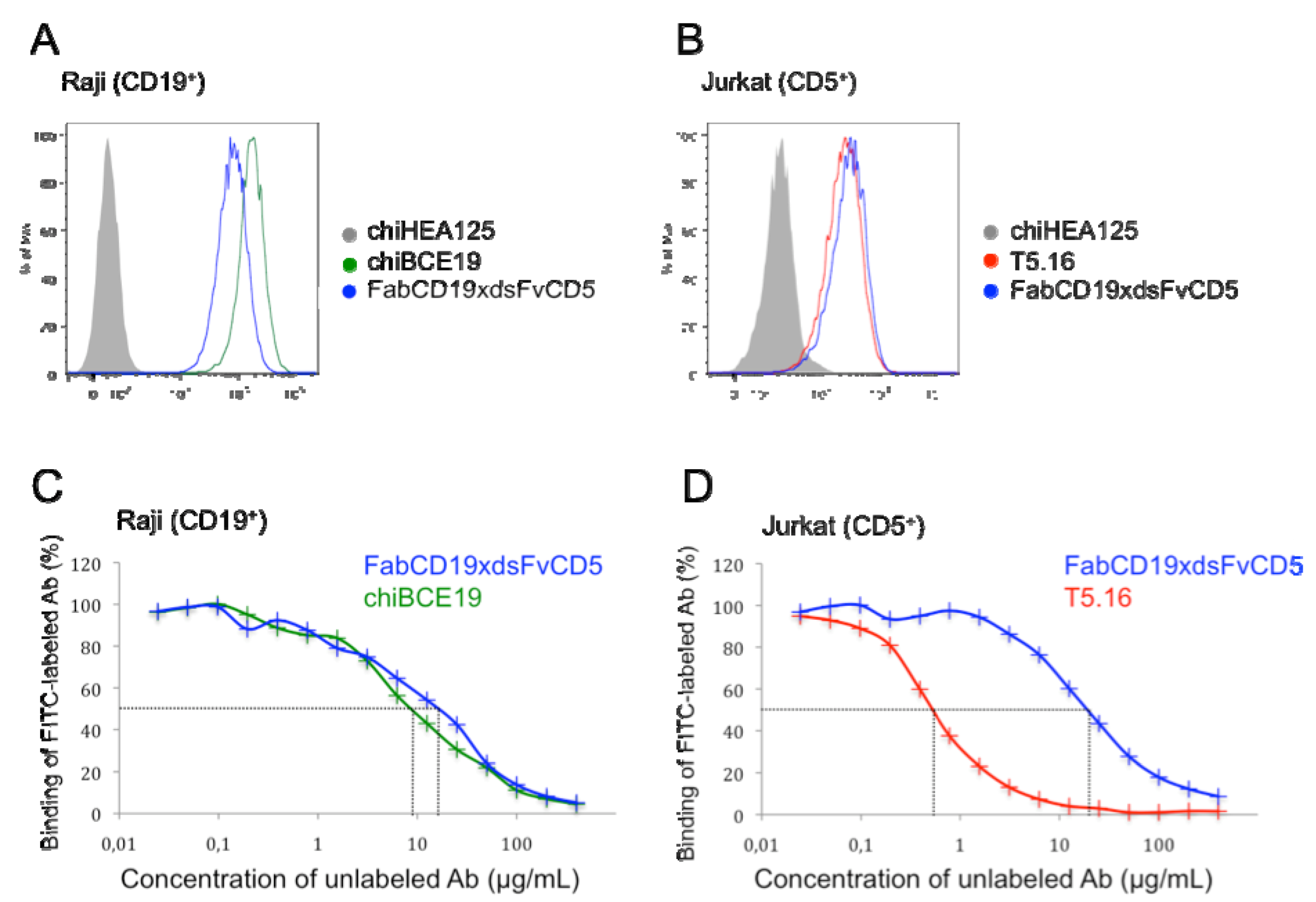
2.5. Flow Cytometry and Binding Competition Assay

Binding of bsAb FabCD19xdsFvCD5 to tumor cell lines was determined by indirect flow cytometry. For this, cells were first incubated with FabCD19xdsFvCD5 followed by a mixture of PE-labeled goat anti-human IgG(H+L) and goat anti-human F(ab)IgG secondary antibodies (Jackson ImmunoResearch).
The gross affinity of bsAb FabCD19xdsFvCD5 was estimated employing a binding competition experiment. Raji and Jurkat target cells were washed twice in FACS buffer (Dulbecco’s PBS with 1% heat-inactivated FCS and 0.1% sodium azide) counted and adjusted to 2 × 10⁷ cells per mL. Fifty μL of cell suspension was given to each well of a 96 well U-bottom microtiter plate to which 50 μL/well of FITC-labeled chiHEA125 antibody was pipetted. Serial dilutions of chiHEA125-Ama or chiHEA125 ranging from 400 μg/mL to 10 ng/mL final dilution were added in triplicates in a volume of 50 μL/well and incubated for 1 h on ice. Subsequently, the plate was centrifuged (2 min at 2,000 rpm) and the supernatant was removed from the wells. The cells were resuspended in 150 μL of FACS buffer and centrifuged again. After two washing steps, the cells were taken up in 100 μL/well of propidium iodide solution (1 mg/mL in FACS buffer) allowing discrimination of dead cells. The analysis was performed on a FACScan cytometer (Becton and Dickinson, Heidelberg, Germany) using CellQuest software.

2.6. Isolation and Activation of Peripheral Blood Mononuclear Cells

PBMC were isolated from heparinized blood of buffy coats obtained from healthy donors by Ficoll-Paque density gradient centrifugation. For pre-activation, culture flasks (150 cm2) were coated with 100 µg mAb OKT3 diluted in 20 mL PBS for 2 h at 37 °C. After removal of unbound mAb, 6 × 10⁷ PBMC were incubated in 30 mL complete RPMI 1640 medium with 10% heat-inactivated FCS containing 2-mercaptoethanol, 150 µg CD28 antibody and 90 U/mL IL-2 for four days. Cells were washed twice and transferred into a new culture flask to get rid of bound antibodies. The activation status of T lymphocytes was monitored by flow cytometry using antibodies recognizing CD25, CD69, CD95 and HLA-DR (all from Becton Dickinson, Heidelberg, Germany) before being employed in mouse treatment model.

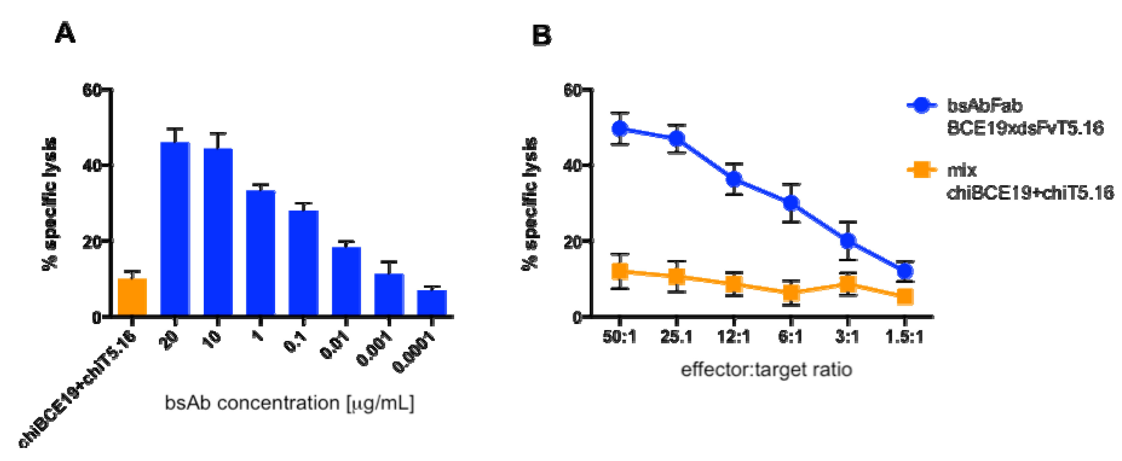
2.7. Cytotoxicity Assay

Cytolytic activity of bsAb was assessed using a standard 51Cr release assay in 96 well U-bottom microtiter plates. Raji target cells (2 × 10⁶) were labeled with 200 μCi sodium 51CrO4 (Perkin Elmer, Rodgau, Germany) followed by 4 washing cycles and resuspended in medium at a concentration of 1 × 10⁵/mL. PBMCs were washed after 4 days of activation, incubated overnight in fresh medium and adjusted to a concentration of 2.5 × 10⁶/mL. Increasing numbers of effector cells in 100 μL were titrated to 5 × 10³ target cells/ well in 50 μL and 50 μL of antibody solution was added to each well. The whole assay was set up in triplicate and incubated for 4 h at 37 °C. 100 μL of supernatant was harvested and assayed for 51Cr-release in a gamma counter. Maximum release was determined by incubation of labeled target cells in 100 μL 10% SDS and spontaneous release was calculated from a sample to which 100 μL of medium was added instead of effector cells. The percentage of specific release was determined as % specific release = (experimental release − spontaneous release) / (maximum release − spontaneous release) × 100.

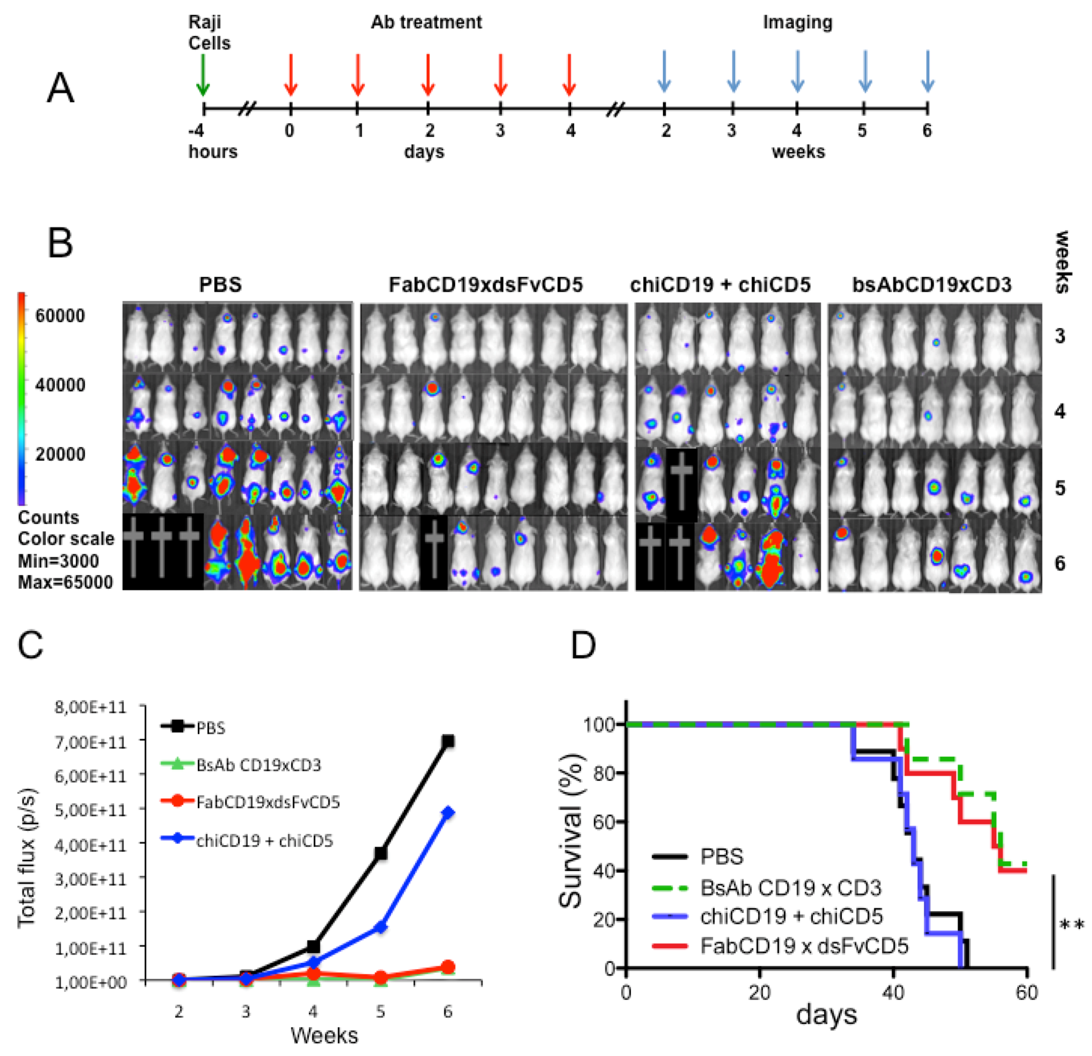
2.8. Raji Lymphoma Xenotransplantation Model and Antibody Treatment

Eight to 10-week-old NOD/SCID CB17 female mice (Charles River Laboratories, Germany) were intravenously injected with 1 × 10⁶ luciferase expressing Raji lymphoma cells together with 7.5 × 10⁶ in vitro preactivated PBMC from healthy donors. They were randomly divided into treatment groups of 6–10 mice. The antibody treatment consisted of daily intraperitoneal injection of 200 µg bispecific antibody or a mixture of parental antibodies 100 µg each in 100 µL PBS per mouse or 100 µL PBS as negative control. Treatment started 4 h after cell mixture inoculation and lasted for 5 days.
Mice were inspected daily for health status, especially for signs of distress or hind-leg paralysis. Moribund mice were euthanized and survival time was taken for the evaluation of therapeutic efficacy. Body weight of mice was determined twice per week as an indicator for tolerability of the treatment Paralysis of the hind legs or weight loss of >25% was used as survival end point. All experiments were performed according to the German animal protection law with permission from the responsible local authorities.

2.9. Bioluminescent Imaging

In vivo bioluminescent imaging allows the non-invasive detection and quantification of tumor spread in the whole mouse. For this, mice were anesthetized by isoflurane and imaged 5 min after the injection of luciferin (Biosynth AG, Switzerland) at a dose of 375 mg/kg body weight. The total flux values, correlating directly with tumor mass, were measured weekly. Imaging was performed on an IVIS Spectrum apparatus (Caliper Life Science, Hopkington, MA, USA) with a 1.8 min exposure time, medium binning, and a 16 f/stop. Multiple images were taken until the luciferase signal intensity reached a plateau, and image parameters were kept identical for the duration of the experiment. Bioluminescence image analyses were performed by Living Image 3.0 (Caliper LifeScience). Luciferase light units were quantified in average total flux (photon/second) of whole mouse.

2.10. Statistical Analysis

Prism software GraphPad was used to analyze tumor growth and luciferase light units and to determine the statistical significance of differences between groups by applying an unpaired Student t test. Kaplan-Meier plots were applied to analyze survival. Comparisons of survival curves were made using the log rank test. P values < 0.05 were considered significant.

3. Results

3.1. Expression and Purification of bsAb FabCD19xdsFvCD5

The aim of our study was to create a bispecific binding molecule with reduced size but strong tumor cell binding capability. An antibody format was chosen in which the Fab portion of the CD19-specific chimeric antibody chiBCE19 was fused to the disulfide-stabilized Fv fragment of the CD5-directed antibody T5.16. The expected size of the molecule was in the range of 80 to 90 kDa. For the gene construct, cDNA from cell line chiBCE19 was used which secretes chimeric antibodies to the pan-B cell antigen CD19. Chimerization was achieved by homologous recombination of the immunoglobulin gene locus within the hybridoma cell line [22]. The CD5 arm was derived as a Fv fragment from the sequence of hybridoma T5.16 that was stabilized by the insertion of an artificial disulfide bond. The design of the expression plasmid and the resulting bispecific construct is depicted in Figure 1.
HEK293T cells were stably transfected with the respective plasmid. After cloning by limiting dilution, the cell line showing the highest immunoglobulin secretion rate was selected and grown in a miniaturized modular bioreactor. Affinity-purified bsAb was analyzed by SDS-PAGE. Under non-reducing conditions bsAb FabCD19xdsFvCD5 exhibited an apparent electrophoretic mobility of 97 kDa whereas under reducing conditions two bands at approximately 60 kDa and 30 kDa were separated, corresponding to the heavy (HC arrowhead) and light chains (LC arrowhead) of the antibody molecule, respectively (Figure 2). These findings let us conclude that the bsAb was correctly assembled and carried the expected chain composition.
Figure 2. Biochemical analysis of bsAb. (A) Separation of purified bsAb by SDS PAGE under reducing (Red) and non-reducing (Non-red) conditions and stained with Coomassie Brilliant Blue. Lane MW contained the molecular weight marker. (B) Western blot analysis of bsAb in comparison to parental chiBCE19 mAb. Lane A; bsAb FabBCE19xdsFvT5.16; lane B; chiBCE19, MW; molecular weight standard. Immunoglobulin bands were detected by anti-hexahistidine, anti-human kappa chain and anti-human IgG/Fc.
Antibodies 02 00338 g002

3.2. Binding Characteristics of bsAb FabCD19xdsFvCD5

Immunoreactivity of the purified bsAb was evaluated by flow cytometry. For this, the Burkitt’s lymphoma cell line Raji (CD19+, CD5) and the T-ALL line Jurkat (CD5+, CD19) served as target cells. Binding of both, the anti-CD19 as well as the anti-CD5 moiety of the bispecific construct could be demonstrated. On Jurkat cells, the bsAb showed a binding pattern virtually identical to the mouse mAb T5.16 (Figure 3B). On Raji cells, binding of the bispecific construct was only slightly decreased when compared to the parental anti-CD19 chimeric antibody chiBCE19 (Figure 3A). These data provided evidence that both arms of the bsAb were functional.
In order to obtain a more comprehensive view on the affinity of both antibody moieties we performed a binding competition assay. For this, Raji cells were incubated with a fixed amount of FITC-labeled mAb chiBCE19 and competed with increasing amounts of unlabeled bsAb and homologous inhibitor mAb chiBCE19. As presented in Figure 3C the binding curves of bsAb and mAb chiBCE 19 are almost congruent. The IC50 value for both samples was approximately 10 to 20 μg/mL indicating a nearly identical avidity of bsAb with the parental anti-CD19 mAb. The same analysis was performed on Jurkat cells using the FITC-labeled mAb T5.16. In this setting a dramatic divergence of binding curves occurred. Whilst the IC50 value for the parental mAb T5.16 was in the range of 0.7 μg/mL, bsAb FabCD19xdsFvCD5 reached an IC50 of approximately 40 μg/mL (Figure 3D). This result clearly evidenced that the CD5-directed arm of bsAb has an approximately 50-fold decreased avidity when compared to the parental anti-CD5 mAb.
Figure 3. Immunoreactivity and avidity of bsAb. (A and B) Binding of bsAb FabCD19xCD5 and its parental antibodies to lymphoma lines Raji and Jurkat was measured by flow cytometry. As a negative control chiHEA125 was used, which is a chimeric anti-EpCAM antibody of human IgG1 isotype and does not react with lymphoma cells (grey histograms). (C and D) Flow cytometric binding competition assay comparing the binding strength of bsAb versus the parental mAb. The IC50 values are indicated by a dotted line.
Antibodies 02 00338 g003

3.3. Cytotoxicity in Vitro

Next, we tested the cytotoxic capacity of preactivated T lymphocytes towards Raji lymphoma cells induced by bsAb FabCD19xdsFvCD5. To this end, PBMC from healthy donors were activated in the presence of immobilized anti-CD3 mAb, soluble anti-CD28 mAb and low-dose IL-2 for four days. After washing steps and cultivation in medium to remove any remaining antibody, the expression of several activation markers was examined by flow cytometry. A strong up regulation of IL-2 receptor alpha-chain (CD25), activation inducer molecule (CD69) and APO-1/Fas antigen (CD95) was noted (data not shown). To determine the optimal concentration of bsAb FabCD19xdsFvCD5 to elicit cytotoxicity we used a fixed effector (E) to target cell (T) ratio of 50:1 and performed a titration of bsAb (Figure 4A). Above a concentration of 1 ng/mL specific lysis of target cells was observed reaching a plateau at 10 μg/mL. The mixture of both parental antibodies chiBCE19 and chiT5.16 at 20 μg/mL resulted in a background cytolysis of approximately 10%. Subsequently, a standard chromium release assay with increasing effector to target cell ratio and a fixed bsAb amount of 10 μg/mL was carried out. At the highest E:T ratio of 50:1 bsAb FabCD19xdsFvCD5 yielded a specific lysis of roughly 50% whereas the mix of parental antibodies serving as a negative control did not exceed 15% cytotoxicity (Figure 4B). It has to be noted that a remarkable variation of cytotoxicity among individual blood donors was observed. Overall, these results provided unambiguous evidence that the bsAb is able to redirect activated T lymphocytes versus CD19-expressing target cells.
Figure 4. Cytotoxicity of activated T lymphocytes against Raji lymphoma cells by bsAb FabCD19xdsFvCD5 in vitro. (A) Dose-response relationship using increasing amounts of bsAb and a constant effector to target cell ratio of 50:1. (B) Specific lysis of lymphoma cells at increasing E:T ratio and a fixed concentration of 10 μg/ml bsAb or a mixture of parental mAb as negative control. Results are presented as mean of triplicate assay and standard deviation.
Antibodies 02 00338 g004

3.4. Antibody Treatment of Raji Lymphoma-Bearing Mice

Luciferase transduced Raji lymphoma cells were injected intravenously together with preactivated human PBMC into immunodeficient mice. The treatment protocol consisted of daily intraperitoneal injection of 200 μg of therapeutic or control antibodies per mouse for five consecutive days (Figure 5A). Four groups of 6 to 10 mice received either PBS (untreated control), a mixture of parental antibodies chiBCE19 and chiT5.16 (100 μg each, negative control), quadroma-derived CD19xCD3 bsAb (positive control) and bsAb FabCD19xdsFvCD5 (experimental). Bioluminescent imaging of tumor growth was performed at weekly intervals. After six weeks of follow-up, 3 out of 8 mice in the PBS group were dead and the remaining 5 showed a severe tumor load (Figure 5B). A comparable outcome was noticed in the control group receiving a mixture of parental antibodies, in all mice except one large tumors had emerged or animals were dead. Interestingly, in the group of mice treated with the novel FabCD19xdsFvCD5 reagent, 5 of 9 animals did not develop any measurable tumor. The death of one mouse in this group was most likely caused by an early tumor manifestation in the brain. The therapeutic efficacy of the bsAb FabCD19xdsFvCD5 was as good as that of the quadroma antibody CD19xCD3 included as a positive control.
Figure 5. Influence of bsAb treatment on lymphoma growth in vivo. (A) Experimental design: NOD/SCID mice were intravenously inoculated with luciferase-transduced Raji lymphoma cells in combination with preactivated PBMC. Mice received five intraperitoneal antibody injections at the subsequent five days. Tumor growth was monitored by bioluminescent imaging for 6 weeks. (B) Luciferase imaging of 4 groups treated with PBS (negative control), bsAb FabBCE19xdsFvCD5 (investigative drug), mixture of mAb chiCD19 and chiCD5 (parental antibody control) and quadroma-derived bsAb CD19xCD3 (positive control). (C) Tumor growth during six weeks of observation measured by quantified bioluminescence (total flux). (D) Kaplan-Meier analysis of survival up to 60 days after tumor inoculation.
Antibodies 02 00338 g005
An integrated view on tumor load within the treatment groups is provided by the total flux of bioluminescence. Whilst PBS and the mixture of parental antibodies had no effect on rapid tumor growth, FabCD19xdsFvCD5 and quadroma bsAb CD19xCD3 caused a dramatic cessation of tumor spreading throughout observation time (Figure 5C). Most strikingly, the Kaplan-Meier plot depicted in Figure 5D revealed an overall survival rate of more than 40% in the groups of mice treated with FabCD19xdsFvCD5 and quadroma bsAb CD19xCD3 that is highly significant when compared to the control groups with p = 0.0057 and p = 0.0035, respectively.

4. Discussion

Although treatment of NHL with antibody therapeutics directed against the CD20 antigen has evolved increasingly successful, there is still an urgent need for novel antibody-based drugs to overcome relapse of disease. There are a couple of promising strategies how to achieve this demanding goal. First, alternative tumor-associated antigens like CD19 can be targeted showing a broader expression pattern than CD20. This would extend the applicability of antibody therapy to lymphoma entities being notoriously low for CD20 expression like B-CLL and B-ALL. Since both antigens are absent from the hematopoietic stem cell only transient immunosuppression and an early reconstitution of the B cell compartment has to be anticipated. Second, molecular biology has provided means to create innovative immunoglobulin molecules as bispecific antibodies able to elicit novel functional properties. During the past two decades, many different recombinant antibody formats were raised and tested in preclinical and clinical settings. Third, our understanding how the immune system is able to control tumor growth and conversely how tumor cells can evade immune surveillance has largely increased.
The key function of a bsAb for tumor therapy is clearly the recruitment of immune effector cells to the tumor site. Cytotoxic T lymphocytes were mostly used as effector cells for tumor attack as they represent the professional killers of the immune system. A variety of bispecific constructs covering different formats recognizing CD19 and CD3 have been described by us and other investigators [26,27,28,29,30,31]. Several of these constructs showed a remarkable anti-tumor efficacy when tested in reconstituted lymphoma-bearing immunodeficient mice. For instance, treatment with a tetravalent CD19xCD3 tandem diabody in combination with CD28 costimulation resulted in the complete elimination of tumors in all animals [30]. Farthermost advanced in clinical evaluation is the so-called bispecific T cell engager blinatumomab, a bispecific single-chain antibody of CD19xCD3 specificity [32,33,34]. In addition to the TCR-associated CD3 molecule other T cell antigens as CD2 [35], CD5 and the TCR itself [36] have been engaged as trigger molecules of bsAb. Our group has recently reported on a quadroma-derived bsAb of the specificity CD19xCD5 that was employed to target an ex vivo expanded and activated T cell subset called cytokine-induced killer (CIK) cells expressing CD5 [37]. Importantly, CD5 targeting bsAb may be particularly useful in combination with adoptive T cell transfer, e.g., in the setting of allogeneic stem cell transplantation, as it neither activates nor induces proliferation of naïve T cells potentially directed against host antigens. Thus, the danger of a graft versus host reaction in the recipient may be diminished.
Intact bsAb like quadroma-derived antibodies carry a Fc portion that potentially binds to Fc-receptors on a variety of accessory cells [38]. This frequently leads to the secretion of various cytokines, an effect that enhance tumor cell elimination as, e.g., in case of the trifunctional bsAb catumaxomab directed against the tumor-associated EpCAM antigen [39,40]. On the other hand, off target binding via Fc receptors may result in dose-limiting toxicity. To overcome such drawbacks we designed a bispecific molecule that lacks a Fc portion to avoid non specific binding to Fc receptor bearing cells and subsequent release of cytokines potentially causing severe side effects. Another advantage of our FabxdsFv format is the reduced size of the antibody. With a mass of approximately 80 kDa it is only half as big as a complete IgG molecule. This is thought to increase tissue penetration and to enhance tumor accessibility. Importantly, the size of the bsAb construct is big enough to avoid rapid renal clearance and thus should warrant a reasonable serum half life. In addition, since the FabCD19xdsFvCD5 was derived from a chimeric version of the anti-CD19 mAb BCE19, we expect a considerable reduction of immunogenicity when applied in humans.
One important functional feature of bsAb is the avidity of both binding moieties. As demonstrated in competition experiments with directly labeled parental mAb, the Fab part of our bsAb has completely retained its high avidity for the lymphoma-associated CD19 antigen. This might be beneficial for the tight association of bsAb with the malignant B cell. On the contrary, the binding strength of the anti-CD5 arm was clearly reduced. Whether this represents a disadvantage or not remains to be investigated. The attraction of activated T cells to the tumor site by relatively mild binding forces delivered by bsAb may prevent activation-induced cell death of the effector cells and thus allows several rounds of cytotoxic interaction of T lymphocytes with target cells. The decreased avidity of the CD5-directed bsAb moiety was largely unexpected since the introduction of interchain disulfide bonds into conserved framework regions are reported to retain specificity, stability and binding characteristics of Fv fragments. Especially, the disulfide bond position vH44-vL100 was observed to enable the most favorable balance of biophysical properties and was employed in our bsAb construct [25,41].

5. Conclusions

A novel bsAb of the FabxdsFv format simultaneously recognizing CD19 and CD5 was designed, expressed and purified. In preliminary tests, this reagent showed favorable binding characteristics to cell lines and was able to redirect activated T lymphocytes towards B lymphoma cells resulting in efficient tumor cell lysis. Most strikingly, bsAb FabCD19xdsFvCD5 led to an impressive retardation of lymphoma growth in tumor-bearing mice. Due to its reduced molecular mass and the replacement of mouse immunoglobulin constant domains by human sequences, an improved tissue penetration together with a diminished immunogenicity is expected when applied to patients. Overall, our preclinical results suggest that bsAb FabCD19xdsFvCD5 may evolve as a promising novel reagent that is worth investigating in early clinical trials for thus still incurable NHL patients.

Acknowledgements

We thank Jessica Kientz, Miriam Soehindrijo and Elvira Hallauer for excellent technical assistance. The authors are grateful to Felix Oden for providing the luciferase expressing Raji cell line. Furthermore, we are indebted to Reinhard Schwartz-Albiez for critical reading of the manuscript. This work was supported by grants from the Helmholtz Alliance on Immunotherapy of Cancer to G.M. and M.L.

References

  1. Siegel, R.; Naishadham, D.; Jemal, A. Cancer Statistics, 2012. CA Cancer J. Clin. 2012, 62, 10–29. [Google Scholar] [CrossRef]
  2. Lim, S.H.; Beers, S.A.; French, R.R.; Johnson, P.W.M.; Glennie, M.J.; Cragg, M.S. Anti-CD20 monoclonal antibodies: historical and future perspectives. Haematologica 2010, 95, 135–143. [Google Scholar] [CrossRef]
  3. Vacchelli, E.; Eggermont, A.; Galon, J.; Sautes-Fridman, C.; Zitvogel, L.; Kroemer, G.; Galluzzi, L. Trial watch: Monoclonal antibodies in cancer therapy. OncoImmunology 2013, 2, e22789. [Google Scholar] [CrossRef]
  4. Rezvani, A.R.; Maloney, D.G. Rituximab resistance. Best Pract. Res. Clin. Haematol. 2011, 24, 203–216. [Google Scholar] [CrossRef]
  5. Chames, P.; Baty, D. Bispecific antibodies for cancer therapy: The light at the end of the tunnel? MAbs 2009, 1, 539–547. [Google Scholar] [CrossRef]
  6. Pezzutto, A.; Dörken, B.; Rabinovitch, P.S.; Ledbetter, J.A.; Moldenhauer, G.; Clark, E.A. CD19 monoclonal antibody HD37 inhibits anti-immunoglobulin-induced B cell activation and proliferation. J. Immunol. 1987, 138, 2793–2799. [Google Scholar]
  7. Tedder, T.F. CD19: A promising B cell target for rheumatoid arthritis. Nat. Rev. Rheumatol. 2009, 5, 572–577. [Google Scholar] [CrossRef]
  8. Suntharalingam, G.; Perry, M.R.; Ward, S.; Brett, S.J.; Castello-Cortes, A.; Brunner, M.D.; Panoskaltsis, N. Cytokine storm in a phase 1 trial of the anti-CD28 monoclonal antibody TGN1412. N. Engl. J. Med. 2006, 355, 1018–1028. [Google Scholar]
  9. Schneider, C.K.; Kalinke, U.; Löwer, J. TGN1412—A regulator’s perspective. Nat. Biotechnol. 2006, 24, 493–496. [Google Scholar] [CrossRef]
  10. Tabbekh, M.; Mokrani-Hammani, M.; Bismuth, G.; Mami-Chouaib, F. T-cell modulatory properties of CD5 and its role in antitumor immune responses. OncoImmunology 2013, 2, e22841. [Google Scholar] [CrossRef]
  11. Baeuerle, P.A.; Reinhardt, C. Bispecific T-cell engaging antibodies for cancer therapy. Cancer Res. 2009, 69, 4941–4944. [Google Scholar] [CrossRef]
  12. Beck, A.; Wurch, T.; Bailly, C.; Corvaia, N. Strategies and challenges for the next generation of therapeutic antibodies. Nat. Rev. Immunol. 2010, 10, 345–352. [Google Scholar] [CrossRef]
  13. Chan, A.C.; Carter, P.J. Therapeutic antibodies for autoimmunity and inflammation. Nat. Rev. Immunol. 2010, 10, 301–316. [Google Scholar] [CrossRef]
  14. Müller, D.; Kontermann, R.E. Recombinant bispecific antibodies for cellular cancer immunotherapy. Curr. Opin. Mol. Ther. 2007, 9, 319–326. [Google Scholar]
  15. Müller, D.; Kontermann, R.E. Bispecific antibodies for cancer immunotherapy. Current perspectives. Biodrugs 2010, 24, 89–98. [Google Scholar] [CrossRef]
  16. Stamova, S.; Koristka, S.; Keil, J.; Arndt, C.; Feldmann, A.; Michalk, I.; Bartsch, H.; Bippes, C.C.; Schmitz, M.; Cartellieri, M.; et al. Cancer Immunotherapy by retargeting of immune effector cells via recombinant bispecific antibody constructs. Antibodies 2012, 1, 172–198. [Google Scholar] [CrossRef]
  17. Weiner, L.M. Building better magic bullets—Improving unconjugated monoclonal antibody therapy for cancer. Nat. Rev. Cancer 2007, 7, 701–706. [Google Scholar] [CrossRef]
  18. Oden, F. Max-Delbruck-Center for Molecular Medicine: Berlin, Germany, 2013; unpublished work.
  19. Zhang, H.; Moldenhauer, G.; Kipriyanov, S.M.; Braunagel, M.; Krauss, J.; Little, M. Construction, sequence and binding properties of an anti-CD5 single chain Fv antibody. Tumor Targeting 1996, 2, 314–321. [Google Scholar]
  20. Moldenhauer, G.; Momburg, F.; Möller, P.; Schwartz, R.; Hämmerling, G.J. Epithelium-specific surface glycoprotein of Mr 34,000 is a widely distributed human carcinoma marker. Br. J. Cancer 1987, 56, 714–721. [Google Scholar] [CrossRef]
  21. Momburg, F.; Moldenhauer, G.; Hämmerling, G.J.; Möller, P. Immunohistochemical study of the expression of a Mr 34,000 human epithelium-specific surface glycoprotein in normal and malignant tissues. Cancer Res. 1987, 47, 2883–2891. [Google Scholar]
  22. Lüttgau, S. German Cancer Research Center and National Center for Tumor Diseases: Heidelberg, Germany, 2013; unpublished work.
  23. Csoka, M.; Strauss, G.; Debatin, K.M.; Moldenhauer, G. Activation of T cell cytotoxicity against autologous common acute lymphoblastic leukemia (cALL) blasts by CD3xCD19 bispecific antibody. Leukemia 1996, 10, 1765–1772. [Google Scholar]
  24. Daniel, P.T.; Kroidl, A.; Kopp, J.; Sturm, I.; Moldenhauer, G.; Dörken, B.; Pezzutto, A. Immunotherapy of B-cell lymphoma with CD3x19 bispecific antibodies: costimulation via CD28 prevents "veto" apoptosis of antibody-targeted cytotoxic T cells. Blood 1998, 92, 4750–4757. [Google Scholar]
  25. Reiter, Y.; Brinkmann, U.; Webber, K.O.; Jung, S.-H.; Lee, B.; Pastan, I. Engineering interchain disulfide bonds into conserved framework regions of Fv fragments: Improved biochemical characteristics of recombinant immunotoxins containing disulfide-stabilized Fv. Protein Eng. 1994, 7, 697–704. [Google Scholar] [CrossRef]
  26. De Gast, G.C.; Van Houten, A.A.; Haagen, I.A.; Klein, S.; De Weger, R.A.; Van Dijk, A.; Philips, J.; Clark, M.; Bast, B.J.E.G. Clinical experience with CD3 x CD19 bispecific antibodies in patients with B cell malignancies. J. Hematother. 1995, 4, 433–437. [Google Scholar] [CrossRef]
  27. Manzke, O.; Tesch, H.; Borchmann, P.; Wolf, J.; Lackner, K.; Gossmann, A.; Diehl, V.; Bohlen, H. Locoregional treatment of low-grade B-cell lymphoma with CD3xCD19 bispecific antibodies and CD28 costimulation: I. clinical phase I evaluation. Int. J. Cancer 2001, 91, 508–515. [Google Scholar] [CrossRef]
  28. Kipriyanov, S.M.; Moldenhauer, G.; Strauss, G.; Little, M. Bispecific CD3xCD19 diabody for T cell-mediated lysis of malignant human B cells. Int. J. Cancer 1998, 77, 763–772. [Google Scholar] [CrossRef]
  29. Kipriyanov, S.M.; Moldenhauer, G; Schuhmacher, J.; Cochlovius, B.; Von der Lieth, C.-W.; Matys, E.R.; Little, M. Bispecific tandem diabody for tumor therapy with improved antigen binding and pharmacokinetics. J. Mol. Biol. 1999, 293, 41–56. [Google Scholar] [CrossRef]
  30. Cochlovius, B.; Kipriyanov, S.M.; Stassar, M.J.J.G.; Christ, O.; Schuhmacher, J.; Strauß, G.; Moldenhauer, G.; Little, M. Treatment of human B cell lymphoma xenografts with a CD3 x CD19 diabody and T cells. J. Immunol. 2000, 165, 888–895. [Google Scholar]
  31. Cochlovius, B.; Kipriyanov, S.M.; Stassar, M.J.J.G.; Schuhmacher, J.; Benner, A.; Moldenhauer, G.; Little, M. Cure of Burkitt’s lymphoma in severe combined immunodeficiency mice by T cells, tetravalent CD3 x CD19 tandem diabody, and CD28 costimulation. Cancer Res. 2000, 60, 4336–4341. [Google Scholar]
  32. Löffler, A.; Kufer, P.; Lutterbüse, R.; Zettl, F.; Daniel, P.T.; Schwenkenbecher, J.M.; Riethmüller, G.; Dörken, B.; Bargou, R.C. A recombinant bispecific single-chain antibody, CD19xCD3, induces rapid and high lymphoma-directed cytotoxicity by unstimulated T lymphocytes. Blood 2000, 95, 2098–2103. [Google Scholar]
  33. Bargou, R.; Leo, E.; Zugmaier, G.; Klinger, M.; Goebeler, M.; Knop, S.; Noppeney, R.; Viardot, A.; Hess, G.; Schuler, M.; et al. Tumor regression in cancer patients by very low doses of a T cell-engaging antibody. Science 2008, 321, 974–977. [Google Scholar] [CrossRef]
  34. Topp, M.S.; Kufer, P.; Gökbuget, N.; Goebler, M.; Klinger, M.; Neumann, S.; Horst, H.-A.; Raff, T.; Viardot, A.; Schmid, M.; et al. Targeted therapy with the T-cell-engaging antibody blinatumomab of chemotherapy-refractory minimal residual disease in B-lineage acute lymphoblastic leukemia patients results in high response rate and prolonged leukemia-free survival. J. Clin. Oncol. 2011, 29, 2493–2498. [Google Scholar] [CrossRef]
  35. Wild, M.K.; Strittmatter, W.; Matzku, S.; Schraven, B.; Meuer, S.C. Tumor therapy with bispecific antibody: the targeting and triggering steps can be separated employing a CD2-based strategy. J. Immunol. 1999, 163, 2064–2072. [Google Scholar]
  36. Ferrini, S.; Prigione, I.; Mammoliti, S.; Colnaghi, M.I.; Ménard, S.; Moretta, A.; Moretta, L. Re-targeting of human lymphocytes expressing the T-cell receptor gamma/delta to ovarian carcinoma cells by the use of bispecific monoclonal antibodies. Int. J. Cancer 1989, 44, 245–250. [Google Scholar] [CrossRef]
  37. Tita-Nwa, F.; Moldenhauer, G.; Herbst, M.; Kleist, C.; Ho, A.D.; Kornacker, M. Cytokine-induced killer cells targeted by the novel bispecific antibody CD19xCD5 (HD37xT5.16) efficiently lyse B-lymphoma cells. Cancer Immunol. Immunother. 2007, 56, 1911–1920. [Google Scholar] [CrossRef]
  38. Nimmerjahn, F.; Ravetch, J.V. Antibodies, Fc receptors and cancer. Curr. Opin. Immunol. 2007, 19, 239–245. [Google Scholar] [CrossRef]
  39. Seimetz, D.; Lindhofer, H.; Bokemeyer, C. Development and approval of the trifunctional antibody catumaxomab (anti-EpCAM x anti-CD3) as a targeted cancer immunotherapy. Cancer Treat. Rev. 2010, 36, 458–467. [Google Scholar] [CrossRef]
  40. Heiss, M.M.; Murawa, P.; Koralewski, P.; Kutarska, E.; Kolesnik, O.O.; Ivanchenko, V.V.; Dudnichenko, A.S.; Aleknaviciene, B.; Razbadauskas, A.; Gore, M.; et al. The trifunctional antibody catumaxomab for the treatment of malignant ascites due to epithelial cancer: results of a prospective randomized phase II/III trial. Int. J. Cancer 2010, 127, 2209–2221. [Google Scholar] [CrossRef]
  41. Weatherill, E.E.; Cain, K.L.; Heywood, S.P.; Compson, J.E.; Heads, J.T.; Adams, R.; Humphreys, D.P. Towards a universal disulphide stabilized single chain Fv format: importance of interchain disulphide bond location and vL-vH orientation. Protein Eng. Des. Sel. 2012, 25, 321–329. [Google Scholar] [CrossRef]

