Article

Influence of Glutamic Acid on the Properties of Poly(xylitol glutamate sebacate) Bioelastomer

The Key Laboratory of Food Colloids and Biotechnology, Ministry of Education, School of Chemical and Material Engineering, Jiangnan University, Wuxi 214122, China
*
Authors to whom correspondence should be addressed.
Polymers 2013, 5(4), 1339-1351; https://doi.org/10.3390/polym5041339
Submission received: 29 September 2013 / Revised: 19 November 2013 / Accepted: 25 November 2013 / Published: 29 November 2013

Abstract

:
In order to further improve the biocompatibility of xylitol based poly(xylitol sebacate) (PXS) bioelastomer, a novel kind of amino acid based poly(xylitol glutamate sebacate) (PXGS) has been successfully prepared in this work by melt polycondensation of xylitol, N-Boc glutamic acid and sebacic acid. Differential scanning calorimetry (DSC) results indicated the glass-transition temperatures could be decreased by feeding N-Boc glutamic acid. In comparison to PXS, PXGS exhibited comparable tensile strength and much higher elongation at break at the same ratio of acid/xylitol. The introduction of glutamic acid increased the hydrophilicity and in vitro degradation rate of the bioelastomer. It was found that PXGS exhibited excellent properties, such as tensile properties, biodegradability and hydrophilicity, which could be easily tuned by altering the feeding monomer ratios. The amino groups in the PXGS polyester side chains are readily functionalized, thus the biomelastomers can be considered as potential biomaterials for biomedical application.

1. Introduction

In recent years, there has been considerable interest in the biodegradable elastomers used for soft tissue regeneration [1,2,3,4,5,6,7]. These materials must be tailored to have suitable biodegradability, mechanical properties, and drug delivery capabilities, as well as adequate biocompatibility. Among them, biodegradable polyester elastomers have been considered in the group of most promising biomaterials because they are easily susceptible to biological attack, their degradation products are non-toxic, and they can enter the metabolic cycles of bio-organisms [8,9]. Besides, some other bio-based elastomers such as soybean-oil-based elastomers are becoming more and more attractive [10]. Recently, many biodegradable polyester elastomers, such as poly(glycerol sebacate) (PGS) [11], poly[(1,2-propanediol-sebacate)-citrate] (PPSC) [12], poly(propylene sebacate) [13], were prepared to be used for soft tissue engineering. Their mechanical properties, degradation profiles and hydrophilicity of the bio-elastomers can be tuned by varying the initial monomer molar ratio and the synthetic conditions. Moreover, the preparation does not involve any harsh solvents and exogenous catalysts that would be harmful to the human body after degradation.
Lately, poly(xylitol sebacate) (PXS) elastomers have been successfully fabricated by polycondensation of xylitol and sebacic acid [14,15]. PXS elastomers are composed of nontoxic monomers endogenous to the mammalian organism, and exhibit excellent properties. Xylitol-based polymers are endotoxin-free and do not impose a potential immunological threat like biological polymers extracted from tissues or produced by bacterial fermentation, including collagen and hyaluronic acid [16,17]. In addition, the mechanical properties of xylitol-based elastomers correspond to biologically relevant values that fall close to those of different tissues, such as acellular peripheral nerves [18], small diameter arteries [19], and cornea [20]. Nevertheless there is still a challenge to further improve the biocompatibility and cell adhesion performance of bio-elastomers in order to satisfy the high demand in bio-tissue fields.
Peptides with unique biological characteristics are increasingly being exploited for the development of new bioactive molecules and biomaterials [21]. Poly(l-glutamic acid) (PGA) is a polypeptide with outstanding biological and physicochemical properties, and exhibits no antigenicity or immunogenicity [22,23]. PGA can biodegrade into glutamic acid, an important component of the body, and has been proven to be a promising polymer for biomedical applications, such as drug-delivery systems [24]. Due to the facile functionalization of the carboxyl groups and the excellent hydrophilicity for easy attachment of cells and exchange of molecules, PGA is considered to be a potential polymer material for tissue engineering.
In this paper, the amine group of l-glutamic acid was firstly protected by reaction of di-tert-butyl dicarbonate to yield Boc-l-glutamic acid. On the basis of xylitol, sebacic acid and Boc-l-glutamic acid, a family of poly(xylitol glutamate sebacate) (PXGS) polymers was formed. The influences of Boc-l-glutamic acid on the thermal and mechanical properties, hydrophilicity and degradation were investigated. A wide range of properties of PXGS was tuned by altering the initial monomers ratios. It was found that PXGS exhibited excellent properties, such as flexibility, biodegradability and hydrophilicity.

2. Experimental Section

2.1. Materials

Xylitol, sebacic acid, l-glutamic acid, di-tert-butyl dicarbonate and other chemicals were purchased from Sinopharm Chemical Reagent Co., Ltd. (Shanghai, China) and used without any further purification.

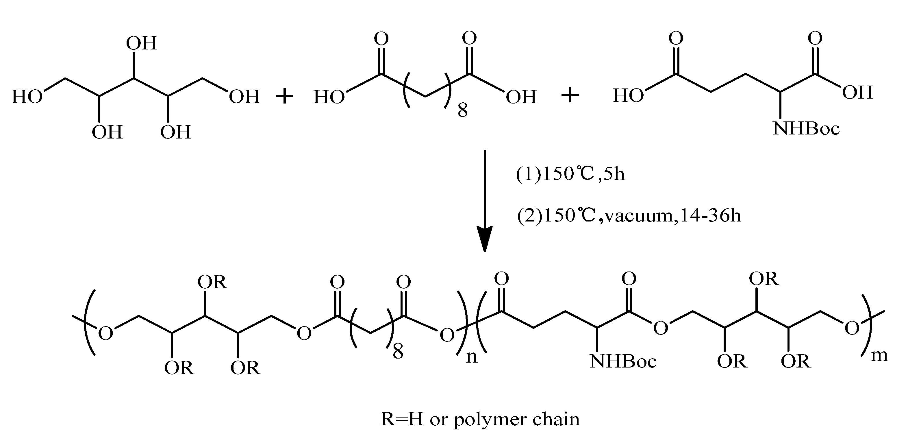
2.2. Preparation of PXGS

Boc-l-glutamic was prepared by reaction of di-tert-butyl dicarbonate and l-glutamic acid according to the literature [25]. Appropriate molar amounts of the xylitol, sebacic and Boc-l-glutamic acid monomer (Table 1) were melted in a round-bottom flask at 150 °C under a blanket of nitrogen gas and stirred for 5 h to prepare the PXGS prepolymers. Then, the samples were transferred into polytetrafluoroethylene molds in a vacuum oven at 150 °C and 1 KPa for a period of time to achieve a crosslinked elastomer plaque. The predetermined time was 14–36 h and depended on the feeding monomer ratios. The synthesis process is described in Scheme 1. In addition, a control sample was carried out, in which the whole procedure was repeated, except for the addition of Boc-l-glutamic acid.
Table 1. The feeding monomer ratio and amount for synthesis of poly(xylitol glutamate sebacate) (PXGS) bioelastomers. PXS x:y and PXGS x:y-z mean the prepolymers with a monomer ratio of xylitol: dicarboxylic acid of x:y, and the content of Boc-l-glutamic acid in dicarboxylic acid was z.
SamplesMonomers ratio (xylitol/sebacic acid/Boc-l-glutamic, mol %)Xylitol (mmol)Sebacic acid (mmol)Boc-l-glutamic (mmol)
PXS 1:1100/100/01010
PXS 1:2100/200/01020
PXS 2:5100/250/01020
PXS 1:3100/300/01030
PXGS 1:1-5%100/95/5109.50.5
PXGS 2:3-5%100/142.5/7.51014.250.75
PXGS 1:2-5%100/190/1010191
PXGS 2:5-5%100/237.5/12.51023.751.25
PXGS 1:3-5%100/285/151028.51.5
PXGS 2:5-10%100/225/251022.502.50
PXGS 2:5-15%100/212.5/37.51021.253.75
Scheme 1. Synthesis of poly(xylitol glutamate sebacate) (PXGS) bioelastomers.
Polymers 05 01339 g009

2.3. Characterization

Attenuated total reflection (ATR) was used to record the infrared spectra mode for 50 scans at a resolution of 1 cm−1 in the range of 4000–600 cm−1 on a Nicolet 6700 FT-IR spectrometer (Thermo Fisher Scientific, Waltham, MA, USA)
1H-NMR spectra were collected on a BUKER400 AVANCEIII spectrometer (Bruker, Coventry, UK). Chloroform was used as solvent and tetramethylsilane (TMS) as internal standard.
Differential scanning calorimeter (DSC) (Mettler Toledo DSC822e, Zurich, Switzerland) was applied to investigate the glass-transition temperature with a heating rate of 10 °C/min from −30 to 150 °C in a nitrogen atmosphere.
The surface hydrophilicity of the elastomer was characterized by measuring the water-in-air contact angle of smooth surfaces of samples using a Germany Kruss (DSA100, Hamburg, Germany).
The gel content of the elastomer was measured by the following tests. A small square sample (1.5 mm thickness and 10 mm diameter) weighing W1 was dipped into tetrahydrofuran (20 mL) for 24 h and was then taken out. The sample was then dried to a constant weight W2 in a vacuum oven. Gel content: G = W2/W1 × 100%. Three samples were conducted for each experiment, and the average value was adopted.

2.4. Tensile Tests

Samples were cut into dumbbell-shaped splines according to the ASTM D412 standard. Tensile tests were conducted by using a Kaiqiang WDT-10 testing machine (Kaiqiangli Testing Instruments Co. Ltd., Shenzhen, China) in triplicate at an elongation speed of 20 mm/min at 20 °C. Error distributions for the mechanical data were less than 15%.

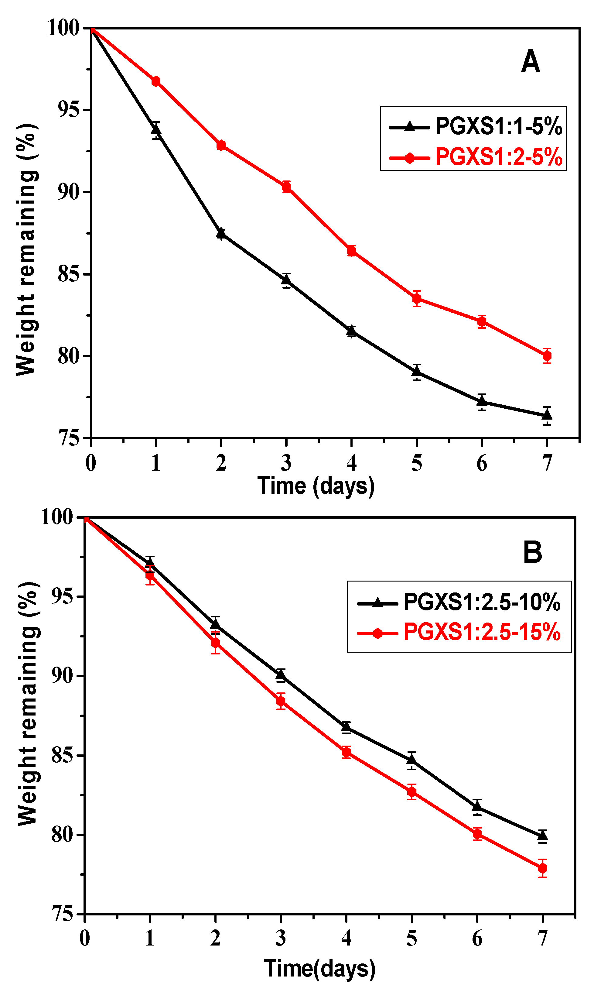
2.5. In Vitro Degradation

The PXGS plaques (10 × 10 × 0.2 mm3) were placed in 25 mL phosphate buffer saline (pH = 7.4) at a concentration of 1 mol/L to rapidly obtain relative degradation rates among samples. The specimens were incubated at 37 ± 1 °C in PBS for predetermined times. After incubation, the plaques were washed thoroughly with distilled water at room temperature, and dried at ambient temperature with vacuum until a constant weight.
The experimental weight values represent the averages of measurements from replicate samples with a standard deviation of 5%. The remaining percentage of weight of the hydrolyzed plaques (Wremaining) was calculated using the plaque weight before (Wbefore) and after (Wafter) hydrolysis:
Wremaining(wt%) = (Wafter/Wbefore) × 100%

3. Results and Discussion

3.1. Characterization of Chemical Structure

Figure 1 displays the 1H-NMR spectrum of N-Boc glutamic acid. The signals at 1.38 ppm were assigned to the –CH3 of N-Boc glutamic acid. The peaks at approximately 2.25 and 2.55 ppm were attributed to the –CH2 and 4.45 ppm was attributed to the –CH. The peak at 5.25 ppm in the 1H-NMR spectrum was assigned to the –NH of glutamic acid. 1H-NMR results suggested N-Boc glutamic acid was successfully synthesized.
Figure 1. 1H-NMR spectrum of N-Boc glutamic acid.
Polymers 05 01339 g001
Figure 2 shows the FT-IR spectroscopy of PXGS with various ratio of dibasic acid to xylitol. It was confirmed that ester bond was formed in all PXGS, with a stretch at 1730 cm−1, which corresponded to ester linkages. The peak at approximately 1170 and 1190 cm−1 was attributed to the –C–O– groups. A broad stretch observed at approximately 3360 cm−1 was attributed to hydrogen-bonded hydroxyl groups. The hydroxyl peak gradually weakened with increasing the amount of dibasic acid, which indicated that the degree of esterification was improved with enhancing the initial amount of acid. Moreover, glutamic acid did not affect FT-IR spectroscopy of PXGS when the content was below 15 wt % (see Figure 2d,f,g).
Figure 2. FT-IR characterization of the poly(xylitol glutamate sebacate) (PXGS): (a) PXGS 1:1-5%; (b) PXGS 2:3-5%; (c) PXGS 1:2-5%; (d) PXGS 2:5-5%; (e) PXGS 1:3-5%; (f) PXGS 2:5-10%; and (g) PXGS 2:5-15%.
Polymers 05 01339 g002

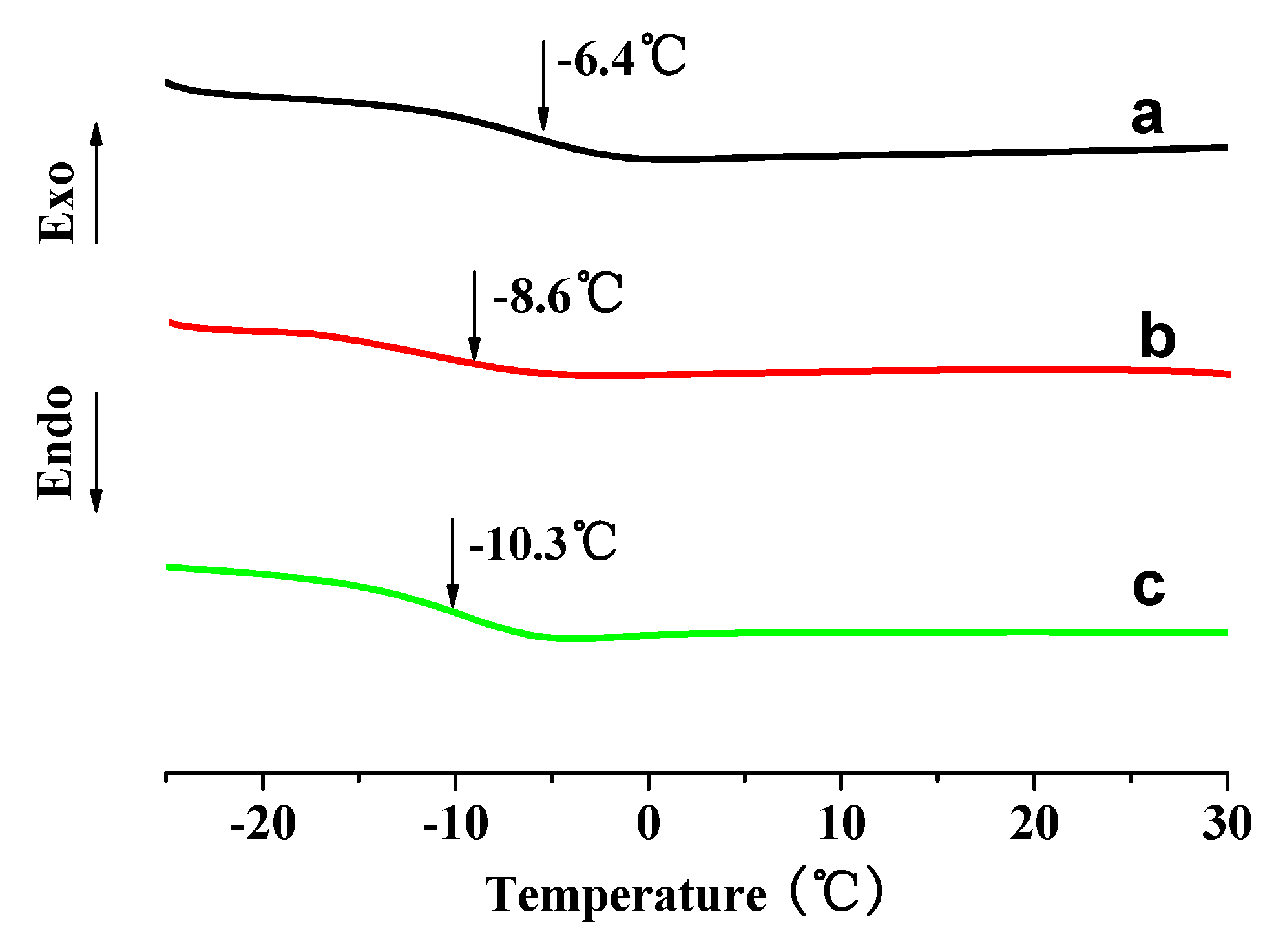
3.2. Thermal Properties

Typical DSC heating scans of PXGS are depicted in Figure 3. It can be seen that all the glass-transition temperatures (Tg) are under room temperature, which suggests that these elastomers are in a rubbery state at room and physiological temperature. Moreover, Tg decreased when increasing the ratio of dicarboxylic acid to xylitol. It can be explained as follows. The dicarboxylic acid was firstly endcapped with xylitol to form a branched structure, then the crosslinked network of the elastomer was formed. Therefore, the larger amount of acid can increase the content of the branched structure, which leads to a decrease of Tg. Compared with data in the literature [14], Tgs of PXGS 1:1-5% (2.4 °C) and 1:2-5% (−5.5 °C) were much lower than those of PXS 1:1 (7.4 °C) and 1:2 (22.9 °C) at the same acid/xylitol ratio. In addition, the Tg was further decreased on increasing the feeding amount of Boc-l-glutamic acid (Figure 4). The reduced Tg was due to the introduction of Boc-l-glutamic acid whose big side chain disturbed the regularity of the molecular chain.
Figure 3. Differential scanning calorimeter (DSC) heating curves of PXGS with various monomer ratios: (a) PXGS 1:1-5%; (b) PXGS 2:3-5%; (c) PXGS 1:2-5%; (d) PXGS 2:5-5%; and (e) PXGS 1:3-5%.
Polymers 05 01339 g003
Figure 4. Differential scanning calorimeter (DSC) heating curves of PXGS with different feeding contents of Boc-l-glutamic acid: (a) PXGS 2:5-5%; (b) PXGS 2:5-10%; and (c) PXGS 2:5-15%.
Polymers 05 01339 g004

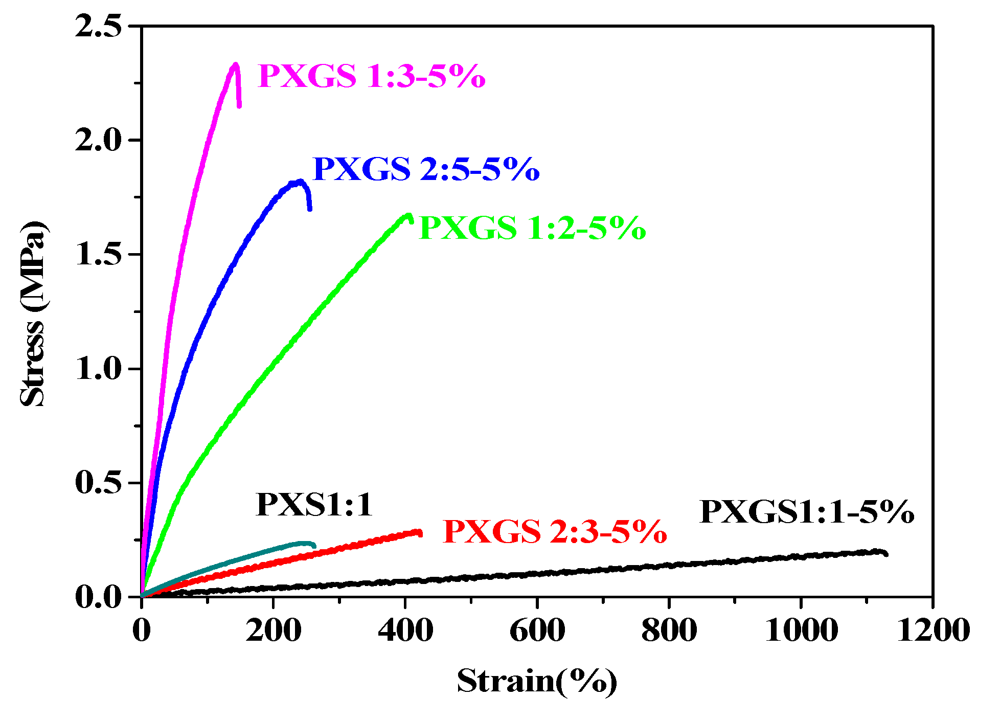
3.3. Mechanical Properties

The data of mechanical properties for PXS and PXGS were summarized in Table 2, and the strain-stress curves were shown in Figure 5. At lower acid/xylitol ratio, the PXGS displayed higher elongation and lower tensile strength, for example the elongation at break and tensile strength of PXGS 1:1-5% were 971% and 0.17 MPa respectively. On the contrary, PXGS showed lower elongation at break and higher tensile strength, which were 148% and 2.35 MPa for PXGS 1:3-5% respectively. In other words, when enhancing the ratio of acid/xylitol, the tensile strength increased, whereas the elongation at break decreased. That was attributed to the higher density of crosslinking on increasing the ratio of acid/xylitol, which was confirmed by the gel content data in Table 2. The tensile permanent deformation of PXGS was decreased from 21% to 5%, when increasing the ratio of acid/xylitol from 1:1 to 3:1. According to the above data, it cand be concluded that there is a large range of tunable mechanical properties for PXGS elastomers available by varying the feeding monomer ratios.
Table 2. Mechanical properties, water contact angle and gel content of different monomer ratios of PXGS and poly(xylitol sebacate) (PXS).
SamplesTensile strength (MPa)Elongations at break (%)Tensile permanent deformation (%)Water contact angle (°)Gel content (%)
PXS 1:10.24 ± 0.18251.7 ± 23.865.5 ± 4.584.0 ± 1.4
PXS 1:20.94 ± 0.15114.3 ± 20.081.5 ± 5.186.1 ± 2.7
PXS 2:52.20 ± 0.14136.6 ± 23.287.6 ± 7.389.6 ± 2.2
PXS 1:32.45 ± 0.2082.0 ± 14.172.7 ± 6.991.0 ± 5.7
PXGS 1:1-5%0.17 ± 0.03971.8 ± 27.82148.9 ± 5.477.5 ± 1.1
PXGS 2:3-5%0.29 ± 0.04462.3 ± 25.01253.0 ± 3.578.8 ± 2.0
PXGS 1:2-5%1.64 ± 0.11409.9 ± 38.8461.5 ± 3.081.4 ± 6.4
PXGS 2:5-5%1.79 ± 0.19226.0 ± 22.2369.6 ± 7.886.2 ± 1.7
PXGS 1:3-5%2.35 ± 0.18148.6 ± 30.5563.9 ± 4.684.5 ± 2.2
PXGS 2:5-10%2.67 ± 0.14349.6 ± 25.7256.5 ± 6.075.5 ± 1.1
PXGS 2:5-15%2.02 ± 0.15471.6 ± 30.1357.0 ± 4.761.0 ± 2.1
Figure 5. Stress-strain curves of PXGS with different monomer ratios and PXS 1:1.
Polymers 05 01339 g005
In comparison to PXS, PXGS exhibited comparable tensile strength and much higher elongation at break at the same ratio of acid/xylitol (Figure 6). When the ratio of acid/xylitol was 1:1, the elongation at break of PXGS (971%) was almost three times higher than PXS (251%). The elongation at break was further improved on increasing the content of Boc-l-glutamic acid due to the lower crosslinking density. Moreover, the addition of Boc-l-glutamic acid had little effect on the tensile permanent deformation. It can be concluded that the introduction of glutamic acid does not diminish the tensile strength but improves the flexibility of the bioelastomer.
Figure 6. The tensile strength (A) and elongation at break (B) of PXS and PXGS are as a function of the ratios of dicarboxylic acid/xylitol.
Polymers 05 01339 g006

3.4. Hydrophilicity

Surface hydrophilicity is one of the important aspects that influences the biocompatibility of the materials in a biological environment. To investigate the hydrophilicity, the static water contact angle of the bioelastomer was determined using a static contact angle measurement. Water contact angle measurements for the PXS and PXGS were performed, and the data are listed in Table 2. The water contact angles for all samples were different from each other (p < 0.05, ANOVA). It was obvious that the water contact angle of PXS and PXGS was lower than 90°, which indicated that the bioelastomers showed hydrophilicity. The water contact angle of PXS found in this work was higher than that of Langer’s results [14]. At the same ratio of acid/xylitol, the contact angle values of PXGS were lower than these of PXS, and further decreased on increasing the content of Boc-l-glutamic acid (Figure 7). The improvement of hydrophilicity of PXGS corresponded to greater hydrophilicity of the Boc-l-glutamic acid segment compared with the sebacic acid one.
Figure 7. Water contact angles of the PXS and PXGS: (A) PXS and PXGS as a function of acid/xylitol ratio; (B) PXGS 1:2.5 with various contents of Boc-l-glutamic acid.
Polymers 05 01339 g007

3.5. In Vitro Degradation

To determine the hydrolytic stability, the in vitro degradation experiments at 37 °C in a phosphate-buffered saline (pH = 7.4) solution were carried out. Figure 8 shows the remaining weight percentage of the PXGS as a function of degradation time. The degradation rates of the PXGS elastomers were different from the feeding monomer ratios. After degradation of seven days, the remaining weight was 75% for PXGS 1:2-5% elastomer, but 80% for PXGS 1:1-5%. It was clear that PXGS with a higher stoichiometric ratio of acid/xylitol showed a lower degradation rate. The reasons for the diminished degradation rate could be due to less hydrophilicity of the sample with the higher acid/xylitol ratio. Furthermore, the degradation rate was enhanced with increasing content of Boc-l-glutamic acid, which was due to the improved hydrophilicity of PXGS with a higher amount of Boc-l-glutamic acid. The results demonstrated that the in vitro-degradation kinetics of xylitol-based elastomers can be tuned by the stoichiometric monomer ratio.
Figure 8. In vitro degradation behavior of PXGS in phosphate-buffered saline (pH = 7.4) at 37 °C for the hydrolysis test. (A) PXGS with different xylitol:dicarboxylic acid ratio; and (B) PXGS with different content of Boc-l-glutamic acid.
Polymers 05 01339 g008

4. Conclusions

A new kind of amino acid based poly(xylitol glutamate sebacate) bioelastomer was successfully prepared by melt polycondensation of xylitol, N-Boc glutamic acid and sebacic acid. The glass transition temperature of the PXGS was decreased due to the introduction of Boc-l-glutamic acid whose large side chain disturbed the regularity of the molecular chain. The introduction of glutamic acid did not reduce tensile strength but led to an increase of elongation at break. The tensile strength of PXGS could be easy tuned from 0.17 to 2.67 MPa, and elongation at break from 148% to 971%, when the initial acid/xylitol ratio was increased from 1:1 to 3:1. The results indicated that a large range of tunable mechanical properties for PXGS elastomers could be obtained by varying the feeding acid/xylitol ratios. As expected, the hydrophilicity of the PXGS was increased by feeding glutamic acid. The degradation experiments revealed that the introduction of Boc-l-glutamic acid increased the degradation rate of PGXS.

Acknowledgments

This work was supported by National Natural Science Foundation of China (51003042, 51173072, 51373070), Program for New Century Excellent Talents in University (NCET-12-0884) and the Research Project of Chinese Ministry of Education (No.113034A).

Conflicts of Interest

The authors declare no conflict of interest.

References

  1. Allen, J.; Khan, S. Characterization of porcine circulating progenitor cells: Toward a functional endothelium. Tissue Eng. A 2008, 14, 183–191. [Google Scholar]
  2. Alperin, C.; Zandstra, P.W. Polyurethane films seeded with embryonic stem cell-derived cardiomyocytes for use in cardiac tissue engineering applications. Biomaterials 2005, 26, 7377–7386. [Google Scholar] [CrossRef]
  3. Nagata, M.; Kanechika, M. Biodegradable network elastomeric polyesters from multifunctional aromatic carboxylic acids and poly(ε-caprolactone) diols. J. Polym. Sci. A Polym. Chem. 2002, 40, 4523–4529. [Google Scholar] [CrossRef]
  4. Kang, Y.; Yang, J. A new biodegradable polyester elastomer for cartilage tissue engineering. J. Biomed. Mater. Res. 2006, 77, 331–339. [Google Scholar] [CrossRef]
  5. Puskas, J.E.; Chen, Y.H. Biomedical application of commercial polymers and novel polyisobutylene-based thermoplastic elastomers for soft tissue replacement. Biomacromolecules 2004, 5, 1141–1154. [Google Scholar] [CrossRef]
  6. Mihye, K.; Bohee, H. Composite system of PLCL scaffold and heparin-based hydrogel for regeneration of partial-thickness cartilage defects. Biomacromolecules 2012, 13, 2287–2298. [Google Scholar] [CrossRef]
  7. Sundback, C.A.; Shyu, J.Y. Biocompatibility analysis of poly(glycerol sebacate) as a nerve guide material. Biomaterials 2005, 26, 5454–5465. [Google Scholar] [CrossRef]
  8. Rizzarelli, P.; Impallomeni, G. Evidence for selective hydrolysis of aliphatic copolyesters induced by lipase catalysis. Biomacromolecules 2004, 5, 433–444. [Google Scholar] [CrossRef]
  9. Lindstrom, A.; Albertsson, A.C. Quantitative determination of degradation products, an effective means to study early stages of egradation in linear and branched poly(butylene adipate) and poly(butylene succinate). Polym. Degrad. Stab. 2004, 83, 487–493. [Google Scholar] [CrossRef]
  10. Wang, Y.; Kim, Y.M.; Langer, R. In vivo degradation characteristics of poly(glycerol sebacate). J. Biomed. Mater. Res. A 2003, 66, 192–197. [Google Scholar] [CrossRef]
  11. Zhao, W.; Xing, Z. Synthesis and characterization of novel soybean-oil-based elastomers with favorable processability and tunable properties. Macromolecules 2012, 45, 9010–9019. [Google Scholar] [CrossRef]
  12. Lei, L.J.; Ding, T. Synthesis, characterization and in vitro degradation of a novel degradable poly[(1,2-propanediol-sebacate)-citrate] bioelastomer. Polym. Degrad. Stab. 2007, 92, 389–396. [Google Scholar] [CrossRef]
  13. Guo, B.C.; Chen, Y.W. Biobased poly(propylene sebacate) as shape memory polymer with tunable switching temperature for potential biomedical applications. Biomacromolecules 2011, 12, 1312–1321. [Google Scholar] [CrossRef]
  14. Bruggeman, J.P.; Bettinger, C.J. Biodegradable xylitol-based polymers. Adv. Mater. 2008, 20, 1922–1927. [Google Scholar] [CrossRef]
  15. Joost, P.; Bruggeman, C.J. Biodegradable xylitol-based elastomers: In vivo behavior and biocompatibility. J. Biomed. Mater. Res. A 2010, 1, 92–104. [Google Scholar]
  16. Ellingsworth, L.R.; DeLustro, F. The human immune response to reconstituted bovine collagen. J. Immunol. 1986, 136, 877–882. [Google Scholar]
  17. Lupton, J.R.; Alster, T.S. Cutaneous hypersensitivity reaction to injectable hyaluronic acid gel. Dermatol. Surg. 2000, 26, 135–137. [Google Scholar] [CrossRef]
  18. Borschel, G.H.; Kia, K.F. Mechanical properties of acellular peripheral nerve. J. Surg. Res. 2003, 114, 133–139. [Google Scholar] [CrossRef]
  19. Clerin, V.; Nichol, J.W. Tissue engineering of arteries by directed remodeling of intact arterial segments. Tissue Eng. 2003, 9, 461–472. [Google Scholar] [CrossRef]
  20. Hjortdal, J.O. Regional elastic performance of the human cornea. J. Biomech. 1996, 29, 931–934. [Google Scholar] [CrossRef]
  21. Kyle, S.; Aggeli, A. Production of self-assembling biomaterials for tissue engineering. Trends Biotechnol. 2009, 27, 423–433. [Google Scholar] [CrossRef]
  22. Chun, L. Poly(l-glutamic acid)-anticancer drug conjugates. Adv. Drug Deliv. Rev. 2002, 54, 695–713. [Google Scholar] [CrossRef]
  23. Markland, P.; Amidon, G.L. Modified polypeptides containing γ-benzyl glutamic acid as drug delivery platforms. Int. J. Pharm. 1999, 178, 183–192. [Google Scholar] [CrossRef]
  24. Cao, B.; Yin, J.B. Porous scaffolds based on cross-linking of poly(l-glutamic acid). Macromol. Biosci. 2011, 11, 427–434. [Google Scholar] [CrossRef]
  25. John, A.W.; Lovina, J.F. Solid-phase syntheses of peptoids using fmoc-protected N-substituted glycines: The synthesis of (retro)peptoids of leu-enkephalin and substance P. Chem. Eur. J. 1998, 4, 1570–1580. [Google Scholar] [CrossRef]

