
Antimicrobial Actions and Applications of Chitosan


Department of Biochemical Science and Technology, College of Life Science, National Taiwan University, Taipei 10617, Taiwan
*
Author to whom correspondence should be addressed.
Polymers 2021, 13(6), 904; https://doi.org/10.3390/polym13060904
Submission received: 18 February 2021
/
Revised: 8 March 2021
/
Accepted: 8 March 2021
/
Published: 15 March 2021
(This article belongs to the Special Issue Functional Chitosan-Based Composites II)
Abstract
:Chitosan is a naturally originating product that can be applied in many areas due to its biocompatibility, biodegradability, and nontoxic properties. The broad-spectrum antimicrobial activity of chitosan offers great commercial potential for this product. Nevertheless, the antimicrobial activity of chitosan varies, because this activity is associated with its physicochemical characteristics and depends on the type of microorganism. In this review article, the fundamental properties, modes of antimicrobial action, and antimicrobial effects-related factors of chitosan are discussed. We further summarize how microorganisms genetically respond to chitosan. Finally, applications of chitosan-based biomaterials, such as nanoparticles and films, in combination with current clinical antibiotics or antifungal drugs, are also addressed.
1. Introduction
Chitin (β-(1–4)-poly-N-acetyl-D-glucosamine) is the second most abundant polysaccharide distributed in nature [1]. Chitin can be easily found in a variety of organisms, particularly in the exoskeletons of insects, lobsters, shrimp, and crabs [1,2]. In addition, chitin (poly-(β-1→4)-2-amino-2-deoxy-d-glucopyranose) is the major source of chitosan, which is obtained by removing the acetyl group (CH3-CO) from chitin [1,2,3,4]. In addition to enzymatic processes, the preparation of chitosan mainly relies on chemical processes to remove the minerals and proteins present in shellfish [1,2,3,4,5,6,7]. Briefly, hydrochloric acid (HCl) is often utilized as the preferred reagent during the demineralization process [4,5]. In the second step, sodium hydroxide (NaOH) is used at 65–100 °C for 0.5–72 h for deproteinization and deacetylation [4,5]. The processes of demineralization and deproteinization profoundly affect the molecular weights (MWs) and distribution of deacetylated chitosan [4,5]. For example, treatment for a long period of time and incubation at high temperatures during deproteinization often produces low molecular weight and highly deacetylated chitosan [1,2,3,4,5,6,7].
Due to its several unique properties, including biodegradability, biocompatibility, and low toxicity, chitosan has been extensively investigated for applications in many fields. For example, chitosan has been used as a flocking agent in water treatment [8,9,10,11,12,13,14,15,16,17,18,19,20,21,22], an elicitor to activate plant defenses [23,24,25,26,27,28,29,30,31,32,33,34,35,36,37], a supplement during food preservation and in food additives [38,39,40,41,42,43,44], a dehydrating agent in cosmetics [45,46,47,48,49,50,51], and a drug delivery carrier [52,53,54,55,56,57,58,59,60,61,62,63,64,65,66,67,68,69,70,71] and a hydrogel film in pharmaceutical areas [59,70,72,73,74,75,76,77,78,79,80]. Furthermore, the broad antimicrobial activity of chitosan against bacteria and fungi has been reported in many articles [72,81,82,83,84,85,86,87,88,89,90,91,92,93,94,95,96,97,98,99,100,101,102,103,104,105,106]. However, the effectiveness of the antimicrobial activity of chitosan is highly dependent on the type of target microorganism [29,36,84,88,90,103,107]. Furthermore, the mechanisms of the antimicrobial activity of chitosan are associated with its physicochemical properties [4,37,43,90,103,108,109,110,111,112]. Thus, this review article highlights the antimicrobial properties of chitosan, the factors that influence its antimicrobial activity, how bacteria and fungi respond to chitosan, and what regulators are involved in this antimicrobial response. Additionally, future perspectives on chitosan, in addition to problems in further applications, are addressed.
2. Antimicrobial Actions of Chitosan
The mechanisms of action of chitosan against bacteria and fungi have been investigated and reported in many articles [72,81,82,83,84,85,86,87,88,89,90,91,92,93,94,95,96,97,98,99,100,101,102,103,104,105,106]. Although the antimicrobial properties of chitosan are highly associated with its structure, physicochemical characteristics, and environmental conditions, in addition to the reactive hydroxyl groups at the C-3 and C-6 positions [4,37,39,43,90,103,105,108,109,110,111,112,113,114,115,116,117,118,119,120], the mode of action of chitosan against microbes can be classified as extracellular effects, intracellular effects, or both based on the targeting site of the antimicrobial effects [36,68,90,103,107,121]. Because high-MW chitosan is generally unable to penetrate the cell wall and cell membrane, its potential antimicrobial effects involved acting as a chelator of essential metals, preventing nutrients from being taken up from cells extracellularly, and altering cell permeability [68,84,90]. However, low-MW chitosan not only has extracellular antimicrobial activity but also intracellular antimicrobial activity, thereby affecting RNA, protein synthesis, and mitochondrial function [68,84,90,122,123]. Furthermore, the mode of antimicrobial action of chitosan is highly dependent on the type of targeted microorganism.
2.1. Antimicrobial Activity against Bacteria
Gram-positive and gram-negative bacteria exhibit remarkable differences in their cell wall structure, in which gram-positive bacteria have thicker peptidoglycans and gram-negative bacteria are enriched in lipopolysaccharide (LPS) [124,125,126,127]. Differences in the cell surface structure of these types of bacteria also lead to distinct susceptibilities to chitosan. For example, gram-negative bacteria present a more negative charge than gram-positive bacteria because LPS is often attached to phosphorylated groups [128,129]. More negatively charged cell surfaces allow the binding of cationic chitosan to phospholipids when the environmental pH is below 6.5 [2,68,90,103,107,121,130,131]. The potential antibacterial action of chitosan is shown in Figure 1A,B. It has been suggested that gram-negative bacteria could be more susceptible to chitosan than gram-positive bacteria [114,132,133,134], but some studies have shown that gram-positive bacteria are more sensitive to chitosan [88].
Teichoic acids in gram-positive bacteria are also negatively charged due to the presence of phosphate groups in their structure [124,125]. However, deletion of the teichoic acid biosynthesis pathway in Staphylococcus aureus resulted in increased resistance to chitosan [107], indicating that the mode of action of chitosan is more complex than simple electrostatic interactions. In addition, unlike gram-negative bacteria, gram-positive bacteria have a thick cell wall, which might prevent chitosan from binding directly to the cell membrane. However, some chitosan oligomers (<5 kDa) penetrate the cell wall and influence DNA/RNA or protein synthesis [68,84,90,122,123]. Interestingly, reports have demonstrated that chitosan (≤50 kDa) can pass through the cell wall and inhibit DNA transcription [68]. Thus, although the molecular size of chitosan plays an important role in targeting, the structure rather than the MW of chitosan also determines its extracellular, intracellular, or both extracellular and intracellular antibacterial activity.
2.2. Antimicrobial Activity Against Fungi
Chitosan has been shown to have fungicidal effects on several fungal pathogens in plants and humans [82,83,108,135,136,137,138,139,140,141,142,143,144,145,146,147]. Its antifungal properties are mainly related to the interaction of chitosan with the cell wall or cell membrane. Nevertheless, the minimum inhibitory concentrations (MICs) of chitosan against fungi vary and are highly associated with the MW and degree of deacetylation (DDA) of chitosan, solvent pH, and the type of fungus being targeted [2,90,107,136,140,144,148,149]. Furthermore, the unsaturated fatty acid contents on the cell membrane might be positively correlated with chitosan susceptibility [150] because a higher content of unsaturated fatty acids exhibits better membrane fluidity, leading to a more negative charge on the cell membrane [151]. For example, the opposite characteristics of chitosan-sensitive and chitosan-resistant Neurospora crassa strains are related to the content of unsaturated fatty acids on the cell membranes [150]. These data may account for, at least in part, why Candida albicans, Candida tropicalis, and other Candida species have remarkable differences in susceptibility to the same chitosan [83,152]. Indeed, C. tropicalis exhibited an increase in susceptibility of more than 1,000-fold to certain chitosans compared with C. albicans [83,152]. Similarly, in addition to its extracellular antifungal effects, low-MW chitosan is able to penetrate the cell wall and cell surface, leading to the inhibition of DNA/RNA and protein synthesis [39,68,82,91,122,125,132]. Interestingly, a previous report has further suggested that chitosan affects mitochondrial activity [123]. The mode of action of chitosan against fungi is shown in Figure 1C.
3. Factors Influencing the Antimicrobial Activity of Chitosan
3.1. pH
A major antimicrobial effect of chitosan is electrostatic interactions between this cationic molecule and negatively charged cell walls [72,81,82,83,84,85,86,87,88,89,90,91,92,93,94,95,96,97,98,99,100,101,102,103,104,105,106]. The pKa values of the amino groups of chitosan are 6.3–6.5, indicating that it is insoluble in alkaline solutions, organic solvents, and water when the pH is higher than 6.5 [36,68,90,103,107,121]. Additionally, the solubility increases with decreasing solution pH, which leads to an increase in the positive charge on the –NH3 groups of chitosan and stronger antimicrobial activity [36,68,90,103,107,121]. In fact, a large number of articles have demonstrated that chitosan exhibits excellent antimicrobial activity under acidic conditions, as summarized in several review articles [36,68,90,103,107,121].
3.2. Molecular Weight
The molecular weight of chitosan determines whether it penetrates the cell surface to exert intracellular antimicrobial activity. Furthermore, the abundance of polysaccharides and a few proteins that compose the complex layers of the cell wall in both bacteria and fungi not only play important roles in pathogenesis, biotic surface adhesion, and abiotic surface adhesion, and induction of the immune response, but also offer mechanical strength and a barrier from the environment [124,153,154,155]. In fact, the rigid cell wall transports molecules across the outer layer barrier via several delicate mechanisms or by simple diffusion [156,157,158], and the cell wall porosity and pore size determine whether a compound or molecule passes through the bacterial or fungal cell wall [156,157,158]. The pore sizes vary between different bacteria and fungi, with a range of 2–4 nm up to 8 nm [154,159,160,161,162,163]. For example, the pore sizes determined by fluorescein-labeled dextran are 2.06 and 2.12 nm in Escherichia coli and Bacillus subtilis, respectively, whereas Pseudomonas aeruginosa exhibits larger pores of 13 ± 5 nm [159,160,161,162]. Additionally, it has been proposed that the cell wall pore sizes in Saccharomyces cerevisiae, C. albicans and Cryptococcus neoformans are approximately 5.8 nm [160,163]. Based on pore size, only ~5 kDa (minimum radius: 1.1 nm) globular molecules or proteins can penetrate most bacterial cell walls, and 50 kDa (minimum radius: 2.4 nm) spherical molecules or proteins should be able to pass through fungal cell walls [164]. However, the hydration state influences sphere size, and the hydrodynamic radii of proteins are usually larger. For example, the radii of beef pancreas ribonuclease A (14 kDa), beef pancreas chymotrypsinogen A (25 kDa), and hen egg ovalbumin (43 kDa) are 1.05, 1.21 and 1.27 nm, respectively, in a nonhydrated state, but these radii increase to 1.64, 2.09 and 3.05 nm, respectively, in their hydrated state [164]. These data suggest that globular proteins with a molecular weight of 30 kDa or less can cross the microbial cell wall under physiological conditions. Similarly, chitosan has a diameter of ~1.1 nm in its linear extended form [165]; however, the hydrodynamic radius of hydrated chitosan (50–190 kDa) is 25.59 nm [107].
Reports have shown that oligo-chitosan (<5 kDa) can penetrate the cell wall, leading to intracellular antimicrobial activity [122]. Therefore, the question is how a ~50 kDa molecular weight chitosan might be able to penetrate the bacterial cell wall to inhibit DNA transcription [68,166]. Several possibilities might explain how larger chitosan molecules could enter cells: (1) Cell walls are dynamic structures that vary during replication, hyphal development, and age [124,153,154,155], and this flexibility may allow various molecules to pass through the cell wall. Indeed, a recent article has provided solid evidence of this phenomenon. Amphotericin B liposomes (AmBisomes) are liposomal delivery systems containing the antifungal drug amphotericin B [163]. Interestingly, AmBisome, which is 60–80 nm, is able to penetrate the cell walls of C. albicans and C. neoformans (pore size of 5.8 nm) [163]. These data suggest that the fungal cell wall is capable of remodeling and that the viscoelastic properties of the cell wall help larger molecules or compounds migrate through the outer layer. (2) Chitosan might affect cell wall porosity. Many reports have shown that environmental conditions and stresses profoundly influence cell wall porosity. For example, in S. cerevisiae, cell wall porosity increases after treatment with polyethylene glycol (PEG), dithiothreitol (DTT), or ethylenediaminetetraacetic acid (EDTA), whereas glucanase-soluble mannoproteins decrease the cell wall porosity of yeasts [160,167,168,169]. In addition to cell wall penetration, AmBisomes also transiently affect the cell wall porosity of C. albicans [163]. Therefore, chitosan may influence the cell wall pore sizes, but there is no evidence to support this hypothesis.
3.3. DDA
Given that the amino group (−NH2) of chitosan is the most important functional group, the DDA of chitosan influences the performance of chitosan in many applications [2,68,90,103,107,121,122,131]. The DDA of chitosan is highly associated with the preparation method, particularly the processing time and temperature used during chemical treatment [4,5]. Longer processing times and higher temperatures usually result in a high DDA [3,5,8,11,12]. Furthermore, chitosan with a high DDA has been shown to exhibit a more positive charge than chitosan with a low DDA in the same acidic environment [68,90,103,107,121,122,170,171]. Thus, chitosan with a high DDA has stronger electrostatic interactions with the microbial cell surface, which often results in better antimicrobial activity. Indeed, studies have shown that high DDAs of chitosan exhibit stronger antimicrobial activity against bacteria [170,171].
3.4. Derivatives
Although the antimicrobial activity of chitosan is affected by the pH, MW, and DDA, the physicochemical characteristics of the C2-NH2, C3-OH (secondary hydroxyl), and C6-OH (primary hydroxyl) functional groups of chitosan also significantly influence the antimicrobial properties. However, the antimicrobial effect of chitosan is observed only in acidic environments. Thus, due to the low solubility and lack of a positive charge at neutral pH, a large number of chitosan derivatives modified with amine (N-modified) and hydroxyl (O-modified) groups by acylation, carboxylation, alkylation, and quaternatization have been developed and investigated [43,58,97,119,120,132,133,142,172,173,174,175,176]. Furthermore, the secondary hydroxyl groups (C3-OH), which are difficult to modify, cause large steric hindrance. Herein, we mainly focus on N-modified chitosan derivatives, given that O-modified (C6-OH) chitosan derivatives are less studied. For example, many N-modified chitosan derivatives, such as acetylphenyl-thiosemicarbazone, N-benzyl and thymine-based chitosan, imino-chitosan, quaternary ammonium chitosan, and alkyl sulfonated derivatives, exhibited stronger antimicrobial activities against Botrytis cinerea, E. coli, S. aureus, P. aeruginosa, Aspergillus nigger, Aspergillus fumigatus, Candida albicans, Colletotrichum gloeosporioides, Alternaria solani, Fusarium oxysporum f. sp. Vasinfectum, Pythium debaryanum, and others [177,178,179,180,181,182]. Furthermore, the carboxymethyl chitosan-zinc complex of either N- or O-derivatives exhibits a better cidal effect against microbes [183]. Interestingly, both N- and O-modified (O-quaternary ammonium N-acyl thiourea) chitosan had greater bacterial growth inhibition than singly modified chitosan and pure chitosan [184].
4. Genetic Responses of Chitosan-Treated Bacteria and Fungi
4.1. Bacterial Responses
There have been few reports regarding the transcriptional responses of bacteria to chitosan. The microarray profile of chitosan-treated S. aureus SG511 showed that 84 genes and 82 genes were significantly upregulated and downregulated, respectively (Table 1) [107]. Chitosan treatment remarkably inhibited bacterial growth through downregulation of the genes involved in growth and metabolism, such as genes for RNA, protein, carbohydrate, amino acid, nucleotide, and lipid biosynthesis [107]. Furthermore, genetic profiles have suggested that chitosan impaired oxygen consumption and preferred anaerobic respiration [107].
A similar finding was found in Bacillus cereus after treatment with either type of polysaccharide or chitosans A and B, in which both chitosans significantly inhibited nitrogen, amino acid, and pyruvate metabolism, and gluconeogenesis (Table 1) [185]. Moreover, several genes involved in ion transport, particularly potassium transport, were upregulated [185]. However, B. cereus deficient in the genes required for potassium transport (the Kdp system) exhibited similar susceptibility to chitosan A and chitosan B compared to the wild-type strain [185], which may have been due to the Kdp system loss in B. cereus not being sufficient to block potassium uptake and enhance chitosan susceptibility.
4.2. Fungal Responses
Compared with bacteria, there have been relatively more studies showing how budding yeast and fungal pathogens respond to chitosan. In S. cerevisiae, as chitosan treatment time increased (15, 30, 60, 120, and 180 min), the number of up- and downregulated genes increased [186]. Functional analysis showed that genes involved in endoplasmic reticulum (ER), cell wall biogenesis, cell membrane biogenesis, and stress adaptation were significantly differentially expressed (Table 2). The ER is a key organelle that synthesizes lipids and membrane-associated proteins for the plasma membrane [186]. Furthermore, chitosan-treated S. cerevisiae exhibited less sensitivity to β-1,3-glucanase [186]. These data suggest that the cell wall and cell membrane are the targets of chitosan. A step further is the understanding of the transcription factor (TF) in S. cerevisiae that are involved in chitosan stress responses, which are Cin5p, Crz1, and Rlm1p. Cin5p is a basic leucine zipper (bZIP) that mediates drug resistance and stress tolerance. Crz1p, a calcineurin-responsive zinc finger, is required for calcium hemostasis and is activated in response to calcium. Rlm1p is a protein kinase involved in cell wall integrity [186]. These data suggest that chitosan may also have intracellular activity that influences gene expression.
Haploinsufficiency (HIP), homozygous deletion (Hop), and multicopy suppression (MSP) fitness assays of chitosan oligosaccharide (COS) combined with microarray analyses, showed that the response to COS is associated with the plasma membrane, respiration, and mitochondrial biogenesis, and 21 genes required for chitosan resistance in budding yeast were successfully identified (Table 2) [187]. Among these, overexpression of ARL1, which encodes a GTPase involved in the regulation of membrane organization and trafficking, resulted in reduced chitosan-induced membrane permeabilization [187]. Interestingly, ARL1 overexpression did not confer resistance to salt and sugar stresses, and exhibited increased sensitivity to antifungal drugs, indicating that the chitosan-induced transcriptional response is distinct from those to antifungals and stresses.
Aspergillus ochraceus is one of the most abundant food-contaminating microorganisms due to mycotoxin production [188]. Chitosan treatment caused A. ochraceus to form abnormal hyphal branches and remarkably influenced cell wall and cell membrane architectures [189]. RNA sequencing analysis further demonstrated that chitosan inhibited genes involved in cell surface integrity and protein biosynthesis [189]. Chitosan upregulated phospholipase-related genes involved in membrane degradation and genes involved in steroid metabolism (Table 2) [189].
In N. crassa, chitosan treatment led to higher levels of intracellular reactive oxygen species (ROS), leading to plasma membrane permeabilization [190]. RNA sequencing analysis revealed that genes associated with mitochondrial function (4, 8, and 16 h treatment), peroxisome organization (4 h treatment), oxidative response (4 h treatment), and fatty acid metabolism (4 h treatment) were induced by chitosan (Table 2). Deletion of either NCU10521, which encodes a glutathione S-transferase involved in ROS detoxification, or NCU07840, which encodes a plasma membrane protein, resulted in increased chitosan susceptibility, which is consistent with the transcriptomic profile [190]. Furthermore, genes associated with the cytoskeleton, cell wall cortex, and vesicle organization were inhibited in response to chitosan (Table 2) [190]. Interestingly, chitosan significantly induced protein synthesis in contrast to the observation in chitosan-treated A. ochraceus [189]. These data suggest that the mode of action of chitosan is greatly dependent on the type of chitosan, the properties of chitosan and the particular fungus.
Interestingly, a recent article showed the potential mechanisms of how a fungus is more resistant to chitosan [191]. Pochonia chlamydosporia is a nematophagous fungus that can be utilized as a biocontrol against the root-knot nematode Meloidogyne javanica [192]. Chitosan not only promotes P. chlamydosporia growth [193] but also improves tomato root colonization by P. chlamydosporia [194]. Furthermore, chitosan in combination with this fungus reduces damage caused by root-knot nematodes [194]. The greater resistance of P. chlamydosporia to chitosan could be due to two mechanisms: (1) The genome of P. chlamydosporia contains more chitosanase genes [195], thereby utilizing chitosan as a nutrient source [196]. (2) Many monosaccharide transport genes of P. chlamydosporia were induced to assimilate chitosan monomers after chitosan was taken up and degraded into monosaccharides. These findings further demonstrate that the antimicrobial activity of chitosan varies among different microorganisms.
C. albicans is the most frequently isolated fungal pathogen in humans [197,198]. Investigation of the mechanisms of chitosan against C. albicans was conducted via mutant library screening [82,123]. These studies identified several genes potentially involved in chitosan resistance (Figure 2). The functions of these genes include adherence, antifungal-related responses, cell surface integrity, stress adaptation, mitochondrial biogenesis, and virulence-associated functions [82,123]. Furthermore, several signaling pathways, such as the Hog1, Cek1/Cek2, Mkc1, Ras1-cAMP, and calcineurin cascades, were proposed to be associated with chitosan tolerance [123]. In particular, chitosan treatment significantly reduced C. albicans cell wall thickness via inhibition of the expression of the Spt-Ada-Gcn5-acetyltransferase (SAGA) complex [82]. Furthermore, chitosan represses mitochondrial function by inhibiting MSS2 [123], which contradicts that observed in N. crassa during the response to chitosan [190]. Finally, several calcineurin components and Crz1 TFs were identified during library screening [82,123]. CRZ1- and calcineurin-associated deletion strains exhibited high sensitivity to both chitosan and high CaCl2 concentrations (unpublished data), suggesting that calcium homeostasis might be associated with chitosan susceptibility. Indeed, in N. crassa, the application of exogenous Ca2+ could minimize damage caused by chitosan [190].
5. Problems Associated with Chitosan
Despite the potential uses of chitosan against microbial infections, there are several concerning issues regarding its properties that may hinder its application: (1) Molecular weight: Chitosan does not have a defined molecular weight, and the molecular weight distribution of each chitosan increases the application difficulty of passing regulatory rules, particularly in the medical field. (2) Purity: Chitosan is made from deacetylated chitin. In general, chitosan with a higher DDA exhibits stronger antimicrobial activity. However, even after treatment with NaOH for a long time and incubation at a high temperature, chitosan with a high DDA (>90%) is produced, indicating that there is less than 10% N-acetylglucosamine in the sample [4,5]. The purity of chitosan might be an issue for application, given that the small amount of N-acetylglucosamine product might affect bioactivity against microbes. (3) Solubility: Chitosan has extremely low solubility under neutral or alkaline pH conditions, and it is dissolved only in acidic environmental conditions [36,68,90,103,107,121], which limits its applications in many areas. Furthermore, a low pH results in more positive charges on chitosan, leading to stronger antimicrobial properties. However, low pH conditions may harm cells, tissues, or organs of the human body.
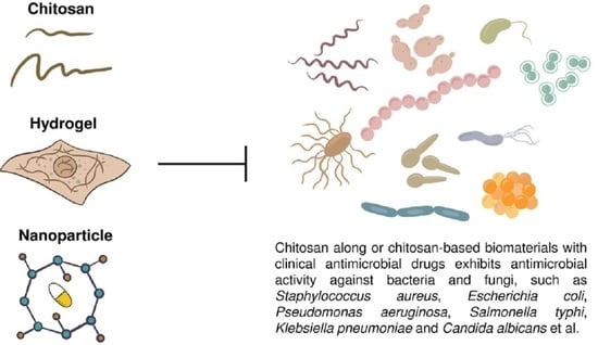
6. Applications of Chitosan-Based Nanoparticles and Films in Combination with Clinical Drugs against Microbes
Chitosan has been widely applied in many areas. However, the antimicrobial effects of pure chitosan and most of its derivatives are still remarkably lower than those of clinical antimicrobial drugs. Several articles have also shown that pure chitosan in combination with clinical drugs exhibits great antimicrobial activity [83,152,199,200,201,202,203]. Thus, this section focuses on the antimicrobial effects of developed chitosan-based biomaterials with current antibacterial and antifungal drugs because chitosan not only has intrinsic antimicrobial properties but is also able to deliver extrinsic antimicrobial drugs (Table 3).
6.1. Nanoparticles
Chitosan nanoparticles are synthesized for different purposes by various methods, such as ionotropic gelation, polyelectrolyte complexation, emulsification solvent diffusion, microemulsion, and reverse micelle formation [204,205]. Moreover, the antimicrobial effects of chitosan-based nanoparticles for drug and drug-free delivery systems have been intensively investigated against bacteria and fungi [56,57,58,60,63,67,68,106,119,203,206,207,208,209,210,211,212]. For example, chitosans of different DDAs and MWs exhibited synergistic activity with sulfamethoxazole against P. aeruginosa [213]. Chitosan nanoparticles loaded with levofloxacin or clarithromycin showed great potential against methicillin-resistant S. aureus (MASA) [211,214]. Additionally, chitosan-silver nanoparticles containing an antibiotic exhibited synergistic effects against fish bacteria [210,211].
6.2. Films
PEG-chitosan hydrogels containing ciprofloxacin improved the growth inhibition of E. coli compared with drug-free hydrogels and sustainably released the antibiotic for 24 hr [70]. Similarly, the high DDA of chitosan films loaded with different antibiotics exhibited better activity against different pathogenic bacteria [55,215,216,217,218]. Fibrin-chitosan loaded with two antibiotics (metroidazole and ciprofloxacin) enhanced anti-Enterococcus faecalis activity [78]. A chitosan hydrogel containing ciprofloxacin and fluconazole nanoparticles exhibited significant antimicrobial activities against C. albicans, E. coli, and S. aureus [219]. Finally, a chitosan gel with metronidazole showed great anti-Candida activity to treat vaginal infection [220].
6.3. Implants
Chitosan-coated titanium containing tetracycline or chlorhexidine digluconate effectively inhibited Actinobacillus actinomycetemcomitans and Staphylococcus epidermidis [221]. Interestingly, a chitosan bar containing gentamicin prepared using crosslinking, solvent evaporation, and a cylinder model cutting technique, which was implanted into rabbit tibias, exhibited significant antibacterial activity, suggesting that this chitosan bar would be effective against chronic osteomyelitis [222].
7. Conclusions
Approximately 30,000 original research and review articles related to chitosan have been reported [223], indicating that this naturally occurring product has great potential applications. This review suggests that chitosan as a natural antimicrobial agent can be applied in agriculture, food, and biomedical areas. Transcriptomic analyses in chitosan-treated microbes have further concluded that the mode of action of chitosan against bacteria or fungi may have multiple intracellular and extracellular effects. Although chitosan shows great promising antimicrobial potential, most of these studies are still at the laboratory level. Furthermore, the low water solubility and the lack of defined molecular weight and purity are the major issues for future application of chitosan. The development of better strategies and optimized conditions against pathogenic bacteria and fungi is necessary.
Author Contributions
Conceptualization, C.-L.K., F.-S.D., C.-Y.C., and C.-H.L. Writing, C.-L.K., F.-S.D., C.-Y.C., and C.-H.L. All of the authors have read and approved the final manuscript. C.-H.L. participated in the coordination and finalization of the manuscript draft. C.-L.K., F.-S.D., and C.-Y.C. contributed equally. All authors have read and agreed to the published version of the manuscript.
Funding
This research was funded by MOST-105-2628-B-002-018-MY3 from the Ministry of Science Technology and by NTU109L7813 from National Taiwan University.
Institutional Review Board Statement
Not Applicable.
Informed Consent Statement
Not Applicable.
Conflicts of Interest
The authors declare no conflict of interest.
References
- Hudson, S.M.; Smith, C. Polysaccharides: Chitin and chitosan: Chemistry and technology of their use as structural materials. In Biopolymers from Renewable Resources; Springer: Berlin/Heidelberg, Germany, 1998; pp. 96–118. [Google Scholar]
- Kumar, M.N.V.R. A review of chitin and chitosan applications. React. Funct. Polym. 2000, 46, 1–27. [Google Scholar] [CrossRef]
- Abdou, E.S.; Nagy, K.S.; Elsabee, M.Z. Extraction and characterization of chitin and chitosan from local sources. Bioresour. Technol. 2008, 99, 1359–1367. [Google Scholar] [CrossRef] [PubMed]
- Younes, I.; Rinaudo, M. Chitin and chitosan preparation from marine sources. Structure, properties and applications. Mar. Drugs 2015, 13, 1133–1174. [Google Scholar] [CrossRef] [Green Version]
- Tolaimate, A.; Rhazi, M.; Alagui, A.; Desbrieres, J.; Rinaudo, M. Valorization of waste products from fishing industry by production of chitin and chitosan. Phys. Chem. News 2008, 42, 120–127. [Google Scholar]
- Nishioka, Y.; Kyotani, S.; Masui, H.; Okamura, M.; Miyazaki, M.; Okazaki, K.; Ohnishi, S.; Yamamoto, Y.; Ito, K. Preparation and release characteristics of cisplatin albumin microspheres containing chitin and treated with chitosan. Chem. Pharm. Bull. 1989, 37, 3074–3077. [Google Scholar] [CrossRef] [Green Version]
- Antonino, R.S.C.M.D.; Fook, B.R.P.L.; Lima, V.A.D.; Rached, R.I.D.; Lima, E.P.N.; Lima, R.J.D.; Covas, C.A.P.; Fook, M.V.L. Preparation and characterization of chitosan obtained from shells of shrimp (Litopenaeus vannamei Boone). Mar. Drugs 2017, 15, 141. [Google Scholar] [CrossRef] [Green Version]
- Abebe, L.S.; Chen, X.; Sobsey, M.D. Chitosan coagulation to improve microbial and turbidity removal by ceramic water filtration for household drinking water treatment. Int. J. Environ. Res. Public Health 2016, 13, 269. [Google Scholar] [CrossRef] [PubMed] [Green Version]
- Altaher, H. The use of chitosan as a coagulant in the pre-treatment of turbid sea water. J. Hazard. Mater. 2012, 233–234, 97–102. [Google Scholar] [CrossRef]
- Ayad, M.; Salahuddin, N.; Fayed, A.; Bastakoti, B.P.; Suzuki, N.; Yamauchi, Y. Chemical design of a smart chitosan-polypyrrole-magnetite nanocomposite toward efficient water treatment. Phys. Chem. Chem. Phys. 2014, 16, 21812–21819. [Google Scholar] [CrossRef] [PubMed]
- Fabris, R.; Chow, C.W.; Drikas, M. Evaluation of chitosan as a natural coagulant for drinking water treatment. Water Sci. Technol. 2010, 61, 2119–2128. [Google Scholar] [CrossRef] [PubMed] [Green Version]
- Li, L.; Luo, C.; Li, X.; Duan, H.; Wang, X. Preparation of magnetic ionic liquid/chitosan/graphene oxide composite and application for water treatment. Int. J. Biol. Macromol. 2014, 66, 172–178. [Google Scholar] [CrossRef]
- Liaw, B.S.; Chang, T.T.; Chang, H.K.; Liu, W.K.; Chen, P.Y. Fish scale-extracted hydroxyapatite/chitosan composite scaffolds fabricated by freeze casting—An innovative strategy for water treatment. J. Hazard. Mater. 2020, 382, 121082. [Google Scholar] [CrossRef] [PubMed]
- Morsi, R.E.; Alsabagh, A.M.; Nasr, S.A.; Zaki, M.M. Multifunctional nanocomposites of chitosan, silver nanoparticles, copper nanoparticles and carbon nanotubes for water treatment: Antimicrobial characteristics. Int. J. Biol. Macromol. 2017, 97, 264–269. [Google Scholar] [CrossRef] [PubMed]
- Nasrollahzadeh, M.; Sajjadi, M.; Iravani, S.; Varma, R.S. Starch, cellulose, pectin, gum, alginate, chitin and chitosan derived (nano)materials for sustainable water treatment: A review. Carbohydr. Polym. 2021, 251, 116986. [Google Scholar] [CrossRef]
- Picos-Corrales, L.A.; Sarmiento-Sanchez, J.I.; Ruelas-Leyva, J.P.; Crini, G.; Hermosillo-Ochoa, E.; Gutierrez-Montes, J.A. Environment-friendly approach toward the treatment of raw agricultural wastewater and river water via flocculation using chitosan and bean straw flour as bioflocculants. ACS Omega 2020, 5, 3943–3951. [Google Scholar] [CrossRef] [PubMed]
- Sekine, M.; Takeshita, A.; Oda, N.; Ukita, M.; Imai, T.; Higuchi, T. On-site treatment of turbid river water using chitosan, a natural organic polymer coagulant. Water Sci. Technol. 2006, 53, 155–161. [Google Scholar] [CrossRef] [PubMed]
- Sun, Y.; Zhang, M.; Bhandari, B.; Yang, C.H. Ultrasound treatment of frozen crayfish with chitosan Nano-composite water-retaining agent: Influence on cryopreservation and storage qualities. Food Res. Int. 2019, 126, 108670. [Google Scholar] [CrossRef] [PubMed]
- Wang, J.P.; Chen, Y.Z.; Yuan, S.J.; Sheng, G.P.; Yu, H.Q. Synthesis and characterization of a novel cationic chitosan-based flocculant with a high water-solubility for pulp mill wastewater treatment. Water Res. 2009, 43, 5267–5275. [Google Scholar] [CrossRef] [PubMed]
- Wibowo, S.; Velazquez, G.; Savant, V.; Torres, J.A. Surimi wash water treatment for protein recovery: Effect of chitosan-alginate complex concentration and treatment time on protein adsorption. Bioresour. Technol. 2005, 96, 665–671. [Google Scholar] [CrossRef] [PubMed]
- Yang, R.; Li, H.; Huang, M.; Yang, H.; Li, A. A review on chitosan-based flocculants and their applications in water treatment. Water Res. 2016, 95, 59–89. [Google Scholar] [CrossRef] [PubMed]
- Yuan, D.; Zhang, W.; Cui, J.; He, L.; Wang, J.; Yan, C.; Kou, Y.; Li, J. Facile fabrication of magnetic phosphorylated chitosan for the removal of Co(II) in water treatment: Separation properties and adsorption mechanisms. Environ. Sci. Pollut. Res. Int. 2020, 27, 2588–2598. [Google Scholar] [CrossRef] [PubMed]
- Cota-Arriola, O.; Cortez-Rocha, M.O.; Burgos-Hernandez, A.; Ezquerra-Brauer, J.M.; Plascencia-Jatomea, M. Controlled release matrices and micro/nanoparticles of chitosan with antimicrobial potential: Development of new strategies for microbial control in agriculture. J. Sci. Food Agric. 2013, 93, 1525–1536. [Google Scholar] [CrossRef]
- Doares, S.H.; Syrovets, T.; Weiler, E.W.; Ryan, C.A. Oligogalacturonides and chitosan activate plant defensive fenes through the octadecanoid pathway. Proc. Natl. Acad. Sci. USA 1995, 92, 4095–4098. [Google Scholar] [CrossRef] [PubMed] [Green Version]
- El Hadrami, A.; Adam, L.R.; El Hadrami, I.; Daayf, F. Chitosan in plant protection. Mar. Drugs 2010, 8, 968–987. [Google Scholar] [CrossRef] [PubMed]
- Fan, G.Z.; Li, X.C.; Wang, X.D.; Zhai, Q.L.; Zhan, Y.G. Chitosan activates defense responses and triterpenoid production in cell suspension cultures of Betula platyphylla Suk. Afr. J. Biotechnol. 2010, 9, 2816–2820. [Google Scholar]
- Hidangmayum, A.; Dwivedi, P.; Katiyar, D.; Hemantaranjan, A. Application of chitosan on plant responses with special reference to abiotic stress. Physiol. Mol. Biol. Plants 2019, 25, 313–326. [Google Scholar] [CrossRef] [PubMed]
- Jia, X.C.; Meng, Q.S.; Zeng, H.H.; Wang, W.X.; Yin, H. Chitosan oligosaccharide induces resistance to Tobacco mosaic virus in Arabidopsis via the salicylic acid-mediated signalling pathway. Sci. Rep. 2016, 6, 26144. [Google Scholar] [CrossRef] [PubMed]
- Li, B.; Shan, C.L.; Ge, M.Y.; Wang, L.; Fang, Y.; Wang, Y.L.; Xie, G.L.; Sun, G.C. Antibacterial mechanism of chitosan and its applications in protection of plant from bacterial disease. Asian J. Chem. 2013, 25, 10033–10036. [Google Scholar] [CrossRef]
- Li, P.Q.; Linhardt, R.J.; Cao, Z.M. Structural characterization of oligochitosan elicitor from Fusarium sambucinum and its elicitation of defensive responses in Zanthoxylum bungeanum. Int. J. Mol. Sci. 2016, 17, 2076. [Google Scholar] [CrossRef] [PubMed] [Green Version]
- Narula, K.; Elagamey, E.; Abdellatef, M.A.E.; Sinha, A.; Ghosh, S.; Chakraborty, N.; Chakraborty, S. Chitosan-triggered immunity to Fusarium in chickpea is associated with changes in the plant extracellular matrix architecture, stomatal closure and remodeling of the plant metabolome and proteome. Plant J. 2020, 103, 561–583. [Google Scholar] [CrossRef] [PubMed]
- Povero, G.; Loreti, E.; Pucciariello, C.; Santaniello, A.; Di Tommaso, D.; Di Tommaso, G.; Kapetis, D.; Zolezzi, F.; Piaggesi, A.; Perata, P. Transcript profiling of chitosan-treated Arabidopsis seedlings. J. Plant Res. 2011, 124, 619–629. [Google Scholar] [CrossRef] [PubMed]
- Thakur, M.; Sohal, B.S. Role of elicitors in inducing resistance in plants against pathogen infection: A review. ISRN Biochem. 2013, 2013, 762412. [Google Scholar] [CrossRef] [PubMed] [Green Version]
- Van Aubel, G.; Cambier, P.; Dieu, M.; Van Cutsem, P. Plant immunity induced by COS-OGA elicitor is a cumulative process that involves salicylic acid. Plant Sci. 2016, 247, 60–70. [Google Scholar] [CrossRef] [PubMed]
- Vanda, G.F.; Shabani, L.; Razavizadeh, R. Chitosan enhances rosmarinic acid production in shoot cultures of Melissa officinalis L. through the induction of methyl jasmonate. Bot. Stud. 2019, 60, 26. [Google Scholar] [CrossRef] [PubMed] [Green Version]
- Varlamov, V.P.; Mysyakina, I.S. Chitosan in biology, microbiology, medicine, and agriculture. Microbiology 2018, 87, 712–715. [Google Scholar] [CrossRef]
- Xing, K.; Zhu, X.; Peng, X.; Qin, S. Chitosan antimicrobial and eliciting properties for pest control in agriculture: A review. Agron. Sustain. Dev. 2015, 35, 569–588. [Google Scholar] [CrossRef] [Green Version]
- Agullo, E.; Rodriguez, M.S.; Ramos, V.; Albertengo, L. Present and future role of chitin and chitosan in food. Macromol. Biosci. 2003, 3, 521–530. [Google Scholar] [CrossRef]
- Dutta, J.; Tripathi, S.; Dutta, P.K. Progress in antimicrobial activities of chitin, chitosan and its oligosaccharides: A systematic study needs for food applications. Food Sci. Technol. Int. 2012, 18, 3–34. [Google Scholar] [CrossRef]
- Morin-Crini, N.; Lichtfouse, E.; Torri, G.; Crini, G. Applications of chitosan in food, pharmaceuticals, medicine, cosmetics, agriculture, textiles, pulp and paper, biotechnology, and environmental chemistry. Environ. Chem. Lett. 2019, 17, 1667–1692. [Google Scholar] [CrossRef] [Green Version]
- Philibert, T.; Lee, B.H.; Fabien, N. Current status and new perspectives on chitin and chitosan as functional biopolymers. Appl. Biochem. Biotechnol. 2017, 181, 1314–1337. [Google Scholar] [CrossRef] [PubMed]
- Shahidi, F.; Arachchi, J.K.V.; Jeon, Y.J. Food applications of chitin and chitosans. Trends Food Sci. Technol. 1999, 10, 37–51. [Google Scholar] [CrossRef]
- Synowiecki, J.; Al-Khateeb, N.A. Production, properties, and some new applications of chitin and its derivatives. Crit. Rev. Food Sci. Nutr. 2003, 43, 145–171. [Google Scholar] [CrossRef] [PubMed]
- Van den Broek, L.A.M.; Knoop, R.J.I.; Kappen, F.H.J.; Boeriu, C.G. Chitosan films and blends for packaging material. Carbohyd. Polym. 2015, 116, 237–242. [Google Scholar] [CrossRef] [PubMed]
- Alves, A.; Sousa, E.; Kijjoa, A.; Pinto, M. Marine-derived compounds with potential use as cosmeceuticals and nutricosmetics. Molecules 2020, 25, 2536. [Google Scholar] [CrossRef]
- Aranaz, I.; Acosta, N.; Civera, C.; Elorza, B.; Mingo, J.; Castro, C.; Gandia, M.D.; Caballero, A.H. Cosmetics and cosmeceutical applications of chitin, chitosan and their derivatives. Polymers 2018, 10, 213. [Google Scholar] [CrossRef] [PubMed] [Green Version]
- Casanova, F.; Santos, L. Encapsulation of cosmetic active ingredients for topical application—A review. J. Microencapsul. 2016, 33, 1–17. [Google Scholar] [CrossRef] [PubMed]
- Jimtaisong, A.; Saewan, N. Utilization of carboxymethyl chitosan in cosmetics. Int. J. Cosmet. Sci. 2014, 36, 12–21. [Google Scholar] [CrossRef] [PubMed] [Green Version]
- Kim, S.K. Marine cosmeceuticals. J. Cosmet. Dermatol. 2014, 13, 56–67. [Google Scholar] [CrossRef] [PubMed]
- Liu, L.P.; Li, K.X.; Ding, H.P.; Lv, J.; Zhang, J. Study on preparation of a chitosan/vitamin C complex and its properties in cosmetics. Nat. Prod. Commun. 2020, 15, 1–9. [Google Scholar]
- Sionkowska, A.; Kaczmarek, B.; Michalska, M.; Lewandowska, K.; Grabska, S. Preparation and characterization of collagen/chitosan/hyaluronic acid thin films for application in hair care cosmetics. Pure Appl. Chem. 2017, 89, 1829–1839. [Google Scholar] [CrossRef]
- Felt, O.; Buri, P.; Gurny, R. Chitosan: A unique polysaccharide for drug delivery. Drug Dev. Ind. Pharm. 1998, 24, 979–993. [Google Scholar] [CrossRef] [PubMed]
- Wang, Z.Y.; Zhang, Q. Study on pulmonary delivery of peptide drugs in rats: Effects of absorption enhancers on cellular membrane fluidity. Yao Xue Xue Bao 2003, 38, 957–961. [Google Scholar] [PubMed]
- Haque, T.; Chen, H.; Ouyang, W.; Martoni, C.; Lawuyi, B.; Urbanska, A.M.; Prakash, S. Superior cell delivery features of poly(ethylene glycol) incorporated alginate, chitosan, and poly-L-lysine microcapsules. Mol. Pharm. 2005, 2, 29–36. [Google Scholar] [CrossRef] [PubMed]
- Fontana, C.R.; dos Santos, D.S., Jr.; Bosco, J.M.; Spolidorio, D.M.; Chierici Marcantonio, R.A. Evaluation of chitosan gel as antibiotic and photosensitizer delivery. Drug Deliv. 2008, 15, 417–422. [Google Scholar] [CrossRef]
- Alonso, M.J.; Coelho, D.; Engwer, C.; Fetzner, S.; Fuenzalida, J.; Goycoolea, F.; Hoffman, S.; Kollenbrock, S.; Menchicchi, B.; Moerschbacher, B.; et al. Chitosan-based nanomaterials for drug delivery and antibiotic-free bacterial control. NSTI-Nanotech. 2013, 3, 217–220. [Google Scholar]
- Ibrahim, H.; El-Bisi, M.; Taha, G.; El-Alfy, E. Chitosan nanoparticles loaded antibiotics as drug delivery biomaterial. J. Appl. Pharm. Sci. 2015, 5, 85–90. [Google Scholar] [CrossRef] [Green Version]
- Ahmed, T.A.; Aljaeid, B.M. Preparation, characterization, and potential application of chitosan, chitosan derivatives, and chitosan metal nanoparticles in pharmaceutical drug delivery. Drug Des. Devel. Ther. 2016, 10, 483–507. [Google Scholar] [CrossRef] [PubMed] [Green Version]
- Alvarez Echazu, M.I.; Olivetti, C.E.; Anesini, C.; Perez, C.J.; Alvarez, G.S.; Desimone, M.F. Development and evaluation of thymol-chitosan hydrogels with antimicrobial-antioxidant activity for oral local delivery. Mater. Sci. Eng. C Mater. Biol. Appl. 2017, 81, 588–596. [Google Scholar] [CrossRef] [PubMed]
- Harris, M.; Alexander, C.; Wells, C.M.; Bumgardner, J.D.; Carpenter, D.P.; Jennings, J.A. Chitosan for the delivery of antibiotics. In Chitosan Based Biomaterials; Woodhead Publishing: Cambridge, UK, 2017; Volume 2, pp. 147–173. [Google Scholar]
- Kiilll, C.P.; Barud, H.D.; Santagneli, S.H.; Ribeiro, S.J.L.; Silva, A.M.; Tercjak, A.; Gutierrez, J.; Pironi, A.M.; Gremiao, M.P.D. Synthesis and factorial design applied to a novel chitosan/sodium polyphosphate nanoparticles via ionotropic gelation as an RGD delivery system. Carbohyd. Polym. 2017, 157, 1695–1702. [Google Scholar] [CrossRef] [PubMed] [Green Version]
- Mohammed, M.A.; Syeda, J.T.M.; Wasan, K.M.; Wasan, E.K. An overview of chitosan nanoparticles and its application in non-parenteral drug delivery. Pharmaceutics 2017, 9, 53. [Google Scholar] [CrossRef] [Green Version]
- Ali, A.; Ahmed, S. A review on chitosan and its nanocomposites in drug delivery. Int. J. Biol. Macromol. 2018, 109, 273–286. [Google Scholar] [CrossRef] [PubMed]
- Darwesh, B.; Aldawsari, H.M.; Badr-Eldin, S.M. Optimized chitosan/anion polyelectrolyte complex based inserts for vaginal delivery of fluconazole: In vitro/in vivo evaluation. Pharmaceutics 2018, 10, 227. [Google Scholar] [CrossRef] [PubMed] [Green Version]
- Quiñones, J.P.; Peniche, H.; Peniche, C. Chitosan based self-assembled nanoparticles in drug delivery. Polymers 2018, 10, 235. [Google Scholar] [CrossRef] [PubMed] [Green Version]
- Rao, K.M.; Kumar, A.; Suneetha, M.; Han, S.S. pH and near-infrared active; chitosan-coated halloysite nanotubes loaded with curcumin-Au hybrid nanoparticles for cancer drug delivery. Int. J. Biol. Macromol. 2018, 112, 119–125. [Google Scholar] [CrossRef]
- Garg, U.; Chauhan, S.; Nagaich, U.; Jain, N. Current advances in chitosan nanoparticles based drug delivery and targeting. Adv. Pharm. Bull. 2019, 9, 195–204. [Google Scholar] [CrossRef] [PubMed]
- Kravanja, G.; Primozic, M.; Knez, Z.; Leitgeb, M. Chitosan-based (Nano) materials for novel biomedical applications. Molecules 2019, 24, 1960. [Google Scholar] [CrossRef] [PubMed] [Green Version]
- Sharif, S.; Abbas, G.; Hanif, M.; Bernkop-Schnurch, A.; Jalil, A.; Yaqoob, M. Mucoadhesive micro-composites: Chitosan coated halloysite nanotubes for sustained drug delivery. Colloids Surf. B. Biointerfaces 2019, 184, 110527. [Google Scholar] [CrossRef]
- Sharma, P.K.; Halder, M.; Srivastava, U.; Singh, Y. Antibacterial PEG-chitosan hydrogels for controlled antibiotic/protein delivery. ACS Appl. Bio Mater. 2019, 2, 5313–5322. [Google Scholar] [CrossRef]
- Schlachet, I.; Moshe Halamish, H.; Sosnik, A. Mixed amphiphilic polymeric nanoparticles of chitosan, poly(vinyl alcohol) and poly(methyl methacrylate) for intranasal drug delivery: A preliminary in vivo study. Molecules 2020, 25, 4496. [Google Scholar] [CrossRef] [PubMed]
- Chen, C.P.; Hsieh, C.M.; Tsai, T.; Yang, J.C.; Chen, C.T. Optimization and evaluation of a chitosan/hydroxypropyl methylcellulose hydrogel containing toluidine blue O for antimicrobial photodynamic inactivation. Int. J. Mol. Sci. 2015, 16, 20859–20872. [Google Scholar] [CrossRef] [Green Version]
- Rajasekaran, P.; Santra, S. Hydrothermally treated chitosan hydrogel loaded with copper and zinc particles as a potential micronutrient-based antimicrobial feed additive. Front. Vet. Sci. 2015, 2, 62. [Google Scholar] [CrossRef] [PubMed] [Green Version]
- Romic, M.D.; Klaric, M.S.; Lovric, J.; Pepic, I.; Cetina-Cizmek, B.; Filipovic-Grcic, J.; Hafner, A. Melatonin-loaded chitosan/Pluronic (R) F127 microspheres as in situ forming hydrogel: An innovative antimicrobial wound dressing. Eur. J. Pharm. Biopharm. 2016, 107, 67–79. [Google Scholar] [CrossRef] [PubMed]
- Hernandez, P.; Lucero-Acuna, A.; Gutierrez-Valenzuela, C.A.; Moreno, R.; Esquivel, R. Systematic evaluation of pH and thermoresponsive poly(n-isopropylacrylamide-chitosan-fluorescein)microgel. e-Polymers 2017, 17, 399–408. [Google Scholar] [CrossRef]
- Wang, Q.; Chen, S.; Chen, D. Preparation and characterization of chitosan based injectable hydrogels enhanced by chitin nano-whiskers. J. Mech. Behav. Biomed. Mater. 2017, 65, 466–477. [Google Scholar] [CrossRef] [PubMed]
- Lopez-Velazquez, J.C.; Rodriguez-Rodriguez, R.; Espinosa-Andrews, H.; Qui-Zapata, J.A.; Garcia-Morales, S.; Navarro-Lopez, D.E.; Luna-Barcenas, G.; Vassallo-Brigneti, E.C.; Garcia-Carvajal, Z.Y. Gelatin-chitosan-PVA hydrogels and their application in agriculture. J. Chem. Technol. Biotechnol. 2019, 94, 3495–3504. [Google Scholar] [CrossRef]
- Aksel, H.; Mahjour, F.; Bosaid, F.; Calamak, S.; Azim, A.A. Antimicrobial activity and biocompatibility of antibiotic-loaded chitosan hydrogels as a potential scaffold in regenerative endodontic treatment. J. Endod. 2020, 46, 1867–1875. [Google Scholar] [CrossRef]
- Iqbal, D.N.; Shafiq, S.; Khan, S.M.; Ibrahim, S.M.; Abubshait, S.A.; Nazir, A.; Abbas, M.; Iqbal, M. Novel chitosan/guar gum/PVA hydrogel: Preparation, characterization and antimicrobial activity evaluation. Int. J. Biol. Macromol. 2020, 164, 499–509. [Google Scholar] [CrossRef]
- Qu, B.; Luo, Y.C. Chitosan-based hydrogel beads: Preparations, modifications and applications in food and agriculture sectors—A review. Int. J. Biol. Macromol. 2020, 152, 437–448. [Google Scholar] [CrossRef] [PubMed]
- Felt, O.; Carrel, A.; Baehni, P.; Buri, P.; Gurny, R. Chitosan as tear substitute: A wetting agent endowed with antimicrobial efficacy. J. Ocul. Pharmacol. Ther. 2000, 16, 261–270. [Google Scholar] [CrossRef] [PubMed]
- Shih, P.Y.; Liao, Y.T.; Tseng, Y.K.; Deng, F.S.; Lin, C.H. A potential antifungal effect of chitosan against Candida albicans is mediated via the inhibition of SAGA complex component expression and the subsequent alteration of cell surface integrity. Front. Microbiol. 2019, 10, 602. [Google Scholar] [CrossRef] [PubMed]
- Lo, W.H.; Deng, F.S.; Chang, C.J.; Lin, C.H. Synergistic antifungal activity of chitosan with fluconazole against Candida albicans, Candida tropicalis, and fluconazole-resistant strains. Molecules 2020, 25, 5114. [Google Scholar] [CrossRef] [PubMed]
- Rabea, E.I.; Badawy, M.E.T.; Stevens, C.V.; Smagghe, G.; Steurbaut, W. Chitosan as antimicrobial agent: Applications and mode of action. Biomacromolecules 2003, 4, 1457–1465. [Google Scholar] [CrossRef] [PubMed]
- Avadi, M.R.; Sadeghi, A.M.M.; Tahzibi, A.; Bayati, K.; Pouladzadeh, M.; Zohuriaan-Mehr, M.J.; Rafiee-Tehrani, M. Diethylmethyl chitosan as an antimicrobial agent: Synthesis, characterization and antibacterial effects. Eur. Polym. J. 2004, 40, 1355–1361. [Google Scholar] [CrossRef]
- Wang, X.H.; Du, Y.M.; Fan, L.H.; Liu, H.; Hu, Y. Chitosan-metal complexes as antimicrobial agent: Synthesis, characterization and structure-activity study. Polym. Bull. 2005, 55, 105–113. [Google Scholar] [CrossRef]
- Hu, Y.; Du, Y.M.; Wang, X.Y.; Feng, T. Self-aggregation of water-soluble chitosan and solubilization of thymol as an antimicrobial agent. J. Biomed. Mater. Res. A 2009, 90A, 874–881. [Google Scholar] [CrossRef]
- Raafat, D.; Sahl, H.G. Chitosan and its antimicrobial potential—A critical literature survey. Microb. Biotechnol. 2009, 2, 186–201. [Google Scholar] [CrossRef] [PubMed] [Green Version]
- Gomez-Estaca, J.; de Lacey, A.L.; Lopez-Caballero, M.E.; Gomez-Guillen, M.C.; Montero, P. Biodegradable gelatin-chitosan films incorporated with essential oils as antimicrobial agents for fish preservation. Food Microbiol. 2010, 27, 889–896. [Google Scholar] [CrossRef] [PubMed]
- Kong, M.; Chen, X.G.; Xing, K.; Park, H.J. Antimicrobial properties of chitosan and mode of action: A state of the art review. Int. J. Food Microbiol. 2010, 144, 51–63. [Google Scholar] [CrossRef] [PubMed]
- Hernandez-Ochoa, L.; Gonzales-Gonzales, A.; Gutierrez-Mendez, N.; Munoz-Castellanos, L.N.; Quintero-Ramos, A. Study of the antibacterial activity of chitosan-based films prepared with different molecular weights including spices essential oils and functional extracts as antimicrobial agents. Rev. Mex. Ing. Quim. 2011, 10, 455–463. [Google Scholar]
- Huang, L.Y.; Dai, T.H.; Xuan, Y.; Tegos, G.P.; Hamblin, M.R. Synergistic combination of chitosan acetate with nanoparticle silver as a topical antimicrobial: Efficacy against bacterial burn infections. Antimicrob. Agents Chemother. 2011, 55, 3432–3438. [Google Scholar] [CrossRef] [PubMed] [Green Version]
- Costa, E.M.; Silva, S.; Pina, C.; Tavaria, F.K.; Pintado, M.M. Evaluation and insights into chitosan antimicrobial activity against anaerobic oral pathogens. Anaerobe 2012, 18, 305–309. [Google Scholar] [CrossRef] [PubMed]
- Badawy, M.E.I.; Rabea, E.I. Synthesis and structure-activity relationship of N-(cinnamyl) chitosan analogs as antimicrobial agents. Int. J. Biol. Macromol. 2013, 57, 185–192. [Google Scholar] [CrossRef] [PubMed]
- Jiang, L.; Wang, F.; Han, F.; Prinyawiwatkul, W.; No, H.K.; Ge, B. Evaluation of diffusion and dilution methods to determine the antimicrobial activity of water-soluble chitosan derivatives. J. Appl. Microbiol. 2013, 114, 956–963. [Google Scholar] [CrossRef] [PubMed] [Green Version]
- Mansilla, A.Y.; Albertengo, L.; Rodriguez, M.S.; Debbaudt, A.; Zuniga, A.; Casalongue, C.A. Evidence on antimicrobial properties and mode of action of a chitosan obtained from crustacean exoskeletons on Pseudomonas syringae pv. tomato DC3000. Appl. Microbiol. Biotechnol. 2013, 97, 6957–6966. [Google Scholar] [CrossRef] [PubMed]
- Mohamed, N.A.; Sabaa, M.W.; El-Ghandour, A.H.; Abdel-Aziz, M.M.; Abdel-Gawad, O.F. Quaternized N-substituted carboxymethyl chitosan derivatives as antimicrobial agents. Int. J. Biol. Macromol. 2013, 60, 156–164. [Google Scholar] [CrossRef] [PubMed]
- Tan, H.L.; Ma, R.; Lin, C.C.; Liu, Z.W.; Tang, T.T. Quaternized chitosan as an antimicrobial agent: Antimicrobial activity, mechanism of action and biomedical applications in orthopedics. Int. J. Mol. Sci. 2013, 14, 1854–1869. [Google Scholar] [CrossRef] [PubMed]
- Fiorani, G.; Saoncella, O.; Kaner, P.; Altinkaya, S.A.; Figoli, A.; Bonchio, M.; Carraro, M. Chitosan-polyoxometalate nanocomposites: Synthesis, characterization and application as antimicrobial agents. J. Clust. Sci. 2014, 25, 839–854. [Google Scholar] [CrossRef] [Green Version]
- Younes, I.; Hajji, S.; Frachet, V.; Rinaudo, M.; Jellouli, K.; Nasri, M. Chitin extraction from shrimp shell using enzymatic treatment. Antitumor, antioxidant and antimicrobial activities of chitosan. Int. J. Biol. Macromol. 2014, 69, 489–498. [Google Scholar] [CrossRef] [PubMed]
- Rahman, P.M.; Muraleedaran, K.; Mujeeb, V.M.A. Applications of chitosan powder with in situ synthesized nano ZnO particles as an antimicrobial agent. Int. J. Biol. Macromol. 2015, 77, 266–272. [Google Scholar]
- Arjunan, N.; Kurnari, H.L.J.; Singaravelu, C.M.; Kandasamy, R.; Kandasamy, J. Physicochemical investigations of biogenic chitosan-silver nanocomposite as antimicrobial and anticancer agent. Int. J. Biol. Macromol. 2016, 92, 77–87. [Google Scholar] [CrossRef]
- Hosseinnejad, M.; Jafari, S.M. Evaluation of different factors affecting antimicrobial properties of chitosan. Int. J. Biol. Macromol. 2016, 85, 467–475. [Google Scholar] [CrossRef]
- Hanpanich, O.; Wongkongkatep, P.; Pongtharangkul, T.; Wongkongkatep, J. Turning hydrophilic bacteria into biorenewable hydrophobic material with potential antimicrobial activity via interaction with chitosan. Bioresour. Technol. 2017, 230, 97–102. [Google Scholar] [CrossRef] [PubMed]
- Ma, Z.X.; Garrido-Maestu, A.; Jeong, K.C. Application, mode of action, and in vivo activity of chitosan and its micro and nanoparticles as antimicrobial agents: A review. Carbohyd. Polym. 2017, 176, 257–265. [Google Scholar] [CrossRef] [PubMed]
- Rivera Aguayo, P.; Bruna Larenas, T.; Alarcon Godoy, C.; Cayupe Rivas, B.; Gonzalez-Casanova, J.; Rojas-Gomez, D.; Caro Fuentes, N. Antimicrobial and antibiofilm capacity of chitosan nanoparticles against wild type strain of Pseudomonas sp. isolated from milk of cows diagnosed with bovine mastitis. Antibiotics 2020, 9, 551. [Google Scholar] [CrossRef]
- Raafat, D.; von Bargen, K.; Haas, A.; Sahl, H.G. Insights into the mode of action of chitosan as an antibacterial compound. Appl. Environ. Microbiol. 2008, 74, 3764–3773. [Google Scholar] [CrossRef] [PubMed] [Green Version]
- Ing, L.Y.; Zin, N.M.; Sarwar, A.; Katas, H. Antifungal activity of chitosan nanoparticles and correlation with their physical properties. Int. J. Biomater. 2012, 2012, 632698. [Google Scholar] [CrossRef] [PubMed]
- Martinez-Camacho, A.P.; Cortez-Rocha, M.O.; Ezquerra-Brauer, J.M.; Graciano-Verdugo, A.Z.; Rodriguez-Felix, F.; Castillo-Ortega, M.M.; Yepiz-Gomez, M.S.; Plascencia-Jatomea, M. Chitosan composite films: Thermal, structural, mechanical and antifungal properties. Carbohyd. Polym. 2010, 82, 305–315. [Google Scholar] [CrossRef]
- Kim, H.J.; Chen, F.; Wang, X.; Rajapakse, N.C. Effect of chitosan on the biological properties of sweet basil (Ocimum basilicum L.). J. Agric. Food Chem. 2005, 53, 3696–3701. [Google Scholar] [CrossRef] [PubMed]
- Kjm, K.M.; Son, J.H.; Kim, S.K.; Weller, C.L.; Hanna, M.A. Properties of chitosan films as a function of pH and solvent type. J. Food Sci. 2006, 71, e119–e124. [Google Scholar]
- Boy, R.; Maness, C.; Kotek, R. Properties of chitosan/soy protein blended films with added plasticizing agent as a function of solvent type at acidic pH. Int. J. Polym. Mater. Polym. Biomater. 2016, 65, 11–17. [Google Scholar] [CrossRef]
- Zohuriaan-Mehr, M.J. Advances in chitin and chitosan modification through graft copolymerization: A comprehensive review. Iran. Polym. J. 2005, 14, 235–265. [Google Scholar]
- No, H.K.; Park, N.Y.; Lee, S.H.; Meyers, S.P. Antibacterial activity of chitosans and chitosan oligomers with different molecular weights. Int. J. Food Microbiol. 2002, 74, 65–72. [Google Scholar] [CrossRef]
- Andres, Y.; Giraud, L.; Gerente, C.; Le Cloirec, P. Antibacterial effects of chitosan powder: Mechanisms of action. Environ. Technol. 2007, 28, 1357–1363. [Google Scholar] [CrossRef]
- Seyfarth, F.; Schliemann, S.; Elsner, P.; Hipler, U.C. Antifungal effect of high- and low-molecular-weight chitosan hydrochloride, carboxymethyl chitosan, chitosan oligosaccharide and N-acetyl-D-glucosamine against Candida albicans, Candida krusei and Candida glabrata. Int. J. Pharm. 2008, 353, 139–148. [Google Scholar] [CrossRef] [PubMed]
- Sadeghi-Kiakhani, M.; Arami, M.; Gharanjig, K. Application of a biopolymer chitosan-poly(propylene)imine dendrimer hybrid as an antimicrobial agent on the wool fabrics. Iran. Polym. J. 2013, 22, 931–940. [Google Scholar] [CrossRef]
- Bhatnagar, A.; Sillanpaa, M. Applications of chitin- and chitosan-derivatives for the detoxification of water and wastewater—A short review. Adv. Colloid Interface Sci. 2009, 152, 26–38. [Google Scholar] [CrossRef] [PubMed]
- Zhao, D.; Yu, S.; Sun, B.; Gao, S.; Guo, S.; Zhao, K. Biomedical applications of chitosan and its derivative nanoparticles. Polymers 2018, 10, 462. [Google Scholar] [CrossRef] [PubMed] [Green Version]
- Azuma, K.; Izumi, R.; Osaki, T.; Ifuku, S.; Morimoto, M.; Saimoto, H.; Minami, S.; Okamoto, Y. Chitin, chitosan, and its derivatives for wound healing: Old and new materials. J. Funct. Biomater. 2015, 6, 104–142, Reprinted in J. Funct. Biomater. 2018, 9, 38. [Google Scholar]
- Cheung, R.C.; Ng, T.B.; Wong, J.H.; Chan, W.Y. Chitosan: An update on potential biomedical and pharmaceutical applications. Mar. Drugs 2015, 13, 5156–5186. [Google Scholar] [CrossRef] [PubMed]
- Sudarshan, N.R.; Hoover, D.G.; Knorr, D. Antibacterial action of chitosan. Food Biotechnol. 1992, 6, 257–272. [Google Scholar] [CrossRef]
- Ke, C.L.; Liao, Y.T.; Lin, C.H. MSS2 maintains mitochondrial function and is required for chitosan resistance, invasive growth, biofilm formation and virulence in Candida albicans. Virulence 2021, 12, 281–297. [Google Scholar] [CrossRef] [PubMed]
- Pasquina-Lemonche, L.; Burns, J.; Turner, R.D.; Kumar, S.; Tank, R.; Mullin, N.; Wilson, J.S.; Chakrabarti, B.; Bullough, P.A.; Foster, S.J.; et al. The architecture of the gram-positive bacterial cell wall. Nature 2020, 582, 294–297. [Google Scholar] [CrossRef]
- Rohde, M. The gram-positive bacterial cell wall. Microbiol. Spectr. 2019, 7. [Google Scholar] [CrossRef] [Green Version]
- Gan, L.; Chen, S.; Jensen, G.J. Molecular organization of gram-negative peptidoglycan. Proc. Natl. Acad. Sci. USA 2008, 105, 18953–18957. [Google Scholar] [CrossRef] [PubMed] [Green Version]
- Beveridge, T.J. Structures of gram-negative cell walls and their derived membrane vesicles. J. Bacteriol. 1999, 181, 4725–4733. [Google Scholar] [CrossRef] [PubMed] [Green Version]
- Kraus, D.; Peschel, A. Molecular mechanisms of bacterial resistance to antimicrobial peptides. Curr. Top. Microbiol. Immunol. 2006, 306, 231–250. [Google Scholar] [PubMed]
- Raetz, C.R.; Reynolds, C.M.; Trent, M.S.; Bishop, R.E. Lipid A modification systems in gram-negative bacteria. Annu. Rev. Biochem. 2007, 76, 295–329. [Google Scholar] [CrossRef] [Green Version]
- Alburquenque, C.; Bucarey, S.A.; Neira-Carrillo, A.; Urzua, B.; Hermosilla, G.; Tapia, C.V. Antifungal activity of low molecular weight chitosan against clinical isolates of Candida spp. Med. Mycol. 2010, 48, 1018–1023. [Google Scholar] [CrossRef] [PubMed] [Green Version]
- Chen, R.H.; Domard, A.; Muzzarelli, R.A.A.; Tokura, S.; Wang, D.M. Advances in chitin/chitosan science and their applications. Carbohyd. Polym. 2011, 84, 695. [Google Scholar] [CrossRef]
- Goy, R.C.; Morais, S.T.B.; Assis, O.B.G. Evaluation of the antimicrobial activity of chitosan and its quaternized derivative on E. coli and S. aureus growth. Rev. Bras. Farmacogn. 2016, 26, 122–127. [Google Scholar] [CrossRef] [Green Version]
- Hassan, M.A.; Omer, A.M.; Abbas, E.; Baset, W.M.A.; Tamer, T.M. Preparation, physicochemical characterization and antimicrobial activities of novel two phenolic chitosan Schiff base derivatives. Sci. Rep. 2018, 8, 11416. [Google Scholar] [CrossRef] [PubMed] [Green Version]
- Chung, Y.C.; Su, Y.P.; Chen, C.C.; Jia, G.; Wang, H.I.; Wu, J.C.G.; Lin, J.G. Relationship between antibacterial activity of chitosan and surface characteristics of cell wall. Acta Pharmacol. Sin. 2004, 25, 932–936. [Google Scholar] [PubMed]
- Muzzalupo, I.; Badolati, G.; Chiappetta, A.; Picci, N.; Muzzalupo, R. In vitro antifungal activity of Olive (Olea europaea) leaf extracts loaded in chitosan nanoparticles. Front. Bioeng. Biotechnol. 2020, 8, 151. [Google Scholar] [CrossRef] [PubMed] [Green Version]
- Lopez-Moya, F.; Suarez-Fernandez, M.; Lopez-Llorca, L.V. Molecular mechanisms of chitosan interactions with fungi and plants. Int. J. Mol. Sci. 2019, 20, 332. [Google Scholar] [CrossRef] [Green Version]
- Ludwig, D.B.; de Camargo, L.E.A.; Khalil, N.M.; Auler, M.E.; Mainardes, R.M. Antifungal activity of chitosan-coated poly(lactic-co-glycolic) acid nanoparticles containing amphotericin B. Mycopathologia 2018, 183, 659–668. [Google Scholar] [CrossRef] [PubMed]
- Lara, H.H.; Guisbiers, G.; Mendoza, J.; Mimun, L.C.; Vincent, B.A.; Lopez-Ribot, J.L.; Nash, K.L. Synergistic antifungal effect of chitosan-stabilized selenium nanoparticles synthesized by pulsed laser ablation in liquids against Candida albicans biofilms. Int. J. Nanomed. 2018, 13, 2697–2708. [Google Scholar] [CrossRef] [Green Version]
- Kalagatur, N.K.; Nirmal Ghosh, O.S.; Sundararaj, N.; Mudili, V. Antifungal activity of chitosan nanoparticles encapsulated with Cymbopogon martinii essential oil on plant pathogenic fungi Fusarium graminearum. Front. Pharmacol. 2018, 9, 610. [Google Scholar] [CrossRef] [PubMed] [Green Version]
- Garcia, L.G.S.; Guedes, G.M.M.; da Silva, M.L.Q.; Castelo-Branco, D.; Sidrim, J.J.C.; Cordeiro, R.A.; Rocha, M.F.G.; Vieira, R.S.; Brilhante, R.S.N. Effect of the molecular weight of chitosan on its antifungal activity against Candida spp. in planktonic cells and biofilm. Carbohydr. Polym. 2018, 195, 662–669. [Google Scholar] [CrossRef] [PubMed]
- Lopez-Moya, F.; Colom-Valiente, M.F.; Martinez-Peinado, P.; Martinez-Lopez, J.E.; Puelles, E.; Sempere-Ortells, J.M.; Lopez-Llorca, L.V. Carbon and nitrogen limitation increase chitosan antifungal activity in Neurospora crassa and fungal human pathogens. Fungal Biol. 2015, 119, 154–169. [Google Scholar] [CrossRef] [PubMed] [Green Version]
- Qiu, M.; Wu, C.; Ren, G.; Liang, X.; Wang, X.; Huang, J. Effect of chitosan and its derivatives as antifungal and preservative agents on postharvest green asparagus. Food Chem. 2014, 155, 105–111. [Google Scholar] [CrossRef] [PubMed]
- Viegas de Souza, R.H.; Takaki, M.; de Oliveira Pedro, R.; dos Santos Gabriel, J.; Tiera, M.J.; de Oliveira Tiera, V.A. Hydrophobic effect of amphiphilic derivatives of chitosan on the antifungal activity against Aspergillus flavus and Aspergillus parasiticus. Molecules 2013, 18, 4437–4450. [Google Scholar] [CrossRef] [PubMed]
- Pena, A.; Sanchez, N.S.; Calahorra, M. Effects of chitosan on Candida albicans: Conditions for its antifungal activity. Biomed. Res. Int. 2013, 2013, 527549. [Google Scholar] [CrossRef] [PubMed] [Green Version]
- Liu, H.; Tian, W.; Li, B.; Wu, G.; Ibrahim, M.; Tao, Z.; Wang, Y.; Xie, G.; Li, H.; Sun, G. Antifungal effect and mechanism of chitosan against the rice sheath blight pathogen, Rhizoctonia solani. Biotechnol. Lett. 2012, 34, 2291–2298. [Google Scholar] [CrossRef] [PubMed]
- Fang, S.W.; Li, C.F.; Shih, D.Y.C. Antifungal activity of chitosan and its preservative effect on low-sugar candied kumquat. J. Food Prot. 1994, 57, 136–140. [Google Scholar] [CrossRef]
- Kendra, D.F.; Hadwiger, L.A. Relative effect of crab shell and fungal wall chitosan on Fusarium growth and induced resistance. Phytopathology 1985, 75, 1303. [Google Scholar]
- Younes, I.; Sellimi, S.; Rinaudo, M.; Jellouli, K.; Nasri, M. Influence of acetylation degree and molecular weight of homogeneous chitosans on antibacterial and antifungal activities. Int. J. Food Microbiol. 2014, 185, 57–63. [Google Scholar] [CrossRef] [PubMed]
- Atai, Z.; Atai, M.; Amini, J.; Salehi, N. In vivo study of antifungal effects of low-molecular-weight chitosan against Candida albicans. J. Oral Sci. 2017, 59, 425–430. [Google Scholar] [CrossRef] [PubMed] [Green Version]
- Palma-Guerrero, J.; Lopez-Jimenez, J.A.; Perez-Berna, A.J.; Huang, I.C.; Jansson, H.B.; Salinas, J.; Villalain, J.; Read, N.D.; Lopez-Llorca, L.V. Membrane fluidity determines sensitivity of filamentous fungi to chitosan. Mol. Microbiol. 2010, 75, 1021–1032. [Google Scholar] [CrossRef]
- Kumariya, R.; Sood, S.K.; Rajput, Y.S.; Saini, N.; Garsa, A.K. Increased membrane surface positive charge and altered membrane fluidity leads to cationic antimicrobial peptide resistance in Enterococcus faecalis. Biochim. Biophys. Aacta 2015, 1848, 1367–1375. [Google Scholar] [CrossRef] [Green Version]
- Ganan, M.; Lorentzen, S.B.; Aam, B.B.; Eijsink, V.G.H.; Gaustad, P.; Sorlie, M. Antibiotic saving effect of combination therapy through synergistic interactions between well-characterized chito-oligosaccharides and commercial antifungals against medically relevant yeasts. PLoS ONE 2019, 14, e0227098. [Google Scholar] [CrossRef] [PubMed]
- Work, E. Biochemistry of the bacterial cell wall. Nature 1957, 179, 841–847. [Google Scholar] [CrossRef] [PubMed]
- Vollmer, W.; Blanot, D.; de Pedro, M.A. Peptidoglycan structure and architecture. FEMS Microbiol. Rev. 2008, 32, 149–167. [Google Scholar] [CrossRef] [Green Version]
- Gow, N.A.R.; Latge, J.P.; Munro, C.A. The Fungal cell wall: Structure, biosynthesis, and function. Microbiol. Spectr. 2017, 5. [Google Scholar] [CrossRef] [Green Version]
- Bajaj, H.; Acosta Gutierrez, S.; Bodrenko, I.; Malloci, G.; Scorciapino, M.A.; Winterhalter, M.; Ceccarelli, M. Bacterial outer membrane porins as electrostatic nanosieves: Exploring transport rules of small polar molecules. ACS Nano 2017, 11, 5465–5473. [Google Scholar] [CrossRef] [PubMed] [Green Version]
- Gademann, K. Controlling protein transport by small molecules. Curr. Drug Targets 2011, 12, 1574–1580. [Google Scholar] [CrossRef] [PubMed] [Green Version]
- Burgstaller, W. Transport of small lons and molecules through the plasma membrane of filamentous fungi. Crit. Rev. Microbiol. 1997, 23, 1–46. [Google Scholar] [CrossRef] [PubMed]
- Demchick, P.; Koch, A.L. The permeability of the wall fabric of Escherichia coli and Bacillus subtilis. J. Bacteriol. 1996, 178, 768–773. [Google Scholar] [CrossRef] [PubMed] [Green Version]
- Scherrer, R.; Louden, L.; Gerhardt, P. Porosity of the yeast cell wall and membrane. J. Bacteriol. 1974, 118, 534–540. [Google Scholar] [CrossRef] [PubMed] [Green Version]
- Turner, R.D.; Hurd, A.F.; Cadby, A.; Hobbs, J.K.; Foster, S.J. Cell wall elongation mode in gram-negative bacteria is determined by peptidoglycan architecture. Nat. Commun. 2013, 4, 1–8. [Google Scholar] [CrossRef] [PubMed] [Green Version]
- Vollmer, W.; Holtje, J.V. The architecture of the murein (peptidoglycan) in gram-negative bacteria: Vertical scaffold or horizontal layer(s)? J. Bacteriol. 2004, 186, 5978–5987. [Google Scholar] [CrossRef] [Green Version]
- Walker, L.; Sood, P.; Lenardon, M.D.; Milne, G.; Olson, J.; Jensen, G.; Wolf, J.; Casadevall, A.; Adler-Moore, J.; Gow, N.A.R. The viscoelastic properties of the fungal cell wall allow traffic of AmBisome as intact liposome vesicles. mBio 2018, 9. [Google Scholar] [CrossRef] [PubMed] [Green Version]
- Erickson, H.P. Size and shape of protein molecules at the nanometer level determined by sedimentation, gel filtration, and electron microscopy. Biol. Proced. Online 2009, 11, 32–51. [Google Scholar] [CrossRef] [PubMed] [Green Version]
- Dmitriev, B.A.; Toukach, F.V.; Holst, O.; Rietschel, E.T.; Ehlers, S. Tertiary structure of Staphylococcus aureus cell wall murein. J. Bacteriol. 2004, 186, 7141–7148. [Google Scholar] [CrossRef] [PubMed] [Green Version]
- Kulikov, S.N.; Tikhonov, V.E.; Bezrodnykh, E.A.; Lopatin, S.A.; Varlamov, V.P. Comparative evaluation of antimicrobial activity of oligochitosans against Klebsiella pneumoniae. Bioorg. Khim. 2015, 41, 67–73. [Google Scholar] [CrossRef]
- Denobel, J.G.; Dijkers, C.; Hooijberg, E.; Klis, F.M. Increased cell-wall porosity in Saccharomyces cerevisiae after treatment with dithiothreitol or EDTA. J. Gen. Microbiol. 1989, 135, 2077–2084. [Google Scholar]
- Denobel, J.G.; Klis, F.M.; Priem, J.; Munnik, T.; Vandenende, H. The gucanase-soluble mannoproteins limit cell-wall porosity in Saccharomyces cerevisiae. Yeast 1990, 6, 491–499. [Google Scholar] [CrossRef] [PubMed]
- Stirke, A.; Celiesiute-Germaniene, R.; Zimkus, A.; Zurauskiene, N.; Simonis, P.; Dervinis, A.; Ramanavicius, A.; Balevicius, S. The link between yeast cell wall porosity and plasma membrane permeability after PEF treatment. Sci. Rep. 2019, 9, 1–10. [Google Scholar] [CrossRef] [Green Version]
- Jung, E.J.; Youn, D.K.; Lee, S.H.; No, H.K.; Ha, J.G.; Prinyawiwatkul, W. Antibacterial activity of chitosans with different degrees of deacetylation and viscosities. Int. J. Food Sci. Technol. 2010, 45, 676–682. [Google Scholar] [CrossRef]
- Foster, L.J.R.; Ho, S.; Hook, J.; Basuki, M.; Marcal, H. Chitosan as a biomaterial: Influence of degree of deacetylation on its physiochemical, material and biological properties. PLoS ONE 2015, 10, e0135153. [Google Scholar] [CrossRef] [Green Version]
- Si, Z.; Hou, Z.; Vikhe, Y.S.; Thappeta, K.R.V.; Marimuthu, K.; De, P.P.; Ng, O.T.; Li, P.; Zhu, Y.; Pethe, K.; et al. Antimicrobial effect of a novel chitosan derivative and its synergistic effect with antibiotics. ACS Appl. Mater. Interfaces 2021, 13, 3237–3245. [Google Scholar] [CrossRef] [PubMed]
- Wang, W.Q.; Meng, Q.Y.; Li, Q.; Liu, J.B.; Zhou, M.; Jin, Z.; Zhao, K. Chitosan derivatives and their application in biomedicine. Int. J. Mol. Sci. 2020, 21, 487. [Google Scholar] [CrossRef] [PubMed] [Green Version]
- Satitsri, S.; Muanprasat, C. Chitin and chitosan derivatives as biomaterial resources for biological and biomedical applications. Molecules 2020, 25, 5961. [Google Scholar] [CrossRef] [PubMed]
- Piegat, A.; Zywicka, A.; Niemczyk, A.; Goszczynska, A. Antibacterial activity of N,O-acylated chitosan derivative. Polymers 2020, 13, 107. [Google Scholar] [CrossRef]
- Verlee, A.; Mincke, S.; Stevens, C.V. Recent developments in antibacterial and antifungal chitosan and its derivatives. Carbohydr. Polym. 2017, 164, 268–283. [Google Scholar] [CrossRef] [PubMed]
- Zhong, Z.; Aotegen, B.; Xu, H. The influence of the different inductivity of acetyl phenyl-thiosemicarbazone-chitosan on antimicrobial activities. Int. J. Biol. Macromol. 2011, 48, 713–719. [Google Scholar] [CrossRef]
- Huang, H.F.; Peng, C.F. Antibacterial and antifungal activity of alkylsulfonated chitosan. Biomark. Genom. Med. 2015, 7, 83–86. [Google Scholar] [CrossRef] [Green Version]
- Abdelwahab, H.E.; Hassan, S.Y.; Yacout, G.A.; Mostafa, M.A.; El Sadek, M.M. Synthesis and biological evaluation of new imine- and amino-chitosan derivatives. Polymers 2015, 7, 2690–2700. [Google Scholar] [CrossRef]
- Kumar, S.; Koh, J.; Kim, H.; Gupta, M.K.; Dutta, P.K. A new chitosan-thymine conjugate: Synthesis, characterization and biological activity. Int. J. Biol. Macromol. 2012, 50, 493–502. [Google Scholar] [CrossRef] [PubMed]
- Rabea, E.I.; Badawy, M.E.I.; Steurbaut, W.; Stevens, C.V. In vitro assessment of N-(benzyl)chitosan derivatives against some plant pathogenic bacteria and fungi. Eur. Polym. J. 2009, 45, 237–245. [Google Scholar] [CrossRef]
- Rabea, E.I.; El Badawy, M.; Rogge, T.M.; Stevens, C.V.; Hofte, M.; Steurbaut, W.; Smagghe, G. Insecticidal and fungicidal activity of new synthesized chitosan derivatives. Pest Manag. Sci. 2005, 61, 951–960. [Google Scholar] [CrossRef] [PubMed]
- Patale, R.L.; Patravale, V.B. O, N-carboxymethyl chitosan-zinc complex: A novel chitosan complex with enhanced antimicrobial activity. Carbohyd. Polym. 2011, 85, 105–110. [Google Scholar] [CrossRef]
- Li, Z.; Yang, F.; Yang, R. Synthesis and characterization of chitosan derivatives with dual-antibacterial functional groups. Int. J. Biol. Macromol. 2015, 75, 378–387. [Google Scholar] [CrossRef] [PubMed]
- Mellegard, H.; Kovacs, A.T.; Lindback, T.; Christensen, B.E.; Kuipers, O.P.; Granum, P.E. Transcriptional responses of Bacillus cereus towards challenges with the polysaccharide chitosan. PLoS ONE 2011, 6, e24304. [Google Scholar] [CrossRef] [Green Version]
- Zakrzewska, A.; Boorsma, A.; Brul, S.; Hellingwerf, K.J.; Klis, F.M. Transcriptional response of Saccharomyces cerevisiae to the plasma membrane-perturbing compound chitosan. Eukaryot. Cell 2005, 4, 703–715. [Google Scholar] [CrossRef] [PubMed] [Green Version]
- Jaime, M.D.L.A.; Lopez-Llorca, L.V.; Conesa, A.; Lee, A.Y.; Proctor, M.; Heisler, L.E.; Gebbia, M.; Giaever, G.; Westwood, J.T.; Nislow, C. Identification of yeast genes that confer resistance to chitosan oligosaccharide (COS) using chemogenomics. BMC Genom. 2012, 13, 1–26. [Google Scholar] [CrossRef] [PubMed] [Green Version]
- Abarca, M.L.; Accensi, F.; Bragulat, M.R.; Cabanes, F.J. Current importance of ochratoxin A-producing Aspergillus spp. J. Food Prot. 2001, 64, 903–906. [Google Scholar] [CrossRef] [PubMed]
- Meng, D.; Garba, B.; Ren, Y.; Yao, M.; Xia, X.; Li, M.; Wang, Y. Antifungal activity of chitosan against Aspergillus ochraceus and its possible mechanisms of action. Int. J. Biol. Macromol. 2020, 158, 1063–1070. [Google Scholar] [CrossRef] [PubMed]
- Lopez-Moya, F.; Kowbel, D.; Nueda, M.J.; Palma-Guerrero, J.; Glass, N.L.; Lopez-Llorca, L.V. Neurospora crassa transcriptomics reveals oxidative stress and plasma membrane homeostasis biology genes as key targets in response to chitosan. Mol. Biosyst. 2016, 12, 391–403. [Google Scholar] [CrossRef] [PubMed] [Green Version]
- Suarez-Fernandez, M.; Sambles, C.; Lopez-Moya, F.; Nueda, M.J.; Studholme, D.J.; Lopez-Llorca, L.V. Chitosan modulates Pochonia chlamydosporia gene expression during nematode egg parasitism. Environ. Microbiol. 2021. [Google Scholar] [CrossRef] [PubMed]
- Forghani, F.; Hajihassani, A. Recent advances in the development of environmentally benign treatments to control root-knot nematodes. Front. Plant Sci. 2020, 11, 1125. [Google Scholar] [CrossRef] [PubMed]
- Palma-Guerrero, J.; Jansson, H.B.; Salinas, J.; Lopez-Llorca, L.V. Effect of chitosan on hyphal growth and spore germination of plant pathogenic and biocontrol fungi. J. Appl. Microbiol. 2008, 104, 541–553. [Google Scholar] [CrossRef] [PubMed]
- Escudero, N.; Lopez-Moya, F.; Ghahremani, Z.; Zavala-Gonzalez, E.A.; Alaguero-Cordovilla, A.; Ros-Ibanez, C.; Lacasa, A.; Sorribas, F.J.; Lopez-Llorca, L.V. Chitosan increases tomato root colonization by Pochonia chlamydosporia and their combination reduces root-knot nematode damage. Front. Plant Sci. 2017, 8, 1415. [Google Scholar] [CrossRef] [PubMed]
- Aranda-Martinez, A.; Lenfant, N.; Escudero, N.; Zavala-Gonzalez, E.A.; Henrissat, B.; Lopez-Llorca, L.V. CAZyme content of Pochonia chlamydosporia reflects that chitin and chitosan modification are involved in nematode parasitism. Environ. Microbiol. 2016, 18, 4200–4215. [Google Scholar] [CrossRef] [Green Version]
- Palma-Guerrero, J.; Gomez-Vidal, S.; Tikhonov, V.E.; Salinas, J.; Jansson, H.B.; Lopez-Llorca, L.V. Comparative analysis of extracellular proteins from Pochonia chlamydosporia grown with chitosan or chitin as main carbon and nitrogen sources. Enzym. Microb. Technol. 2010, 46, 568–574. [Google Scholar] [CrossRef]
- Achkar, J.M.; Fries, B.C. Candida infections of the genitourinary tract. Clin. Microbiol. Rev. 2010, 23, 253–273. [Google Scholar] [CrossRef] [PubMed] [Green Version]
- Kullberg, B.J.; Arendrup, M.C. Invasive candidiasis. N. Engl. J. Med. 2015, 373, 1445–1456. [Google Scholar] [CrossRef] [PubMed] [Green Version]
- Charway, G.N.A.; Park, S.; Yu, D.; Je, J.Y.; Kim, D.H.; Jung, W.K.; Kim, Y.M. In vitro antibacterial and synergistic effect of chitosan-phytochemical conjugates against antibiotic resistant fish pathogenic bacteria. Indian J. Microbiol. 2019, 59, 116–120. [Google Scholar] [CrossRef] [PubMed]
- Asghar, M.A.; Yousuf, R.S.; Shoaib, M.H.; Asghar, M.A.; Ansar, S.; Zehravi, M.; Rehman, A.A. Synergistic combinations of chitosans and antibiotics in Staphylococcus aureus. Int. J. Nanomed. 2020, 13, 7841–7859. [Google Scholar] [CrossRef] [PubMed]
- Erman, A.; Hergouth, V.K.; Blango, M.G.; Kos, M.K.; Mulvey, M.A.; Veranic, P. Repeated treatments with chitosan in combination with antibiotics completely eradicate uropathogenic Escherichia coli from infected mouse urinary bladders. J. Infect. Dis. 2017, 216, 375–381. [Google Scholar] [CrossRef] [Green Version]
- Yano, K.; Yamada, T.; Banno, Y.; Sekiya, T.; Nozawa, Y. Modification of lipid composition in a dimorphic fungus, Candida albicans during the yeast cell to hypha transformation. Jpn. J. Med. Mycol. 1982, 23, 159–165. [Google Scholar] [CrossRef]
- Brasil, M.; Filgueiras, A.; Campos, M.; Neves, M.; Eugênio, M.; Sena, L.; Sant’Anna, C.; da Silva, V.; Diniz, C.; Sant’Ana, A. Synergism in the antibacterial action of ternary mixtures involving silver nanoparticles, chitosan and antibiotics. J. Braz. Chem. Soc. 2018, 29, 2026–2033. [Google Scholar] [CrossRef]
- Seyam, S.; Nordin, N.A.; Alfatama, M. Recent progress of chitosan and chitosan derivatives-based nanoparticles: Pharmaceutical perspectives of oral insulin delivery. Pharmaceuticals 2020, 13, 307. [Google Scholar] [CrossRef] [PubMed]
- Naskar, S.; Sharma, S.; Kuotsu, K. Chitosan-based nanoparticles: An overview of biomedical applications and its preparation. J. Drug Deliv. Sci. Technol. 2019, 49, 66–81. [Google Scholar] [CrossRef]
- Ong, T.H.; Chitra, E.; Ramamurthy, S.; Ling, C.C.S.; Ambu, S.P.; Davamani, F. Cationic chitosan-propolis nanoparticles alter the zeta potential of S. epidermidis, inhibit biofilm by modulating gene expression and exibit synergism with antibiotics. PLoS ONE 2019, 14, e0213079. [Google Scholar] [CrossRef]
- Zhang, C.; Hui, D.; Du, C.; Sun, H.; Peng, W.; Pu, X.; Li, Z.; Sun, J.; Zhou, C. Preparation and application of chitosan biomaterials in dentistry. Int. J. Biol. Macromol. 2020. [Google Scholar] [CrossRef] [PubMed]
- Idrees, H.; Zaidi, S.Z.J.; Sabir, A.; Khan, R.U.; Zhang, X.; Hassan, S.U. A review of biodegradable natural polymer-based nanoparticles for drug delivery applications. Nanomaterials 2020, 10, 1970. [Google Scholar] [CrossRef] [PubMed]
- Saha, P.; Goyal, A.K.; Rath, G. Formulation and evaluation of chitosan-based ampicillin trihydrate nanoparticles. Trop. J. Pharm. Res. 2010, 9, 483–488. [Google Scholar] [CrossRef] [Green Version]
- Barakat, K.M.; Gohar, Y.M. Nanosilver-marine fungal chitosan as antibiotic synergizers against sepsis fish bacteria. Iran. J. Microbiol. 2015, 7, 324–332. [Google Scholar] [PubMed]
- Asghar, M.A.; Yousuf, R.I.; Shoaib, M.H.; Asghar, M.A.; Ansar, S.; Zehravi, M.; Abdul Rehman, A. Synergistic nanocomposites of different antibiotics coupled with green synthesized chitosan-based silver nanoparticles: Characterization, antibacterial, in vivo toxicological and biodistribution studies. Int. J. Nanomed. 2020, 15, 7841–7859. [Google Scholar] [CrossRef] [PubMed]
- Golmohamadi, M.; Ghorbani, H.R.; Otadi, M. Synthesis of chitosan nanoparticles loaded with antibiotics as drug carriers and the study of antibacterial activity. J. Nanoanal. 2019, 6, 72–79. [Google Scholar]
- Tin, S.; Sakharkar, K.R.; Lim, C.S.; Sakharkar, M.K. Activity of chitosans in combination with antibiotics in Pseudomonas aeruginosa. Int. J. Biol. Sci. 2009, 5, 153–160. [Google Scholar] [CrossRef] [PubMed] [Green Version]
- Shahiwala, A.F.; Khan, G.A.; Bostanooei, N.M. Efficacy of levofloxacin, chitosan and EDTA combination against methicillin resistant Staphylococcus aureus skin infections: In vitro and in vivo evaluations. Int. J. Clin. Med. Microbiol. 2017, 2, 119. [Google Scholar] [CrossRef]
- Smith, J.K.; Bumgardner, J.D.; Courtney, H.S.; Smeltzer, M.S.; Haggard, W.O. Antibiotic-loaded chitosan film for infection prevention: A preliminary in vitro characterization. J. Biomed. Mater. Res. B Appl. Biomater. 2010, 94, 203–211. [Google Scholar] [CrossRef] [PubMed]
- Grimling, B.; Karolewicz, B.; Nawrot, U.; Wlodarczyk, K.; Gorniak, A. Physicochemical and antifungal properties of clotrimazole in combination with high-molecular weight chitosan as a multifunctional excipient. Mar. Drugs 2020, 18, 591. [Google Scholar] [CrossRef] [PubMed]
- Goller, S.; Turner, N.J. The antimicrobial effectiveness and cytotoxicity of the antibiotic-loaded chitosan: ECM scaffolds. Appl. Sci. 2020, 10, 3446. [Google Scholar] [CrossRef]
- Noel, S.P.; Courtney, H.; Bumgardner, J.D.; Haggard, W.O. Chitosan films: A potential local drug delivery system for antibiotics. Clin. Orthop. Relat. Res. 2008, 466, 1377–1382. [Google Scholar] [CrossRef] [Green Version]
- Nimal, T.R.; Mohandas, A.; Riju, R.M.; Arun, S.M.; Raja, B.; Rangasamy, J. Ciprofloxacin- and fluconazole-containing fibrin-nanoparticle-incorporated chitosan bandages for the treatment of polymicrobial wound infections. ACS Appl. Bio Mater. 2018, 2, 243–254. [Google Scholar]
- Perinelli, D.R.; Campana, R.; Skouras, A.; Bonacucina, G.; Cespi, M.; Mastrotto, F.; Baffone, W.; Casettari, L. Chitosan loaded into a hydrogel delivery system as a strategy to treat vaginal co-infection. Pharmaceutics 2018, 10, 23. [Google Scholar] [CrossRef] [Green Version]
- Norowski, P.A.; Courtney, H.S.; Babu, J.; Haggard, W.O.; Bumgardner, J.D. Chitosan coatings deliver antimicrobials from titanium implants: A preliminary study. Implant Dent. 2011, 20, 56–67. [Google Scholar] [CrossRef] [PubMed]
- Chen, A.; Chunlin, H.; Bao, J.; Zhou, T.; Dong, Z. Antibiotic loaded chitosan bar. An in vitro, in vivo study of a possible treatment for osteomyelitis. Clin. Orthop. Relat. Res. 1998, 366, 239–247. [Google Scholar]
- Bellich, B.; D’Agostino, I.; Semeraro, S.; Gamini, A.; Cesaro, A. “The good, the bad and the ugly” of chitosans. Mar. Drugs 2016, 14, 99. [Google Scholar] [CrossRef] [PubMed] [Green Version]
Figure 1.
Potential antimicrobial actions of chitosan against (A) gram-positive bacteria, (B) gram-negative bacteria, and (C) fungi. Given the negative charges of teichoic acids in gram-positive bacteria, lipopolysaccharide (LPS) in gram-negative bacteria and the phosphorylated mannosyl side in fungi, electrostatic interactions occur between the positively charged chitosan and the cell surface of the microorganism. Furthermore, chitosan chelates the environmental ions and nutrients required for bacterial survival. Low-molecular weight (MW) chitosan and oligo-chitosan might affect DNA/RNA or protein synthesis after passing through the cell wall and cell membrane into the cytoplasm. Additionally, low-MW chitosan and oligo-chitosan inhibit mitochondrial function and ATP production.

Figure 2.
Potential signaling cascades and factors required for chitosan tolerance in C. albicans. Mutant library screening has revealed that the Hog1, Cek1/Cek2, Mkc1, Ras1-cAMP, and calcineurin cascades as well as the Ssn3-mediated Sef1 pathway and other factors are involved in chitosan resistance. The roles of these pathways and factors in mediating cell wall integrity might affect mitochondrial function. Furthermore, low-MW chitosan directly represses mitochondrial function, leading to ATP production inhibition.


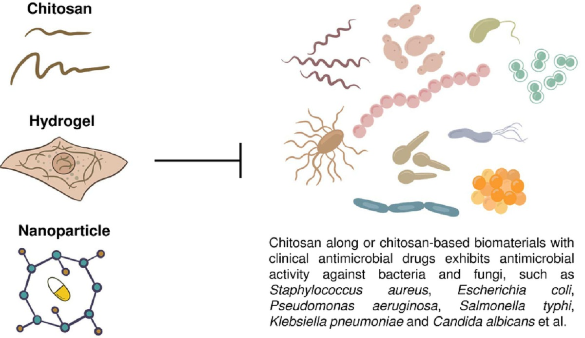
Table 1.
Genetic responses of chitosan-treated bacteria.
| Bacteria | Functional Categories | Conclusions | Ref. | |
|---|---|---|---|---|
| S. aureus | Upregulation | 84 genes Membrane Bioenergetics Cell Division Metabolism of Carbohydrates Metabolism of Amino Acids Regulation of RNA Synthesis Protein Folding Adaptation to Atypical Conditions Phage-Related Functions | Chitosan binds to teichoic acids Chitosan increases membrane permeability and causes membrane depolarization Chitosan might be involved in energy metabolism | [107] |
| Downregulation | 82 genes Transport/Binding Proteins and Lipoproteins Metabolism of Nucleotides and Nucleic Acids Metabolism of Lipids | |||
| B. cereus | Upregulation | 57 genes Potassium Transport System Membrane Protein-Associated Genes | Chitosan caused the permeabilizing effect, resulting in the leakage of intracellular potassium Deletion of the kdp gene (an ATP-driven K+ transport system) exhibited no significant difference in the minimum inhibitory concentration (MIC) and minimum bactericidal concentration (MBC) | [185] |
| Downregulation | 51 genes Chitin Binding Protein Metabolism of Amino Acids and Other Cellular Constituents Gluconeogenesis | |||
Table 2.
Genetic responses of chitosan-treated fungi.
| Fungus | Functional Categories | Conclusion | Ref. | |
|---|---|---|---|---|
| S. cerevisiae | Upregulation | Treated for 15 min/46 genes ER Integral to Membrane Treated for 30 min/97 genes ER Integral to Membrane Cell Wall Cell Wall Organization and Biogenesis Vacuoles Plasma Membrane Treated for 60 min/97 genes ER Integral to Membrane Cell Wall Cell Wall Organization and Biogenesis Vacuoles Treated for 120 min/234 genes ER Integral to Membrane Cell Wall Cell Wall Organization and Biogenesis Vacuoles Stress Response Treated 180 min/432 genes ER Integral to Membrane Cell Wall Cell Wall Organization and Biogenesis Vacuoles Response to Stress | Chitosan may be representative of other plasma membrane-perturbing compounds Chitosan stress decreases translational activity Calcineurin-dependent pathway is involved Deletion of CIN5, CRZ1, or RIM1 exhibits high sensitivity to chitosan | [186] |
| Downregulation | Not available rRNA Processing Ribosomes | |||
| S. cerevisiae | Upregulation | 589 genes Transcription Cell Cycle Protein Modification Stress Response RAS Signal Transduction | COS does not have specific gene targets Membrane permeability is increased in the COS-treated budding yeast Synergistic antifungal activity between chitosan and fluconazole was found | [187] |
| Downregulation | 631 genes Protein Folding Protein Complex Assembly Mitochondrial Electron Transport | |||
| A. ochraceus | Upregulation | 309 genes Starch and Sucrose Metabolism Glycerophospholipid Metabolism Ether Lipid Metabolism Steroid Biosynthesis Mitochondrial Electron Transport | Chitosan damages the integrity of the cell surface architecture and affects membrane fluidity Chitosan affects protein biosynthesis Chitosan is an alternative compound to control fungal pathogens | [189] |
| Downregulation | 26 genes Ribosome Biogenesis in Eukaryotes Glycerophospholipid Metabolism Ether Lipid Metabolism Steroid Biosynthesis | |||
| N. crassa | Upregulation | 237 genes in total (4, 8, and 16 h of treatment) Peroxisome Organization, ROS Degradation and Fatty Acid Catabolism (4 h) Mitochondrial Function (4 h, 8 h and 16 h) Ribosome and Ribosome Biogenesis (16 h) Nucleolus (8 h and 16 h) Structural Molecule Activity (16 h) | A MFS transporter (NCU04534) and a glutathione transferase (NCU10521) are the targets of chitosan Chitosan treatment causes an increase in intracellular ROS Chitosan affects protein biosynthesis ΔNCU03639 (lipase), ΔNCU04537 (monosaccharide transporter), ΔNCU10521 (glutathione S-transferase), ΔNCU08907 Clock controller gene 13 (ccg-13) and ΔNCU07840 (plasma membrane protein with a het domain) are sensitive to chitosan The presence of Ca2+ increases chitosan tolerance | [190] |
| Downregulation | 291 genes in total (4, 8, and 16 h treatment) Peroxisome Organization, ROS Degradation and Fatty Acid Catabolism (16 h) Cell Cortex (4, 8, and 16 h) Vesicle Organization (8 and 16 h) Conjugation (4, 8, and 16 h) G Protein Receptor Signaling Pathway (16 h) Microtubule Organizing Center (4, 8, and 16 h) Ribosome and Ribosome Biogenesis (4 h) Nucleolus (4 h) Structural Molecule Activity (4 h) | |||
| P. chlamydosporia | Upregulation | 46 genes Redox Processes Carbohydrate Catabolism Proteolysis Carbohydrate Transport Cell Cycle Energetic Metabolism Lipid Metabolism Protein Synthesis and Modification Chitin and Chitosan Degradation Structural Constituent of Cell Wall | Chitosan activates the expression of cytochrome P450 ClCP1 and thioredoxin-like proteins P. chlamydosporia is more resistance to chitosan because it may contain more chitosanase genes Chitosan induces many monosaccharide transport genes Chitosan could be a non-toxic additive to reduce root-knot nematode parasitism | [191] |
| Downregulation | 90 Genes Oxidation–Reduction Metabolism Cellular Protein Metabolic Process Macromolecule Biosynthetic Process Small Molecule Metabolic Process Metal Transport | |||
Table 3.
Applications of chitosan-based biomaterials containing clinical antimicrobial drugs.
| Chitosan/Antimicrobial Drug | Chitosan-Based Biomaterial | Findings | Microorganism(s) | Ref. |
|---|---|---|---|---|
| Chitosan/Sulfamethoxazole | Nanoparticle | Synergistic activity with sulfamethoxazole | P. aeruginosa | [213] |
| Chitosan/Amoxicillin Cefixime Levofloxacin | Nanoparticle | Significant antibacterial activity | P. aeruginosa E. coli S. aureus Salmonella typhi Klebsiella pneumoniae | [211] |
| Chitosan/Amikacin Rifampicin | Nanoparticle | Antibacterial activity against resistant strains | Aeromonas hydrophila Edwardsiella tarda Pasteurella piscicida P. aeruginosa Streptococcus faecium Streptococcus iniae Vibrio ordalli Yersinia ruckeri | [210] |
| Chitosan/Ciprofloxacin Chlortetracycline Hydrochloride Gentamycin sulfate | Nanoparticle | Inhibits the growth of gram-positive and gram-negative bacteria | E. coli S. aureus | [57] |
| Chitosan/Azithromycin Levofloxacin Tetracycline | Nanoparticle | Shows significant antibacterial effects | E. coli S. aureus | [203] |
| Chitosan/Rifampicin Ciprofloxacin Vancomycin Doxycycline Gentamicin | Nanoparticle | Inhibits bacterial biofilm and exhibits synergism with antibiotics | S. epidermidis | [206] |
| Chitosan/Clarithromycin | Nanoparticle | Shows antibacterial activity | S. aureus | [212] |
| Chitosan/Ciprofloxacin | Hydrogel | Inhibits bacterial growth | E. coli | [70] |
| Chitosan/Clindamycin | Hydrogel | Enhances the antibacterial properties | E. faecalis | [78] |
| Chitosan/Ciprofloxacin Fluconazole | Hydrogel(Bandage) | Shows significant antimicrobial activity | C. albicans E. coli S. aureus | [219] |
| Chitosan/Minocycline Rifampicin | Hydrogel | Provides bactericidal activity directly to the wound site | E. coli S. aureus | [217] |
| Chitosan/Tetracycline | Hydrogel | Has potential applications for antimicrobial action | S. aureus | [55] |
| Chitosan/Amikacin Daptomycin | Film | Effectively inhibits the growth of bacteria | S. aureus | [218] |
| Chitosan/Daptomycin Vancomycin | Film | Shows activity against gram-positive bacteria | S. aureus | [215] |
| Chitosan/Clotrimazole | Solid Mixtures | Acts synergistically with clotrimazole against non-albicans Candida strains | Candida glabrata | [216] |
| Chitosan/Levofloxacin | Hydroxypropyl methyl cellulose (HPMC) gel | Antibacterial activity against resistant strains | Methicillin-resistant S. aureus | [214] |
| Chitosan/Metronidazole | HPMC Gel | Anti-Candida activity | Candida species | [220] |
| Chitosan/Tetracycline Chlorhexidine | Chitosan-Coated Titanium Pins | Inhibits pathogen growth | A. actinomycetemcomitans S. epidermidis | [221] |
| Chitosan/Gentamicin | Chitosan Bar | Shows significant antibacterial activity | Microbes | [222] |
Publisher’s Note: MDPI stays neutral with regard to jurisdictional claims in published maps and institutional affiliations. |
© 2021 by the authors. Licensee MDPI, Basel, Switzerland. This article is an open access article distributed under the terms and conditions of the Creative Commons Attribution (CC BY) license (http://creativecommons.org/licenses/by/4.0/).
Share and Cite
MDPI and ACS Style
Ke, C.-L.; Deng, F.-S.; Chuang, C.-Y.; Lin, C.-H. Antimicrobial Actions and Applications of Chitosan. Polymers 2021, 13, 904. https://doi.org/10.3390/polym13060904
AMA Style
Ke C-L, Deng F-S, Chuang C-Y, Lin C-H. Antimicrobial Actions and Applications of Chitosan. Polymers. 2021; 13(6):904. https://doi.org/10.3390/polym13060904
Chicago/Turabian StyleKe, Cai-Ling, Fu-Sheng Deng, Chih-Yu Chuang, and Ching-Hsuan Lin. 2021. "Antimicrobial Actions and Applications of Chitosan" Polymers 13, no. 6: 904. https://doi.org/10.3390/polym13060904
Note that from the first issue of 2016, this journal uses article numbers instead of page numbers. See further details here.