Article

Spectroscopy, Crystal and Molecular Structures of New 4-Acylpyrazolone Dinitrophenylhydrazones

by
Omoruyi G. Idemudia
1,* and
Eric C. Hosten
2
1
Chemistry Department, University of Fort Hare, Private Bag X1314, Alice 5700, South Africa
2
Chemistry Department, Nelson Mandela Metropolitan University, Summerstrand Campus, P.O Box 77000, Port Elizabeth 6031, South Africa
*
Author to whom correspondence should be addressed.
Crystals 2016, 6(10), 127; https://doi.org/10.3390/cryst6100127
Submission received: 16 August 2016 / Revised: 13 September 2016 / Accepted: 26 September 2016 / Published: 19 October 2016

Abstract

:
Still looking at the development of new materials with unique properties and taking advantage of the nucleophilic properties of amines to form stable azomethines, dinitrophenylhydrazine was reacted with 4-ethyl-5-methyl-2-phenyl-pyrazol-3-one and then with 4-propyl-5-methyl-2-phenyl-pyrazol-3-one to get 4-ethyl-5-methyl-2-phenyl-pyrazol-3-one dinitrophenylhydrazone (Empp-Dh) and 4-propyl-5-methyl-2-phenyl-pyrazol-3-one dinitrophenylhydrazone (Pmpp-Dh), respectively, via a simple condensation reaction in a one-pot synthesis system. Careful interpretations of results from elemental analysis, mass and NMR spectroscopy were in agreement with single-crystal X-ray diffraction data. Reported Schiff bases in their single-crystal solid state exist in imine keto tautomer form, each crystallizing in a monoclinic crystal system, with a space group of C2/c (No. 15) in Empp-Dh and P21/c (No. 14) in Pmpp-Dh. They have extensive intra- and inter-molecular hydrogen as well as C–H···π-ring interactions.

Graphical Abstract

1. Introduction

Schiff bases are imine compounds which may be represented by the general formula R3R2C=NR1, where R1, R2 and R3 can/may be an alkyl, aryl, heteroaryl and/or hydrogen. They were first prepared by a German-born chemist and physicist known as Hugo Schiff [1]. Their versatile behavioral properties, remarkable reactivity and wide applications still continue to attract a lot of research attention [1,2]. They are synthesized easily by way of a condensation reaction between a carbonyl group (aldehydes or ketones) and an amine in a basic system with the elimination of water molecules as a by-product [3,4]. Acylpyrazolones have been reported to be a very useful chemical compound [5,6,7,8] and they form interesting Schiff bases on reaction with different amine groups by way of functional group substitution and synthetic modifications [9,10]. Their good chelating potentials towards transition metal ions have made them useful analytical reagents. As such, 2,4-dinitrophenylhydrazine is a nitro-substituted phenylhydrazine with an intense reddish orange color. It was employed for the quantitative identification of carbonyl functional groups [11], a characteristic that is responsible for its function as a nucleophilic amine for Schiff base synthesis. Schiff bases derived from N-(1-phenyl-2-hydroxy-2-phenyl ethylidine)-2,4-dinitrophenyl hydrazine (PDH) have been reported to positively affect (reduce) the average weight of the esophageal squamous carcinoma(ECA) tumor cells in a research study on mice [12]. We have synthesized a benzoyl derivative of a 2,4-dinitrophenylhydrazone with a solvent molecule distortion, titled, 4-{[2-(2,4-Dinitrophenyl)hydrazinylidene]-(phenyl)methyl}-5-methyl-2-phenyl-1H-pyrazol-3-(2H)-one ethanol monosolvate [13], as well as a group of other benzoyl derivatives of 4-acylpyrazolone Schiff bases which have shown interesting biological properties both as ligands and as transition metal complexes [14]. Our work on the development of new Schiff bases with novel properties [15,16], taking advantage of functional group modifications, continues in this paper. The single-crystal structure of 4-ethyl and 4-propyl pyrazolone derivatives of 2,4-dinitrophenylhydrazones alongside their spectroscopic properties are presented and discussed.

2. Results

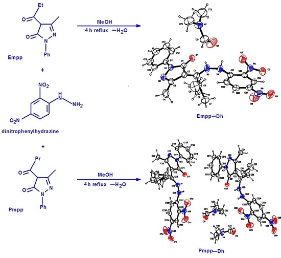
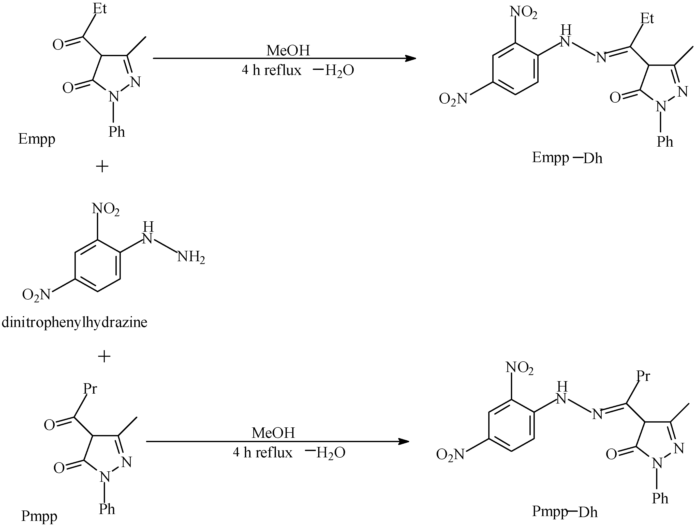
2.1. Synthesis and Elemental Analysis

Acylpyrazolone precursors have been synthesized previously [17], and with method modifications 4-ethyl-5-methyl-2-phenyl-pyrazol-3-one (Empp) and 4-propyl-5-methyl-2-phenyl-pyrazol-3-one (Pmpp) were obtained. In a simple condensation reaction with 2,4 dinitrophenylhydrazine, 4-ethyl-5-methyl-2-phenyl-pyrazol-3-one dinitrophenylhydrazone (Empp-Dh) and 4-propyl-5-methyl-2-phenyl-pyrazol-3-one dinitrophenylhydrazone (Pmpp-Dh) were synthesized in a one-pot synthesis set-up observing all reaction conditions as shown in Figure 1. The new ligands precipitated in good yield with a distinct orange color, which was followed by successive washing with methanol as well as with diethyl ether to remove all traces of unreacted starting materials and possible hydration from solvent water. They were recrystallized from hot methanol and a small melting point range was in agreement with their isolation in their pure forms. Single crystals of ligands were later grown from the dry precipitates. Experimental Carbon Hydrogen and Nitrogen elemental analysis for the reported synthesized ligands was in agreement with the calculated values which supported their spectroscopic results.

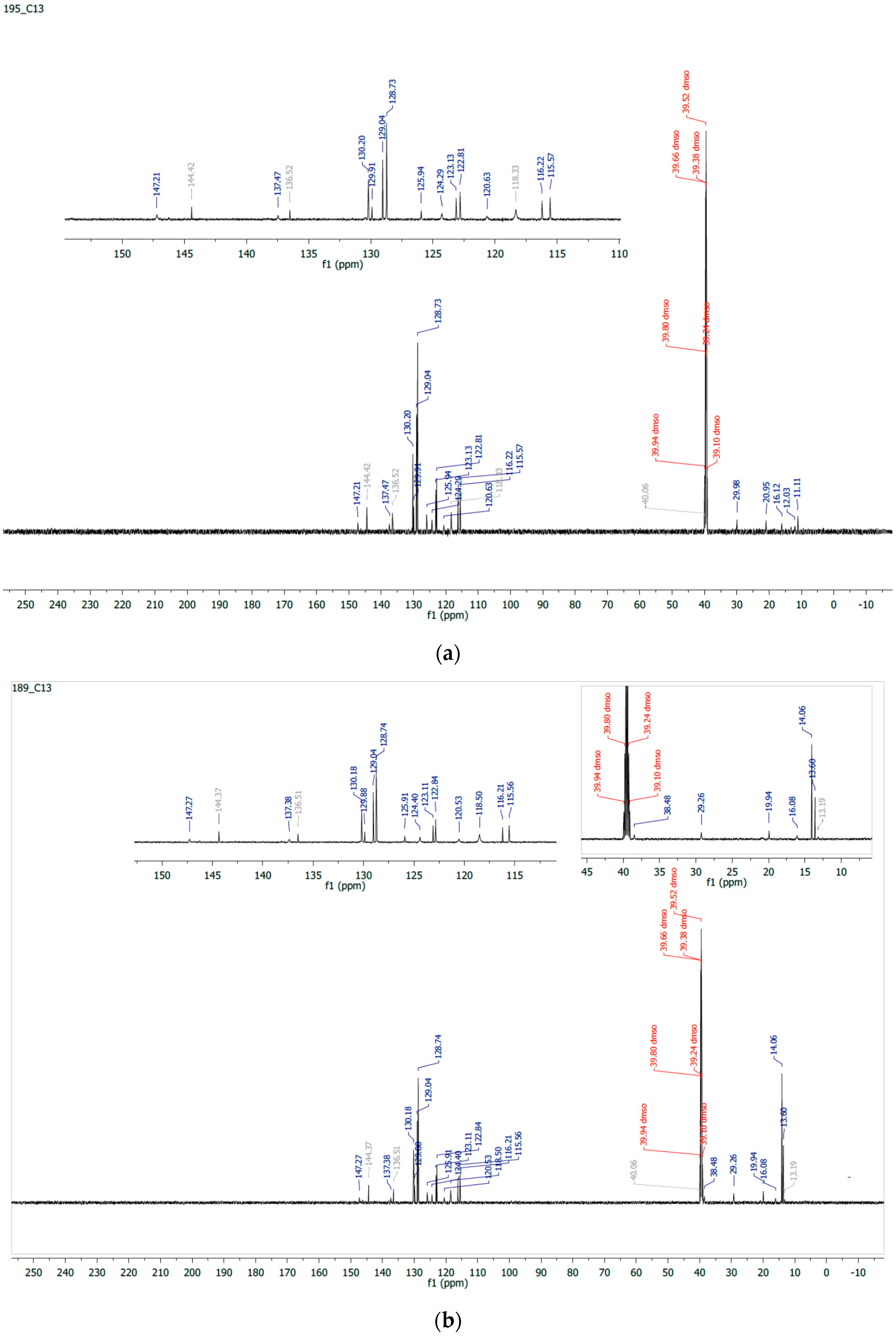
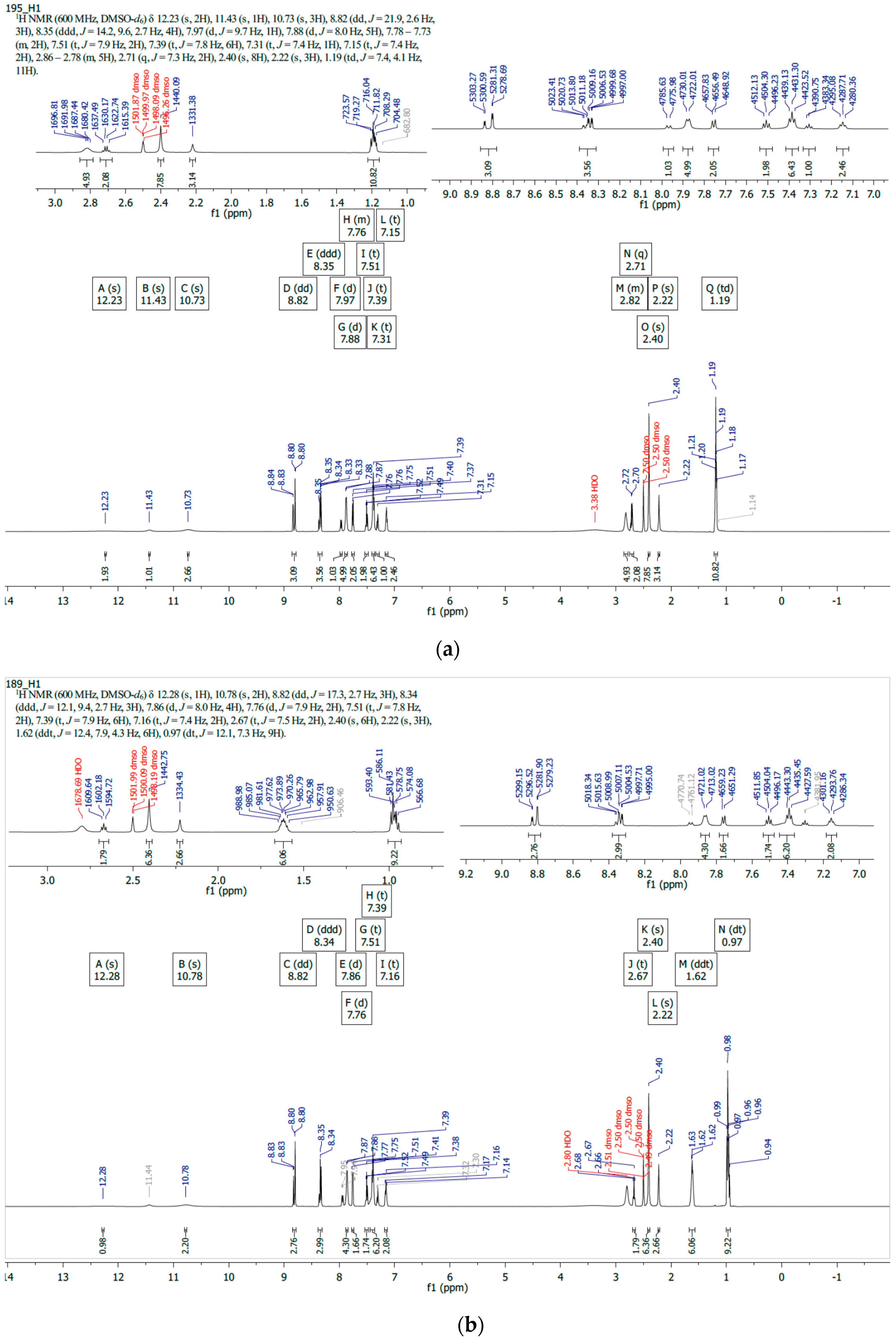
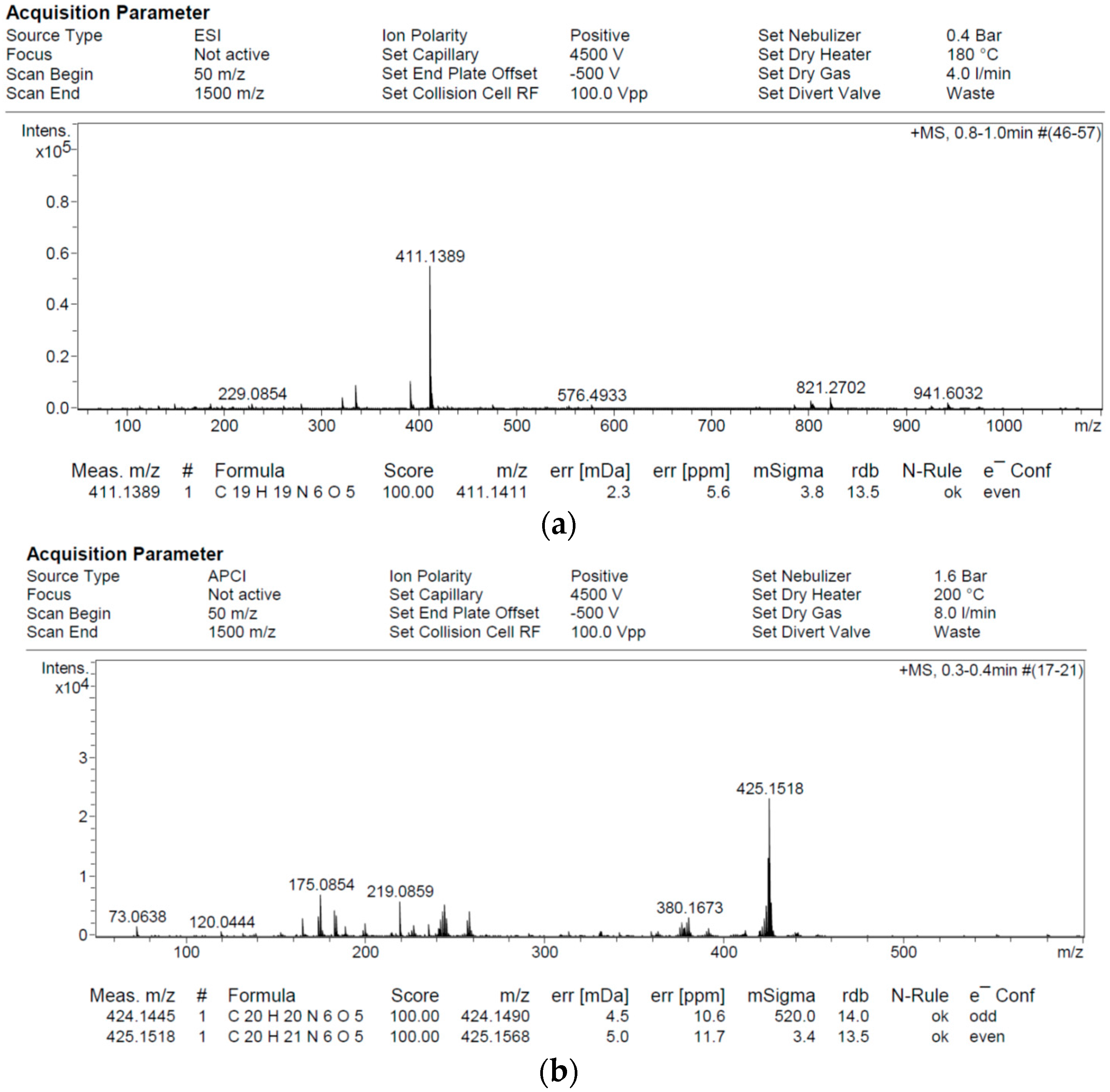
2.2. 1H/13C NMR and Mass Spectroscopy

The dinitrophenylhydrazones reported have exhibited common resonance signals at almost the same chemical shifts in their 13C NMR spectra. The presense of an aliphatic region resonance band at 38.50 ppm in Pmpp-Dh, which is absent in the spectrum of Empp-Dh, may be attributed to the extra –CH– group associated with the propyl derivative, as shown in Figure 2. This can also be corroborated by the prominent sharp signal at around 14.06 ppm. Similarly, the presence of the carbonyl C=O and the azomethine C=N, resonating at 147.21 and 144.42 ppm in Empp-Dh, is observed at 147.27 and 144.37 ppm in Pmpp-Dh, respectively [14,18].
In the 1H NMR spectra in Figure 3, the resonance signals observed at the downfield region from 12.28 to 10.73 ppm are attributed to the –NH proton and the azomethine group hydrogen atom C=NH. The aromatic hydrogens Ar-H are observed between 8.84 and 7.14 ppm while the aliphatic –CH2 and –CH3 resonate at 2.22 and 0.98 ppm.
The calculated formula mass of the reported 2,4-dinitrophenylhydrazones is in agreement with the molecular ion peak plus one proton (M+ + 1) as observed in their mass spectra, shown in Figure 4.

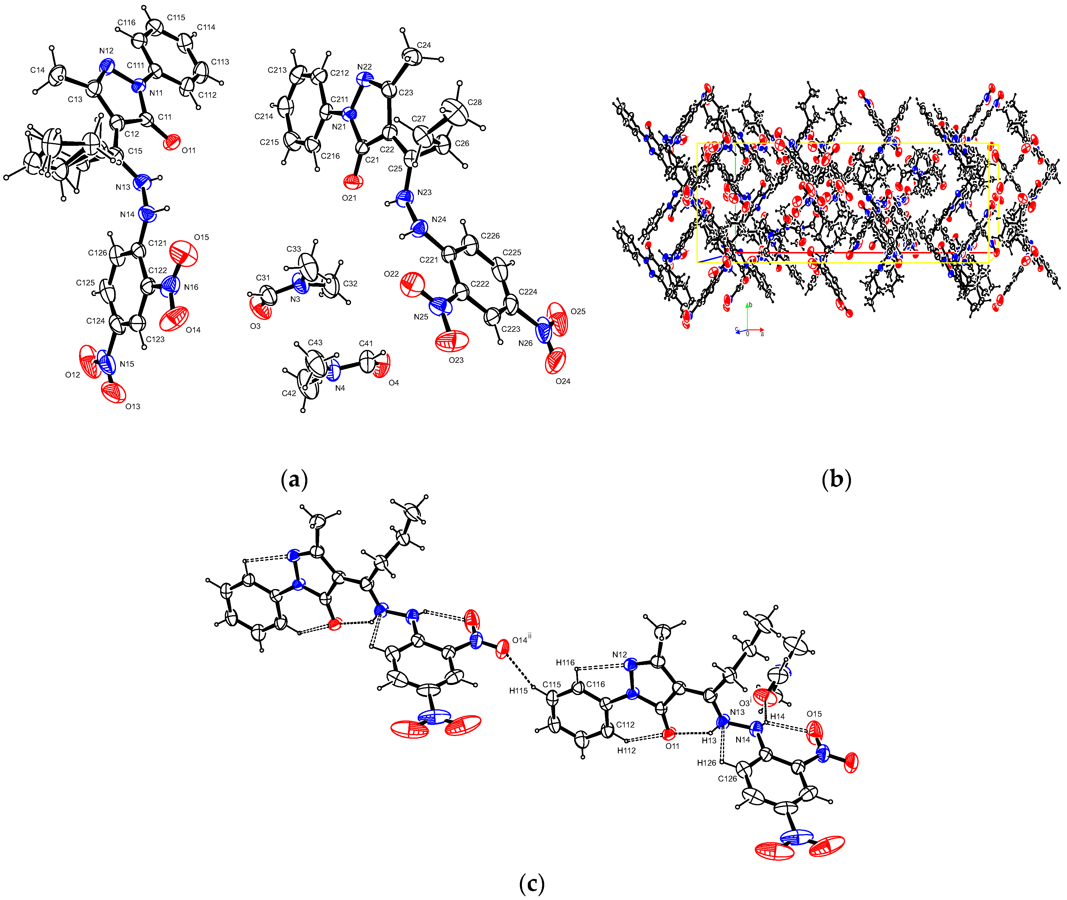
2.3. X-Ray Crystallography

A summary of the crystal data of Schiff base ligands is presented in Table 1, and their molecular and crystal structures are presented in Figure 5 and Figure 6. In the crystal structure of Empp-Dh, the asymmetrical unit cell contains one molecule of the compound, and one solvent dimethylformamide DMF molecule. The ethyl group on the Empp-Dh ligand has rotational disorder in a 0.87:0.13 ratio.
In the methyl-phenyl pyrazolone group, the phenyl ring plane makes an angle of 13.95(6)° with the methyl-pyrazolone plane, which in turn makes an angle of 74.23(2)° with the dinitrophenylhydrazine group.
There are a number of N–H···O, C–H···O and C–H···N hydrogen interactions [13]. The shortest intra-molecular interaction is N3–H3…O1 with a length of 1.881(17) Å, while the shortest inter-molecular interaction is between the titled molecule and the solvent DMF N4–H4···O7 with a length of 2.065(19) Å. There is also a C–H···π-ring interaction between DMF and the phenyl of the phenylhydazone group with a C73–H73A to centroid distance of 2.68 Å. The shortest ring π···π interaction is between the phenyl of the phenylhydazone group and an adjacent dinitrophenyl group with a centroid-to-centroid distance of 3.8096(8) Å.
The asymmetric unit cell of the Pmmp-Dh crystal structure contains two molecules of Pmmp-Dh, and two solvent dimethylformamide DMF molecules, shown in Figure 6. The two Pmmp-Dh molecules are identical except for the rotation around the N–N bond, and at the propyl group of one of the molecules there is a rotational disorder in a 0.88:0.12 ratio. After the inversion of one of the molecules and overlaying, the Root-mean-square deviation of atomic positions (RMSD values) is only 0.0847 and the largest positional difference between two non-hydrogen atoms, O14 and O23, is 0.2923 Å. The average dihedral angle between the two least square planes through the phenazone groups and the dinitrophenylhydrazine groups is 80.8(2) Å.
There are a number of N–H···O, C–H···O and C–H···N intra-molecular hydrogen interactions with N13–H13···O11 and N23–H23···O21 being the shortest with lengths of 1.89(3) and 1.95(3) Å, respectively. There are also a number of inter-molecular hydrogen interactions directly between the titled molecules. C213–H213···O21 and C115–H115···O11 are the shortest with lengths of 2.55 and 2.58 Å, respectively. The DMF solvent molecules have inter-molecular hydrogen interactions with the Pmmp-Dh ligand molecules with N14–H14···O3 having a length of 2.02(4) and N24–H24···O4 having a length of 2.06(3) Å, which is the shortest. The crystal shortest π- ring interactions are C–H···Cg (π- ring)-type interactions between the methyl groups of the DMF molecules and the phenyl rings of the phenylhydrazone groups, where the shortest interactions are C33–H33C···Cg (C111–C116) and C43–H43C···Cg (C211–C216) with hydrogen-to-centroid lengths of 2.84 and 2.90 Å, respectively.

3. Experimental

3.1. General Methods and Synthesis

Melting point was determined using the GallenKamp melting point apparatus (Northampton, UK). CHN elemental analyses were carried out on a LECO.TRUSpec Micro CHNS analyzer (St. Joseph, MI, USA). 1H and 13C NMR spectra in deuterated DMSO were recorded on a Varian Unity Inova 600 NMR spectrometer (Lyon, Rhône-Alpes, France) with a 1H frequency of 600 MHz and a 13C frequency of 150 MHz using trimethylsilane TMS as internal standard. A 5 mm dual channel IDpfg probe was used to collect the spectra and chemical shifts are given in ppm (δ scale). Mass spectra were determined by the Bruker micrOTOF-Q II 10390 mass spectrometer (Billerica, MA, USA). They were analyzed with Atmospheric-pressure chemical ionization (APCI) and Electrospray Ionization (ESI) using a direct insertion probe (DIP). An external calibration with sodium formate was performed to attain the correct accurate mass. Single-crystal X-ray diffraction studies were performed at 200 K using a Bruker Kappa Apex II diffractometer (Madison, WI, USA) with graphite monochromated Mo Kα radiation (λ = 0.71073 Å). All reagents, solvents; dinitrophenylhydrazine and 3-methyl-1-phenyl-2-pyrazolin-5-one were of analytical grade as supplied by Aldrich. Schiff base precursors, 4-ethyl-5-methyl-2-phenyl-pyrazol-3-one Empp and 4-propyl-5-methyl-2-phenyl-pyrazol-3-one Pmpp were synthesized, isolated and purified as reported earlier [14,18].

3.2. 4-Ethyl-5-Methyl-2-Phenyl-Pyrazol-3-one Dinitrophenylhydrazone, C19H18N6O5 Empp-Dh

A solution of 4-ethyl-5-methyl-2-phenyl-pyrazol-3-one Empp (2 mmol, 0.46 g) in methanol (40 mL) was added in drops to a solution of 2,4-dinitrophenylhydrazine DNPH (2 mmol, 0.40 g) in hot methanol (40 mL), in a 250 ml round bottom flask while stirring under reflux. An orange precipitate of the ligand was obtained after 2 h reflux period. The solution was filtered and the precipitate washed with methanol and diethyl ether, and then dried at room temperature. Slow evaporation of a solution of the resultant solid in dimethylformamide DMF afforded orange crystals of the titled 4-ethyl-5-methyl-2-phenyl-pyrazol-3-one-2,4-dinitrophenylhydrazone with m.pt 188–190 °C, suitable for X-ray crystallography. (2.59g, 90% yield). 1H NMR (600 MHz, DMSO-d6) δ 12.23(s, 2H), 11.43 (s, 1H), 10.73 (s, 3H), 8.82 (dd, J = 21.9, 2.6 Hz, 3H) 8.35 (ddd, J = 14.2, 9.6, 2.7 Hz, 4H), 7.97 (d, J = 9.7 Hz, 1H), 7.88 (d, J = 8.0 Hz, 5H), 7.78–7.73 (m, 2H), 7.51 (t, J = 7.9 Hz, 2H), 7.39 (t, J = 7.8 Hz, 6H), 7.31 (t, J = 7.4 Hz, 1H), 7.15 (t, J = 7.4 Hz, 2H), 2.86–2.78 (m, 5H), 2.71 (q, J = 7.3 Hz, 2H), 2.40 (s, 8H), 2.22 (s, 3H), 1.19 (td, J = 7.4, 4.1 Hz, 11H). 13C NMR (150 MHz, DMSO-d6) 147.2 (C=O), 144.4 (C=N), 137.5–115.6 (Aromatic C.), 30.0–21.0 (–CH–), 16.1–11.1 (–CH2–CH3). MS (ESI): m/z = 411.14 (calculated. 410.38 for C19H18N6O5, equivalent to M+ + 1). Elemental Analysis Calculated: C, 55.61%; H, 4.42%; N, 20.48%. Found: C, 55.20%; H, 4.08%; N, 20.56%.

3.3. 4-Propyl-5-Methyl-2-Phenyl-Pyrazol-3-One Dinitrophenylhydrazone, C20H20N6O5 Pmpp-Dh

A solution of 4-propyl-5-methyl-2-phenyl-pyrazol-3-one Pmpp (2 mmol, 0.49 g) in methanol (40 mL) was added in drops to a solution of 2,4-dinitrophenylhydrazine DNPH (2 mmol, 0.40 g) in hot methanol (40 mL), inside a 250 ml round bottom flask while stirring under reflux. An orange precipitate of the required ligand was obtained after 2 hours of reflux. The solution was filtered and the precipitate obtained was washed with methanol/diethyl ether, and then dried at room temperature. Slow evaporation of a solution of the resultant solid in dimethylformamide DMF afforded orange crystals of the new 4-propyl-5-methyl-2-phenyl-pyrazol-3-one-2, 4-dinitrophenylhydrazone with m.pt 191–193 °C, suitable for X-ray crystallography. (2.27g, 94%). 1H NMR (600 MHz, DMSO-d6) δ 12.28 (s, 1H), 10.78 (s, 2H), 8.82 (dd, J = 17.3, 2.7 Hz, 3H), 8.34 (ddd, J = 12.1, 9.4, 2.7 Hz, 3H), 7.86 (d, J = 8.0 Hz, 4H), 7.76 (d, J = 7.9 Hz, 2H), 7.51 (t, J = 7.8 Hz, 2H), 7.39 (t, J = 7.9 Hz, 6H), 7.16 (t, J = 7.4 Hz, 2H), 2.67 (t, J = 7.5 Hz, 2H), 2.40 (s, 6H), 2.22 (s, 3H), 1.62(ddt, J = 12.4, 7.9, 4.3 Hz, 6H), 0.97 (dt, J = 12.1, 7.3 Hz, 9H). 13C NMR (600 MHz, DMSO-d6) 147.3 (C=O), 144.4 (C=N), 137.4–115.6 (Aromatic C.), 29.3–19.4 (–CH–), 16.1–13.2 (–CH2–CH3). MS (APCI): m/z = 425.15 (calculated. 424.41 for C20H20N6O5, equivalent to M+ + 1). Elemental Analysis Calculated: C, 56.60%; H, 4.75%; N, 19.80%. Found: C, 56.33%; H, 4.36%; N, 19.96%.

3.4. X-Ray Diffraction Studies

Single-crystal X-ray diffraction studies were performed at 200 K using a Bruker Kappa Apex II diffractometer with graphite monochromator, Mo Kα radiation (λ = 0.71073 Å). Data collection was carried out with the help of the Bruker Instrument Service v2011.4.0.0 (Madison, WI, USA) and cell refinement/data reduction was made possible with the APEX2 v2011.4-1 (Madison, WI, USA) [19]. The structure was solved using SHELXL2014/7 program [20]. SHELXL2014/7 was also used for structure refining alongside the ShelXle [21] as a graphical interface. All non-hydrogen atoms were refined anisotropically. Molecular graphics was ensured with the ORTEP-3 for Windows [22], while the PLATON [23] as well as the Mercury [24] were used for crystal materials preparation.

4. Conclusions

The nucleophilic amine properties of dinitrophenyl hydrazine have been utilized to design/prepare the new N, O donor bidentate dinitrophenylhydrazones with interesting spectroscopic and solid-state structural attributes. Preliminary studies of the nitrogen-rich dinitrophenylhydrazone derivatives reported earlier showed significant bioactivities and, as such, the anticancer studies of these new, chemically stable 2,4-dinitrophenylhydrazones reported herein is ongoing.

Acknowledgments

This project was made possible by the National Research Foundation (NRF) and Sasol Inzalo Foundation (SIF) South Africa, through the award of a post-doctoral fellowship (Grant UID: 92275). GMRDC, the University of Fort Hare is also recognized for their contributions.

Author Contributions

Omoruyi G. Idemudia conceived the research, developed methods and experiments, synthesized the reported compounds Empp-Dh and Pmpp-Dh, and wrote the research paper. Eric C. Hosten diffracted the crystal structures and prepared their discussion.

Conflicts of Interest

The authors declare no conflict of interest.

Appendix

Crystallographic data for these dinitrophenylhydrazones have been deposited with the Cambridge Crystallographic Data Centre. Empp-Dh is assigned a CCDC # of 1431261 and Pmpp-Dh, CCDC # of 1431267. The files contains the supplementary crystallographic data which can be obtained free of charge via http://www.ccdc.cam.ac.uk/conts/retrieving.html, or from the Cambridge Crystallographic Data Centre, 12 Union Road, Cambridge CB2 IEZ, UK; Fax: +44 1223 336033; e-mail: [email protected].

References

  1. Qin, W.; Long, S.; Panunzio, M.; Biondi, S. A Short Survey on an Evergreen Chemistry Tool. Molecules 2013, 18, 12264–12289. [Google Scholar] [CrossRef] [PubMed]
  2. Subbaiyan, S.; Chinnasamy, J. Synthesis, characterization, DNA binding and cleavage activity of Ruthenium(II) complexes with heterocyclic substituted thiosemicarbazones. J. Chil. Chem. Soc. 2013, 58, 1637–1642. [Google Scholar]
  3. Mohamed, G.G.; Zayed, M.A.; Abdallah, S.M. Metal Complexes of a Novel Schiff Base Derived from Sulphametrole and Varelaldehyde. Synthesis, Spectral, Thermal Characterization and Biological Activity. J. Mol. Struct. 2010, 979, 62–71. [Google Scholar] [CrossRef]
  4. Gharamaleki, J.A.; Akbari, F.; Karbalaei, A.; Ghiassi, K.B.; Olmstead, M.M. Synthesis, Characterization and Crystal Structure of a New Schiff Base Ligand from a Bis(Thiazoline) Template and Hydrolytic Cleavage of the Imine Bond Induced by a Co(II) Cation. Open J. Inorg. Chem. 2016, 6, 76–88. [Google Scholar] [CrossRef]
  5. Castagnolo, D.; De Logu, A.; Radi, M.; Bechi, B.; Manetti, F.; Magnani, M.; Supino, S.; Meleddu, R.; Chisu, L.; Botta, M. Synthesis, biological evaluation and SAR study of novel pyrazole analogues as inhibitors of Mycobacterium tuberculosis. Bioorg. Med. Chem. 2008, 16, 8587–8591. [Google Scholar] [CrossRef] [PubMed]
  6. Marchetti, F.; Pettinari, C.; Pettinari, R. Acylpyrazolone ligands: Synthesis, structures, metal coordination chemistry and applications. Coord. Chem. Rev. 2005, 249, 2909–2945. [Google Scholar] [CrossRef]
  7. Marchetti, F.; Pettinari, R.; Pettinari, C. Recent advances in acylpyrazolone metal complexes and their potential applications. Coord. Chem. Rev. 2015, 303, 1–31. [Google Scholar] [CrossRef]
  8. Idemudia, O.G.; Sadimenko, A.P.; Hosten, E.C. Crystal structure of bis(4-benzoyl-3-methyl-1-phenyl pyrazol-5-one)-bis(N,N-dimethylformamide)cobalt(II), C40H40CoN6O6. Z. Kristallogr. New. Cryst. Struct. 2015, 230, 83–84. [Google Scholar] [CrossRef]
  9. Jadeja, R.N.; Parihar, S.; Vyas, K.; Gupta, V.K. Synthesis and crystal structure of a series of pyrazolone based Schiff base ligands and DNA binding studies of their copper complexes. J. Mol. Struct. 2013, 1013, 86–94. [Google Scholar] [CrossRef]
  10. Idemudia, O.G.; Sadimenko, A.P.; Afolayan, A.J. Potential Therapeutic Mn2+ and Ni2+ Complexes of New 4-Acetyl-3-Methyl-1-Phenyl-2-Pyrazoline-5-One Phenylhydrazone Ampp-Ph. Macromol. Symp. 2015, 351, 61–68. [Google Scholar] [CrossRef]
  11. Brady, O.L.; Elsmie, G.V. The use of 2,4-dinitrophenylhydrazine as a reagent for aldehydes and ketones. Analyst 1926, 51, 77–78. [Google Scholar] [CrossRef]
  12. Jesmin, M.; Ali, M.M.; Khanam, J.A. Antitumour activities of some Schiff bases derived from benzoin, salicylaldehyde, amino phenol and 2,4 dinitrophenyl hydrazine. Thai J. Pharm. Sci. 2010, 34, 20–31. [Google Scholar]
  13. Idemudia, O.G.; Sadimenko, A.P.; Hosten, E.C. 4-{[2-(2,4-Dinitrophenyl) hydrazinylidene] (phenyl)methyl}-5-methyl-2-phenyl-1H-pyrazol-3(2H)-one ethanol monosolvate. Acta Cryst. 2012, E68, o3380. [Google Scholar] [CrossRef] [PubMed]
  14. Idemudia, O.G.; Sadimenko, A.P.; Afolayan, A.J.; Hosten, E.C. Synthesis and Characterization of Bioactive Acylpyrazolone Sulfanilamides and Their Transition Metal Complexes: Single Crystal Structure of 4-Benzoyl-3-methyl-1-phenyl-2-pyrazolin-5-one Sulfanilamide. Bioinorg. Chem. Appl. 2015, 2015, 717089. [Google Scholar] [CrossRef] [PubMed]
  15. Idemudia, O.G.; Sadimenko, A.P.; Hosten, E.C. Crystal structure of 4-benzoyl-3-methyl-1-phenyl-2-pyrazolin-5-one sulfadiazine dimethylformamide monosolvate, C30H29N7O4S. Z. Kristallogr. New. Cryst. Struct. 2014, 229, 455–457. [Google Scholar] [CrossRef]
  16. Idemudia, O.G.; Sadimenko, A.P.; Hosten, E.C. Crystal structure of 4-propyl-3-methyl-1-phenyl-2-pyrazolin-5-one thiosemicarbazone, C15H19N5OS. Z. Kristallogr. New. Cryst. Struct. 2015, 230, 81–82. [Google Scholar] [CrossRef]
  17. Parihar, S.; Pathan, S.; Jadeja, R.N.; Patel, A.; Gupta, V.K. Synthesis and Crystal Structure of an Oxovanadium(IV) Complex with a Pyrazolone Ligand and Its Use as a Heterogeneous Catalyst for the Oxidation of Styrene under Mild Conditions. Inorg. Chem. 2012, 51, 1152–1161. [Google Scholar] [CrossRef] [PubMed]
  18. Idemudia, O.G.; Sadimenko, A.P.; Hosten, E.C. Metal Complexes of New Bioactive Pyrazolone Phenylhydrazones; Crystal Structure of 4-Acetyl-3-Methyl-1-Phenyl-2-Pyrazoline-5-One Phenylhydrazone Ampp-Ph. Int. J. Mol. Sci. 2016, 17, 1–24. [Google Scholar] [CrossRef] [PubMed]
  19. Bruker. APEXII, SAINT, SADABS, Bruker AXS Inc.: Madison, WI, USA, 2007.
  20. Sheldrick, G.M. A short history of SHELX. Acta Cryst. 2008, 112–122. [Google Scholar] [CrossRef] [PubMed]
  21. Hübschle, C.B.; Sheldrick, G.M.; Dittrich, B. ShelXle: A Qt graphical user interface for SHELXL. J. Appl. Cryst. 2011, 44, 1281–1284. [Google Scholar] [CrossRef] [PubMed]
  22. Farrugia, L.J. ORTEP-3 for Windows—A Version of ORTEP-III with a Graphical User Interface (GUI). J. Appl. Cryst. 1997, 30, 565–566. [Google Scholar] [CrossRef]
  23. Spek, A.L. Single-crystal structure validation with the program PLATON. J. Appl. Cryst. 2003, 36, 7–13. [Google Scholar] [CrossRef]
  24. Macrae, C.F.; Bruno, I.J.; Chisholm, J.A.; Edgington, P.R.; McCabe, P.; Pidcock, E.; Rodriguez-Monge, L.; Taylor, R.; van de Streek, J.; Wood, P.A. Mercury CSD 2.0—New Features for the Visualization and Investigation of Crystal Structures. J. Appl. Cryst. 2008, 41, 466–470. [Google Scholar] [CrossRef]
Figure 1. Synthesis scheme for Empp-Dh and Pmpp-Dh.
Crystals 06 00127 g001
Figure 2. (a) 13C NMR spectrum of Empp-Dh; (b) 13C NMR spectrum of Pmpp-Dh.
Crystals 06 00127 g002
Figure 3. (a) 1H NMR spectrum of Empp-Dh; (b) 1H NMR spectrum of Pmpp-Dh.
Crystals 06 00127 g003
Figure 4. (a) Mass spectrum of Empp-Dh; (b) Mass NMR spectrum of Pmpp-Dh.
Crystals 06 00127 g004
Figure 5. X-ray crystal structures of Emmp-Dh with ellipsoids drawn at 50% probability level: (a) Ortep diagram of Emmp-Dh; (b) Unit cell contents viewed normal to (001); (c) Hydrogen bonding with symmetry element i: x, y, 1 + z. The colored lines are the unit cell axis orientation and the red and blue circles represent oxygen and nitrogen, respectively.
Crystals 06 00127 g005
Figure 6. X-ray crystal structures of Pmmp-Dh with thermal ellipsoids drawn at 50% probability level: (a) Ortep diagram of Pmmp-Dh; (b) Unit cell contents of Pmmp-Dh viewed normal to (001); (c) Selected hydrogen bonding with symmetry elements i: x, 1 + y, z; ii: x, y, −1 + z. The colored lines are the unit cell axis orientation and the red and blue circles represent oxygen and nitrogen, respectively.
Crystals 06 00127 g006
Table 1. Crystal data of Empp-Dh and Pmpp-Dh.
CompoundEmpp-PhPmpp-Dh
FormulaC19H18N6O5, C3H7NOC20H20N6O5, C3H7NO
Crystal color and formOrange/BlockOrange/Block
Formula weight483.49497.51
Crystal systemMonoclinicMonoclinic
Space groupC2/cP21/c
a31.6116(9) (Å)32.2379(16) (Å)
b9.8297(3) (Å)10.0527(5) (Å)
c14.9770(5) (Å)15.2972(8) (Å)
α90o90o
β92.114(2)o103.420(2)o
γ90o90o
V4650.7(2) (Å3)4822.1(4) (Å3)
Z88
D(calc)1.381 (Mg cm−1)1.371 (Mg cm−1)
F(000)20322096
θ range2.2–28.3 (°)0.6–28.4 (°)
Crystal size0.21 × 0.26 × 0.45 (mm)0.21 × 0.41 × 0.60 (mm)
Reflections measured575311985
Independent/observed0.018/44720.032/9541
Mu(MoKa)0.103 (/mm)0.102 (/mm)
Temperature200 (K)200 (K)
Parameters348695

