Article

Innovative Investigation of Zinc Oxide Nanoparticles Used in Dentistry

by
Ajay Kumar Tiwari
1,
Saket Jha
2,
Abhimanyu Kumar Singh
3,*,
Sheo Kumar Mishra
4,
Ashok Kumar Pathak
5,
Rudra Prakash Ojha
6,
Raghvendra Singh Yadav
7 and
Anupam Dikshit
2
1
Department of Physics, Nehru Gram Bharati (Deemed to be University), Prayagraj 221505, India
2
Biological Product Laboratory, Department of Botany, University of Allahabad, Prayagraj 211002, India
3
Department of Physics, Shyama Prasad Mukherjee Government Degree College, University of Allahabad, Prayagraj 211013, India
4
Department of Physics, Indira Gandhi National Tribal University, Amarkantak 484886, India
5
Department of Physics, Ewing Christian College, University of Allahabad, Prayagraj 211003, India
6
Department of Zoology, Nehru Gram Bharati (Deemed to be University), Prayagraj 221505, India
7
Centre of Polymer Systems, Tomas Bata University in Zlin, Trida Tomase Bati 5678, 76001 Zlin, Czech Republic
*
Author to whom correspondence should be addressed.
Crystals 2022, 12(8), 1063; https://doi.org/10.3390/cryst12081063
Submission received: 1 July 2022 / Revised: 25 July 2022 / Accepted: 27 July 2022 / Published: 29 July 2022

Abstract

:
Dental caries is a major lifestyle concern as dental components affect the face of an individual. The issue of tooth decay occurs in every age group throughout the globe. Researchers are probing incipient implements and techniques to develop filling agents for decayed teeth. Zinc oxide (ZnO) powder is utilized mostly as a filling agent. Nanotechnology enhanced the efficiency of compounds of metal oxides utilized for dental caries. The present study aims to investigate the properties of ZnO nanoparticles (NPs) synthesized chemically (using ZnCl2 and NaOH) as well as biologically (using aqueous leaf extract of Murraya paniculata). The XRD patterns confirm that ZnO NPs have a hexagonal crystalline structure with particle sizes of 47 nm and 55 nm for chemically and biologically synthesized NPs, respectively. The FE-SEM data confirm the nanorod and spherical/cubical shape morphologies for the chemically and biologically synthesized ZnO NPs, respectively. FTIR data show the peaks between 4000 and 450 cm−1 of the functional groups of –OH, C-O, –C-H-, and Zn-O bonds. The UV–Vis absorption study indicates a peak around 370 nm and a hump around 360 nm corresponding to the chemically and biologically synthesized ZnO NPs, respectively. An antibacterial bioassay was performed and compared with commercially available ZnO bulk powder against tooth decaying pathogens, viz., Streptococcus mutans, Staphylococcus aureus, E. coli, and Lactobacillus fermentum, and found that both ZnO NPs had results closer to those of the standard drug (rifampicin). Thus, the synthesized ZnO NPs may be utilized as nano-drugs for the application of tooth decaying filling agents. Even biologically synthesized ZnO NPs may be considered more environmentally friendly and less toxic to human health concerns.

1. Introduction

Teeth play a generalized role in the individual personality. They not only help in verbalizing, but also affect one’s countenance. Dental caries adversely affects an individual’s personality and is a major concern in human welfare. Tooth decay is now becoming an incurable disease and is found to be a major concern for every age group. The people of India and abroad are affected by this disease throughout their lifetime [1]. Researchers and medical practitioners are perpetually probing for novel concepts to deal with dental caries.
Cavities are created due to bacterial incursions as well as due to contingent causes, but there is always the pressure of having smooth and white teeth. Because of these exigencies, sundry tooth filling agents have been utilized as per their availability. These filling powders have always been regulated as they degrade and conventionally lack antibacterial properties. Several antibiotics have been used to cope with dental pathogens, but they have some limitations such as resistant microbes, antibiotic degradation, or loss of antimicrobial property [2]. The incorporation of nanotechnology resolves these issues and provides advanced implements and techniques for materials used as filling agents. Their nano-sized structure and sizably voluminous surface-to-volume ratio transmutes their properties compared with their bulk materials/compounds [3,4]. The bulk materials of calcium, silicon, gold, zinc, etc., have been widely utilized as filling agents and tooth implants.
Nanoparticles are considered to have more potential as they never lose their properties and are not degradable; however, they still need to be explored further due to health concerns [5,6,7,8,9,10]. Sundry applications have proved that these nanoparticles, associated with polymers [8] or having a surface coating [11,12], show maximum antimicrobial efficacy against tooth decaying pathogens [13,14,15]. Among several metal oxide nano-biomaterials, ZnO NPs have been found to be a good tooth filling agent; they are widely utilized because of their white color and because they show more antimicrobial properties [16,17,18]. These metal oxide nanoparticles protect from dental pathogens and never fade or become decolorized. Thus, a congruous implant with these nanoparticles provides a perpetual approach to tooth implantation and tooth fillings [19]. Their nano-sized structure provides the capability to enter inside the microbial cell membranes and facilely alter their metabolic pathways, leading to the death of or a decrease in microbial populations. The only difficulties with these nanoparticles are those regarding human health concerns. Many researchers believe that, due to their enhanced proprieties and nano-size, these nanoparticles may alter the biological metabolic pathways of human cells and lead to causing some cancers, rigorous diseases, or mutagenic irradiations [20].
Researchers are working on the establishment of nanoparticles with lesser toxicological activity against humans but that trigger more lethality towards pathogens. Many promising methodologies are being developed that still need to be further explored [21,22,23,24,25,26,27,28,29,30]. Simple methods were chemically synthesized with a hydrothermal decomposition reaction [31,32] and biologically with plant extracts, utilizing their secondary metabolites to minimize the size of the metal. These simple methods are convenient, environmentally friendly, and have a low cost [33,34].
In the present investigation, both biological and chemical synthesis techniques were performed to prepare ZnO NPs. Various characterization techniques, such as field emission scanning electron microscopy (FE-SEM), X-ray diffraction (XRD), Fourier transform infrared (FTIR) spectroscopy, and ultraviolet–visible (UV–Vis) spectroscopy, were used to evaluate their shape, particle size, bond stretching, and optical properties, respectively. Further, the synthesized ZnO NPs using a chemical method and a biological method were checked against major dental caries pathogens by utilizing the broth microdilution technique recommended by CLSI M7A8 [35]. Further, the antibacterial results of both the ZnO NPs were compared with the results of ZnO bulk powder.

2. Materials and Methods

2.1. Collection of ZnO Bulk Powder

The ZnO bulk powder (Dental products of India, Bombay Burmah Corporation Ltd., Mumbai, India) was collected from dentists of Sidhi Vinayak Dental Clinic at Naini, Prayagraj, India.

2.2. Chemical Synthesis of ZnO Nanoparticles

For the synthesis of ZnO NPs, an aqueous solution of 19.99 gm of sodium hydroxide (Merck, Mumbai, India) was integrated in the aqueous solution of 5.5 gm zinc chloride (Merck, Mumbai, India) with perpetual stirring (1200 rpm) at the temperature of 60 °C for 2 h. The white precipitate was filtered with Whatman filter paper no. 42, washed thrice with double distilled water, and left for 24 h in an oven at 200 °C [34,35]. Finally, the ZnO NPs were obtained in powder form.

2.3. Biological Synthesis of ZnO Nanoparticles

The preparation of ZnO NPs was performed as prescribed by Bela et al. [36] with some modifications. The 50 mL solution of zinc acetate (0.45 M) was integrated dropwise in the aqueous solution of 5 gm of leaves of Murraya paniculata with perpetual stirring (1200 rpm) at the temperature of 60 °C for 2 h. The precipitate was centrifuged at 6000 rpm for 15 min and washed thrice with ethanol and double distilled water. The brown color precipitate was kept for 24 h in a hot air oven at 100 °C. Finally, ZnO nanoparticles were obtained in powder form.

2.4. Inoculum Preparation

The tooth decaying pathogens were Streptococcus mutans (MTCC- 497), Staphylococcus aureus (MTCC-3160), Lactobacillus fermentum (MTCC-1745), and E. coli (MTCC-723). The inocula were prepared from 24 h old cultures and the suspension was compared with 0.5 McFarland standard solution using a spectrophotometer at an optical density of 625 nm with a value of 0.08–0.13 as prescribed by CLSI guidelines [37]. The inoculants having a concentration of 5 × 10⁵ CFU/mL were utilized further for the broth microdilution technique for the drug susceptibility test.

2.5. Antibacterial Bioassay

The ZnO NPs synthesized by chemical as well as biological techniques and ZnO bulk powder were comparatively studied with their antibacterial property against tooth decaying pathogens, viz., S. mutans, S. aureus, L. fermentum, and E. coli. The broth microdilution method was performed to evaluate the antibacterial efficacy with standard protocol as prescribed by CLSI [35,37,38,39,40,41,42]. Rifampicin was taken as a standard drug and the assay was performed on 96-microtiter plates. The experimental plates were incubated at 37 ± 2 °C in a BOD incubator for 24 h and data were calculated on a Spectramax Plus 384 molecular device.

3. Results and Discussion

The chemically and biologically synthesized ZnO NPs were characterized with X-ray diffractometer (Rikagu, Japan) for their crystalline sizes and structures, field emission scanning electron microscopy (FE-SEM) (Make: JEOL India Pvt. Ltd., Delhi, India) for their morphology, Fourier transform infrared (FTIR) spectrometer (Perkin Elmer, Buckinghamshire, UK) for their stability, and ultraviolet–visible (UV–Vis) spectrophotometer (Spectra Max, San Jose, CA, USA) for their optical properties; furthermore, the antibacterial property of ZnO NPs was assessed and compared with that of ZnO bulk powder (obtained from a dental clinic) against different bacterial strains.

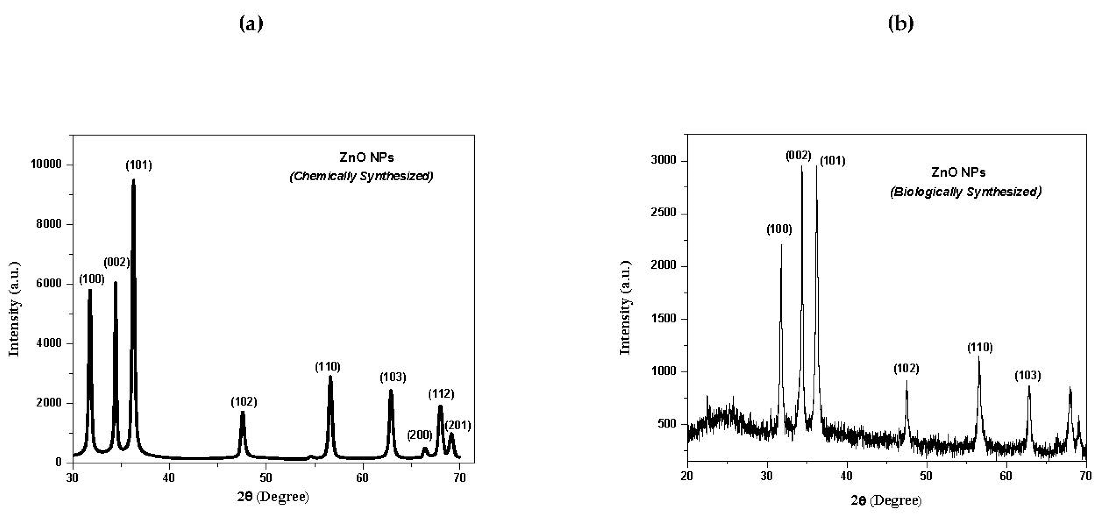
3.1. X-ray Diffraction (XRD) Analysis of ZnO NPs

Figure 1a,b show the XRD patterns of ZnO NPs synthesized by chemical and biological methods, respectively. All peaks in Figure 1a,b matched with JCPDS Cat. No. 36-1451 and 79-0207 [43], confirming the formation of ZnO NPs. The XRD 2ϴ values were found for crystallography peaks at (100), (002), (101), (102), (110), (103), (200), (112), and (201) for the chemically as well as the biologically synthesized ZnO NPs. The grain sizes of XRD patterns were calculated from the Debye–Scherrer formula:
D = 0.9λ/βcosθ
where D is the grain size, β is the full width at half maximum (FWHM) in radians of the XRD pattern, λ is the X-ray wavelength, and ϴ is the Bragg diffraction angle of the XRD peak. The particle sizes of the ZnO NPs synthesized by the chemical method (Figure 1a) and biological method (Figure 1b) were calculated from the above formula and found to be 47 nm and 55 nm, respectively, along with the high intensity orientation (002). Both NPs had hexagonal crystalline structures with space group P63mc (186). The appearance of some noise in the XRD pattern of the biologically synthesized NPs may be due to some impurities of other elements present in the leaves of Murraya paniculata.

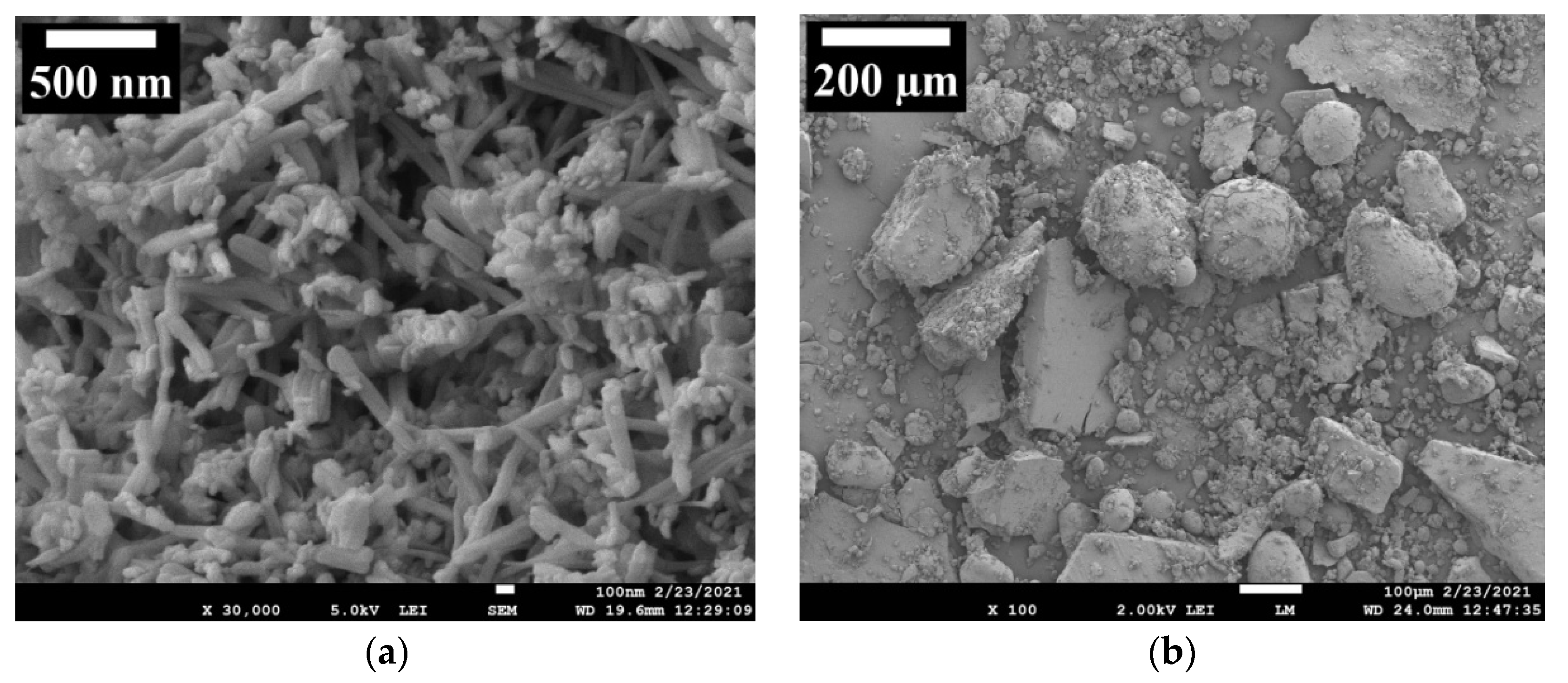
3.2. Field Emission Scanning Electron Microscopy (FE-SEM) Analysis of ZnO NPs

Figure 2a,b show FE-SEM images of ZnO NPs synthesized by chemical and biological methods, respectively. The FE-SEM images reveal the crystal shapes as being nanorods for the chemically synthesized ZnO NPs (Figure 2a) and spherical and cubical for the biologically synthesized ZnO NPs (Figure 2b). The thickness and length of the nanorods were calculated by the “image J” software as shown in the SEM image of Figure 2a. The thickness/diameter of the nanorods was 25–65 nm and the length of the nanorods was 250–600 nm. From the FE-SEM image of Figure 2a, it is clearly seen that the nanorods are uniformly distributed in the whole space. However, in the FE-SEM image of Figure 2b, it is clearly seen that the particle sizes vary from a very small size to a larger size and some particles are of spherical, cubical, and other shapes. There are clear differences in the morphology of the obtained nanoparticles by different synthesis methods. The formation of nanorods occurs by chemical reaction. In the case of biological synthesis, the processes of the formation of nanoparticles have a more complex mechanism than in the case of chemical synthesis.

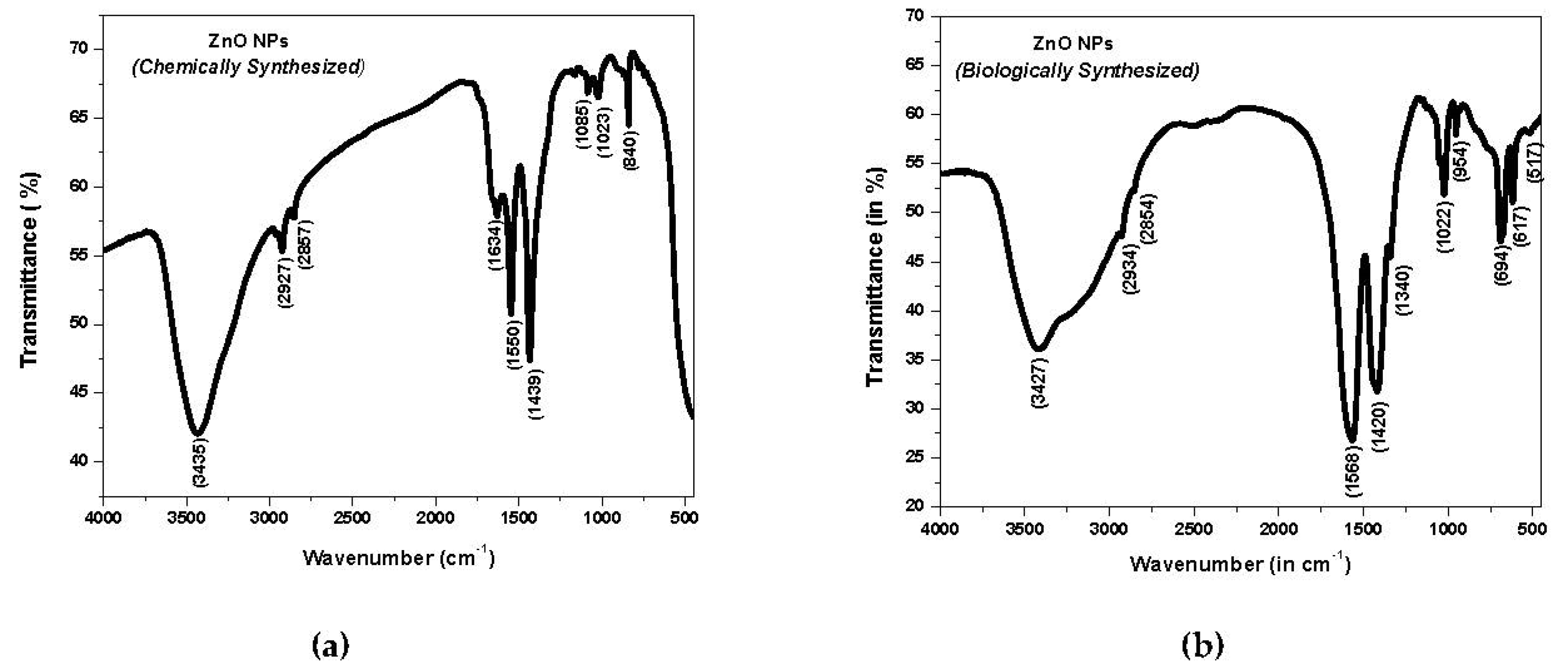
3.3. Fourier Transform Infrared (FTIR) Spectroscopy Analysis of ZnO NPs

Figure 3a,b show the FTIR spectra of ZnO NPs synthesized chemically and biologically, respectively. The FTIR spectra were recorded in the form of pellets utilizing spectroscopic grade KBr in the wave number range of 4000 to 450 cm−1 in the diffuse reflectance mode. The spectrum of the chemically synthesized ZnO NPs (Figure 3a) showed bands of functional groups located at 3435, 2927, 2857, 1634, 1550, 1439, 1085, 1023, and 840 cm−1. The spectrum of the biologically synthesized ZnO NPs (Figure 3b) showed bands of functional groups located at 3427, 2934, 2854, 1568, 1420, 1340, 1022, 954, 694, 617, and 517 cm−1. The strong broad bands in the higher region at 3435 and 3427 cm−1 are due to the stretching vibration of hydroxyl (OH) groups. The absorption bands at 2927, 2934, 2857, and 2854 cm−1 are due to the stretching vibration of CH groups. The peaks around 1634, 1550, and 1568 cm−1 correspond to the C=O amide group. The –C-H- bending vibration band arises at 1439, 1420, and 1340 cm−1. The C-O stretching vibration band arises at 1085, 1023, and 1022 cm−1. The peaks around 840, 954, and 694 cm−1 are due to the bending vibration of =C-H groups. The peak around 617 cm−1 is ascribed to the bending vibration of the O-H group. The absorption band at 517 cm−1 is attributed to the stretching vibration of Zn-O bonds and confirms the formation of materials [44]. The peaks from 4000 to 450 cm−1 observed in the spectra indicate the presence of –OH, C-O, and –C-H- residues, which may be due to the precursors used in the reaction.

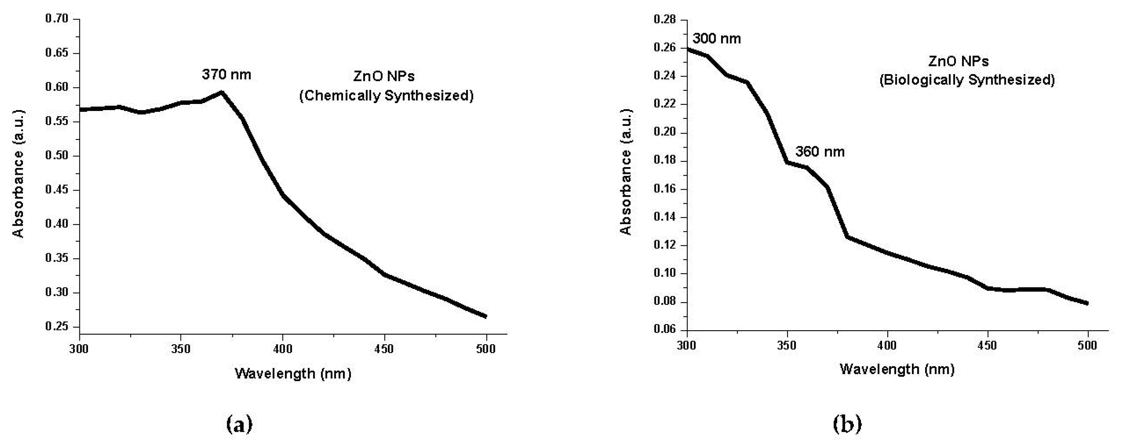
3.4. UV–Visible Spectroscopy Analysis of ZnO NPs

To explore the optical properties of the ZnO NPs, UV–visible spectroscopy is the best technique. The absorption spectrum of ZnO NPs is shown in Figure 4a,b. The high and stable peak found around 370 nm, corresponding to the chemically synthesized ZnO NPs (Figure 4a), confirms the formation of ZnO NPs. The hump around 360 nm, corresponding to the biologically synthesized ZnO NPs (Figure 4b), is evidence of the formation of ZnO NPs. The prepared ZnO nanoparticles exhibit an absorbance peak around 300 nm in Figure 1b, which corresponds to the formation of smaller-sized particles [45]. The smaller-sized particles are also seen in the SEM picture (Figure 2b), which also supports the above statement. It is also evidence that the paramount absorption of sharp ZnO NPs designates the mono disappeared nature of the NPs distribution [43]. The average particle size of the ZnO NPs can be calculated from the UV–Vis absorption spectra using an effective mass model (Figure 3a,b) [46,47]. The band gap enlargement is expected for particle radii less than about 10 nm for biologically synthesized ZnO NPs and 20 nm for chemically synthesized ZnO NPs [48,49,50].

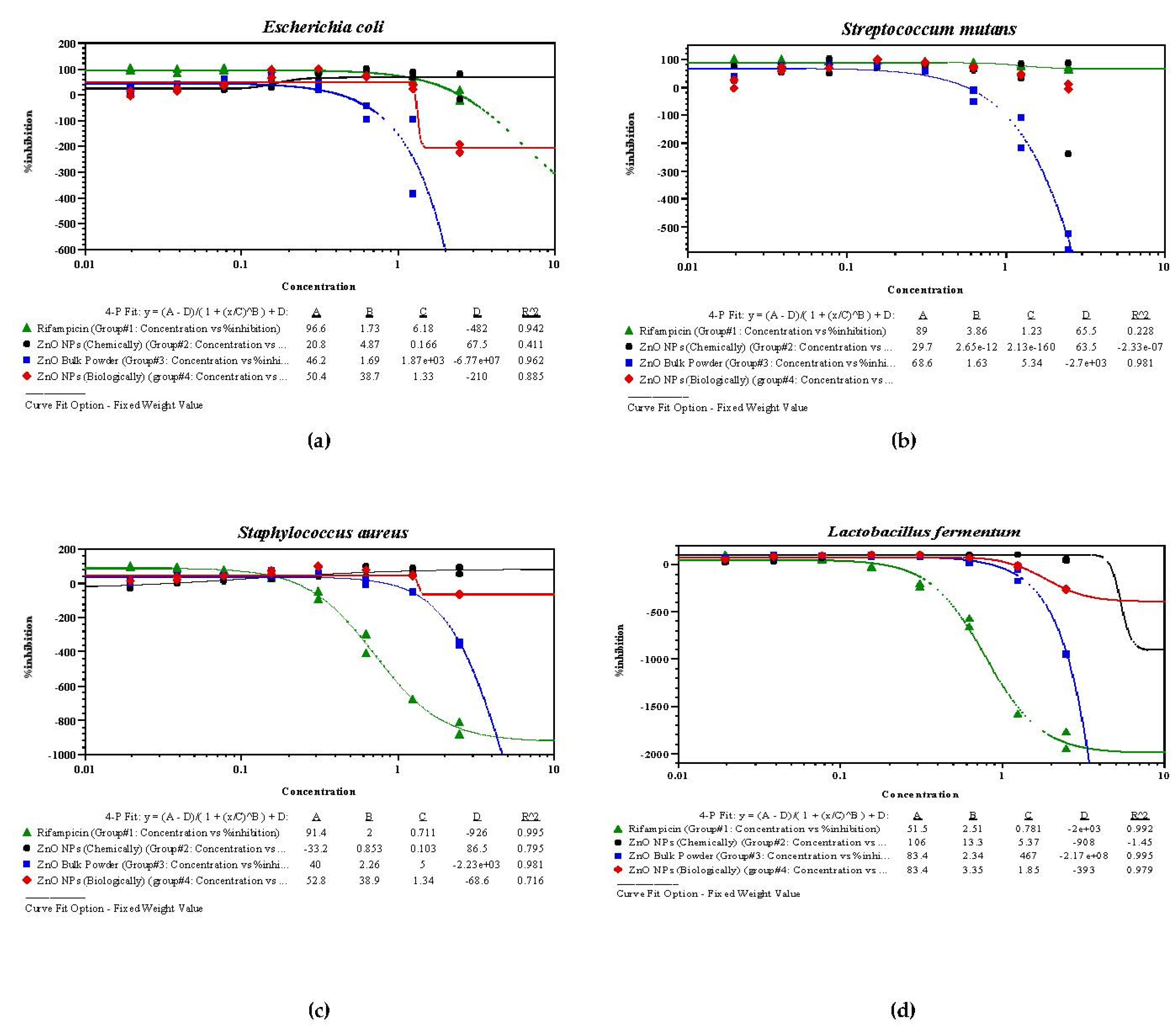
3.5. Antibacterial Bioassay of Synthesized ZnO NPs

The biologically synthesized ZnO NPs had minimum inhibitory concentration (MIC) values of 0.261 µg/mL for E. coli, 0.293 µg/mL for S. mutans, 0.134 µg/mL for S. aureus, and 0.426 µg/mL for L. fermentum. The chemically synthesized ZnO NPs were evaluated with MIC values of 0.193 µg/mL for E. coli, 0.308 µg/mL for S. mutans, 0.296 µg/mL for S. aureus, and 0.407 µg/mL for L. fermentum. Both the NPs were found to more efficacious as compared to ZnO bulk powder. The biologically synthesized ZnO NPs were more efficacious against S. mutans and S. aureus, whereas the chemically synthesized ZnO NPs showed higher antibacterial efficacy against E. coli and L. fermentum. The MIC and half-maximal inhibitory concentration (IC50) values of both NPs were closer to the values of the standard drug rifampicin. Moreover, both NPs were found to be more effective compared to the ZnO bulk powder, leading to potency for the drug formulations against tested pathogens (Table 1, Figure 5 and Figure 6a–d). Both nanomaterials were found to much less effective against S. mutans and L. fermentum compared to the standard drug.
Zinc has been widely used in human welfare since ancient times in the form of tooth powder and toothpaste [51,52]. Zinc has antibacterial properties and is a major constituent after calcium and magnesium in tooth formation. The present study was performed with ZnO NPs and compared with commercially available ZnO bulk power against tooth decaying pathogens utilizing the broth microdilution technique. The data were analyzed and found high antibacterial efficacy compared with the ZnO bulk powder [53,54]. The biologically synthesized ZnO NPs were found to have the highest antibacterial property against S. aureus and were less effective against L. fermentum. The chemically synthesized ZnO NPs were found to have more lethality against E. coli and were less toxic to S. aureus. Both nano-biomaterials were found to have a potential antibacterial property compared to the standard drug (rifampicin) and ZnO bulk powder. The antibacterial properties of ZnO NPs were also reported by other researchers [53,54,55]. The MIC values of the ZnO NPs synthesized chemically as well as biologically were found to be similar to those from other, earlier reports [56]. For the herbal (phyto)synthesis, more bioactive components were found and they showed less toxicity to human health [56,57]. Some researchers reported mechanisms wherein these nanoparticles trigger the release of free radicals which rupture the microbial cell membranes, causing their death [57,58]. Both synthesized ZnO NPs showed better proximity to the standard drug compared to ZnO powder against all four bacterial species. This may be due to the ZnO NPs having highly penetrable, large surface areas and stable properties compared to the ZnO bulk powder. The nano form of ZnO leads to alteration in cell membrane permeability, which is responsible for leakage of the components of the intracellular compartment [59,60].

4. Conclusions

ZnO nanoparticles were successfully synthesized by chemical as well as biological methods. Biological synthesis of ZnO NPs using the aqueous leaf extract of Murraya paniculata as a reducing agent provides an environmentally friendly, simple, and efficient route for the synthesis of nanoparticles. The XRD patterns confirm that ZnO NPs have a hexagonal crystalline structure with space group P63mc (186). The particle sizes were found to be 47 nm and 55 nm for the chemically and biologically synthesized ZnO NPs, respectively, along with the high intensity orientation (002). FE-SEM data confirm the rod shape morphology with a thickness of ~25–65 nm and length of ~250–600 nm for the chemically synthesized ZnO NPs and spherical and cubical shapes for the biologically synthesized ZnO NPs. FTIR data show that the peaks between 4000 and 450 cm−1 observed in the spectra indicate the presence of the functional groups –OH, C-O, and –C-H-. The absorption band at 517 cm−1 is attributed to the stretching vibration of Zn-O bonds, which confirms the formation of materials for the biologically synthesized ZnO NPs. The UV–Vis absorption spectra of the ZnO NPs indicate that the high and stable peak was found around 370 nm, corresponding to the chemically synthesized ZnO NPs, which confirms the formation of ZnO NPs. The hump around 360 nm, corresponding to the biologically synthesized ZnO NPs, is evidence of the formation of ZnO NPs, and an absorbance peak around 300 nm corresponds to the formation of smaller-sized particles. Both ZnO NPs and ZnO bulk powder were comparatively studied with their antibacterial property with the broth microdilution method. The MIC and IC50 values of both the synthesized ZnO NPs showed better proximity to the values of the standard drug compared to those of the ZnO powder against tooth decaying pathogens, i.e., S. mutans, S. aureus, L. fermentum, and E. coli. This may be due to the ZnO NPs having a highly penetrable, large surface area and stable properties compared to the ZnO bulk powder. The present study has potentially shown the antibacterial efficacy of ZnO NPs against major tooth decaying pathogens, opening the door for biologically synthesized ones as an alternative that is more cost-effective and less toxic to human health. Finally, we conclude that the present study focused on the environmentally friendly and low-cost synthesized nanomaterials of other tooth filling elements, such as Ti, Ag, and Ca, which may be utilized as filling agents with high antibacterial efficacy. Moreover, the amalgamation of tooth filling nanomaterial powder can be further utilized in dentistry as well as other pharmacological applications. This study is helpful and useful to the scientific community for using ZnO NPs in dentistry, antimicrobial, and other pharmacological applications for commercial biomedical applications.

Author Contributions

Experimental design and data evaluated by A.K.T., S.K.M. and S.J.; discussion and data compiling by A.K.S., A.K.P., R.P.O., R.S.Y. and A.D. All authors have read and agreed to the published version of the manuscript.

Funding

This research received no external funding.

Institutional Review Board Statement

Not applicable.

Informed Consent Statement

Not applicable.

Data Availability Statement

Data will be made available upon request.

Acknowledgments

Authors are thankful to the director Subodh Kumar, Birbal Sahni Institute of Paleoscience, Lucknow, for SEM and valuable suggestions; R. R. Awasthi, Lucknow University for XRD; V. Bhadoria, Ewing Christian College, University of Allahabad for FTIR; the Head of the Department of Botany, University of Allahabad for providing the lab facility and UGC, New Delhi for financial Assistance to Anupam Dikshit as BSR Faculty.

Conflicts of Interest

The authors declare no conflict of interest.

References

  1. Selwitz, R.H.; Ismail, A.I.; Pitts, N.B. Dental caries. Lancet 2007, 369, 51–59. [Google Scholar] [CrossRef]
  2. Aviv, M.; Berdicevsky, I.; Zilberman, M. Gentamicin-loaded bioresorbable films for prevention of bacterial infections associated with orthopedic implants. J. Biomed. Mater. Res. A 2007, 83, 10–19. [Google Scholar] [CrossRef] [PubMed]
  3. Bonzonga, J.C.J.; Kopelevich, D.; Bitton, G. Assessment of the Environmental Impacts of Nanotechnology on Organisms and Ecosystem; Progress Report; United States Environmental Protection Agency: Washington, DC, USA, 2007. [Google Scholar]
  4. Buzea, C.; Pacheco, I.I.; Robbie, K. Nanomaterials and nanoparticles: Sources and toxicity. Biointerphases 2007, 2, MR17–MR71. [Google Scholar] [CrossRef] [PubMed] [Green Version]
  5. Cho, E.J.; Holback, H.; Liu, K.C.; Abouelmagd, S.A.; Park, J.; Yeo, Y. Nanoparticle characterization: State of the art, challenges, and emerging technologies. Mol. Pharm. 2013, 10, 2093–2110. [Google Scholar] [CrossRef] [Green Version]
  6. Ramos, A.P.; Cruz, M.A.E.; Tovani, C.B.; Ciancaglini, P. Biomedical applications of nanotechnology. Biophys. Rev. 2017, 9, 79–89. [Google Scholar] [CrossRef]
  7. Sirelkhatim, A.; Mahmud, S.; Seeni, A.; Kaus, N.H.M.; Ann, L.C.; Bakhori, S.K.M.; Hasan, H.; Mohamad, D. Review on zinc oxide nanoparticles: Antibacterial activity and toxicity mechanism. Nano-Micro Lett. 2015, 7, 219–242. [Google Scholar] [CrossRef] [Green Version]
  8. Saafan, A.; Zaazou, M.H. Assessment of Photodynamic Therapy and Nanoparticles Effects on Caries Models. Maced. J. Med. Sci. 2018, 6, 1289–1295. [Google Scholar] [CrossRef] [Green Version]
  9. Cao, W.; Zhang, Y. Development of a novel resin-based dental material with dual biocidal modes and sustained release of Ag+ ions based on photo-curable core-shell AgBr/cationic polymer nanocomposites. J. Mater. Sci. Mater. Med. 2017, 28, 103. [Google Scholar] [CrossRef]
  10. Magalhaes, A.P.; Moreira, F.C. Silver nanoparticles in resin luting cements: Antibacterial and Physiochemicalproperties. J. Clin. Exp. Dent. 2016, 8, 415–422. [Google Scholar] [CrossRef]
  11. McNamara, K.; Tofail, S.A.M. Nanoparticles in biomedical applications. Adv. Phys. 2017, 2, 54–88. [Google Scholar] [CrossRef]
  12. Madubuonu, N.; Aisida, S.O.; Ali, A.; Ahmad, I.; Zhao, T.K.; Botha, S.; Maaza, M.; Ezema, F.I. Biosynthesis of iron oxide nanoparticles via a composite of Psidiumguavaja-Moringaoleifera and their antibacterial and photocatalytic study. J. Photochem. Photobiol. B Biol. 2019, 119, 111601. [Google Scholar] [CrossRef]
  13. Aisida, S.O.; Akpa, P.A.; Ahmad, I.; Zhao, T.-K.; Maaza, M.; Ezema, F.I. Bio-inspired encapsulation and functionalization of iron oxide nanoparticles for biomedical applications. Eur. Polym. J. 2020, 122, 109371. [Google Scholar] [CrossRef]
  14. Cao, W.; Zhang, Y. Novel resin-based dental material with anti-biofilm activity and improved mechanical property by incorporating hydrophilic cationic copolymer functionalized nanodiamond. J. Mater. Sci. Mater. Med. 2018, 29, 162. [Google Scholar] [CrossRef]
  15. Kasraei, S.; Sami, L. Antibacterial properties of composite resins incorporating silver and zinc oxide nanoparticles on Streptococcus mutans and Lactobacillus. Restore. Dent. Endod. 2014, 39, 109–114. [Google Scholar] [CrossRef] [Green Version]
  16. Fernandes, G.L.; Delbem, A.C.B. Nanosynthesis of Silver-Calcium Glycerophosphate: Promising Associationagainst Oral Pathogens. Antibiotics 2018, 7, 52. [Google Scholar] [CrossRef] [Green Version]
  17. Fang, M.; Chen, J.H.; Xu, X.L.; Yang, P.H.; Hildebrand, H.F. Antibacterial activities of inorganic agents on six bacteria associated with oral infections by two susceptibility tests. Int. J. Antimicrob. Agents 2006, 27, 513–517. [Google Scholar] [CrossRef]
  18. Burguera-Pascu, M.; Rodríguez-Archilla, A.; Baca, P. Substantivity of zinc salts used as rinsing solutions and their effect on the inhibition of Streptococcus mutans. J. Trace Elem. Med. Biol. 2007, 21, 92–101. [Google Scholar] [CrossRef]
  19. Sevinc, B.A.; Hanley, L. Antibacterial activity of dental composites containing zinc oxide nanoparticles. J. Biomed. Mater. Res. 2011, 94, 22–31. [Google Scholar]
  20. Jiang, J.; Pi, J.; Cai, J. The advancing of zinc oxide nanoparticles for biomedical applications. Bioinorg. Chem. Appl. 2018, 2018, 1062562. [Google Scholar] [CrossRef]
  21. Tokumoto, M.S.; Pulcinelli, S.H.; Santilli, C.V.; Briois, V. Catalysis and temperature dependence on the formation of ZnO nanoparticles and of zinc acetate derivatives prepared by the sol-gel route. J. Phys. Chem. B 2003, 107, 568–574. [Google Scholar] [CrossRef]
  22. Singhal, M.; Chhabra, V.; Kang, P.; Shah, D.O. Synthesis of ZnO nanoparticles for varistor application using Znsubstituted aerosol OT microemulsion. Mater. Res. Bull. 1997, 32, 239–247. [Google Scholar] [CrossRef]
  23. Rataboul, F.; Nayral, C.; Casanove, M.J.; Maisonnat, A.; Chaudret, B. Synthesis and characterization of monodisperse zinc and zinc oxide nanoparticles from the organometallic precursor [Zn(C6H11)2]. J. Organomet. Chem. 2002, 643, 307–312. [Google Scholar] [CrossRef]
  24. Okuyama, K.; Lenggoro, W.W. Preparation of nanoparticles via spray route. Chem. Eng. Sci. 2003, 58, 537–547. [Google Scholar] [CrossRef]
  25. Moghaddam, A.B.; Nazari, T.; Badraghi, T.J.; Kazemzad, M. Synthesis of ZnO nanoparticles and electrodeposition of poly-pyrrole/ZnO nanocomposite film. Int. J. Electrochem. Sci. 2009, 4, 247–257. [Google Scholar]
  26. Wei, Y.L.; Chang, P.C. Characteristics of nano zinc oxide synthesized under ultrasonic conditions. J. Phys. Chem. Solids 2008, 69, 688–692. [Google Scholar] [CrossRef]
  27. Hu, X.L.; Zhu, Y.J.; Wang, S.W. Sono-chemical and microwave-assisted synthesis of linked single-crystalline ZnO rods. Mater. Chem. Phys. 2004, 88, 421–426. [Google Scholar] [CrossRef]
  28. Wu, J.J.; Liu, S.C. Low-temperature growth of well-aligned ZnO nanorods by chemical vapor deposition. Adv. Mater. 2002, 14, 215–218. [Google Scholar] [CrossRef]
  29. Zhai, H.J.; Wu, W.H.; Lu, F.; Wang, H.S.; Wang, C. Effects of ammonia and cetyl-trimethyl-ammonium bromide (CTAB) on morphologies of ZnO nano- and micro-materials under solvothermal process. Mater. Chem. Phys. 2008, 112, 1024–1028. [Google Scholar] [CrossRef]
  30. Bitenc, M.; Marinsek, M.; Crnjak, O.Z. Preparation and characterization of zinc hydroxide carbonate and porous zinc oxide particles. J. Eur. Ceram. Soc. 2008, 28, 2915–2921. [Google Scholar] [CrossRef]
  31. Ajmal, H.M.S.; Khan, F.; Nam, K.; Kim, H.Y.; Kim, S.D. Ultraviolet Photodetection Based on High-Performance Co-Plus-Ni Doped ZnO Nanorods Grown by Hydrothermal Method on Transparent Plastic Substrate. Nanomaterials 2020, 10, 1225. [Google Scholar] [CrossRef]
  32. Ajmal, H.M.S.; Khan, F.; Huda, N.U.; Lee, S.; Nam, K.; Kim, H.Y.; Eom, T.-H.; Kim, S.D. High-Performance Flexible Ultraviolet Photodetectors with Ni/Cu-Codoped ZnO Nanorods Grown on PET Substrates. Nanomaterials 2019, 9, 1067. [Google Scholar] [CrossRef] [PubMed] [Green Version]
  33. Westermark, K.; Rensmo, H.; Siegbahn, H.; Keis, K.; Hagfeldt, A.; Ojamäe, L.; Persson, P. PES studies of Ru (dcbpyH2) 2 (NCS) 2 adsorption on nanostructured ZnO for solar cell applications. J. Phys. Chem. B 2002, 106, 10102–10107. [Google Scholar] [CrossRef]
  34. Luna, I.Z.; Hilary, L.N.; Chowdhury, A.M.S.; Gafur, M.A.; Khan, N.; Khan, R.A. Preparation and Characterization of Copper Oxide Nanoparticles Synthesized via Chemical Precipitation Method. Open Access Libr. J. 2015, 2, 1409. [Google Scholar] [CrossRef]
  35. Jha, S.; Singh, R.; Pandey, A.; Bhardwaj, M.; Tripathi, S.K.; Mishra, R.K.; Dikshit, A. Bacterial toxicological assay of calcium oxide nanoparticles against some plant growth-promoting rhizobacteria. Int. J. Res. Appl. Sci. Eng. Technol. 2018, 6, 460–466. [Google Scholar]
  36. Bala, N.; Saha, S.; Chakraborty, M.; Maiti, M.; Das, S.; Basu, R.; Nandy, P. Green synthesis of zinc oxide nanoparticle using Hibiscus subdariffa leaf extract: Effect of temperature on synthesis, antibacterial activity, and antidiabetic activity. RSC Adv. 2015, 5, 4993–5003. [Google Scholar] [CrossRef]
  37. Clinical and Laboratory Standards Institute (CLSI). Methods for Dilution Antimicrobial Susceptibility Tests for Bacteria that Grow Aerobically Approved Standard, 8th ed.; CLSI: Wayne, PA, USA, 2009. [Google Scholar]
  38. Cos, P.; Vlietinck, A.J.; Berghe, D.V.; Maes, L. Anti-infective potential of natural products: How to develop a stronger in vitro “proof-of-concept”. J. Ethnopharmacol. 2006, 106, 290–302. [Google Scholar] [CrossRef]
  39. Novy, P.; Kloucek, P.; Rondevaldova, J.; Havlik, J.; LenkaKourimska, L.; Kokoska, L. Thymoquinone vapor significantly affects the results of Staphylococcus aureus sensitivity tests using the standard broth microdilution method. Fitoterapia 2014, 94, 102–107. [Google Scholar] [CrossRef]
  40. Singh, R.; Jha, S.; Pandey, A.; Dikshit, A.; Mishra, R.K. Comparison of Antibacterial Activities of essential oils of JuniperuscommunisL., Pinusroxburghii Sarg. and Taxodiumdistichum L. against Klebsiellapneumoniea. J. Emerg. Technol. Innov. Res. 2018, 5, 661–669. [Google Scholar]
  41. Tripathi, S.K.; Singh, R.; Jha, S.; Pandey, A.; Dikshit, A. Comparison of antibacterial activities leaf extracts of Eclipta alba against Klebsiellapneumoniae. J. Emerg. Technol. Innov. Res. 2018, 5, 349–352. [Google Scholar]
  42. Khoshhesab, Z.M.; Sarfaraz, M.; Asadabad, M.A. Preparation of ZnO nanostructures by chemical precipitation method. Synth. React. Inorg. Met. Nano-Metal Chem. 2011, 41, 814–819. [Google Scholar] [CrossRef]
  43. Talam, S.; Karumuri, S.R.; Gunnam, N. Synthesis, Characterization, and Spectroscopic Properties of ZnO Nanoparticles. ISRN Nanotechnol. 2012, 2012, 372505. [Google Scholar] [CrossRef] [Green Version]
  44. Jayarambabu, N.; Kumari, S.; Rao, K.V.; Prabhu, Y.T. Germination and Growth Characteristics of Mungbean Seeds (Vignaradiata L.) affected by Synthesized Zinc Oxide Nanoparticles. Int. J. Curr. Eng. Technol. 2014, 4, 3411. [Google Scholar]
  45. Melo, M.A.S.; Guedes, S.F.F.; Xu, H.H.K.; Rodrigues, L.K.A. Nanotechnology-based restorative materials for dental caries management. Trends Biotechnol. 2013, 31, 459–467. [Google Scholar] [CrossRef] [Green Version]
  46. Shionoya, S.; Yen, W.M. (Eds.) Phosphor Handbook; CRC Press: Boca Raton, FL, USA, 1998. [Google Scholar]
  47. Berger, L.I. Semiconductor Materials; CRC Press: Boca Raton, FL, USA, 1997. [Google Scholar]
  48. Vanheusden, K.; Warren, W.L.; Seager, C.H.; Tallant, D.R.; Voigt, J.A.; Gnade, B.E. Mechanisms behind green photoluminescence in ZnO phosphor powders. J. Appl. Phys. 1996, 79, 7983–7990. [Google Scholar] [CrossRef]
  49. Fan, X.M.; Lian, J.S.; Zhao, L.; Liu, Y.H. Single violet luminescence emitted from ZnO films obtained by oxidation of Zn film on quartz glass. Appl. Surf. Sci. 2005, 252, 420–424. [Google Scholar] [CrossRef]
  50. Brus, L. Electronic wave functions in semiconductor clusters: Experiment and theory. J. Phys. Chem. 1986, 90, 2555–2560. [Google Scholar] [CrossRef]
  51. Phan, T.N.; Buckner, T.; Sheng, J.; Baldeck, J.D.; Marquis, R.E. Physiologic actions of zinc related to inhibition of acid and alkali production by oral streptococci in suspensions and biofilms. Oral Microbiol. Immunol. 2004, 19, 31–38. [Google Scholar] [CrossRef]
  52. Giertsen, E. Effects of mouth rinses with triclosan, zinc ions, copolymer, and sodium lauryl sulphate combined with fluoride on acid formation by dental plaque in vivo. Caries Res. 2004, 38, 430–435. [Google Scholar] [CrossRef]
  53. Turner, R.D.; Mesnage, S.; Hobbs, J.K.; Foster, S.J. Molecular imaging of glycan chains couples cell-wall polysaccharide architecture to bacterial cell morphology. Nat. Commun. 2018, 9, 1263. [Google Scholar] [CrossRef]
  54. Villegas, N.A.; Compagnucci, M.J.S.; Aja, M.S.; Rocca, D.M.; Becerra, M.C.; Molina, G.F.; Palma, S.D. Novel antibacterial resin-based filling material containing nanoparticles for the potential one-step treatment of caries. J. Healthc. Eng. 2019, 2019, 6367919. [Google Scholar] [CrossRef] [Green Version]
  55. Niu, L.; Fang, M.; Jiao, K.; Tang, L.H.; Xiao, Y.H.; Shen, L.J.; Chen, J.H. Tetrapod-like zinc oxide whisker enhancement of resin composite. J. Dent. Res. 2010, 89, 746–750. [Google Scholar] [CrossRef]
  56. Hojati, S.T.; Alaghemand, H.; Hamze, F.; Babaki, F.A.; Rajab-Nia, R.; Rezvani, M.B.; Kaviani, M.; Atai, M. Antibacterial, physical and mechanical properties of flowable resin composites containing zinc oxide nanoparticles. Dent. Mater. 2013, 29, 495–505. [Google Scholar] [CrossRef]
  57. Mahendra, R.; Ranjita, S. Metal Nanoparticles in Pharma, 1st ed.; Springer: Berlin/Heidelberg, Germany, 2017. [Google Scholar]
  58. Zhang, L.; Jiang, Y.; Ding, Y.; Povey, M.; York, D. Investigation into the antibacterial behavior of suspensions of ZnO nanoparticles (ZnO nanofluids). J. Nanoparticle Res. 2006, 9, 479–489. [Google Scholar] [CrossRef]
  59. Hamad, A.M.; Atiyea, Q.M. In vitro study of the effect of zinc oxide nanoparticles on Streptococcus mutans isolated from human dental caries. J. Phys. Conf. Ser. 2021, 1879, 022041. [Google Scholar] [CrossRef]
  60. Qi, K.; Xing, X.; Zada, A.; Li, M.; Wang, Q.; Liu, S.; Lin, H.; Wang, G. Transition metal doped ZnO nanoparticles with enhanced photocatalytic and antibacterial performances: Experimental and DFT studies. Ceram. Intern 2020, 46, 1494–1502. [Google Scholar] [CrossRef]
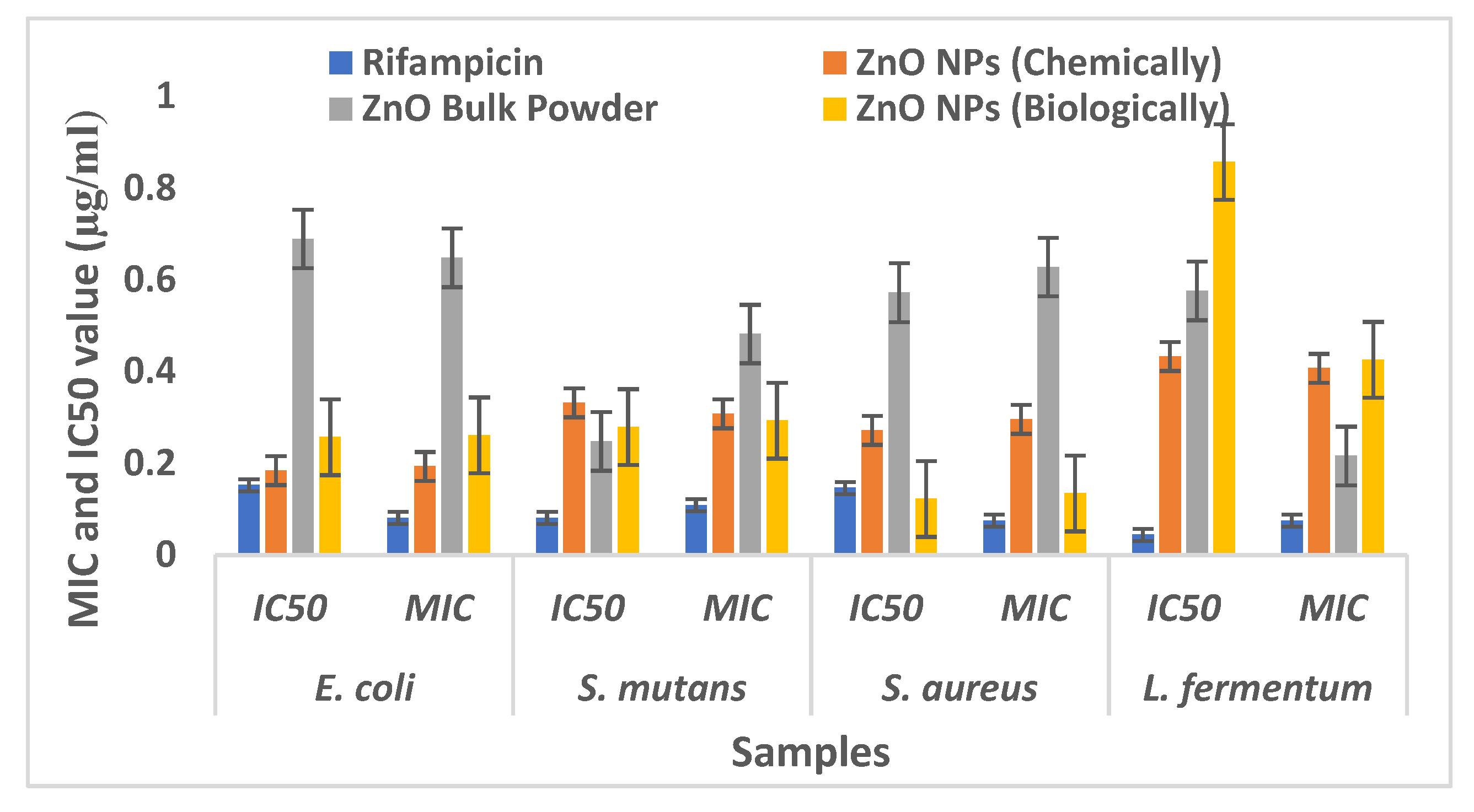
Figure 1. X-ray diffraction patterns of: (a) chemically synthesized ZnO NPs and (b) biologically synthesized ZnO NPs.
Crystals 12 01063 g001
Figure 2. FE-SEM image of: (a) chemically synthesized ZnO NPs and (b) biologically synthesized ZnO NPs.
Crystals 12 01063 g002
Figure 3. FTIR spectra of: (a) chemically synthesized ZnO NPs and (b) biologically synthesized ZnO NPs.
Crystals 12 01063 g003
Figure 4. UV–Visible spectra of: (a) chemically synthesized ZnO NPs and (b) biologically synthesized ZnO NPs.
Crystals 12 01063 g004
Figure 5. Comparative evaluation of antibacterial bioassay of synthesized ZnO NPs (chemically and biologically) with ZnO bulk powder against tooth decaying pathogens.
Crystals 12 01063 g005
Figure 6. Antibacterial bioassay of synthesized ZnO NPs (chemically and biologically), ZnO bulk powder, and rifampicin against: (a) E. coli; (b) S. mutans; (c) S. aureus; and (d) L. fermentum.
Crystals 12 01063 g006
Table 1. Antibacterial bioassay of synthesized ZnO NPs against tooth decaying pathogens.
S. No.Drugs/SamplesE. coliS. mutansS. aureusL. fermentum
IC50MICIC50MICIC50MICIC50MIC
1Rifampicin0.1520.0810.0810.1080.1460.0750.0440.075
2ZnO NPs (chemically)0.1840.1930.3320.3080.2720.2960.4330.407
3ZnO bulk powder0.6890.6480.2480.4820.5720.6280.5760.516
4ZnO NPs (biologically)0.2570.2610.2790.2930.1220.1340.4570.426
Note: All the values are in MIC (in µg/mL) and IC50 (in µg/mL).
Publisher’s Note: MDPI stays neutral with regard to jurisdictional claims in published maps and institutional affiliations.

