Article

In Vitro and Electrochemical Characterization of Laser-Cladded Ti-Nb-Ta Alloy for Biomedical Applications

by
Raj Soni
1,
Sarang Pande
1,*,
Sachin Salunkhe
2,
Harshad Natu
3,
Emad Abouel Nasr
4,
Ragavanantham Shanmugam
5 and
Hussein Mohammed Abdel Moneam Hussein
6,7
1
Department of Mechanical Engineering, Marwadi University, Rajkot 360003, India
2
Department of Mechanical Engineering, Vel Tech Rangarajan Sagunthala R & D Institute of Science and Technology, Chennai 600062, India
3
Magod Fusion Technologies Pvt. Ltd., Pune 411026, India
4
Department of Industrial Engineering, College of Engineering, King Saud University, Riyadh 11421, Saudi Arabia
5
School of Engineering, Mathematics and Technology, Navajo Technical University, Crownpoint, NM 87312, USA
6
Mechanical Engineering Department, Faculty of Engineering and Technology, Future University in Egypt, New Cairo 11835, Egypt
7
Mechanical Engineering Department, Faculty of Engineering, Helwan University, Cairo 11732, Egypt
*
Author to whom correspondence should be addressed.
Crystals 2022, 12(7), 954; https://doi.org/10.3390/cryst12070954
Submission received: 31 May 2022 / Revised: 23 June 2022 / Accepted: 4 July 2022 / Published: 8 July 2022
(This article belongs to the Special Issue Laser Melting of Metals and Metal Matrix Composites)

Abstract

:
Titanium (Ti) and its alloys are predominant choices for use as biomaterials in human implants. Research has shown the adverse effects of using commercial Ti alloy Ti-6Al-4V in the human body, and this presents a need for viable alternatives. In this study, Ti alloy Ti-17Nb-6Ta was manufactured by laser cladding—a prominent additive manufacturing (AM) technology. Laser cladded specimens were evaluated for their in vitro and electrochemical behavior. A human osteosarcoma cell line (MG-63 cells) was used for in vitro investigations. Cell proliferation was good in the physiological medium, and cells were alive when in contact with the laser cladded alloy, even after two to three weeks, indicating good cell viability and compatibility with this alloy. Electrochemical characterization was carried out in Ringer’s solution, and noticeably lower corrosion current density and corrosion rate values were observed. The lower amounts of these parameters indicated the passivation behavior due to multi-layer Ti, Nb, and Ta alloy oxide films. These oxide films also enhanced osseointegration. Thus, the Ti-17Nb-6Ta alloy can be an ideal biocompatible alternative to Ti-6Al-4V.

1. Introduction

Titanium (Ti) and its alloys (Ti alloys) have shown enormous potential to be used to make human body implants and other metallic biomaterials. A rise in patient-specific implants has been observed in recent years due to novel inventions in technology and increased human life expectancy [1]. However, the materials currently being used have compatibility issues, and there has been a concurrent rise in searching for viable alternatives. Ti-6Al-4V is a leading metallic biomaterial used in orthopedic implants and has been standardized by the ASTM F136-13 committee [2]. It has substantial mechanical properties, exceptional resistance to corrosion, and a good amount of biocompatibility [3,4]. However, it also has a high Young’s modulus compared to human bone, which causes increased stress-shielding with the surrounding bone [5]. Apart from it, the Al- and V- in Ti-6Al-4V allegedly cause adverse tissue reactions and other neurological disorders [6,7,8].
Three-dimensional printing or additive manufacturing (AM) has proven its importance in the healthcare sector in recent years. With the use of AM, researchers are trying to increase the research into implantable devices. Selective laser melting (SLM), electron beam melting (EBM), and laser cladding are the leading technologies in AM and are the most commonly used to develop healthcare devices.
Past research has explored the compatibility of Ti alloys determined by conventional methods such as arc melting and powder metallurgy for biomedical applications [9,10]. The present study describes the manufacturing of a Ti alloy with laser cladding to analyse its biomedical applications in the human body. Laser cladding produces net-shape parts from a computer-generated 3D model using a layer-by-layer method with the help of laser power [11,12]. The setup of the laser cladding unit is illustrated in Figure 1. Metallic/alloy material in the powder form is directly supplied from the coaxial nozzle, which is then melted, solidified, and used to create a layer with the help of laser power on the substrate. The substrate is scanned several times with the powder feed, and multiple layers are created to form the entire object [13,14].
Dilution is an unavoidable phenomenon in laser cladding. Dilution is calculated from the percentage of fused material from the substrate. Dilution is directly proportional to laser power. Apart from laser power, the feed rate and scan speed also play a significant role in dilution. The optimization of all of these parameters is indispensable to achieving low dilution [15,16]. Low dilution is desired to achieve perfect metallurgical bonding between the clad material and the substrate. However, an excess amount of it leads to poor bonding.
Titanium is an allotropic element with many crystallographic forms such as α, α + β, and β phases. The metallic elements used in enhancing the α-phase are known as α-stabilizers, and those used for enhancing the β-phase are known as β-stabilizers. In this study, a Ti alloy comprising tantalum and niobium, Ti-17Nb-6Ta, was manufactured by AM, which had not been previously attempted to the best of the authors’ knowledge. Similar to titanium, metals such as tantalum (Ta) and niobium (Nb) are also highly corrosion-resistant and inert in body fluids, which are responsible for enhancing the β phase [17,18]. The alloys in the β phase are more stable and have a low Young’s modulus value. In vitro and corrosion characterization was also performed to explore possible uses of the manufactured alloy in biomedical applications.

2. Materials and Methods
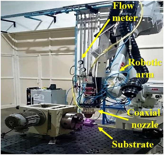
2.1. Laser Cladding

Laser cladding produces a material directly from raw powder and thus possesses the possibility to change the composition of the alloying constituents at any given point in time. This flexibility in material selection is not possible in any other AM technique. By maintaining the flow rate, control is gained over the percentage of alloying elements and can even be used for gradient manufacturing. Additionally, medium entropy (MEA) and high entropy alloys (HEA) with refractory metals as constituents can easily be fabricated using this technique, something that is difficult for other AM techniques [19]. Xiang et al. [20,21] studied CoCrNiTi and CrFeNiTi MEA coatings and CoCrFeNiNbx HEA coatings on a Ti sheet by pulsed laser cladding. They obtained a higher amount of hardness and wear resistance than the substrate.
Powder bed techniques such as SLM require more raw material to fill the build platform to begin the manufacturing process. Unlike SLM, laser cladding only requires a tiny amount of the raw powder material, which is directly supplied and melted. Hence, this technology is economically viable in comparison to others.
Titanium powder was procured from Parshvamani Metals, Mumbai, India, for this study. Tantalum and niobium powder was procured from Aritech Chemazone Pvt. Ltd., Kurukshetra, India. Laser cladding was performed with a 4 KW diode laser on ABB robotic arm at Magod Fusion Technologies Pvt. Ltd., Pune, India. The alloy powders were mixed in preset proportions. It was poured into a laser cladding unit after sieving and thorough mechanical shaking. To reduce cracking susceptibility, preheating is generally used. By preheating, the temperature difference between the substrate and the clad material is reduced, and the residual stress generation becomes negligible. A pure titanium (CP-Ti) plate was used as the substrate and was pre-heated to 200 °C to obtain uniform cladding. The cladding was performed in a closed chamber with an inert argon atmosphere to eliminate oxidation.
For proper melting, a minimum amount of energy needs to be applied to the powder, which is known as the laser energy density (Ed). This energy is dependent on the laser power (P), laser spot diameter (d), and scan speed (V) [12]. The relationship is best represented by Equation (1), as shown below.
E d = P Vd
The parameters used for cladding are highlighted in Table 1. Laser cladding specimens were ground using an ELB Optimal 4250 surface grinding machine and cut using a Charmiless Robofil 190 CNC wire cut spark erosion machine at the Indo German Tool Room (IGTR), Ahmedabad, India. The sample sizes of 20 × 20 × 5 mm3 and 10 × 10 × 5 mm3 were used for further experimentation.

2.2. In Vitro Characterization
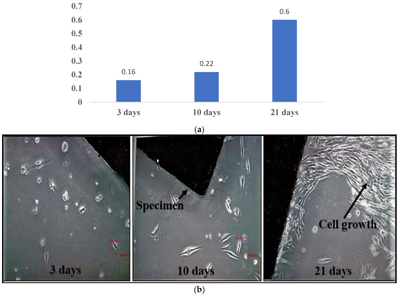
In vitro characterization was performed at the Biotechnology Lab at Gujarat Technological University, Ahmedabad, India. A total of 6 specimens were used for in vitro tests. Specimens were first autoclaved at 121 °C for 30 min. Plain human osteosarcoma cell lines (MG-63) were used as controls. The MG-63 cells were poured onto the specimens and placed in 6-well plates. The initial cell density was measured to be 200,000 cells per well. A 3 mL aliquot of Dulbecco’s modified eagle medium (enhanced with 2.5 µg/mL amphotericin B, 50 µg/mL gentamicin, and 10% fetal bovine serum) was poured into each well. Cultures were incubated at 37 °C under a 5% CO2 environment [22]. The incubation medium was replaced every 48 h for the duration of the experiment. The specimens were examined at 3, 10, and 21 days of incubation.

MTT Assay

The MTT (3-(4,5-dimethylthiazol-2-yl)-2,5-diphenyl-2H-tetrazolium bromide) test was performed to evaluate the toxicity of the samples. Under beneficial conditions, MTT is transformed into formazan by the mitochondrial dehydrogenases and is a good indicator of mitochondrial activity and cell viability. A 100 µL aliquot of MTT was poured into the wells containing each specimen for this study. The supernatant was removed in a CO2 atmosphere after 4 h of incubation, and 100 µL of dimethyl sulfoxide was added to dissolve the formazan crystals. An absorption reading was taken at 570 nm with a microplate reader after shaking the mixture for 10 min. The results were obtained as corrected optical densities (OD).

2.3. Electrochemical Characterization
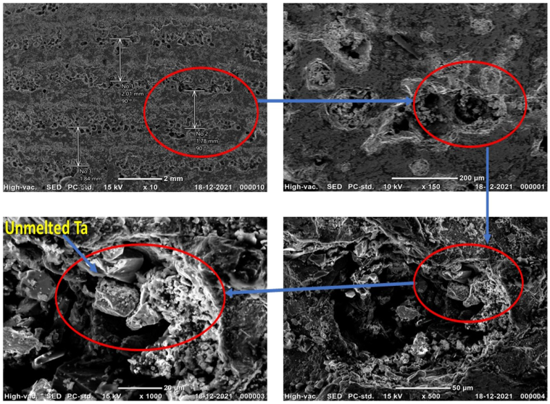
The electrochemical characterization of the laser cladding samples was carried out at the MEMS Lab, Indian Institute of Technology-Bombay, Mumbai, India. The working setup was a 3-electrode setup with the specimen as a working electrode, platinum as the counter electrode, and saturated calomel as the reference electrode [23]. Potentiodynamic polarization readings were obtained from −745 mVSCE to 75 mVSCE with a 1 mV/s scan rate in the Potentiostat (PARSTAT 3000A). Current (I) and voltage (VSCE) values were measured, and Tafel curves were plotted using Origin Pro software. Triplicate analysis was performed to minimize errors in the readings. The specimens were immersed in Ringer’s solution to simulate a human body-like environment. Ringer’s solution was made by adding 9 g NaCl, 0.43 g KCl, 0.2 g NaHCO3, and 0.24 g CaCl2 to 1 L of distilled water followed by autoclaving at 121 °C for 15 min [24].

3. Results

Laser cladding specimens as-cladded and cut to the required sizes, are shown in Figure 2. An SEM-EDX analysis was conducted to analyze the metal proportion and microstructures of the manufactured alloy. Figure 3 illustrates the SEM images, wherein the cladding layer and unmelted Ta are visible. Additionally, the pass-layer distance could be verified from the measurements. Some porosity was also observed in the SEM images, which could indicate residual gases entrapped at cladding. The partial boiling of Ti particles may also be another reason for the porosity, as there was no significant difference between the boiling point of titanium (3287 °C) and the melting point of tantalum (3017 °C) [25]. The EDX result of the manufactured alloy is provided in Figure 4.

3.1. In Vitro Characterization

The MTT assay results exhibited an increased proliferation of MG-63 cells on the specimen samples. The cell densities on the specimens evaluated after 3, 10, and 21 days are illustrated in Figure 5a. Initially, there was less proliferation, possibly due to there being lower attachment with the alloy chemistry. Over time, the cells were possibly habituated to the new alloy material and gradually differentiated. After 21 days, noticeable cell growth and proliferation were seen, as shown in Figure 5b.

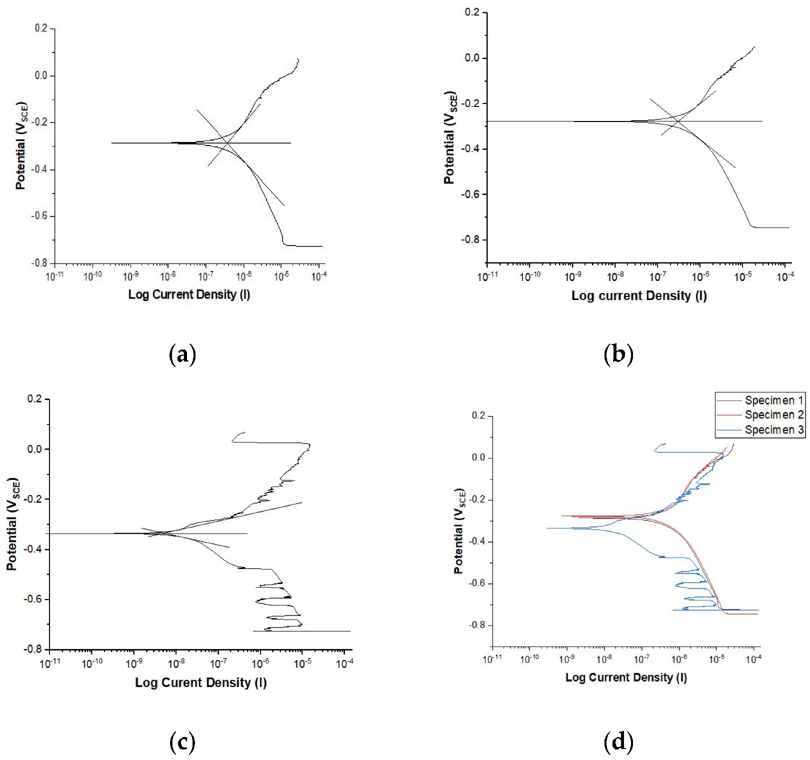
3.2. Electrochemical Characterization

The corrosion resistance of the cladded alloy was examined from the Icorr (corrosion current density) and Ecorr (corrosion potential) values [26]. The obtained Icorr and Ecorr values are highlighted in Table 2. The Tafel plots for the values are illustrated in Figure 6. The rate of corrosion (CR) can be obtained using Equation (2) (ASTM standards G01) [27,28].
CR = K 1 ( Icorr EW ) ρ
where Icorr = the corrosion current density (µA/cm2), K1 = 3.27 × 10⁻³ (mm g/μA cm yr), ρ = the material density (g/cm3), EW = the equivalent weight, and CR = the corrosion rate in mmpy.
From the calculations, the corrosion rates for this Ti-17Nb-6Ta alloy were found to be 0.002 mm/year, which is well under the outstanding corrosion category (0.02 mm/year) [29].

4. Discussion

In this work, a Ti-17Nb-6Ta alloy was fabricated using the laser cladding technique. In vitro and electrochemical characterization were performed to evaluate the behavior of the fabricated alloy when exposed to an artificial human body environment. In the vitro tests, the cell attachment to the fabricated alloy was increased with increased culture time. Significant cell growth was observed after two weeks. No adverse effects from the specimens were observed on the culture growth, as indicated by the third week’s cell density. This indicates the biocompatible nature of the newly fabricated alloy.
In electrochemical characterization, current density, and potential values were determined after testing. Tafel plots were plotted, and the Icorr and Ecorr values were found. The Icorr values evaluated from the Tafel curves were low (in the magnitude of 10−5 to 10−7), which depicts the passive behavior of the Ti-17Nb-6Ta alloy. This passivity is mainly due to the formation of an oxide film on the alloy surface. The surface oxide films inhibit the release of the alloying component into the human body, thus inhibiting the ions from immersing in the bloodstream. Passivation is a natural phenomenon that occurs on the titanium alloy surface. It prevents the further oxidation of the alloy by making a protective layer on the surface. Titanium has a high affinity to oxygen according to which the oxide layer is built on the titanium and its alloy surface, and it enhances the corrosion resistance and osseointegration [30]. The inclusion of alloying elements such as Nb and Ta improves the passivation by forming Nb2O5 and Ta2O5, respectively, which are super stable in the human body and provide superior corrosion resistance [31]. The Ti-17Nb-6Ta alloy forms multi-layer oxide films such as TiO2, Nb2O5, and Ta2O5 on the material surface, which additionally help in increasing corrosion inhibition [32,33,34]. Additionally, the oxide layers also enhance osseointegration [35]. A low CR in the biomaterial is desirable, as it lowers the discharge of metallic ions into the human body, thus reducing the chance of allergic reactions and adverse events due to ionic contamination [27].

5. Conclusions

There has been an increased requirement for human body implants in recent years. The number of revision surgeries has also increased due to the incompatibility of the material that is into the body. Additive manufacturing offers the fastest route for making customized implants compared to any other manufacturing process. Titanium alloys have long been promising candidates for making customized biocompatible implants. Therefore, a titanium alloy Ti-17Nb-6Ta was manufactured in this study using laser cladding, one of the prominent AM techniques for metal fabrication. During post-manufacturing, the alloy was ground and cut into the required sizes for in vitro and electrochemical characterization. No adverse effect was found in the presence of the Ti-17Nb-6Ta alloy, with good cell growth being observed even after two to three weeks. A meager corrosion rate (0.002 mm per year) was observed, along with a low Icorr (0.226 µA/cm2) value. The lower values can mainly be attributed to the multi-layer oxide films (TiO2, Nb2O5, and Ta2O5) generated on the alloy surface, which inhibit metallic ions release into the human body. Hence, the alloy does not cause any cytotoxic symptoms. It can be concluded that the new Ti alloy fabricated using laser cladding possesses good biocompatibility, which is the prime requirement for human body implants.

Author Contributions

Conceptualization and methodology, R.S. (Raj Soni), R.S. (Ragavanantham Shanmugam), S.P., S.S. and H.N.; writing—original draft preparation, R.S. (Raj Soni), S.P., S.S. and H.N.; writing—review and editing, R.S. (Raj Soni), S.P., S.S., E.A.N., R.S. (Ragavanantham Shanmugam) and H.M.A.M.H. All authors have read and agreed to the published version of the manuscript.

Funding

This research has received funding from King Saud University through Researchers Supporting Project number (RSP-2021/164), King Saud University, Riyadh, Saudi Arabia.

Institutional Review Board Statement

Not applicable.

Informed Consent Statement

Not applicable.

Data Availability Statement

Not applicable.

Acknowledgments

The authors extend their appreciation to King Saud University for funding this work through Researchers Supporting Project number (RSP-2021/164), King Saud University, Riyadh, Saudi Arabia. The authors gratefully acknowledge the help provided by Mahendra Motiwala, IGTR-Ahmedabad, India, in carrying out the wire cutting, Vaibhav Bhatt, AIC (GTU)—Ahmedabad, India, in performing the in vitro experimentation, and Rahul Agrawal, MEMS Lab, IIT-Bombay, India, in performing the electrochemical characterization.

Conflicts of Interest

The authors declare no conflict of interest.

References

  1. Giner, M.; Chicardi, E.; Costa, A.D.; Santana, L.; Vázquez-Gámez, M.Á.; García-Garrido, C.; Colmenero, M.A.; Olmo-Montes, F.J.; Torres, Y.; Montoya-García, M.J. Biocompatibility and Cellular Behavior of TiNbTa Alloy with Adapted Rigidity for the Replacement of Bone Tissue. Metals 2021, 11, 130. [Google Scholar] [CrossRef]
  2. Standard Specification for Wrought Titanium-6Aluminum-4Vanadium ELI (Extra Low Interstitial) Alloy for Surgical Implant Applications (UNS R56401). Available online: https://www.astm.org/f0136-13r21e01.html. (accessed on 16 January 2022).
  3. Kuroda, D.; Niinomi, M.; Morinaga, M.; Kato, Y.; Yashiro, T. Design and mechanical properties of new β type titanium alloys for implant materials. Mater. Sci. Eng. A 1998, 243, 244–249. [Google Scholar] [CrossRef]
  4. Kumari, R.; Scharnweber, T.; Pfleging, W.; Besser, H.; Majumdar, J.D. Laser Surface Textured Titanium Alloy (Ti-6Al-4V)-Part II-Studies on Bio-compatibity. Appl. Surf. Sci. 2015, 357, 750. [Google Scholar] [CrossRef]
  5. Liu, J.; Chang, L.; Liu, H.; Li, Y.; Yang, H.; Ruan, J. Microstructure, mechanical behavior and biocompatibility of powder metallurgy Nb-Ti-Ta alloys as biomedical material. Mater. Sci. Eng. C. 2017, 71, 512–519. [Google Scholar] [CrossRef] [PubMed]
  6. Sumitomo, N.; Noritake, K.; Hattori, T.; Morikawa, K.; Niwa, S.; Sato, K.; Niinomi, M. Experiment study on fracture fixation with low rigidity titanium alloy. J. Mater. Sci. Mater. Med. 2008, 19, 1581–1586. [Google Scholar] [CrossRef]
  7. Choe, H.C. Nanotubular surface and morphology of Ti-binary and Ti-ternary alloys for biocompatibility. Thin Solid Films 2011, 519, 4652–4657. [Google Scholar] [CrossRef]
  8. Bocchetta, P.; Chen, L.Y.; Tardelli, J.D.; Reis, A.C.; Almeraya-Calderón, F.; Leo, P. Passive Layers and Corrosion Resistance of Biomedical Ti-6Al-4V and -Ti Alloys. Coatings 2021, 11, 487. [Google Scholar] [CrossRef]
  9. Park, S.Y.; Jo, C.I.; Choe, H.C.; Brantley, W.A. Hydroxyapatite deposition on micropore-formed Ti-Ta-Nb alloys by plasma electrolytic oxidation for dental applications. Surf. Coat. Technol. 2016, 294, 1152–1157. [Google Scholar] [CrossRef]
  10. Zhukova, Y.S.; Pustov, Y.A.; Konopatsky, A.S.; Filonov, M.R. Characterization of electrochemical behavior and surface oxide films on superelastic biomedical Ti–Nb–Ta alloy in simulated physiological solutions. J. Alloys Compd. 2014, 586, S535. [Google Scholar] [CrossRef]
  11. Bandyopadhyay, A.; Krishna, B.V.; Xue, W.; Bose, S. Application of Laser Engineered Net Shaping (LENS) to manufacture porous and functionally graded structures for load bearing implants. J. Mater. Sci. Mater. Med. 2009, 20, 29–34. [Google Scholar] [CrossRef]
  12. Bhardwaj, T.; Shukla, M.; Paul, C.P.; Bindra, K.S. Direct Energy Deposition—Laser Additive Manufacturing of Titanium-Molybdenum alloy: Parametric studies, microstructure and mechanical properties. J. Alloys Compd. 2019, 787, 1238–1248. [Google Scholar] [CrossRef]
  13. Chen, J.; Zhou, Y.; Shi, C.; Mao, D. Microscopic Analysis and Electrochemical Behavior of Fe-Based Coating Produced by Laser Cladding. Metals 2017, 7, 435. [Google Scholar] [CrossRef] [Green Version]
  14. Xue, W.; Krishna, B.V.; Bandyopadhyay, A.; Bose, S. Processing and biocompatibility evaluation of laser processed porous titanium. Acta Biomater. 2007, 3, 1007–1018. [Google Scholar] [CrossRef] [PubMed]
  15. Amado, J.M.; Rodríguez, A.; Montero, J.N.; Tobar MJYáñez, A. A comparison of laser deposition of commercially pure titanium using gas atomized or Ti sponge powders. Surf. Coat. Technol. 2019, 374, 253–263. [Google Scholar] [CrossRef]
  16. Weng, F.; Chen, C.; Yu, H. Research status of laser cladding on titanium and its alloys: A review. Mater. Mater. Des. 2014, 58, 412–425. [Google Scholar] [CrossRef]
  17. Okazaki, Y. Effect of friction on anodic polarization properties of metallic biomaterials. Biomaterials 2002, 23, 2071–2077. [Google Scholar] [CrossRef]
  18. Liu, J.; Ruan, J.; Chang, L.; Yang, H.; Ruan, W. Porous Nb-Ti-Ta alloy scaffolds for bone tissue engineering: Fabrication, mechanical properties and in vitro/vivo biocompatibility. Mater. Sci. Eng. C 2017, 78, 503–512. [Google Scholar] [CrossRef]
  19. Dobbelstein, H.; Thiele, M.; Gurevich, E.L.; George, E.P.; Ostendorf, A. Direct metal deposition of refractory high entropy alloy MoNbTaW. Phys. Procedia 2016, 83, 624–633. [Google Scholar] [CrossRef] [Green Version]
  20. Xiang, K.; Chen, L.Y.; Chai, L.; Guo, N.; Wang, H. Microstructural characteristics and properties of CoCrFeNiNbx high-entropy alloy coatings on pure titanium substrate by pulsed laser cladding. Appl. Surf. Sci. 2020, 517, 146214. [Google Scholar] [CrossRef]
  21. Xiang, K.; Chai, L.; Zhang, C.; Guan, H.; Wang, Y.; Ma, Y.; Li, Y. Investigation of microstructure and wear resistance of laser-clad CoCrNiTi and CrFeNiTi medium-entropy alloy coatings on Ti sheet. Opt. Laser Technol. 2022, 145, 107518. [Google Scholar] [CrossRef]
  22. Kim, S.E.; Jeong, H.W.; Hyun, Y.T.; Lee, Y.T.; Jung, C.H.; Kim, S.K.; Lee, J.H. Elastic Modulus and In Vitro Biocompatibility of Ti-xNb and Ti-xTa Alloys Met. Mater. Int. 2007, 13, 145–149. [Google Scholar] [CrossRef]
  23. Assis SLd Wolynec, S.; Costa, I. Corrosion characterization of titanium alloys by electrochemical techniques. Electrochim. Acta 2006, 51, 1815. [Google Scholar] [CrossRef]
  24. Karayan, A.I.; Park, S.W.; Lee, K.M. Corrosion behavior of Ti–Ta–Nb alloys in simulated physiological media. Mater. Lett. 2008, 62, 1843–1845. [Google Scholar] [CrossRef]
  25. Balla, V.K.; Banerjee, S.; Bose, S.; Bandyopadhyay, A. Direct Laser Processing of Tantalum Coating on Titanium for Bone Replacement Structures. Acta Biomater. 2010, 6, 2329–2334. [Google Scholar] [CrossRef] [PubMed] [Green Version]
  26. Hussein, A.H.; Gepreel MA, H.; Gouda, M.K.; Hefnawy, A.M.; Kandil, S.H. Biocompatibility of new Ti–Nb–Ta base alloys. Mater. Sci. Eng. C Mater. Biol. Appl. 2016, 61, 574. [Google Scholar] [CrossRef]
  27. Giatmana, D.D.; Affi, J.; Fonna, S.; Niinomi, M.; Nakai, M. Corrosion resistance of new beta type titanium alloy, Ti-29Nb-13Ta-4.6Zr in artificial saliva solution. IOP Conf. Ser. Mater. Sci. Eng. 2018, 352, 012008. [Google Scholar]
  28. du Plooy, R.; Akinlabi, E.T. Analysis of laser cladding of Titanium alloy. Mater. Today Proc. 2018, 5, 19594–19603. [Google Scholar] [CrossRef]
  29. Bhola, R.; Bhola, S.M.; Mishra, B.; Olson, D.L. Electrochemical Behavior of Titanium and Its Alloys as Dental Implants in Normal Saline. Adv. Phys. Chem. 2009, 2009, 574359. [Google Scholar] [CrossRef] [Green Version]
  30. Eliaz, N. Corrosion of Metallic Biomaterials: A Review. Materials 2019, 12, 407. [Google Scholar] [CrossRef] [Green Version]
  31. Geetha, M.; Singh, A.K.; Asokamani, R. Ti based biomaterials, the ultimate choice for orthopaedic implants—A review. Prog. Mater. Sci. 2009, 54, 397–425. [Google Scholar] [CrossRef]
  32. Chen, Y.H.; Chuang, W.S.; Huang, J.C.; Wang, X.; Chou, H.S.; Lai, Y.J.; Lin, P.H. On the bio-corrosion and biocompatibility of TiTaNb medium entropy alloy films. Appl. Surf. Sci. 2020, 508, 145307. [Google Scholar] [CrossRef]
  33. Kobayashi, E.; Wang, T.J.; Doi, H.; Hamanaka, H. Mechanical properties and corrosion resistance of Ti–6Al–7Nb alloy dental castings. J. Mater. Sci. Mater. Med. 1998, 9, 567–574. [Google Scholar] [CrossRef] [PubMed]
  34. Zhou, Y.L.; Niinomi, M.; Akahori, T.; Fukui, H.; Toda, H. Corrosion resistance and biocompatibility of Ti–Ta alloys for biomedical applications. Mater. Sci. Eng. A 2005, 398, 28–36. [Google Scholar] [CrossRef]
  35. Kirmanidou, Y.; Sidira, M.; Drosou, M.E.; Bennani, V.; Bakopoulou, A.; Tsouknidas, A.; Michalakis, K. New Ti-Alloys and Surface Modifications to Improve the Mechanical Properties and the Biological Response to Orthopedic and Dental Implants: A Review. BioMed Res. Int. 2016, 2016, 2908570. [Google Scholar] [CrossRef] [PubMed] [Green Version]
Figure 1. Setup of the laser cladding unit.
Crystals 12 00954 g001
Figure 2. (a) As-cladded specimen; (b) specimens after cutting into required sizes.
Crystals 12 00954 g002
Figure 3. SEM images of manufactured alloy Ti-17Nb-6Ta.
Crystals 12 00954 g003
Figure 4. EDX analysis of manufactured alloy Ti-17Nb-6Ta.
Crystals 12 00954 g004
Figure 5. The increase in (a) optical density and (b) cell density of specimens at different time intervals.
Crystals 12 00954 g005
Figure 6. (ac) Tafel curves 1, 2, 3, and (d) combined Tafel curve.
Crystals 12 00954 g006
Table 1. Cladding parameters.
ParametersValues
Power1200 watt
Cladding powder flow12 g/min
Cladding travel speed30 mm/s
Bed temp (preheat)200 °C
Powder carrier gas15 lpm
Nozzle shielding gas15 lpm
Focusing lens300 mm
Spot size (diameter)3 mm
Pass layer distancearound 1.5 mm
Table 2. Values of Icorr, Ecorr, and CR obtained from the Tafel curve.
123Avg
Icorr (µA/cm2)3.75 × 10−12.99 × 10−15.18 × 10−32.26 × 10−1
Ecorr (VSCE)−2.867 × 10⁻¹−2.771 × 10⁻¹−3.348 × 10⁻¹−3.00 × 10⁻¹
CR (mmpy)3.30 × 10−32.64 × 10−34.57 × 10−52.00 × 10−3
Publisher’s Note: MDPI stays neutral with regard to jurisdictional claims in published maps and institutional affiliations.

