Article

Antimicrobial Activity of Commercial Photocatalytic SaniTise™ Window Glass

1
Institute of Physics, University of Tartu, W. Ostwaldi Str 1, 50411 Tartu, Estonia
2
Institute of Molecular and Cell Biology, University of Tartu, Riia 23, 51010 Tartu, Estonia
3
Department of Chemistry and Biotechnology, Tallinn University of Technology, Akadeemia tee 15, 12618 Tallinn, Estonia
4
Laboratory of Environmental Toxicology, National Institute of Chemical Physics and Biophysics, Akadeemia tee 23, 12618 Tallinn, Estonia
*
Author to whom correspondence should be addressed.
Catalysts 2022, 12(2), 197; https://doi.org/10.3390/catal12020197
Submission received: 30 December 2021 / Revised: 29 January 2022 / Accepted: 1 February 2022 / Published: 5 February 2022
(This article belongs to the Special Issue Photocatalytic Nanomaterials for Abatement of Microorganisms)

Abstract

:
Photocatalytic and antibacterial properties of TiO2-based SaniTise™ glass by Pilkington were studied with an aim to benchmark this first commercial UVA-activated antimicrobial glass and to evaluate its efficacy in indoor-like conditions. For comparison, the antibacterial and photocatalytic activity of self-cleaning BIOCLEAN® glass and photocatalytically inactive clear float PLANICLEAR® control glass were analysed. The presence of an anatase TiO2 layer was demonstrated on the surface of SaniTise™ and BIOCLEAN®. Photocatalytic degradation of organic model dye and antibacterial activity against Escherichia coli and Staphylococcus aureus were higher on SaniTise™ than on BIOCLEAN®. In a liquid antibacterial assay corresponding to ISO 27447 format, 4 h exposure of bacteria to the SaniTise™ surface under UVA resulted in >2.8 log decrease in E. coli and >2.5 log decrease in S. aureus viable cell counts. In experiments with the more application-relevant “dry droplet method”, significantly higher antibacterial activity was observed up to the level where during 4 h at ≤50% RH complete inactivation of bacteria was observed also on PLANICLEAR® control glass. The latter raises concerns about the real-life relevancy of the standard test conditions and suggests that at low air humidity conditions, shorter exposure periods than suggested by current antimicrobial testing protocols should be targeted by photocatalytically active antibacterial surfaces.

Graphical Abstract

1. Introduction

TiO2 and especially its anatase crystal phase is a material with well-known photocatalytic activity used in a variety of self-cleaning products [1]. The self-cleaning property of TiO2 relies on two complementary processes during which TiO2 photocatalytically first degrades organic contamination and then induces superhydrophilic properties that enables the overflowing water to remove the degraded organic material from the surface [2,3,4]. Therefore, the self-cleaning properties of TiO2 are most often used on windows and external glass surfaces that frequently come into contact with water. The photocatalytic activity of TiO2 has, however, also been utilized for the purpose of disinfection [5,6]. Indeed, both Gram-negative and Gram-positive bacteria [7] but also unicellular and filamentous fungi, algae, and protozoa [6] have been shown to be affected by TiO2 and especially its nanostructures. The mechanism of antimicrobial activity of TiO2 involves degradation of the cell wall and cytoplasmic membrane due to reactive oxygen species produced during the photocatalytic process [8,9]. The latter leads to leakage of cellular contents and cell lysis [10]. Most of the studies on antimicrobial effects of TiO2 have, however, concentrated on nanoparticles, and significantly less has been done with thin films ([11,12,13,14] and references therein).
In early 2001, Pilkington plc (now owned by Nippon Sheet Glass Co, Ltd., Minato-ku, Japan) began producing TiO2 thin film coated glass called Pilkington ActivTM as a first commercial self-cleaning glass product [15]. Pilkington ActivTM comprised approximately 15 nm film of nanocrystalline TiO2 layer that was applied onto clear float glass by an on-line chemical vapour deposition process [16]. During the following years, more TiO2 thin layer based commercial self-cleaning glasses for windows were marketed, e.g., BIOCLEAN® (Saint-Gobain, Courbevoie, France), AQUACLEANTM (Saint-Gobain), and Sunclean™ (PPG Industries, Pittsburgh, PA, USA) [17]. These glasses were not officially marketed as “antimicrobial” or “antibacterial”, however, it is reasonable to propose that due to the self-cleaning properties those also exhibit activity against microbes. Previously, antimicrobial activity of similar systems, thin anatase TiO2 films on glass substrates, has been demonstrated and discussed [4,18].
At the end of 2020, Pilkington introduced SaniTise™—a new pyrolytically TiO2 coated glass that officially introduces antimicrobial activity [19]. Pilkington states that the product “helps protecting against enveloped viruses and bacteria according to tests carried out by leading universities and independent laboratories” [20]. The principal difference between self-cleaning and antimicrobial glasses is that the former are used on outer surfaces that are exposed to UV from sunlight and rainwater, enabling the combined effect of photocatalysis and superhydrophilicity. The latter, however, are usually used on internal surfaces of the buildings and rely primarily on UV-induced reactive oxygen species (ROS) production enabled by airborne moisture and following antimicrobial activity [10].
The aim of the current work was to study the photocatalysis-driven antibacterial effect of SaniTise™ glass and to demonstrate its potential efficacy if used in indoor conditions as recommended by the producer. As SaniTiseTM is currently the sole antibacterial glass product on the market, we find it important to independently assess its efficacy in application-relevant conditions for the initial benchmarking of the efficacy of this surface. Antibacterial assays were carried out with two model bacteria, Escherichia coli and Staphylococcus aureus, and under testing conditions that resemble the real-life usage of the glass. In parallel, a classical dye degradation test for photocatalytic activity assessment of SaniTiseTM glass was used. For comparison, antibacterial and photocatalytic activity of self-cleaning glass BIOCLEAN® by Saint Gobain Recherche (Courbevoie, France) [21], which has an advertised formulation similar to SaniTise™ but is marketed as self-cleaning and not antibacterial, and of a clear float PLANICLEAR® glass with no TiO2 on its surface, were studied.

2. Results and Discussion

2.1. Characterization of Glass Surfaces

The three glass surfaces—SaniTise™, BIOCLEAN®, and PLANICLEAR® (control glass)—were characterized for their surface morphology and composition. On SaniTise™ and BIOCLEAN® glasses, the chemical state and crystal structure of the TiO2 coating was also analysed. Finally, change in hydrophilicity of the glass surfaces under UVA irradiation was determined using the sessile drop method.
Surface morphology was imaged using SEM and AFM (Figure 1). For SaniTise™ glass, nanosized structures were revealed under both SEM and AFM (Figure 1A,C,F). In the case of BIOCLEAN®, the surface structures were not visible under SEM (Figure 1B), and due to the strong charging caused by low conductivity of the uncoated glass sample, it was not possible to image PLANICLEAR® under SEM. According to AFM, SaniTise™ has well-resolved surface features several nanometers in height with an overall root mean square (RMS) roughness of 1.9 nm. At the same time, the surface of BIOCLEAN® has rather regular flat ring-like features a few tens of nanometers in diameter, with height on the order of 1 nm (Figure 1D,G) and RMS roughness of 0.2 nm. The same RMS roughness (0.2 nm) was also measured on the PLANICLEAR® control glass; however, in contrast to BIOCLEAN®, the surface of the PLANICLEAR® had a significant amount of randomly distributed particle-like structures of different sizes (up to tens of nanometers) that are likely caused by ambient particulates. Three-dimensional AFM images (Figure 1F–H) help to illustrate the differences among the three samples. Note that the Z-scales on all 3D images are stretched four times relative to the X and Y scales to emphasize surface features.
To further study the elemental composition and chemical state of TiO2 covered glass surfaces, XPS was used. Surface composition analysis of the uppermost region (6 ± 2 nm) [22] of glasses with XPS indicated clear spectra with O 1s and Ti 2p photolines, confirming the presence of TiO2 [23] on both SaniTiseTM (Figure 2A,B) and BIOCLEAN® (Figure 2C,D) glasses. No other Ti oxidation states or chemical species besides TiO2 were detected in the high-resolution Ti 2p spectra (Figure 2B,D) [24]. Peak asymmetry in the Ti 2p spectrum of the BIOCLEAN® glass surface (Figure 2D) can be explained by charging effects. The survey spectrum (Figure 2A) revealed that the SaniTise™ glass surface layer contains Ti, O, C, Ca, and Si, and the BIOCLEAN® surface layer contains Ti, O, C, and Ca (Figure 2C). The survey spectrum of the reference glass (uncoated side of BIOCLEAN®) revealed the presence of O, C, Si, and Ca (Figure 2E). We suggest that Ca on the surface of BIOCLEAN® and SaniTise™ glass originated from Ca ions diffused from the base glass substrate.
Raman spectroscopy was used to confirm the crystal structure of the TiO2 coating on SaniTise™ and BIOCLEAN® glasses (Figure 3). Characteristic Raman bands of anatase (144, 198, 397, 516, 639 cm−1) [25] were observed for both glasses, confirming an anatase crystal structure of the TiO2 coatings. No Raman bands characteristic to the rutile crystal phase of TiO2 were observed. The exploitation of the anatase phase TiO2 as a photocatalytic coating is reasonable, as it is well known that the anatase crystal phase is significantly more photocatalytically effective than the rutile phase; however, clear reasons for this phenomenon are still under discussion [26,27]. In addition to anatase specific peaks, the Raman spectra of SaniTise™ and BIOCLEAN® show an observable background that indicates the existence of amorphous TiO2 in addition to the anatase crystal phase [28].
Hydrophilicity of the glass surfaces was measured using the sessile drop method, where the water contact angle is measured. In addition to original or “virgin” glasses, we also measured the hydrophilicity of all the three glasses after UVA pre-activation, as commonly suggested by manufacturers of TiO2-based photoactive surfaces. The concept of UV pre-activation of photocatalytic surfaces prior to their use is also included in internationally standardized procedures, e.g., in ISO 10678 [29]. Pilkington, which produces SaniTise™ glasses, suggests for their photocatalytic Pilkington ActivTM glasses [30]: “It’s worth noting that when self-cleaning glass has been stored in a warehouse for a long period prior to installation, it may take up to seven days to become fully activated. This is because the activation of the glass occurs via a chemical reaction between UV rays from natural daylight, oxygen and the coating. After that, it will continue to work as long as it’s exposed to daylight, even in dull winter weather.” To test whether UV pre-activation was also required for BIOCLEAN® and SaniTise™ glasses, these along with the PLANICLEAR® control glass were irradiated with UVA (22–25 W/m2) over 72 h, and surface hydrophilicity followed. UVA pre-activation of SaniTise™ and BIOCLEAN® clearly increased their hydrophilic properties (Figure 4) and decreased the initial water contact angle of 60° to 20–30° after 4 h and to ≤10°, i.e., to superhydrophilic, after 24 h (Figure 4A). This suggests that the realization of superhydrophilicity and the self-cleaning effect of anatase-TiO2-based glass surfaces indeed requires UVA pre-activation. Interestingly, when UVA irradiation of SaniTise™ and BIOCLEAN® was finished (i.e., after 72 h), the hydrophilicity of the glass surfaces was gradually lost (Figure 4B), and it reverted to its initial level 48 h after the end of pre-activation. UVA irradiation of PLANICLEAR® control glass did not cause any change in water contact angle, and therefore recovery of the contact angle in dark conditions (Figure 4B) was not measured (Figure 4A,B). In order to utilize the superhydrophilicity-related properties of SaniTise™ and BIOCLEAN® glasses, all the three glass types were tested for their photocatalytic activity and antibacterial effect in “virgin” form and also immediately after 72 h pre-activation.

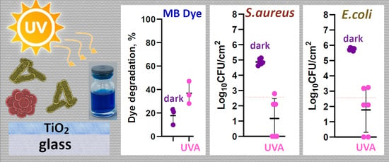
2.2. Photocatalytic Activity of Glass Surfaces

Assuming that the antibacterial effect of SaniTise™ glass is directly related to its ability to produce reactive oxygen species due to UVA irradiation, we first determined the photocatalytic activity of all the selected glasses. Methylene blue dye (MB, model organic contaminant) degradation during 4 h of dark exposure or UVA irradiation were used as a measure for photocatalytic activity of SaniTise™, BIOCLEAN®, and PLANICLEAR® surfaces. To understand the effect of UVA pre-activation on photocatalytic activity, both virgin and UVA pre-activated glasses were measured, and the latter were also evaluated for photocatalytic activity after their 2 h and 24 h recovery in the dark (Figure 5). Our results showed that, in general, the glasses had very low photocatalytic effect in the experimental conditions used, and the only glass surface for which photocatalytic activity was significantly different from the control was UVA pre-activated SaniTise™ glass (Figure 5). Interestingly, this significant photocatalytic effect of SaniTise™ glass was retained after 2 h and 24 h dark recovery post pre-activation, suggesting that photocatalytic degradation of organics can be expected at least 24 h post UVA pre-activation. Therefore, as expected, the photocatalytic effect of TiO2-based surfaces and surface hydrophilicity determined by contact angle method do not always coincide. The reason is that the photocatalysis and hydrophilicity can take place simultaneously on the same surface, even though the mechanisms are completely different [31]. Intriguingly, BIOCLEAN® glasses that are advertised as photocatalytically active glasses did not cause any significant MB degradation in the experimental conditions. We suggest that one of the reasons behind such a difference is the thickness of TiO2 layer on those different glasses as shown by Raman analysis. Expectedly, the PLANICLEAR® glass that served as a control also did not demonstrate any photocatalytic effect after UVA pre-activation and further UVA irradiation.

2.3. Antibacterial Effect of Glass Surfaces

The photocatalytic antibacterial properties of SaniTise™ glass along with BIOCLEAN® and PLANICLEAR® (control) glasses were evaluated using the ISO 27447 [32] standard test format most deployed in the industry and also using the “dry droplet method” in which small droplets of bacterial suspension on the test surface are incubated at variable relative humidity (RH).
The ISO 27447 method uses a small volume of bacterial inoculum spread over the surface in a thin layer, covered by UVA-transmissive film and exposed to UVA in saturated RH conditions to prevent drying of the inoculum. A Gram-positive (Staphylococcus aureus ATCC 6538) and Gram-negative (Escherichia coli ATCC 8739) bacterial strain differing in their cell wall composition were used as model species due to their potentially different susceptibility. Similar to photocatalytic effects, antibacterial activity of all three glasses was evaluated during 4 h in dark conditions and under UVA irradiation in their virgin state and after pre-activation.
Clearly, the highest antibacterial effect for both E. coli and S. aureus was observed on SaniTise™ glass under UVA irradiation (Figure 6). Additionally, SaniTise™ in combination with UVA irradiation was the only surface to decrease the viable count of E. coli. After 4 h under UVA, virgin and UVA pre-activated SaniTise™ glass demonstrated a 2.1 (p < 0.0001) to over 2.8 (p < 0.0001) log decrease in E. coli viable count compared to the control surface (Figure 6A). The effect of SaniTise™ glass on S. aureus regardless of its UVA pre-activation status was over 2.5 log decrease in viable counts. In the case of S. aureus, in addition to SaniTise™ glass, the UVA pre-activated BIOCLEAN® also showed a low (0.8 log) but significant (p = 0.03) decrease in viable counts (Figure 6B). Interestingly, the virgin SaniTise™ glasses seem to also have a minor antibacterial effect of 0.8 log decrease (p = 0.03) on the more susceptible S. aureus in dark conditions. The 0.6 log decrease effect of UVA pre-activated SaniTise™ glasses in dark conditions did not prove to be statistically significant (p = 0.3) but followed the trend. These results show a significantly higher effect in SaniTise™ glass compared with BIOCLEAN® glass, and in the case if E. coli, no antibacterial effect on the BIOCLEAN® glass, are in concordance with the results from photocatalytic activity where only SaniTise™ glass had significant photocatalytic effect. In general, no statistically significant or biologically meaningful differences could be detected between antibacterial activity of virgin and UVA pre-activated glasses regardless of material or bacterial species used (Figure 6A,B).
The “dry droplet method“ uses small droplets of the inoculum that are placed onto the test surface and exposed at predefined conditions, such as RH, to mimic possible application-relevant end-use conditions that cannot be evaluated in the ISO format due to very restrictive quality criteria for bacterial viability on control surfaces. Here we used RH levels to represent high (90%), moderate (50%), and low (20%) RH conditions. Results indicated that already a small change from saturated RH in the ISO test format to inoculum droplets incubated at 90% RH significantly enhanced antibacterial activity of the BIOCLEAN® glass towards both test species. At high RH and low-intensity UVA irradiation, the BIOCLEAN® glass demonstrated over 1.6 (p < 0.0001) log reduction in E. coli viability (Figure 6C) compared to no antibacterial effect in the wet conditions in the ISO test format. High RH conditions offered an additional 1.7 (p < 0.0001) log reduction in S. aureus viability on UVA pre-activated BIOCLEAN® glasses compared to the moderate 0.8 log decrease in the ISO test format (Figure 6D). Importantly, less than quantifiable amounts of viable bacteria (2.6 log CFU/cm2) resulting in over 3 log reduction in viable counts was detected on all 4 h UVA-exposed surfaces at 50% and 20% RH (Figure 6C,D), including control surfaces. Complete inactivation of bacterial cells at 50% and 20% RH under UVA irradiation was also demonstrated by no visible growth after 48 h in tubes containing PLANICLEAR® surfaces submerged in growth-supporting toxicity neutralizing medium used for harvesting the bacteria for plate counts. Therefore, despite it being evident that decreasing RH leads to increased photocatalytic antibacterial effect of TiO2-based surfaces, lower RH also results in higher inactivation of bacteria on the control surfaces and complicates antimicrobial activity evaluation of the test surfaces. Although the drastic decrease in viable counts at medium and low RH level on the control surface would fail the pass criteria of an ISO test, it clearly demonstrates that after 4 h of low-intensity UVA exposure, the SaniTiseTM and BIOCLEAN® glasses offered no antibacterial benefit over the non-photocatalytic PLANICLEAR® glass. Although it is possible that less phototoxicity and higher bacterial viability on control glass in dry ambient conditions could be achieved with substantially shorter UVA exposures, the same light exposure causing phototoxicity is needed for the antibacterial action of the photocatalytic glasses in question. This in turn raises doubts about the usefulness of installing such materials in dry ambient environments without further evaluation of the materials in more application-relevant conditions to demonstrate superiority over conventional materials.

3. Materials and Methods

3.1. Glass Materials

A 4 mm thick Pilkington SaniTiseTM glass, which, according to producer’s (Nippon Sheet Glass Co Ltd., Minato City, Tokyo, Japan) information is pyrolytically coated TiO2 coated glass, was used as an antimicrobial glass. A 6 mm thick BIOCLEAN® (Saint-Gobain, Courbevoie, France) that is covered with TiO2 anatase thin film was used as an example self-cleaning glass. According to information from producers, the thickness of the TiO2 layer on such covered commercial glasses is typically small and in the range of ~15–20 nm [21,33]. Additionally, a 4 mm thick PLANICLEAR® (Saint-Gobain, Courbevoie, France, basic soda-lime silicate glass produced using the float procedure) was used as a non-coated control. These glass surfaces were analysed for their photocatalytic and antimicrobial effects in their original form or after UVA pre-activation. The original form is further referred to as “virgin” glass and pre-activated form is further called “UVA pre-activated”. Pre-activation was applied following the suggestion of glass producers.
For the analysis, the glasses were cut into 25 × 25 mm squares. As an exception, 5 × 8 mm size glass pieces were used for XPS analysis.

3.2. Characterization of the Glass Surfaces

Scanning electron microscopy (SEM) imaging. For SEM, 25 × 25 mm samples of SaniTise™ and BIOCLEAN® mounted onto a standard SEM pin stub were used. The samples were partly covered with conductive silver paint to improve conductivity. Imaging was performed using FEI Nova NanoSEM 450 (Lincoln, NE, USA) at 10 kV.
Atomic force microscopy (AFM) analysis. Topography of the samples was measured by AFM (Dimension Edge, Bruker, Billerica, MA, USA) in tapping mode using Standard Tapping Mode AFM Probe PPP-NCHR (Nanosensors, Neuchatel, Switzerland). Surface roughness and average size of the surface structures were calculated from AFM data using Gwyddion version 2.56 (http://gwyddion.net/; Department of Nanometrology, Czech Metrology Institute) data visualization and analysis software.
X-ray photoelectron spectroscopy (XPS) analysis. XPS measurements were conducted using a surface station equipped with an electron energy analyser(SCIENTA SES 100, Uppsala, Sweden) and a non-monochromatic twin anode X-ray tube (Thermo XR3E2, East Grinstead, UK), with characteristic energies of 1253.6 eV (Mg Kα1,2 FWHM 0.68 eV) and 1486.6 eV (Al Kα1,2 FWHM 0.83 eV). All XPS measurements were conducted in ultra-high vacuum (UHV) conditions. Reference XPS spectrum (uncoated glass) was measured from the uncoated side of the BIOCLEAN® glass. The binding energy scales for the XPS experiments were referenced to the binding energy of Ti4+ 2p3/2 (458.6 eV) and/or C 1s (285 eV) photoemission line. The raw data were processed using Casa XPS software version 2.3.12 (Teignmouth, UK) [34]. Data processing involved removal of Kα and Kβ satellites, removal of the background, and fitting of components. Background removal was carried out using Tougaard background; for fitting, the Gauss-Lorentz hybrid function (GL 95, Gauss 5%, Lorentz 95%) was used for SaniTise™ and the asymmetric Gauss-Lorentz hybrid function (A(0.35,0.8,0)GL(95), Gauss 5%, Lorentz 95%) was used for BIOCLEAN® samples. UHV compatible silver paste was used to reduce charging effects during the XPS experiment. However, the absolute amounts of different compounds and elements have to be considered cautiously, and are given to outline trends and estimates only. Due to the possible surface region deviation from chemical homogeneity in the working range of photoelectron spectroscopy (surface region with a thickness of up to three electron mean free paths), some signals might be amplified or suppressed.
Raman scattering analysis. Raman scattering experiments for all the glasses were carried out with Renishaw inVia micro-Raman spectrometer (New Mills, UK) equipped with an optical microscope. The measurements were made with 50× objective, permitting measurements with spatial resolution down to ~1 μm. The excitation source used was a 514 nm solid state laser with an edge filter. The measurements were done in two steps. First, the Raman scattering signal from the TiO2 film side of the glass substrate was measured. Thereafter the acquired spectra were corrected by subtracting the background signal from the uncoated side of the glass substrate.
Contact angle measurements. The sessile drop method [35] was used to measure water drop contact angles on TiO2 covered glass samples using a moving platform(based on Thorlab DT12 (Newton, NJ, USA) dovetail translation stage). A 2 μL drop of deionized water was pipetted onto a dry glass surface. After 10 s, the water droplet was photographed with a Canon EOS 650d camera (Ota City, Tokyo, Japan) using a MP-E 65 mm f/2.8 1–5× Macro focus lens. Image analysis software (ImageJ 1.8.0 172 (NIH, Madison, WI, USA) for Windows, plugin for contact angle measurement [36]) was used to determine the contact angle formed by the liquid drop on the glass surface. The average of contact angles measured from four water droplets per sample was calculated.
Contact angles were measured either on virgin or on UVA pre-activated surfaces. To create UVA pre-activated glasses, SaniTiseTM, BIOCLEAN®, and PLANICLEAR® glasses were irradiated with UVA generated by a self-built fluorescent lamp containing four fluorescent Hg light bulbs (JW Sales GmbH (Stuttgart, Germany); 15 W iSOLde Cleo, λcentral = 355 nm). The emission spectrum of this lamp measured with SpectraPro 2000i monochromator (Princeton, NJ, USA) and H8259 photon (Hamamatsu, Japan) counting head is shown in Figure 7. The intensity of UVA light (measured using Delta Ohm UVA probe (Caselle, Italy) at 315–400 nm spectral range) was 22–25 W/m2 at the position of sample activation.
Photocatalytic activity of the glasses. For assessment of photocatalytic activity, model organic contaminant methylene blue (MB) was used. A volume of 15 µL of 0.002 M MB aqueous solution was pipetted onto 25 × 25 mm virgin or UVA pre-activated glass samples and covered with 20 × 20 × 0.05 mm polyethylene thin film for controlled and even distribution of the dye solution. The samples were placed on Petri dishes containing wet filter papers to decrease drying effects and pre-conditioned in a climatic chamber (Memmert CTC 256, Schwabach, Germany) at 22 °C and 90% relative humidity (RH) in dark conditions for 20 min to establish adsorption equilibrium of MB. After that, the glasses in three parallels were exposed to UVA (2.4–2.5 W/m2 according to Delta Ohm UVA probe) or kept in dark conditions for 4 h in climatic chamber at 22 °C, 90% RH. After UVA irradiation or dark exposure, the dye solution was washed off using 3 mL of water and its UV-Vis absorbance at 633 nm was measured using a spectrophotometer (Agilent Cary UV-Vis-NIR 5000, Santa Clara, CA, USA). Decrease in UV-Vis absorbance reflected increasing photocatalytic activity.

3.3. Evaluation of Antibacterial Efficacy of TiO2 Coated Glass Surfaces

Antibacterial efficacy assessment using modified ISO 22196/ISO 27447 test. Antibacterial testing was carried out using a modified test protocol from ISO 22196 and ISO 27447. The procedures were modified due to the need for higher throughput to study several materials and time points. Escherichia coli DSM1576 (ATCC8739; German Collection of Microorganisms and Cell Cultures (DSMZ), Braunschweig, Germany) and Staphylococcus aureus ATCC6538 (National Institute of Chemical Physics and Biophysics (Tallinn, Estonia)) were used in the tests as model Gram-positive and Gram-negative organisms.
Bacteria were cultivated overnight on nutrient agar plates (NA: 5 g/L meat extract, 10 g/L peptone, 5 g/L NaCl, 15 g/L agar), collected using a sterile inoculation loop and suspended in exposure medium (500-fold diluted nutrient broth, NB: 3 g/L meat extract, 10 g/L peptone, 5 g/L NaCl), followed by cell density adjustment until an OD600 value of 0.074 (E. coli) and 0.070 (S. aureus), i.e., approximately 3.5 × 10⁵ CFU/cm2. Next, 20 μL of the prepared inoculum suspension was applied to 25 × 25 mm virgin or UVA pre-activated sample surfaces, covered by 20 × 20 × 0.05 mm polyethylene (PE) film, and incubated in the dark or under UVA. PE cover film was used to attain even coverage, even UVA irradiation, and similar humidity conditions of bacterial suspension on the predefined test area. Samples were placed onto a bent glass U-rod over sterile wet filter paper covered by a Petri dish cover in dark conditions or 1.1 mm thick UVA-transmissive borosilicate moisture preservation glass under UVA illumination to attain a humid environment around the samples. Bacterial tests were carried out in a climate chamber at 22 °C either in the dark or under 2.43 ± 0.11 W/m2 UVA irradiation (Delta Ohm UVA probe). After 4 h dark exposure or UVA irradiation, bacteria were harvested from the surfaces by submerging the surfaces in 15 mL of toxicity neutralizing soybean casein digest broth with lecithin and polyoxyethylene sorbitan monooleate (SCDLP, undiluted: 17 g/L casein peptone, 3 g/L soybean peptone, 5 g/L NaCl, 2.5 g/L Na2HPO4, 2.5 g/L glucose, 1.0 g/L lecithin, 7.0 g/L nonionic surfactant Tween80) in 50 mL tubes, followed by vortexing the tubes for 30 sec at full power. Serial dilutions in phosphate buffered saline (PBS: 8 g/L NaCl, 0.2 g/L KCl, 1.63 g/L Na2HPO4 × 12H2O, 0.2 g/L KH2PO4; pH 7.1) were drop-plated on NA plates and incubated at 30 °C. Colonies of E. coli and S. aureus were counted after 24 h and 48 h, respectively, and results are expressed as log10-transformed CFU counts. Antibacterial activity is expressed as log reduction on viable counts. The tubes with growth-supporting toxicity-neutralizing SCDLP medium and surfaces were incubated at 30 °C for 48 h, and growth was evaluated visually.
Antibacterial efficacy assessment at different relative humidity. Bacterial suspensions were prepared as described above but applied to glass surfaces in 20 × 1 μL droplets to achieve the same final cell counts per surface and even coverage without using PE cover film. The surfaces were incubated in a climate chamber in the same lighting conditions (UVA irradiation) and temperature as above but kept at different relative air humidity (15%, 50%, 90% RH). At each humidity condition, bacteria were collected from surfaces after 4 h UVA exposure as above. Bacterial number was assessed as above and results are expressed as log10-transformed CFU counts.

3.4. Statistical Analysis

One-way ANOVA analysis followed by Tukey post-hoc test was used to detect significant differences in multiple comparisons at 0.05 significance level using GraphPad Prism 9.3.0 (San Diego, CA, USA). Log10-transformed data at or above the detection limit of three colonies counted in undiluted plated drop was used for the analysis of CFU counts. Only statistically significant (p < 0.05) differences and log reductions are noted in the text unless stated otherwise.

4. Conclusions

In this paper we present data on photocatalysis-driven antibacterial effect of SaniTise™ glass surfaces marketed by Pilkington as the first commercial antibacterial glass product. In parallel to SaniTise™, we also tested the antibacterial effects of commercially advertised photocatalytic BIOCLEAN® (Saint Gobain) and non-photocatalytic PLANICLEAR® (Saint Gobain) glass surfaces.
We confirmed the presence of anatase TiO2 layer on the surface of SaniTise™ and BIOCLEAN® but not on PLANICLEAR®. Therefore, both SaniTise™ and BIOCLEAN® were expected to exhibit photocatalytic properties, but, surprisingly only SaniTise™ caused significant UVA-induced photocatalytic degradation of a model organic dye. In a similar line, the SaniTise™ glass surface was also more antibacterially active compared with BIOCLEAN®. This was an expected result, as SaniTise™ glass has been marketed with an antibacterial statement.
Antibacterial tests carried out with SaniTise™ and BIOCLEAN® surfaces in a liquid environment as suggested in ISO 27447 protocol and using a more application-relevant “dry droplet method” showed significantly higher efficiency of the latter testing conditions. Whereas in the liquid environment, BIOCLEAN® had no effect on E. coli during 4 h exposure under UVA, in the dry droplet method at 90% RH, >1.6 log reduction of viable colonies was observed. S. aureus showed almost complete inhibition on BIOCLEAN® surface at 90% RH. Furthermore, the dry droplet method combined with RH ≤ 50% also resulted in complete inactivation of bacteria on the control surface of PLANICLEAR®. This specifies the need for more application-relevant test conditions and points to the fact that at low air humidity conditions, shorter exposure periods than recommended in the standard protocols should be targeted by photocatalytically active antibacterial surfaces. One co-result of exploiting photocatalytic antimicrobial glasses could be increased indoor air quality via photocatalytic degradation of volatile organic compounds (VOCs) [37]. However, both, the real-life relevant antimicrobial efficacy as well as improvement of ambient air quality need to be assessed and proven in future studies.

Author Contributions

Conceptualization, V.K. and A.I.; methodology, M.V., M.R., R.P., S.V., M.K. and S.L.; software, M.R., M.V., D.D., R.P. and M.K.; formal analysis, M.R., M.V., D.D., R.P., M.K., S.V. and S.L.; investigation, M.R., M.V., D.D., S.V. and S.L.; resources, A.I. and V.K.; data curation, M.R.; writing—original draft preparation, V.K. and A.I.; writing—review and editing, M.R., M.V., S.L., R.P., M.K. and S.V.; project administration and funding acquisition, A.I. and V.K. All authors have read and agreed to the published version of the manuscript.

Funding

The authors gratefully acknowledge the financial support by the Estonian Research Council Grants (EAG20, PRG1496, COVSG2, MOBTP145), Estonian Centre of Excellence in Research project “Advanced materials and high-technology devices for sustainable energetics, sensorics and nanoelectronics” TK141 (2014−2020.4.01.15−0011), and University of Tartu Development Fund (grant PLTFYARENG53). The research was conducted using the NAMUR+ core facility funded by projects “Center of nanomaterials technologies and research” (2014-2020.4.01.16-0123) and TT13. M.K. acknowledges support by Graduate School of Functional materials and technologies receiving funding from the European Regional Development Fund in University of Tartu, Estonia (project code 2014−2020.4.01.16−0027). S.V. acknowledges ERA Chair MATTER from the European Union’s Horizon 2020 research and innovation programme under grant agreement No 856705.

Data Availability Statement

The data supporting this study are available in the data repository of University of Tartu (dataDOI): http://dx.doi.org/10.23673/re-302 (date of deposition 4 January 2022).

Acknowledgments

The authors would like to thank Glassolutions Baltiklaas Tartu and Artekom OÜ.

Conflicts of Interest

The authors declare no conflict of interest. The funders had no role in the design of the study; in the collection, analyses, or interpretation of data; in the writing of the manuscript, or in the decision to publish the results.

References

  1. Padmanabhan, N.T.; Honey, J. Titanium dioxide based self-cleaning smart surfaces: A short review. J. Environ. Chem. Eng. 2020, 8, 104211. [Google Scholar] [CrossRef]
  2. Fujishima, A.; Zhang, X.; Tryk, D. TiO2 photocatalysis and related surface phenomena. Surf. Sci. Rep. 2008, 63, 515–582. [Google Scholar] [CrossRef]
  3. Banerjee, S.; Dionysiou, D.D.; Pillai, S.C. Self-cleaning applications of TiO2 by photo-induced hydrophilicity and photocatalysis. Appl. Catal. B Environ. 2015, 176–177, 396–428. [Google Scholar] [CrossRef] [Green Version]
  4. Nakata, K.; Fujishima, A. TiO2 photocatalysis: Design and applications. J. Photochem. Photobiol. C Photochem. Rev. 2012, 13, 169–189. [Google Scholar] [CrossRef]
  5. Gamage, J.; Zhang, Z. Applications of Photocatalytic Disinfection. Int. J. Photoenergy 2010, 764870. [Google Scholar] [CrossRef] [Green Version]
  6. Chong, M.N.; Jin, B.; Chow, C.W.K.; Saint, C. Recent developments in photocatalytic water treatment technology: A review. Water Res. 2010, 44, 2997–3027. [Google Scholar] [CrossRef]
  7. De Castillo, C.D.; Correa, M.G.; Martinez, F.B.; Streitt, C.; Galotto, M.J. Antimicrobial Effect of Titanium Dioxide Nanoparticles. In Antimicrobial Resistance—A One Health Perspective; IntechOpen: London, UK, 2020. [Google Scholar] [CrossRef] [Green Version]
  8. Kubacka, A.; Diez, M.S.; Rojo, D.; Bargiela, R.; Ciordia, S.; Zapico, I.; Albar, J.P.; Barbas, C.; Martins dos Santos, V.A.; Fernández-García, M.; et al. Understanding the Antimicrobial Mechanism of TiO2-Based Nanocomposite Films in a Pathogenic Bacterium. Sci. Rep. 2014, 4, 4134. [Google Scholar] [CrossRef] [Green Version]
  9. Kiwi, J.; Nadtochenko, V. Evidence for the mechanism of photocatalytic degradation of the bacterial wall membrane at the TiO2 interface by ATR-FTIR and laser kinetic spectroscopy. Langmuir 2005, 21, 4631–4641. [Google Scholar] [CrossRef]
  10. Foster, H.A.; Ditta, I.B.; Varghese, S.; Steele, A. Photocatalytic disinfection using titanium dioxide: Spectrum and mechanism of antimicrobial activity. Appl. Microbiol. Biotechnol. 2011, 90, 1847–1868. [Google Scholar] [CrossRef]
  11. Joost, U.; Juganson, K.; Visnapuu, M.; Mortimer, M.; Kahru, A.; Nõmmiste, E.; Joost, U.; Kisand, V.; Ivask, A. Photocatalytic antibacterial activity of nano-TiO2 (anatase)-based thin films: Effects on Escherichia coli cells and fatty acids. J. Photochem. Photobiol. B Biol. 2015, 142, 178–185. [Google Scholar] [CrossRef]
  12. Alotaibi, A.M.; Williamson, B.A.D.; Sathasivam, S.; Kafizas, A.; Alqahtani, M.; Sotelo-Vazquez, C.; Buckeridge, J.; Wu, J.; Nair, S.P.; Scanlon, D.O.; et al. Enhanced Photocatalytic and Antibacterial Ability of Cu-Doped Anatase TiO2 Thin Films: Theory and Experiment. ACS Appl. Mater. Interfaces 2020, 12, 15348–15361. [Google Scholar] [CrossRef] [PubMed] [Green Version]
  13. Cushnie, T.P.T.; Robertson, P.K.J.; Officer, S.; Pollard, P.M.; Prabhu, R.; Mccullagh, C.; Robertson, J.M.C. Photobactericidal effects of TiO2 thin films at low temperatures—A preliminary study. J. Photochem. Photobiol. A Chem. 2010, 216, 290–294. [Google Scholar] [CrossRef]
  14. Sunada, K.; Watanabe, T.; Hashimoto, K. Studies on photokilling of bacteria on TiO2 thin film. J. Photochem. Photobiol. A Chem. 2003, 156, 227–233. [Google Scholar] [CrossRef]
  15. Mills, A.; Lepre, A.; Elliott, N.; Bhopal, S.; Parkin, I.P.; O’Neill, S.A. Characterisation of the photocatalyst Pilkington Activ™: A reference film photocatalyst? J. Photochem. Photobiol. A Chem. 2003, 160, 213–224. [Google Scholar] [CrossRef]
  16. Garlisi, C.; Palmisano, G. Radiation-free superhydrophilic and antifogging properties of e-beam evaporated TiO2 films on glass. Appl. Surf. Sci. 2017, 420, 83–93. [Google Scholar] [CrossRef]
  17. Dunnill, C.W.; Aiken, Z.A.; Kafizas, A.; Pratten, J.; Wilson, M.; Morgan, D.J.; Parkin, I.P. White light induced photocatalytic activity of sulfur-doped TiO2 thin films and their potential for antibacterial application. J. Mater. Chem. 2009, 19, 8747. [Google Scholar] [CrossRef] [Green Version]
  18. Yu, J.C.; Tang, H.Y.; Yu, J.; Chan, H.C.; Zhang, L.; Xie, Y.; Wang, H.; Wong, S.P. Bactericidal and photocatalytic activities of TiO2 thin films prepared by sol–gel and reverse micelle methods. J. Photochem. Photobiol. A Chem. 2002, 153, 211–219. [Google Scholar] [CrossRef]
  19. Nippon Sheet Glass Co. Ltd. Pilkington SaniTise™ Brochures. Available online: https://www.pilkington.com/en/global/products/product-categories/health-applications/pilkington-sanitise#brochures (accessed on 23 December 2021).
  20. Nippon Sheet Glass Co. Ltd. Pilkington SaniTise™ Technical Bulletin. Available online: https://assetmanager-ws.pilkington.com/fileserver.aspx?cmd=get_file&ref=GL112&cd=cd (accessed on 23 December 2021).
  21. Peruchon, L.; Puzenat, E.; Girard-Egrot, A.; Blum, L.; Herrmann, J.M.; Guillard, C. Characterization of self-cleaning glasses using Langmuir–Blodgett technique to control thickness of stearic acid multilayers. J. Photochem. Photobiol. A Chem. 2008, 197, 170–176. [Google Scholar] [CrossRef]
  22. Powell, C.J.; Jablonski, A. NIST Electron Inelastic-Mean-Free-Path Database, Version 1.2, SRD 71; National Institute of Standards and Technology: Gaithersubrg, MD, USA, 2010. Available online: https://www.nist.gov/system/files/documents/srd/SRD71UsersGuideV1-2.pdf (accessed on 31 January 2022).
  23. Pärna, R.; Joost, U.; Nõmmiste, E.; Käämbre, T.; Kikas, A.; Kuusik, I.; Hirsimäki, M.; Kink, I.; Kisand, V. Effect of cobalt doping and annealing on properties of titania thin films prepared by sol–gel process. Appl. Surf. Sci. 2011, 257, 6897–6907. [Google Scholar] [CrossRef]
  24. Biesinger, M.C.; Lau, L.W.M.; Gerson, A.R.; Smart, R.S.C. Resolving surface chemical states in XPS analysis of first row transition metals, oxides and hydroxides: Sc, Ti, V, Cu and Zn. Appl. Surf. Sci. 2010, 257, 887–898. [Google Scholar] [CrossRef]
  25. Zhu, K.-R.; Zhang, M.-S.; Chen, Q.; Yin, Z. Size and phonon-confinement effects on low-frequency Raman mode of anatase TiO2 nanocrystal. Phys. Lett. A 2005, 340, 220–227. [Google Scholar] [CrossRef]
  26. Luttrell, T.; Halpegamage, S.; Tao, J.; Kramer, A.; Sutter, E.; Batzill, M. Why is anatase a better photocatalyst than rutile?—Model studies on epitaxial TiO2 films. Sci. Rep. 2014, 4, 4043. [Google Scholar] [CrossRef] [PubMed] [Green Version]
  27. Zhang, J.; Zhou, P.; Liu, J.; Yu, J. New understanding of the difference of photocatalytic activity among anatase, rutile and brookite TiO2. Phys. Chem. Chem. Phys. 2014, 16, 20382–20386. [Google Scholar] [CrossRef] [PubMed]
  28. Chang, H.; Huang, P.J. Thermo-Raman studies on anatase and rutile. J. Raman Spectrosc. 1998, 29, 97–102. [Google Scholar] [CrossRef]
  29. ISO 10678:2010; Fine Ceramics (Advanced Ceramics, Advanced Technical Ceramics)—Determination of Photocatalytic Activity of Surfaces in an Aqueous Medium by Degradation of Methylene Blue. International Organization for Standardization: Geneva, Switzerland, 2010.
  30. Nippon Sheet Glass Co. Ltd. Ten Top Tips for Installing Self-Cleaning Glass. Available online: https://www.pilkington.com/en-gb/uk/news-insights/featured-articles/ten-top-tips-for-installing-self-cleaning-glass-not-published (accessed on 23 December 2021).
  31. Kaishu, G. Relationship between photocatalytic activity, hydrophilicity and self-cleaning effect of TiO2/SiO2 films. Surf. Coat. Technol. 2005, 191, 155–160. [Google Scholar]
  32. ISO 27447:2019; Fine Ceramics (Advanced Ceramics, Advanced Technical Ceramics)—Test Method for Antibacterial Activity of Semiconducting Photocatalytic Materials. International Organization for Standardization: Geneva, Switzerland, 2019.
  33. Maiorov, V.A. Self-Cleaning Glass. Glass Phys. Chem. 2019, 45, 161–174. [Google Scholar] [CrossRef]
  34. Fairley, N.; Fernandez, V.; Richard-Plouet, M.; Guillot-Deudon, C.; Walton, J.; Smith, E.; Flahaut, D.; Greiner, M.; Biesinger, M.; Tougaard, S.; et al. Systematic and collaborative approach to problem solving using X-ray photoelectron spectroscopy. Appl. Surf. Sci. Adv. 2021, 5, 100112. [Google Scholar] [CrossRef]
  35. Van Oss, C.J. Acid–base interfacial interactions in aqueous media. Colloids Surf. A Physicochem. Eng. 1993, 78, 1–49. [Google Scholar] [CrossRef]
  36. Schneider, C.A.; Rasband, W.S.; Eliceiri, K.W. NIH Image to ImageJ: 25 years of image analysis. Nat. Methods 2012, 9, 671–675. [Google Scholar] [CrossRef]
  37. Shah, K.W.; Li, W. A Review on Catalytic Nanomaterials for Volatile Organic Compounds VOC Removal and Their Applications for Healthy Buildings. Nanomaterials 2019, 9, 910. [Google Scholar] [CrossRef] [Green Version]
Figure 1. SEM (A,B), AFM (CE), and AFM 3D (FH) images of SaniTiseTM, BIOCLEAN®, and PLANICLEAR® (control glass). The Z-scales on all 3D images are stretched four times relative to the X and Y scales to emphasize surface features. All scale bars correspond to 500 nm.
Catalysts 12 00197 g001
Figure 2. XPS survey spectra of SaniTise™ (A), BIOCLEAN®, (C) and reference (uncoated side of BIOCLEAN® glass) (E); Ti 2p high-resolution spectra of SaniTise™ (B) and BIOCLEAN® (D). Dotted lines represent the spectral components of experimental spectra obtained by fitting. XPS photoelectron lines are marked as element symbol and respective core level. Auger peaks are marked as element symbol and Auger transition series. Spin orbitally split Ti 2p photoline sub-bands are marked as Ti 2p3/2 and 2p1/2. The TiO2 shake up satellite is marked as well.
Catalysts 12 00197 g002
Figure 3. Raman spectra of SaniTise™ and BIOCLEAN® glasses. The spectrum of BIOCLEAN® is shifted in intensity scale for clarity; inset shows initial intensities. Characteristic Raman bands of anatase (marked with arrows) are observed for both glasses.
Catalysts 12 00197 g003
Figure 4. Time and treatment-dependent changes in hydrophilicity of the glass surfaces according to contact angle measurements: (A) contact angle variation after pre-activation with high-intensity (22–25 W/m2) UVA; (B) contact angle recovery in dark conditions (note that PLANICLEAR® control glass was not measured as there was no change in contact angle according to (A)). Dotted line represents immeasurable contact angle value (10°), below which the surfaces were considered superhydrophilic.
Catalysts 12 00197 g004
Figure 5. Photocatalytic effect of SaniTise™, BIOCLEAN®, and PLANICLEAR® (control) glasses according to 4 h methylene blue degradation assay. Virgin and UVA pre-activated glass surfaces (0 h: UVA pre-activated surface used immediately; 2: 2 h recovery of the surface after pre-activation; 72: 72 h recovery of the surface after pre-activation). The ‘V’ denotes significant difference from virgin surface of the same type in the same conditions (p < 0.05); ‘C’ denotes significant difference from PLANICLEAR® control surfaces in the same conditions (p < 0.05).
Catalysts 12 00197 g005
Figure 6. Antibacterial activity of the glass surfaces in ISO test format (A,B) and at different relative humidity conditions (C,D) after 4 h low-intensity UVA irradiation or dark exposure. Decrease in viable counts of Escherichia coli (A,C) and Staphylococcus aureus (B,D) are shown. Values at or above the detection limit of at least three colonies counted in all undiluted plated drops (red dotted line) was used for the statistical analysis of CFU counts. Lower values are shown to demonstrate that growth of viable bacteria was detected in at least some repetitions.
Catalysts 12 00197 g006
Figure 7. Emission spectrum of the used UVA light bulb 15 W iSOLde Cleo. Band gap (Eg) of the TiO2 anatase crystal phase, UVA, and UVB spectral regions are also marked on the figure.
Catalysts 12 00197 g007
Publisher’s Note: MDPI stays neutral with regard to jurisdictional claims in published maps and institutional affiliations.

