
Ratiometric Electrochemical Biosensing of Methyltransferase Activity
by
Cong Wang
1, 
Zhihua Guo
2,*,
Ruifen Tian
3,*,
Keying Zhang
1,
Hongyan Wang
1,
Fajun Li
1,
Hongwei Shi
1 and
Zhicheng Wang
1 1
Anhui Key Laboratory of Spin Electron and Nanomaterials, School of Chemistry & Chemical Engineering, Suzhou University, Suzhou 234000, China
2
School of Biology and Food Engineering, Suzhou University, Suzhou 234000, China
3
College of Chemistry and Materials Science, Anhui Normal University, Wuhu 241000, China
*
Authors to whom correspondence should be addressed.
Catalysts 2022, 12(11), 1362; https://doi.org/10.3390/catal12111362
Submission received: 29 August 2022
/
Revised: 29 October 2022
/
Accepted: 1 November 2022
/
Published: 4 November 2022
(This article belongs to the Section Electrocatalysis)
Abstract
:In this work, a novel ratiometric electrochemical readout platform was proposed and developed for the fast and flexible analysis of M.SssI methyltransferase (MTase) activity. In this platform, two hairpin DNAs (H1 and H2) were designed. H1 contains the palindromic sequence of 5′-CCGG-3′ in its stem which could be methylated and hybridize with H2 labeled by methylene blue (MB) as one of the signal reporters on a gold electrode (GE) in the presence of M.SssI MTase. Additionally, a specific immunoreaction was introduced by conjugating an anti-5-methylcytosine antibody, a DNA CpG methylation recognition unit, with 1,3-ferrocenedicarboxylic acid (Fc) as the second signal reporter. The results showed that when the Fc tag approaches, the MB tag was far from the gold electrode surface, resulting in a decrease in the oxidation peak current of MB (IMB) and an increase in the oxidation peak current of Fc (IFc). The ratiometric electrochemical method above shows the linear range of detection was 0 U/mL 40 U/mL with a detection limit of 0.083 U/mL (the mean signal of blank measures þ3s).
1. Introduction
In the past few decades, the field of epigenetics, that is, heritable changes in gene expression that are not caused by changes in DNA sequences [1], but mainly consist of DNA methylation, RNA-associated silencing, and histone modification [2], has received more and more attention. Among these systems, DNA methylation, as the most well-known epigenetic modification [3,4], has been demonstrated to have an essential role in a broad spectrum of biological processes, including genomic imprinting, gene expression, and X-chromosome inactivation [5,6,7]. Interestingly, only 5′-CG-3′ sequences (cpgs) in DNA are methylated in mammalian animals, in which the transfer of a methyl group from S-adenosyl-methionine (SAM) to the carbon 5 position of cytosine can be catalyzed by DNA methyltransferases (MTase) [8,9]. It should also be noted that a moderate alteration in MTase activity is likely to cause aberrant DNA methylation patterns and those patterns are closely linked to several genetical diseases such as diabetes, hypertension, and cardiopathy, and various types of cancer such as lymphoma, breast cancer, and lung cancer [10,11]. Most importantly, DNA MTase has been shown to be a potential target for the diagnosis and treatment of some diseases, especially as a new biomarker for cancer [11,12,13]. In light of this, it is of great significance to establish a sensitive and accurate assay of MTase activity for the early diagnosis of diseases.
Traditional methods reported in the literature to detect MTase involve quite a wide range of detection methods, such as high-performance liquid chromatography [14], methylation-specific polymerase chain reaction [15], and gel electrophoresis [16]. Though widely used, the majority of these methods suffer from both a laborious and time-consuming nature as well as a requirement for radioactive materials, which is undesirable for routine and real-time monitoring. To overcome the above-as-mentioned problems, some new approaches with real-time and nonradioactive characteristics have been developed such as the colorimetric [17,18,19], fluorescent [20,21,22,23,24,25], photoelectrochemical [26,27], and electrochemical methods [28,29,30,31,32,33,34,35,36,37]. Among them, electrochemical methods, due to their built-in features, such as simplicity, high sensitivity, good selectivity, and low cost [31,38,39], have been proven to be a highly promising method in DNA MTase activity assay. Electrochemical methods can be roughly categorized into two types of strategies, “signal-off” and “signal-on”. Wang and co-workers [34] reported the signal-off of electrochemical DNA methylation biosensors based on the reduction of the electrochemical signal due to the reduction of the adsorption amount of Ruhex. Wang et al. [30] developed a “signal-on” electrochemical biosensor based on signal amplification of DNA AuNPs for high-sensitivity detection of DNA methylation, methyltransferase activity, and inhibitor screening. For the former, the “signal-off” sensor is limited by the signal capacity, with only 100% maximum signal suppression being achieved under any experimental conditions [40]. For the latter, although the nanomaterials incorporated endowed the sensing platform with high sensitivity, the platform still has a long way to go, as there are two stumbling blocks ahead: the tedious and complex materials preparation and the assembling process; and the fact that these electrochemical biosensors were based on a single response mechanism, which may involve the “false” positive response. Therefore, it is essential to develop a novel electrochemical method with high sensitivity, good selectivity, and diversification of data analysis for the accurate detection of methyltransferase activity.
As an alternative to the methods discussed above, ratiometric detection techniques, especially the ratiometric electrochemical biosensing platform, due to the advantages of low detection limit, wide linear range, good reliability and reproducibility, and low background noise [41,42,43,44,45,46,47,48], and thus possessing the best characteristics of both “signal-on” and “signal-off” strategies, have gained considerable attention and have already been applied for a diverse range of analytes, such as Adenosine-5′-triphosphate disodium salt (ATP) [49], sequence of H5N1 virus [50], protein [46], and approximate target mismatch location [51]. To the best of our knowledge, applying a ratiometric electrochemical biosensing strategy to the custom design for DNA MTase activity detection has not been reported.
Herein, a novel dual-signal ratiometric electrochemical biosensing platform for DNA MTase is constructed in a simple and sensitive manner for the first time. The basis of the platform is the changes in the dual signals ratios—methylene blue (MB) and 1, 1′-ferrocenedicarboxylic acid—in the presence of M.SssI MTase. In the presence of M.SssI MTase, the pre-designed DNA sequence was firstly methylated and the degree of methylation then leads to the conformational change of hairpin DNA to a duplex structure, resulting in an increase in the oxidation peak current of Fc (IFc) and a decrease in that of MB (IMB). Notably, the proposed strategy exhibits high sensitivity and specificity towards M.SssI MTase, and it effectively avoids extra separation steps and materials preparations and involves only one step conformational change of DNA, which shows great potential in point-of-care testing and bioanalysis.
2. Results and Discussion
2.1. Principle of the Proposed Method
Scheme 1 showed the principle of the analytical method for ratiometric detection of DNA methylation. The system contains two hairpins marked H1 and H2 respectively. Firstly, the MB-labeled hairpin probe (MB-H2) was immobilized on the GE by Au-S bond, where MB was confined near the electrode surface, generating a strong electrochemical signal. Meanwhile, in the presence of M.SssI MTase, H1 containing the palindrome sequences of 5′-CCGG-3′ in its stem was methylated. Then, with the complementary base-pair specific interreaction [52], methylated H1 would hybridize with H2 and the MB tag is pushed far away from the electrode surface. The methylated H1 would conjugate with Fc-Ab through the specific immunoreaction between methylated cytosine and anti-5-methylcytosine antibody [53]. As a result, the two Fc tags were dragged closer to the electrode surface than in the absence of M.SssI MTase. With the increasing methylated extent, this would lead to an increase in the Fc signal (IFc) and a decrease in the MB signal (IMB). The dual-peak current ratiometry would provide precise and sensitive measurements.
2.2. Feasibility of the Ratiometric Electrochemical Biosensor
In order to prove the feasibility of the proposed biosensor, the SWV responses of different modified electrodes were studied. As shown in Figure 1, in the absence of M.SssI MTase, only one peak appears, originating from the oxidation of MB, and no oxidation peaks of Fc are observed in the SWV curves (curve a) after the hybridization process of H1 and H2. However, in the presence of M.SssI MTase, the MB tags move away from the gold electrode surface, and the MB oxidation peak current decreases (curve b). Then, with the addition of Fc-Ab, the oxidation peak current of MB further decreases but that of Fc appears, which can be attributed to Fc assembled near the electrode after the Fc-Ab recognized the methylation site, and after the methylated process (curve c). Additionally, the cross-reactivity between the ferro/ferri cyanide redox pair and MB was studied. The CV graph in Figure S2 shows that the impact was negligible. These results clearly showed that the designed electrochemical strategy can open up a new approach for the detection of M.SssI MTase, which is consistent with previous similar studies [54,55].
2.3. Characterization of the Ratiometric Electrochemical Biosensor
As one of the most effective methods for interface research, electrochemical impedance spectroscopy (EIS) is a common method to study the modification process of an electrode [56]. In typical EIS, the high-frequency semicircle corresponds to the charge transfer limiting process, and the increase in semicircle diameter reflects the increase of interfacial charge transfer resistance (Rct) [57]. Figure 2A shows the EIS results of the Au electrode at different modification stages. It is obvious that the bare gold electrode exhibits a very small semicircular domain (Rct = 500 Ω, curve a), which indicates that its charge transfer process is very fast. The self-assembly layer with negative charge H2 on the Au electrode surface effectively repels the [Fe(CN)6]3−/4− anions, resulting in an increased charge-transfer resistance (Rct = 1500 Ω, curve b). After the H2/GE was treated with methylated H1, the diameter of the semicircle of the curve increases sharply, and the corresponding Rct value is about 2700 Ω (curve d). The conjugation of Fc-Ab leads to a significant improvement in the Rct value of the electrode (4200 Ω, curve c), which may be attributed to the immunoreaction between methylated cytosine and Fc-Ab. In order to further verify the step-by-step modification process, the electrochemical biosensor was characterized by cyclic voltammetry in a probe of [Fe(CN)6]3−/4− solution. As shown in Figure 2B, the peak current of the H2/GE (curve b) decreases obviously and the peak-to-peak separation increases when compared with the bare gold electrode (curve a). Correspondingly, the peak current decreases (curve c) when H1 hybridized with H2 onto the electrode. After further treatment in the presence of M.SssI MTase, similarly, the Fc-Ab was modified on the electrode and the peak current decreased (curve d), proving the implementation of the proposed strategy. The self-assembly layer with negatively-charged DNA (H2) produces Coulombic repulsion of ferricyanide, and the DNA hybridization coupled with conformational change also generates additional repulsion with ferricyanide [58,59]. The decrease in the peak current of the ferricyanide redox probe was attributed to an increase in impedance for electron transfer. These results are consistent with those observed in the EIS investigation (Figure 2A), strongly demonstrating that the electrochemical biosensor was successfully fabricated according to Scheme 1.
2.4. Optimization of Experimental Conditions for DNA Biosensor
The detection sensitivity of the proposed biosensing protocols was influenced by the experimental condition. The detection mechanism of the ratiometric electrochemical relied on the hybridization of H1 and H2. Temperature is of great importance in the rate-determining step of DNA hybridization, thus the temperature under which H1 and H2 hybridized should be firstly optimized. As shown in Figure 3A, the electrochemical response enhances with increasing temperature from 20 °C to 37 °C and then decreases with further increasing temperature to 44 °C. Thus, a temperature of the hybridization of 37 °C achieves a better electrochemical response.
The incubation time of the immunoreactions was another important parameter affecting the analytical performance. Hence, the biosensor was prepared with 5 μL Fc-Ab. The value of IFc was obtained by performing Fc-Ab assembly on the biosensor with different incubation times. As shown in Figure 3B, the electrochemical response increases quickly with extending the hybridization time from 30 to 120 min. Although a longer incubation time makes a slight enhancement in the electrochemical response, considering the detection efficiency, a 2 h optimum incubation time was chosen.
pH plays a significant role in DNA hybridization, especially for complementary nucleobase interaction and duplex stability; therefore, it is highly advisable to optimize reaction pH to enhance the analytical performance of the biosensor. As shown in Figure 3C, as the reaction pH increased from 6.0 to 7.4, the electrochemical response first increased and then decreased with the further increase of the reaction pH. The maximum value of IFc of around 4.876 μA occurs at the pH of 7.4 with an assembly time of 2 h at 37 °C. Thus, pH 7.4 was chosen for the subsequent experiments.
2.5. Assay Performance
The analytical performance of the biosensor system was further studied under optimized experimental conditions. Notably, as shown in Figure 4A, the oxidation peak current of MB decreased while that of Fc increased with increasing M.SssI MTase concentration. For the individual Fc or MB signal, the correlation of oxidation peak current variation of MB (ΔIMB) or Fc (ΔIFc) on the M.SssI MTase concentration is shown in Figure 4B,C, respectively. It can be observed clearly that the value of IFc(or IMB) increases (or decreases) with the increase of M.SssI MTase concentration in the range of 0 to 40 U/mL. There is a linear relationship between current and concentration in the range of 0 to 40 U/mL. Figure 4D shows the calibration plot of the electrochemical ratiometric assay of M.SssI MTase using IFc/|IMB| as the signal. The value of IFc/|IMB| is linear with the concentration of M.SssI MTase in the range from 0 to 40 U/mL and the linear regression equation is IFc/|IMB| = 1.4694CM.SssI + 1.4735 (R2 = 0.9894). The detection limit of 0.083 U/mL was obtained in terms of 3 times the deviation of the blank sample, which was satisfying in comparison to previously reported methods (Table 1). With reference to the linear response of ratiometric signal toward the concentration of M.SssI, IFc/|IMB| = 1.4694CM.SssI + 1.4735, the limit of detection (LOD) and limit of quantification (LOQ) were calculated by using LOD = 3 Sa/b = 0.083 U/mL, LOQ = 10 Sa/b = 0.278 U/mL, respectively, where Sa is the standard deviation of the response and b is the slope of the calibration curve.
2.6. Reproducibility, Stability, and Specificity of the Electrochemical Biosensor
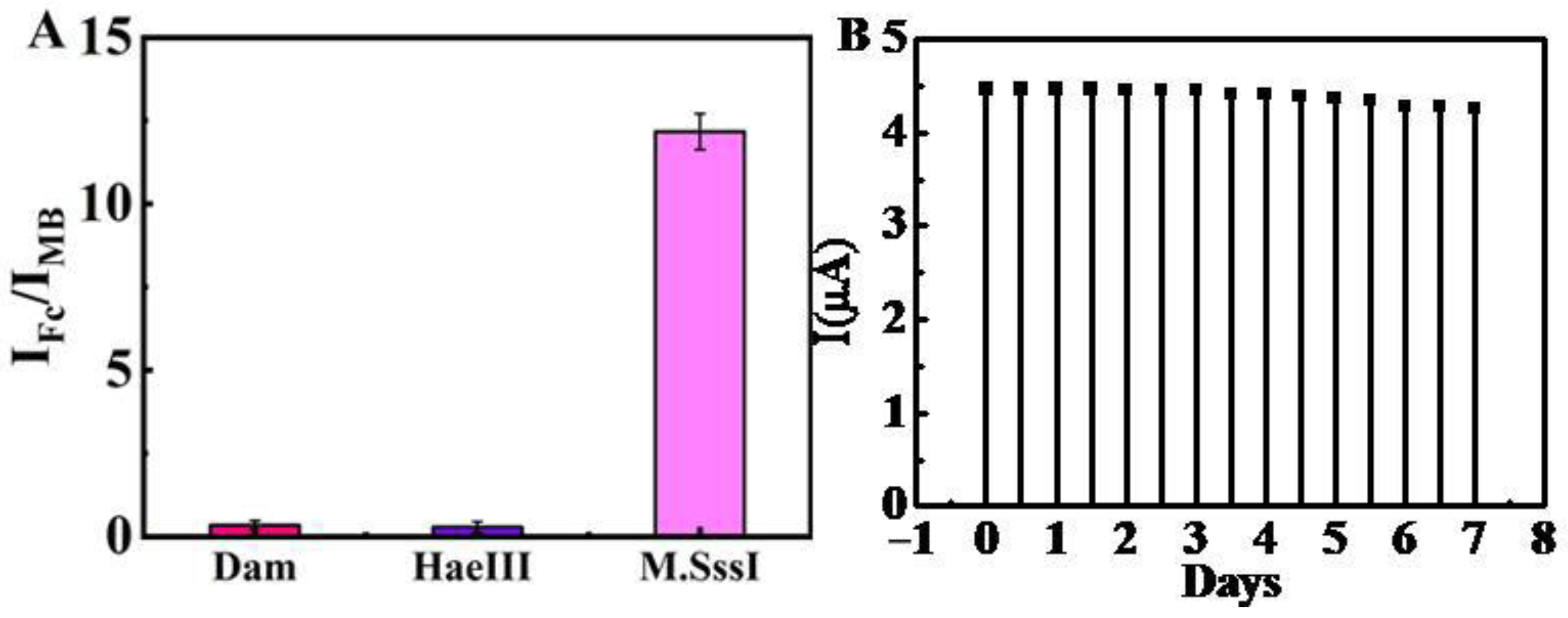
Reproducibility is a significant indicator for biosensors. Thus, six parallel measurements of the IFc/|IMB| under the same condition were carried out, and the relative standard deviation (RSD) was found to be 4.35% for M. SssI MTase of the same concentration, indicating that the presented biosensor has good reproducibility. To confirm the ability of the designed biosensor for M.SssI MTase, the selectivity of the proposed method was evaluated by using SWV analysis with a comparison of IFc/|IMB| values (Figure S3). For specificity assay, Dam MTase, together with HaeIII MTase enzyme were chosen as controls [52,53]. The sensor could selectively methylate the adenine residues in the double-strand DNA symmetric 5′-GATC-3′ sequence.
As expected, the values of IFc/|IMB| for Dam MTase and HaeIII MTase are only about 2.29% and 2.33% of that for M.SssI MTase at the same concentration (Figure 5A). These results indicate that the electrochemical biosensor developed in this work has good specificity for the detection of M.SssI MTase. The stability of the modified electrode has also been checked using SWV (Figure S4). The result is shown in Figure 5B, demonstrating that the response rate of the modified electrode could be maintained at about 95.4% after being refrigerated at 4 °C for 1 week.
3. Materials and Methods
3.1. Materials and Reagents
Anti-5-methylcytosineantibody was obtained from Abcam (Cambridge, UK). S-adenosylmethionine and M.SssI MTase was purchased from New England BioLabs (Ipswich, MA, USA). 1,1′-Ferrocenedicarboxylic acid (FcDA), N-hydroxysuccinimide (NHS), 1-ethyl-3-(3-dimethylaminopropyl)carbodiimide (EDC), phosphate-buffered saline (PBS) were obtained from Sigma-Aldrich (St. Louis, MO, USA). (Hydroxymethyl)aminomethane (Tris) was obtained from Aladdin (Shanghai, China). The oligonucleotides used in this study were obtained from Shanghai Sangon Biotechnology Co. (Shanghai, China) and the base sequences were as follows:
H1: 5′-CCGGGTAGAGCTCCCTTCAATCCAACATGATACCCGG-3′;
H2: 5′-SH-CCGGGTATCATGTTGGATTGAAGGGAGCTCTACCCGG-MB-3′
The stock buffer solutions used in this study are as follows: M.SssI: 10 mM Tris-HCl, 10 mM MgCl2, 50 mM NaCl (pH 7.4), and 30% glycerinum. Anti-5-methylcytosine antibody: 30% glycerinum and 0.01 M PBS (pH 7.4). M.SssI: 10 mM Tris-HCl, 50 mM NaCl, 10 mM MgCl2 (pH 7.4), and 30% glycerinum. DNA hybridization: 10 mM Tris-HCl, 1.0 M NaCl (pH 7.4), and 1.0 mM EDTA.
3.2. Apparatus
All electrochemical experiments were carried out using a CHI-660D electrochemical workstation (Chenhua Instrument Company of Shanghai, Shanghai, China). In the electrochemical measurements, a conventional three-component electrochemical cell was used, in which the bare electrode or the modified gold electrode was used as the working electrode, a saturated calomel electrode (SCE) was used as the reference electrode, and a platinum wire was used as the auxiliary electrode.
3.3. Preparation of FcDA Labeled Antibody (Fc-Ab)
Covalent conjugation of Fc-Ab was achieved via amide bond formation between an NH2 group of antibodies and a COOH group of FcDA using EDC as the coupling agent and NHS as the activator. Briefly, 20 mM EDC and 32 mM NHS were added to 10 mL of 0.1 M PBS buffer (pH 7.4) containing 2 mM FcDA. Herein, EDC activated the terminal carboxyl groups in FcDA to couple it with the primary amines of Ab. The active intermediate of the amine is stabilized by adding NHS to improve the efficiency of the EDC-activated coupling reaction. The reactant was shaken at room temperature for 2 h. After that, 2 mL of the obtained product was dispersed again in a mixture containing 10 μL 10 mg/mL AB in 4 mL PBS buffer (pH 7.4). The suspension was again shaken at room temperature for an additional 12 h and then incubated overnight at 4 °C to obtain Fc-Ab. Consequently, the ferrocene derivative, ferrocenedicarboxylic acid, was covalently bound to Ab to achieve Fc-Ab conjugates.
3.4. Methylation of Hairpin Probe
For the methylation of the hairpin probe, 0.5 μM H1 was incubated in the 200 μL M.SssI stock buffer, which contains 160 μM SAM and different concentrations of M.SssI MTase, under humid conditions at 37 °C for 2 h, and then rinsed three times with 10 mM Tris-HCl (pH 7.4).
3.5. Fabrication of the Ratiometric Electrochemical Biosensor
Before the modification, the surface of the bare GE with a diameter of 3 mm was polished into a mirror shape with alumina suspension, and then was continuously ultrasonically cleaned with 95% ethanol and ultrapure water for 5 min. Prior to attachment to the GE surface, 100 μL of 100 μM thiolated hairpin H2 was incubated with 0.1 μL of 100 mM tris(2-carboxyethyl)phosphine (TCEP) for 1 h to reduce disulfide bonds and subsequently diluted with phosphate buffer to 0.5 μM. Five microliters of thiolated H2 (0.5 μM) were dropped on the cleaned GE for 2 h at room temperature in the dark. During this process, the H2 was assembled onto the GE via the Au–S bond. After rinsing with ultrapure water, the H2-modified GE was incubated with 1.0 mM 6-mercaptohexanolin 10 mM Tris-HCl buffer (pH 7.4) for 1 h at room temperature to block the remaining bare region. Then 5 μL methylated H1 was dropped on the H2/GE. When H1 wasn’t methylated, we obtained an H1/H2/GE, then, after 2 h incubation, a methylated H1/H2/GE was obtained. Five microliters of Fc-Ab were further dropped on the surface of the methylated H1/H2/GE and incubated for 1 h. Finally, the resultant Fc-Ab/H1/H2/GE was washed with 0.1 M PBS buffer to remove nonspecific DNA adsorption. The reaction was terminated by thorough washing. The above process is shown in Scheme 1.
3.6. Measurement Procedure
The electrochemical performance of the electrode was studied by square wave voltammetry (SWV). The conditions were as follows: a potential of −0.80 to 0.80 V was scanned in 10 mM PBS buffer (pH 7.4), the step potential was 4 mV, the frequency was 25 Hz, and the amplitude was 25 mV. In Tris-HCl buffer solution (pH 7.4) containing 5 mM [Fe(CN)6]3−/4−, cyclic voltammetry was performed using a step potential of 0.05 V at a scan rate of 50 mV/s in the potential range of −0.6 to 0.2 V. Electrochemical impedance spectroscopy (EIS) was carried out in Tris-HCl (pH 7.4) containing 5 mM Fe(CN)63−/Fe(CN)64− and 0.1 M KCl in the frequency range from 0.1 Hz to 100 kHz with 5 mV as the amplitude at a polarization potential of 0.18 V.
4. Conclusions
In summary, on the basis of the dual-signaling electrochemical ratiometric method, a novel, practical, and sensitive electrochemical DNA biosensor has been developed. In this method, the target M.SssI MTase catalyzes the methylation of hairpin H1, which leads to more Fc approaching the electrode surface and MB moving far away from the electrode surface, so that the ratio of IFc/|IMB| changes, providing a significant enhancement of the system for M.SssI MTase detection down to 0.083 U/mL. Although our presented method by using a hairpin probe labeled with MB achieved excellent detection of M.SssI MTase, the tedious labeling process suggests a new direction of research by developing label-free probes for sensitive bio-analysis. This method is also selective towards M.SssI MTase against other control proteins. The developed electrochemical biosensor can provide better flexibility for target sequence selection and provide a potential platform for DNA detection in the field of DNA methylation.
Supplementary Materials
The following supporting information can be downloaded at: https://www.mdpi.com/article/10.3390/catal12111362/s1, Figure S1: Cyclic voltammetry curves of 10 mM PBS buffer (pH 7.4) at the Fc-Ab/H1/H2/GE, scan rate: 20 (a), 50 (b), 100 (c), 200 (d), 300 (e); Figure S2: Cyclic voltammograms of the different modified electrodes in 0.1 M Tris-HCl buffer solution containing 5 mM (1:1) [Fe(CN)6]3−/4−: the MB labeled Fc-Ab/H1/H2/GE (red curve), the MB unlabeled Fc-Ab/H1/H2/GE (black curve); Figure S3: SWV curves for the same concentration of Dam MTase HaeIII MTase and M.SssI MTase; Figure S4: SWV curves for the different storage time of the modified electrode in the same M.SssI MTase solution.
Author Contributions
Conceptualization and methodology, C.W. and Z.G.; data curation, Z.W.; formal analysis, investigation and visualization, H.W. and H.S.; software, K.Z. and F.L.; writing—original draft preparation, writing—review and editing, C.W., Z.G. and R.T.; All authors have read and agreed to the published version of the manuscript.
Funding
This research was funded by the Funded Project of Anhui Province Cultivate Outstanding Talent (gxbjZD2020090), the Natural Science Research Project of the Education Department of Anhui Province (KJ2019A0671, KJ2020A0736, KJ2020A0730, KJ2019A0669), the College Students’ Innovative Entrepreneurial Training Plan Program of China (202110379037, S202010379023).
Data Availability Statement
Not applicable.
Conflicts of Interest
The authors declare no conflict of interest.
References
- Holliday, R. The inheritance of epigenetic defects. Science 1987, 238, 163–170. [Google Scholar] [CrossRef]
- Egger, G.; Liang, G.; Aparicio, A.; Jones, P.A. Epigenetics in human disease and prospects for epigenetic therapy. Nature 2004, 429, 457–463. [Google Scholar] [CrossRef]
- Heithoff, D.M.; Sinsheimer, R.L.; Low, D.A.; Mahan, M.J. An essential role for DNA adenine methylation in bacterial virulence. Science 1999, 284, 967–970. [Google Scholar] [CrossRef]
- Robertson, K.D. DNA methylation and human disease. Nat. Rev. Genet. 2005, 6, 597–610. [Google Scholar] [CrossRef]
- Razin, A.; Cedar, H. DNA methylation and gene expression. Microbiol. Rev. 1991, 55, 451–458. [Google Scholar] [CrossRef]
- Li, E.; Beard, C.; Jaenisch, R. Role for DNA methylation in genomic imprinting. Nature 1993, 366, 362–365. [Google Scholar] [CrossRef]
- Csankovski, G.; Nagy, A.; Jaenisch, R.J. Synergism of Xist RNA, DNA methylation, and histone hypoacetylation in maintaining X chromosome inactivation. Cell Biol. 2001, 153, 773–784. [Google Scholar] [CrossRef]
- Branciamore, S.; Chen, Z.X.; Riggs, A.D.; Rodin, S.N. Rodin, CpG island clusters and pro-epigenetic selection for CpGs in protein-coding exons of HOX and other transcription factors. Proc. Natl. Acad. Sci. USA 2010, 107, 15485–15490. [Google Scholar] [CrossRef] [Green Version]
- Badran, A.H.; Furman, J.L.; Ma, A.S.; Comi, T.J.; Porter, J.; Ghosh, R.I. Global CpG Methylation Status Native DNA Utilizing Bipartite Split-Luciferase Sensor. Anal. Chem. 2011, 83, 7151–7157. [Google Scholar] [CrossRef] [Green Version]
- Robertson, K.D. DNA methylation, methyltransferases, and cancer. Oncogene 2001, 20, 3139–3155. [Google Scholar] [CrossRef]
- Lyko, F.; Brown, R.J. DNA methyltransferase inhibitors and the development of epigenetic cancer therapies. Natl. Cancer Inst. 2005, 97, 1498–1506. [Google Scholar] [CrossRef] [Green Version]
- Choy, J.S.; Wei, S.; Lee, J.Y.; Tan, S.; Chu, S.; Lee, T.H. DNA methylation increases nucleosome compaction and rigidity. J. Am. Chem. Soc. 2010, 132, 1782–1783. [Google Scholar] [CrossRef] [Green Version]
- Frigola, J.; Song, J.; Stirzaker, C.; Hinshelwood, R.A.; Peinado, M.A.; Clark, S.J. Epigenetic remodeling in colorectal cancer results in coordinate gene suppression across an entire chromosome band. Nat. Genet. 2006, 38, 540–549. [Google Scholar] [CrossRef]
- Lopez Torres, A.; Yanez Barrientos, E.; Wrobel, K. Selective Derivatization of Cytosine and Methylcytosine Moieties with 2-Bromoacetophenone for Submicrogram DNA Methylation Analysis by Reversed Phase HPLC with Spectrofluorimetric Detection. Anal. Chem. 2011, 83, 7999–8005. [Google Scholar] [CrossRef]
- Lyko, F.; Ramsahoye, B.H.; Jaenisch, R. DNA methylation in Drosophila melanogaster. Nature 2000, 408, 538–540. [Google Scholar] [CrossRef]
- McLaughlin, L.W.; Benseler, F.; Graeser, E.; Piel, N.; Scholtissek, S. Effects of functional group changes in the EcoRI recognition site on the cleavage reaction catalyzed by the endonuclease. Biochemistry 1987, 26, 7238–7245. [Google Scholar] [CrossRef]
- Li, Z.M.; Zhang, X.; Pi, T.; Bu, J.; Deng, R.H.; Chi, B.Z.; Zheng, X.J. Colorimetric determination of the activity of methyltransferase based on nicking enzyme amplification and the use of gold nanoparticles conjugated to graphene oxide. Microchim. Acta 2019, 186, 594. [Google Scholar] [CrossRef]
- Kermani, H.A.; Hosseini, M.; Miti, A.; Dadmehr, M.; Zuccheri, G.; Hosseinkhani, S.; Ganjali, M.R. A colorimetric assay of DNA methyltransferase activity based on peroxidase mimicking of DNA template Ag/Pt bimetallic nanoclusters. Anal. Bioanal. Chem. 2018, 410, 4943–4952. [Google Scholar] [CrossRef]
- Bi, S.; Zhao, T.; Luo, B.; Zhu, J.J. Hybridization chain reaction-based branched rolling circle amplification for chemiluminescence detection of DNA methylation. Chem. Commun. 2013, 49, 6906–6908. [Google Scholar] [CrossRef]
- Wu, M.; Zhang, M.; Fan, Z.; Qin, X.; Zhu, X.; Ji, H.; Qin, Y.; Wang, Q.; Wu, L. Ultrasensitive DNA methyltransferase activity sensing and inhibitor evaluation with highly photostable upconversion nanoparticle transducer. Microchim. Acta 2021, 188, 169. [Google Scholar] [CrossRef]
- Chen, J.; Wang, Y.; Li, W.Y.; Zhou, H.P.; Li, Y.X.; Yu, C. Nucleic Acid-Induced Tetraphenylethene Probe Noncovalent Self-Assembly and the Superquenching of Aggregation-Induced Emission. Anal. Chem. 2014, 86, 9866–9872. [Google Scholar] [CrossRef]
- Zhang, X.; Zhong, X.-L.; Jiang, W.-W.; Zeng, S.-H.; Pi, T.; Zheng, X.-J.; Li, Z.-M. Fluorescence-based Polymerase Amplification for the Sensitive Detection of DNA Methyltransferase Activity. Anal. Sci. 2018, 34, 959–964. [Google Scholar] [CrossRef] [Green Version]
- Quach, Q.H.; Chung, B.H. A signal-on fluorescent assay for DNA methyltransferase activity using a methylation-resistant endonuclease. Analyst 2014, 139, 2674–2677. [Google Scholar] [CrossRef]
- Wang, Y.; Chen, J.; Chen, Y.; Li, W.Y.; Yu, C. Polymer-Induced Perylene Probe Excimer Formation and Selective Sensing of DNA Methyltransferase Activity through the Monomer–Excimer Transition. Anal. Chem. 2014, 86, 4371–4378. [Google Scholar] [CrossRef]
- Hu, H.; Zhou, F.; Wang, B.J.; Chang, X.; Dai, T.Y.; Tian, R.F.; Wan, Y.F.; Wang, X.Y.; Wang, G.F. Autonomous operation of 3D DNA walkers in living cells for microRNA imaging. Nanoscale 2021, 13, 1863–1868. [Google Scholar] [CrossRef]
- Shen, Q.M.; Han, L.; Fan, G.C.; Abdel-Halim, E.S.; Jiang, L.P.; Zhu, J.J. Highly sensitive photoelectrochemical assay for DNA methyltransferase activity and inhibitor screening by exciton energy transfer coupled with enzyme cleavage biosensing strategy. Biosens. Bioelectron. 2015, 64, 449–455. [Google Scholar] [CrossRef]
- Zhou, Y.L.; Xu, Z.N.; Wang, M.; Sun, B.; Yin, H.S.; Ai, S.Y. DNA methyltransferase activity assay based on visible light-activated photoelectrochemical biosensor. Biosens. Bioelectron. 2014, 53, 263–267. [Google Scholar] [CrossRef]
- Wang, Y.; Fang, X.; Yin, H.S.; Zhou, Y.L.; Yang, Y.; Ai, S.Y. Photoelectrochemical immunosensor for methylated RNA detection based on WS2 and poly(U) polymerase–triggered signal amplification. Microchim. Acta 2020, 187, 596. [Google Scholar] [CrossRef]
- Deng, H.M.; Yang, X.J.; Yeo, S.P.X.; Gao, Z.Q. Highly Sensitive Electrochemical Methyltransferase Activity Assay. Anal. Chem. 2014, 86, 2117–2123. [Google Scholar] [CrossRef]
- Wang, C.; Guo, Z.H.; Zhang, L.; Zhang, N.; Zhang, K.Y.; Xu, J.G.; Wang, H.Y.; Shi, H.W.; Qin, M.; Ren, L. DNA based signal amplified molecularly imprinted polymer electrochemical sensor for multiplex detection. RSC Adv. 2016, 6, 49597–49603. [Google Scholar] [CrossRef]
- Li, W.; Wu, P.; Zhang, H.; Cai, C.X. Signal Amplification of Graphene Oxide Combining with Restriction Endonuclease for Site-Specific Determination of DNA Methylation and Assay of Methyltransferase Activity. Anal. Chem. 2012, 84, 7583–7590. [Google Scholar] [CrossRef]
- Bhattacharjee, R.; Moriam, S.; Umer, M.; Nguyen, N.T.; Shiddiky, M.J.A. DNA methylation detection: Recent developments in bisulfite free electrochemical and optical approaches. Analyst 2018, 143, 4802–4818. [Google Scholar] [CrossRef]
- Muren, N.B.; Barton, J.K. Electrochemical Assay for the Signal-On Detection of Human DNA Methyltransferase Activity. J. Am. Chem. Soc. 2013, 135, 16632–16640. [Google Scholar] [CrossRef] [Green Version]
- Wang, G.F.; Wan, J.; Zhang, X.J. TTE DNA–Cu NPs: Enhanced fluorescence and application in a target DNA triggered dual-cycle amplification biosensor. Chem. Commun. 2017, 53, 5629–5632. [Google Scholar] [CrossRef]
- Wang, M.; Xu, Z.N.; Chen, L.J.; Yin, H.S.; Ai, S.Y. Electrochemical Immunosensing Platform for DNA Methyltransferase Activity Analysis and Inhibitor Screening. Anal. Chem. 2012, 84, 9072–9078. [Google Scholar] [CrossRef]
- Lu, L.; Liu, B.; Leng, J.; Ma, X. Electrochemical determination of the activity of DNA methyltransferase based on the methyl binding domain protein and a customized modular detector. Microchim. Acta 2019, 186, 229. [Google Scholar] [CrossRef]
- Zhang, Y.; Hao, L.J.; Zhao, Z.; Yang, X.Y.; Wang, L.; Liu, S.F. Immuno-DNA binding directed template-free DNA extension and enzyme catalysis for sensitive electrochemical DNA methyltransferase activity assay and inhibitor screening. Analyst 2020, 145, 3064–3072. [Google Scholar] [CrossRef]
- Ricci, F.; Plaxco, K.W. E-DNA sensors for convenient, label-free electrochemical detection of hybridization. Microchim. Acta 2008, 163, 149–155. [Google Scholar] [CrossRef]
- Bakker, E.; Qin, Y. Electrochemical Sensors. Anal. Chem. 2006, 78, 3965–3984. [Google Scholar] [CrossRef] [Green Version]
- Yu, Z.G.; Lai, R.Y. A reagentless and reusable electrochemical DNA sensor based on target hybridization-induced stem-loop probe formation. Chem. Commun. 2012, 48, 10523–10525. [Google Scholar] [CrossRef]
- Cheng, Y.; Huang, Y.; Lei, J.P.; Zhang, L.; Ju, H.X. Design and Biosensing of Mg2+ Dependent DNAzyme Triggered Ratiometric Electrochemiluminescence. Anal. Chem. 2014, 86, 5158–5163. [Google Scholar] [CrossRef]
- Liu, L.; Yang, Q.H.; Lei, J.P.; Xu, N.; Ju, H.X. DNA-regulated silver nanoclusters for label-free ratiometric fluorescence detection of DNA. Chem. Commun. 2014, 50, 13698–13701. [Google Scholar] [CrossRef]
- Zhang, X.X.; Xiao, K.Y.; Cheng, L.W.; Chen, H.; Liu, B.H.; Zhang, S.; Kong, J.L. Visual and Highly Sensitive Detection of Cancer Cells by a Colorimetric Aptasensor Based on Cell-Triggered Cyclic Enzymatic Signal Amplification. Anal. Chem. 2014, 86, 5567–5572. [Google Scholar] [CrossRef]
- Yu, P.; Zhou, J.W.; Wu, L.; Xiong, E.H.; Zhang, X.H.; Chen, J.H. A ratiometric electrochemical aptasensor for sensitive detection of protein based on aptamer–target–aptamer sandwich structure. J. Electroanal. Chem. 2014, 732, 61–65. [Google Scholar] [CrossRef]
- Du, Y.; Lim, B.J.; Li, B.L.; Jiang, Y.S.; Sessler, J.L.; Ellington, A.D. Ellington, Reagentless, Ratiometric Electrochemical DNA Sensors with Improved Robustness and Reproducibility. Anal. Chem. 2014, 86, 8010–8016. [Google Scholar] [CrossRef]
- Ren, K.W.; Wu, J.; Yan, F.; Ju, H.X. Ratiometric electrochemical proximity assay for sensitive one-step protein detection. Sci. Rep. 2014, 4, 4360–4365. [Google Scholar] [CrossRef] [Green Version]
- Ren, K.W.; Wu, J.; Yan, F.; Zhang, Y.; Ju, H.X. Immunoreaction-triggered DNA assembly for one-step sensitive ratiometric electrochemical biosensing of protein biomarker. Biosens. Bioelectron. 2015, 66, 345–349. [Google Scholar] [CrossRef]
- Xiong, E.H.; Wu, L.; Zhou, J.W.; Yu, P.; Zhang, X.H.; Chen, J.H. A ratiometric electrochemical biosensor for sensitive detection of Hg2+ based on thymine–Hg2+–thymine structure. Anal. Chim. Acta 2015, 853, 242–248. [Google Scholar] [CrossRef]
- Wu, L.; Zhang, X.H.; Liu, W.; Xiong, E.H.; Chen, J.H. Sensitive Electrochemical Aptasensor by Coupling “Signal-on’’ and “Signal-off’’ Strategies. Anal. Chem. 2013, 85, 8397–8402. [Google Scholar] [CrossRef]
- Grabowska, I.; Malecka, K.; Stachyra, A.; Góra-Sochacka, A.; Sirko, A.; Zagórski-Ostoja Radecka, W.H.; Radecki, J. Single Electrode Genosensor for Simultaneous Determination of Sequences Encoding Hemagglutinin and Neuraminidase of Avian Influenza Virus Type H5N1. Anal. Chem. 2013, 85, 10167–10173. [Google Scholar] [CrossRef]
- Yang, W.W.; Lai, R.Y. Integration of two different sensing modes in an electrochemical DNA sensor for approximation of target mismatch location. Electrochem. Commun. 2011, 13, 989–992. [Google Scholar] [CrossRef]
- Miodek, A.; Regan, E.M.; Bhalla, N.; Hopkins, N.A.E.; Goodchild, S.A.; Estrela, P. Optimisation and Characterisation of Anti-Fouling Ternary SAM Layers for Impedance-Based Aptasensors. Sensors 2015, 15, 25015–25032. [Google Scholar] [CrossRef] [PubMed] [Green Version]
- Luo, X.L.; Xu, Q.; James, T.; Davis, J.J. Redox and Label-Free Array Detection of Protein Markers in Human Serum. Anal. Chem. 2014, 86, 5553–5558. [Google Scholar] [CrossRef] [PubMed]
- Žutić, V.; Svetličić, V.; Clavilier, J.; Chevalet, J. Supramolecular phenomena in organic redox films at electrodes: Part II. The methylene blue/leucomethylene blue redox couple at the gold electrode. J. Electroanal. Chem. Interfacial Electrochem. 1987, 219, 183–195. [Google Scholar] [CrossRef]
- Zhan, R.; Song, S.; Liu, Y.; Dong, S. Mechanisms of methylene blue electrode processes studied by in situ electron paramagnetic resonance and ultraviolet–visible spectroelectrochemistry. J. Chem. Soc. Faraday FTrans. 1990, 86, 3125–3127. [Google Scholar] [CrossRef]
- Ahmad, H.M.N.; Dutta, G.; Csoros, J.; Si, B.; Yang, R.; Halpern, J.M.; Seitz, W.R.; Song, E. Stimuli-Responsive Templated Polymer as a Target Receptor for a Conformation-Based Electrochemical Sensing Platform. ACS Appl. Polym. Mater. 2021, 3, 329–341. [Google Scholar] [CrossRef]
- Cao, Y.; Zhu, S.; Yu, J.C.; Zhu, X.J.; Yin, Y.M.; Li, G.X. Protein Detection Based on Small Molecule-Linked DNA. Anal. Chem. 2012, 84, 4314–4320. [Google Scholar] [CrossRef]
- Koo, K.M.; Carrascosa, L.G.; Shiddiky, M.J.; Trau, M. Poly(A) extensions of miRNAs for amplification-free electrochemical detection on screen-printed gold electrodes. Anal. Chem. 2016, 88, 2000–2005. [Google Scholar] [CrossRef] [Green Version]
- Ferapontova, E.E. Hybridization Biosensors Relying on Electrical Properties of Nucleic Acids. Eletroanalysis 2017, 29, 6–13. [Google Scholar] [CrossRef]
Scheme 1.
Schematic illustration of the ratiometric electrochemical biosensor for DNA M. SssI detection.
Scheme 1.
Schematic illustration of the ratiometric electrochemical biosensor for DNA M. SssI detection.
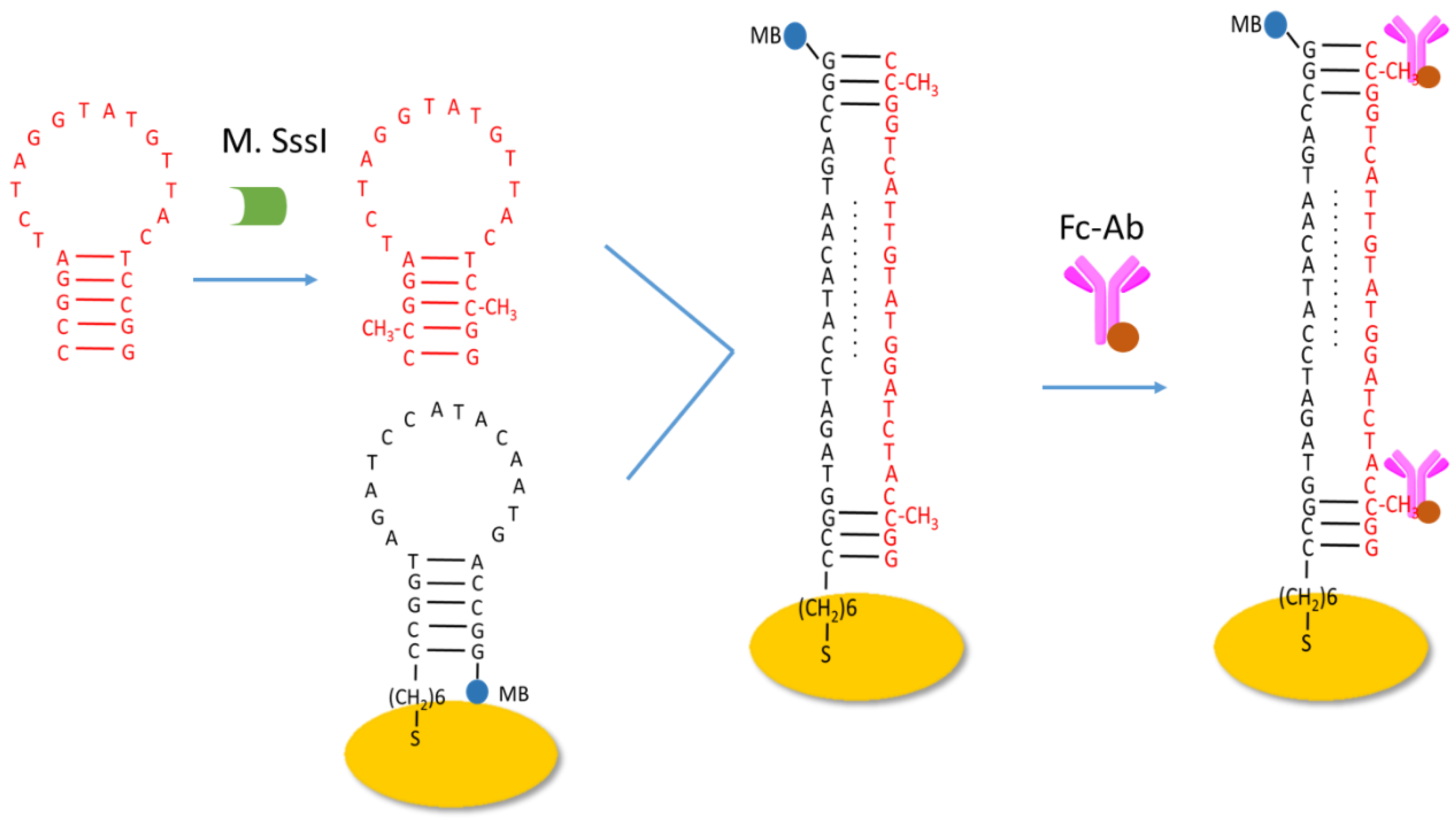
Figure 1.
SWV responses of the developed biosensor at different experimental conditions: (a) H1/H2/GE, (b) methylated H1/H2/GE, and (c) Fc-Ab/H1/H2/GE.
Figure 1.
SWV responses of the developed biosensor at different experimental conditions: (a) H1/H2/GE, (b) methylated H1/H2/GE, and (c) Fc-Ab/H1/H2/GE.
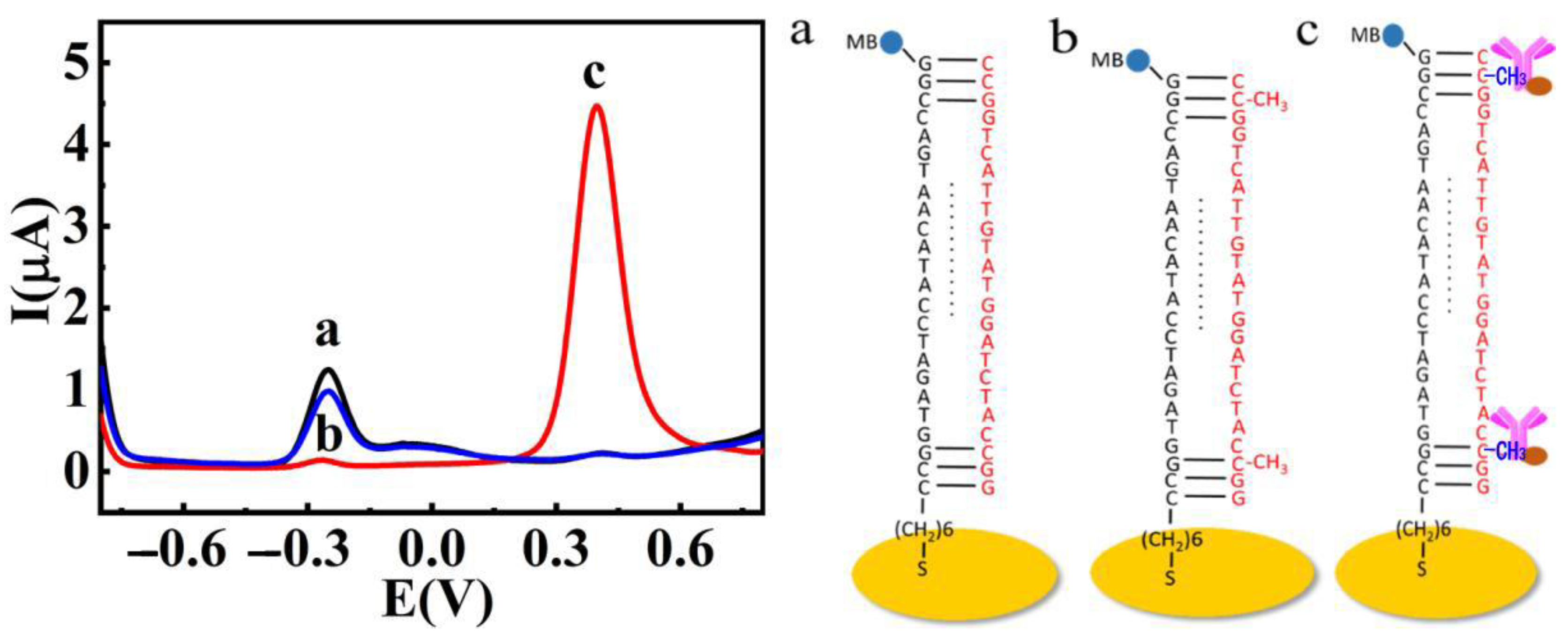
Figure 2.
Electrochemical impedance spectra (Nyquist plots) (A) and Cyclic voltammograms (B) of the different modified electrodes in 0.1 M Tris-HCl buffer solution containing 5 mM (1:1) [Fe(CN)6]3−/4−: (a) Bare GE, (b) H2/GE, (c) H1/H2/GE, and (d) Fc-Ab/H1/H2/GE.
Figure 2.
Electrochemical impedance spectra (Nyquist plots) (A) and Cyclic voltammograms (B) of the different modified electrodes in 0.1 M Tris-HCl buffer solution containing 5 mM (1:1) [Fe(CN)6]3−/4−: (a) Bare GE, (b) H2/GE, (c) H1/H2/GE, and (d) Fc-Ab/H1/H2/GE.
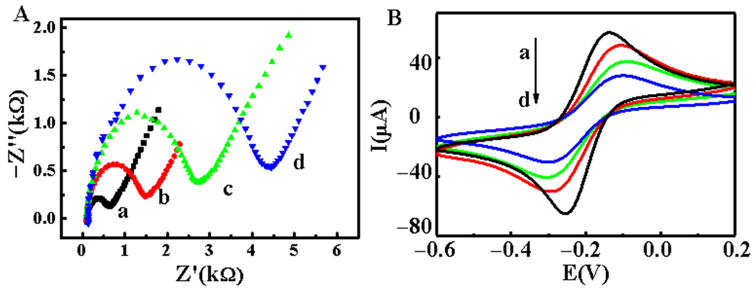
Figure 3.
Effects of the temperature (A), incubation time (B), and pH (C) in the presence of 5 μL Fc-Ab.
Figure 3.
Effects of the temperature (A), incubation time (B), and pH (C) in the presence of 5 μL Fc-Ab.

Figure 4.
(A) SWV curves for the different concentrations of M.SssI MTase. The concentrations are (from a to h) 0 U/mL, 0.25 U/mL, 0.375 U/mL, 0.5 U/mL, 1 U/mL, 10 U/mL, 20 U/mL, and 40 U/mL. Dependence of IMB (B) and IFc (C) on M.SssI MTase concentration. Insets in (B,C) are the linear relationship between the logarithmic function of IMB and IFc, respectively. (D) Dependence of IFc/|IMB| on M.SssI MTase concentration. Insets show the linear fit plots of IFc, or IMB, and IFc/|IMB| as a function of the M.SssI MTase concentration. Insets are the linear relationship between IFc/|IMB| and the concentration of M.SssI MTase ranging from 0 to 2 U/mL.
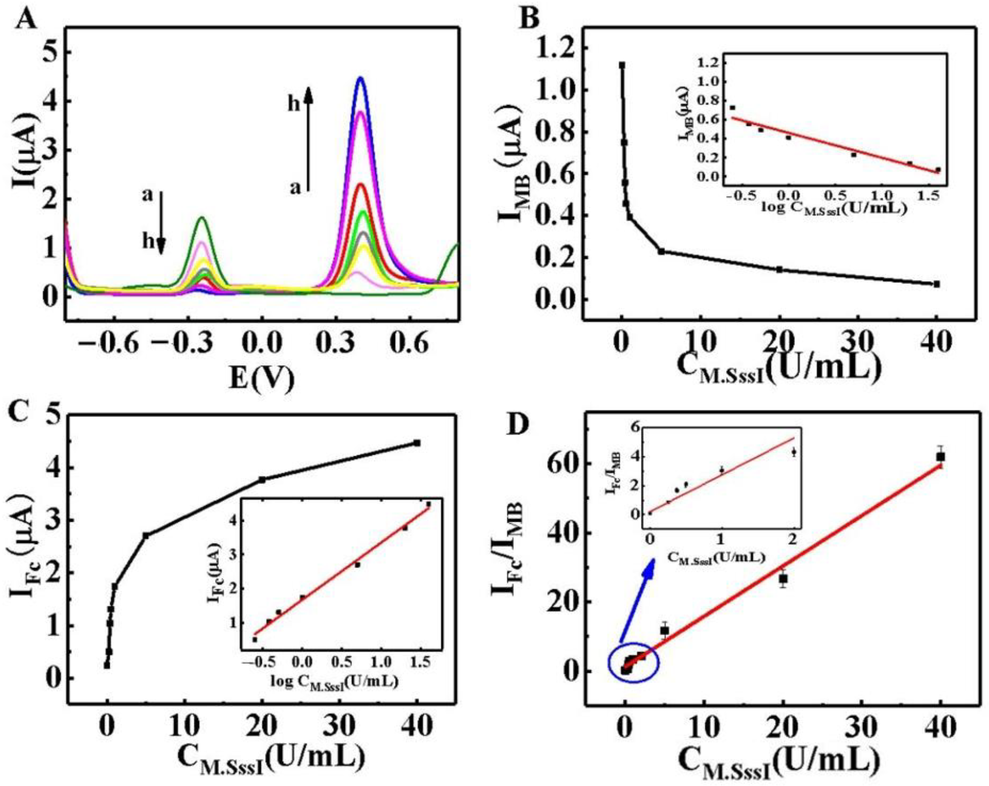
Figure 5.
(A) Selectivity of the system for M.SssI MTase analysis; Error bars represent standard deviations of three parallel experiments. (B) The time dependence of the signal change of the proposed method in the 7 days measurement.
Figure 5.
(A) Selectivity of the system for M.SssI MTase analysis; Error bars represent standard deviations of three parallel experiments. (B) The time dependence of the signal change of the proposed method in the 7 days measurement.

Table 1.
A comparison of detection performance in the current work with previously reported methods for DNA MTase.
Table 1.
A comparison of detection performance in the current work with previously reported methods for DNA MTase.
| Strategy | Detection Method | Signal | Detection Limit (U/mL) | Linear Range (U/mL) | Reference |
|---|---|---|---|---|---|
| HRP mimicking DNA zyme | Colorimetry | Signal on | 0.4 | 0.8–24 | [17] |
| HRP mimicking DNA zyme | Colorimetry | Signal on | 6 | 6–100 | [18] |
| RCA | Chemiluminescence | Signal on | 0.52 | 1–10 | [19] |
| Nicking enzyme-assisted signal amplification | Fluorescence | Signal on | 0.06 | 0.1–4 | [20] |
| without any amplification | Electrochemistry | Signal off | 0.18 | 0.25–10 | [36] |
| This work | Electrochemistry | Dual signal | 0.083 | 0–40 |
Publisher’s Note: MDPI stays neutral with regard to jurisdictional claims in published maps and institutional affiliations. |
© 2022 by the authors. Licensee MDPI, Basel, Switzerland. This article is an open access article distributed under the terms and conditions of the Creative Commons Attribution (CC BY) license (https://creativecommons.org/licenses/by/4.0/).
Share and Cite
MDPI and ACS Style
Wang, C.; Guo, Z.; Tian, R.; Zhang, K.; Wang, H.; Li, F.; Shi, H.; Wang, Z. Ratiometric Electrochemical Biosensing of Methyltransferase Activity. Catalysts 2022, 12, 1362. https://doi.org/10.3390/catal12111362
AMA Style
Wang C, Guo Z, Tian R, Zhang K, Wang H, Li F, Shi H, Wang Z. Ratiometric Electrochemical Biosensing of Methyltransferase Activity. Catalysts. 2022; 12(11):1362. https://doi.org/10.3390/catal12111362
Chicago/Turabian StyleWang, Cong, Zhihua Guo, Ruifen Tian, Keying Zhang, Hongyan Wang, Fajun Li, Hongwei Shi, and Zhicheng Wang. 2022. "Ratiometric Electrochemical Biosensing of Methyltransferase Activity" Catalysts 12, no. 11: 1362. https://doi.org/10.3390/catal12111362
Note that from the first issue of 2016, this journal uses article numbers instead of page numbers. See further details here.