
Photocatalytic Bacterial Inactivation of Acinetobacter baumannli on Cu/TiO2/Diatomite
by
 ,
,
Xiaolin Xu
1,†,
Yacong Yang
1,2,†,
Yingchun Miao
1,*,
Kaiquan Liu
1,
Fujian Lv
1,*,
Liping Zhou
1,
Xuqi Tang
1,
Yanmi Liu
1 and
Xinchun Guo
1 1
College of Chemistry and Environmental Science, Qujing Nomal University, Qujing 655011, China
2
The Education Ministry Key Laboratory of Resource Chemistry, Shanghai Key Laboratory of Rare Earth Functional Materials, Shanghai Normal University, Shanghai 200234, China
*
Authors to whom correspondence should be addressed.
†
These authors contributed equally to this work.
Catalysts 2022, 12(10), 1217; https://doi.org/10.3390/catal12101217
Submission received: 12 September 2022
/
Revised: 6 October 2022
/
Accepted: 7 October 2022
/
Published: 12 October 2022
(This article belongs to the Special Issue Recent Progress of Catalysis in “Dual Carbon Targets”)
Abstract
:Cu4Ti2O/TiO2/diatomite with double interface Cu4Ti2O/TiO2 and rutile/anatase heterojunction were fabricated, which demonstrated good antibacterial activity (100%) against Acinetobacter baumannii. Cu/TiO2/diatomite prepared under optimum preparation conditions (added diatomite, 0.005 g; Cu, 0.005 g; reaction temperature, 180 °C; reaction time, 8 h) exhibited high antibacterial activity (100%) against A. baumannii. For the Cu/TiO2/diatomite powders, their structural, compositional, optical and morphological traits were characterized by XRD, SEM, TEM, XPS, BET, FTIR, Mapping, and DRS. It was shown that Cu/TiO2/diatomite under optimum conditions consisted of the double interface Cu4Ti2O/TiO2 and rutile/anatase heterojunction with the narrowest band gap and largest BET surface area, pore size, and pore volume. Then, it could exhibit the best photocatalytic activity.
1. Introduction
Given the fast worldwide spread of multidrug-resistant lineages of A. baumannii (Acinetobacter baumannii), a high-risk Gram-negative pathogen, it has become one of the bacteria with strongest antibiotic resistance on a global scale [1,2]. Infection with A. baumannii manifests itself as varying diseases, including nosocomial and community-acquired pneumonia, wound and burn infections, bloodstream infections, infections of skin and soft tissue, urinary tract infections, meningitis, as well as endocarditis. Management of nosocomial meningitis is especially utterly challenging, which is an unpopular neurosurgical complication induced by A. baumannii that possesses broad drug resistance [3,4]. Due to the significant increase in the antimicrobial resistance of Acinetobacter baumannii, many new technologies to block the progression of infection were strongly needed. With “Dual Carbon” targets becoming catchphrases, the photocatalytic antibacterial technology has been widely studied for its simple operation, no secondary pollution as well as the ability to convert solar energy into chemical energy for the mineralization of organic matter and bacteria. AgVO3/AgI graphene microtube displayed excellent sterilization against E. coli (Escherichia coli), with a bacteriostatic efficiency of 100% [5]. Inactivation activity of an all-organic composite PDINH (photocatalytic nanomaterial C3N4/perylene-3,4,9,10-tetracarboxylic diimide) heterostructure was outstanding against the Gram-positive and -negative bacteria [6]. Compared with the properties of many photocatalytic antibacterial agents, TiO2 has more excellent properties. Researchers have been modifying the energy levels by organic and inorganic compounds, various cations and anions, etc., causing the narrowing of TiO2 band gap or photoinduced charge transfer to achieve high antibacterial performance.
The photocatalytic sterilization behavior of Cu-TiO2 nanofibers was carried out in viral/bacterial systems and viruses under visible light [7]. During the degradation of pollutants and inactivation of bacteria in aqueous phase, a photocatalytic reaction appeared in the TiO2/CuxOy nanotube arrays [8]. It was discovered that the toxicity of films containing Cu ions was much higher than that of Ag, Zn, Co, Al and Hg ions, and copper and copper alloys have a considerable cost advantage over precious metals such as silver. Therefore, TiO2 has been modified by Cu ion to improve its absorption of visible light and inhibit the photoelectron–hole recombination, which was an active research direction in order to prepare new, stable antibacterial materials [9]. The photocatalytic bactericidal action was strengthened against A. baumannii by the hybrid Cu2O/TiO2 nanocomposites [10]. Although a series of copper compounds/TiO2 catalysts with visible light response have been prepared, the antibacterial efficiency could be significantly improved. The above-mentioned improvements were limited by changing the energy level structure and shape control of the photocatalyst, etc. To enhance the bactericidal action of copper compound/TiO2 photocatalyst in practical application, the effects of the photocatalyst’s morphology, composition, dispersion and combination form on its antibacterial activity should be explored more in-depth, which could be crucially conducive to the development and control of copper compound/TiO2 photocatalyst [11,12]. Recently, we have adjusted the shape of the porous catalyst and the construction of a novel heterostructure to improve its photocatalytic activity. A novel hydrophilic copper/titanium dioxide composite composed of Cu2Cl(OH)3, Cu2+1O and anatase TiO2 was successfully prepared, and the relationship between the surface micromorphology, crystalline phase and the antibacterial activity of E. coli was investigated [13].
TiO2 exists in the following 3 polymorphs: anatase, brookite (orthorhombic system), as well as rutile (tetragonal system). Most research concerning the synthesis of TiO2-supported Cu compounds for photocatalysis discovered the formation of a mere anatase phase [14], whose photocatalytic potential was best compared to the remaining TiO2 crystalline phase. Recently, some studies demonstrated the better photocatalytic potential of hybrid phases (anatase/brookite, rutile/anatase) compared to the pure rutile or anatase phase because of the interface heterojunction of rutile/anatase, ultimately leading to the electron (e−)–hole(h+) recombination reduction [15,16,17]. Under the same concept, diatomite was selected as a porous and natural mineral support for the compound of porous TiO2 and copper compounds, not only because the diatomaceous was composed of a porous diatomite framework with a Si-O tetrahedral network that was high in specific surface area and porosity and low in density [18], but also because the diatomite could control the rutile/anatase phase ratio in TiO2. In the present study, the “super bacteria” Acinetobacter baumannii was used as the probe target. The different composites materials Cu/TiO2/diatomite comprising varying amounts of diatomite, Cu compound and anatase/rutile TiO2 were fabricated, so that the photocatalytic potential for the removal of Acinetobacter baumannii was optimizable.
2. Results and Discussion
2.1. XRD Analysis
The wide angle XRD spectra for pure diatomite, TiO2/diatomite, Cu/TiO2 and Cu/TiO2/diatomite were shown in Figure 1a. The diatomite was amorphous, and the broad peak of its diffraction pattern centered at 2θ = 17–28° was assigned to amorphous SiO2 [3]. In the Cu/TiO2 diffraction pattern, the peaks at 25.3° and 48.0° corresponded to the (101) and (200) anatase facets of TiO2 (JCPDS No. 21-1272) and the peak at 27.4° was consistence with the (110) facet of rutile TiO2 (JCPDS No. 21-1276). In the TiO2/diatomite diffraction pattern, the peaks at 25.3°, 37.8°, 48.0°, 53.8°, and 55.1° corresponded to the (101), (004), (200), (105) and (211) facets of anatase TiO2 (JCPDS No. 21-1272) and the peak at 27.4°, 62.9°, 69.0°, and 74.4° corresponded to the (110), (002), (301), and (320) facet of rutile TiO2/diatomite (JCPDS No. 21-1276). For Cu/TiO2/diatomite, five strong diffraction peaks were noted at 25.3°, 37.8°, 48.0°, 53.8° and 55.1°, which corresponded to the (101), (004), (200), (105) and (211) planes for the TiO2 anatase phase (JCPDS No. 21-1272), while the rutile phase evidenced by only two peaks with veer low intensity at 62.9° and 74.4°. For Cu/TiO2/diatomite and TiO2/diatomite, it was not shown that the broad amorphous peak of amorphous SiO2 due to little diatomite. According to the above result, TiO2 were the mixed phases (rutile/anatase) in TiO2/diatomite, Cu/TiO2, and Cu/TiO2/diatomite, and these catalysts could exhibit higher photocatalytic potential compared to the pure rutile or anatase phase because of the interface heterojunction of rutile/anatase, ultimately leading to the electron (e−)-hole(h+) recombination reduction [16]. No characteristic peaks of Cu metal or oxide phases were found in the Cu/TiO2 and Cu/TiO2/diatomite diffraction patterns, which indicated that the primary crystalline phase in the composite samples was TiO2. Therefore, in line with the prior observational report [19], Cu was highly dispersed on the TiO2 surface or existed in an amorphous phase. Compared with the other three samples, the Cu/TiO2/diatomite sample had high intensity and its diffraction peaks were narrower than those of the other samples, indicating the formation of larger TiO2 crystallites and a higher degree of crystallinity. These were important factors that can affect photocatalytic efficiency [20]. On the other hand, the crystallite size for Cu/TiO2/diatomite sample was about 0.48 µm, which were calculated via Scherrer equation based on (1 0 1) principal diffraction peak at 2θ = 25.3°.
To clarify Cu’s crystal morphology, we prepared Cu/TiO2/diatomite-30 catalyst, and the wide-angle XRD patterns of Cu/TiO2/diatomite and Cu/TiO2/diatomite-30 were shown in Figure 1b. In the Cu/TiO2/diatomite-30 diffraction pattern, the peaks at 35.0° and 60.9° corresponded to the (101) and (110) anatase facets of Cu4Ti2O (JCPDS No. 21-0199). Additionally, by comparing these two composites, the Cu/TiO2/diatomite-30‘s characteristic diffraction peak of TiO2 is stronger than Cu/TiO2/diatomite.
2.2. BET Analysis
Figure 2 displays the pore structural parameter and BET-specific surface area statistics for the Cu/TiO2/diatomite and Cu/TiO2 composites, as well as the pure diatomite based on the N2 adsorption−desorption approach. As shown in Figure 2a, the textural analysis revealed that Cu/TiO2 depicted a type I isotherm (IUPAC) characteristic of a microporous material. The sample of Cu/TiO2/diatomite and pure diatomite exhibited a typical type IV isotherm with an H3 hysteresis loop that was characteristic of a mesoporous material in accordance with IUPAC classification. Compare with pure diatomite, the adsorption branch of Cu/TiO2/diatomite rises slowly when the pressure scope was low within P/P0 < 0.8, while it could exhibit a sharp rise when P/P0 > 0.8. The results showed that Cu/TiO2/diatomite had more dense mesopores [5]. According to the reference, the reaction system used diatomite as the carrier, and the added TiO2 also has the effect of creating pores [6], increasing the pore capacity. Figure 2b depicts the distributions of BJH pore sizes for the pure diatomite, Cu/TiO2, as well as Cu/TiO2/diatomite. The cumulative pore volume of Cu/TiO2 as the pore diameter increases, the pore volume hardly changed. The cumulative pore volume of diatomite was based on the pore diameter of 2.5 nm as the watershed, presenting a trend of increasing first and then decreasing. However, the cumulative pore volume of Cu/TiO2/diatomite was based on the pore diameter of 15.0 nm as the watershed, showing an initial increase and subsequent decrease trend. The result was consistent with to the reference [6]. The reaction system was used diatomite as the carrier, and the added TiO2 also has the effect of creating pores, which can increase the pore capacity. The above results were also consistent with the pore structure parameters for the pure diatomite, Cu/TiO2/diatomite and Cu/TiO2 detailed in Table 1. Compared to the Cu/TiO2 and pure diatomite, the BET surface area, pore size and pore volume of Cu/TiO2/diatomite were larger, which corroborated the remarkable enhancement of porous structure by the TiO2 nanoparticle loading. Both Cu/TiO2/diatomite and pure diatomite were mesoporous materials, and Cu/TiO2 was a microporous material.
2.3. Energy Dispersive Spectrometer (EDS) and SEM Mapping
Figure 3 presents an energy dispersive spectrometer (EDS) in the diatomite and Cu/TiO2/diatomite samples. As shown in Figure 3a, diatomite contains Si, P, C, N and O elements. Compared to the EDS of diatomite, Cu/TiO2/diatomite had more Cu and Ti. As shown in Figure 3b, the Cu and Ti were very evenly dispersed in the Cu/TiO2/diatomite sample, demonstrating the effectiveness of diatomite as a carrier.
2.4. SEM and TEM Analysis
Figure 4 presented the SEM and TEM of Cu/TiO2/diatomite. Figure 4a showed the SEM images of Cu/TiO2/diatomite. It could be seen that Cu/TiO2/diatomite has stacked with nanoparticles (about 0.5 µm). The TEM of the Cu/TiO2/diatomite was shown in Figure 4b, and TiO2’s distinct lattice fringe revealed a 0.36-nm interplanar gap, showing agreement with the (101) plane of anatase TiO2. In addition, the clear lattice fringes of Cu4Ti2O calibrated by TEM showed a 0.24 nm lattice gap, which coincided with the (110) plane, indicating that Cu-Ti bonds might be formed. The obtained results were highly consistent with the XRD analysis results.
2.5. FTIR Analysis
Structural traits about the Cu/TiO2/diatomite and pure diatomite were further analyzed using FTIR spectra in Figure 5. For both diatomite and Cu/TiO2/diatomite, the bands at about 3378 and 1642 cm−1 could be attributed to the stretching vibration of hydroxyl and water, respectively [21]. The peaks at 1094 and 801 cm−1 are derived from the stretching vibration of Si-O-Si. The peak in the 466 cm−1 vicinity was ascribed to the antisymmetric flexural vibration of Si−O−Si in [SiO4] tetrahedron [22]. According to Ref. [23], the absorption bands in the 473–695 cm−1 vicinity corresponded to the [TiO6] vibrations and the characteristic absorption peaks of TiO2 for the Cu/TiO2/diatomite, suggesting the formation of TiO2. The absorption peak at 956 cm−1 was ascribed to Ti−O−Si’s eigen peaks. Summarizing the foregoing analysis, new chemical bonds were established among Ti, O and Si in the composites [24]. The 575 cm−1 bands were ascribed to the Cu–O stretching vibration from Cu4Ti2O [25]. These results were highly consistent with the XRD analysis results.
2.6. XPS Analysis
Chemical state elucidations based on XPS analysis were presented in Figure 6 for the pure diatomite and Cu/TiO2/diatomite. It can be observed from the XPS analysis that there was only one O1s characteristic peak at the binding energy of 532.25 eV in diatomite in Figure 6a. However, the binding energy of O1s in Cu/TiO2/diatomite was 528.85 eV, 530.35 eV and 531.60 eV with three characteristic peaks in Figure 6b. The binding energy was the characteristic peak of O1s in diatomite corresponding to the characteristic peak of 531.60 eV, while the peak is shifted to the direction of low binding energy. It demonstrated that diatomite and Cu/TiO2 are closely combined. The characteristic peaks 528.85 and 530.35 eV in respective binding energies, corresponding to O in Cu4Ti2O and TiO2 in the Cu/TiO2/diatomite composite, respectively. This implied the transfer of electrons between the Si and Ti atoms, which was accomplished through the motion of the intermediate O atom, ultimately resulting in the Si-O-Ti bond establishment [26]. As shown in Figure 6c, Cu 2p peaks appeared at 934.15 eV. According to reports [27], the peaks near 934.00 eV and 954.00 eV were assigned to Cu 2p. Therefore, the Cu 2p peak at a high binding energy (about 934.15 eV) indicated that the main valence state of Cu in Cu/TiO2/diatomite was Cu2+.
2.7. DRS Analysis
Figure 7a displays the Uv-vis absorption patterns for TiO2/diatomite, Cu/TiO2 and Cu/TiO2/diatomite. As we all have known, the energy band structure of semiconductors had an important influence on their optical properties [28]. As shown in Figure 7a, 200–400 nm was the absorption of TiO2 in the ultraviolet region. Cu/TiO2, Cu/TiO2/diatomite and TiO2/diatomite had obvious absorption values in the ultraviolet region. Among them, the absorption of pure diatomite was weak within the Vis-light region probably [28]. In comparison with diatomite, Cu/TiO2, and TiO2/diatomite, Cu/TiO2/diatomite showed stronger visible-light absorption, enabling better utilization of solar energy and helping to obtain high photocatalytic activity. Using the Kubelka–Munk function, (Figure 7b) the band gap energies of Cu/TiO2/diatomite, Cu/TiO2 and TiO2/diatomite calculated from the Tauc curve were 3.11 eV, 3.51 eV and 3.36 eV. The photocatalyst whose band gap was narrower was universally considered to attain a unit-time acquisition of more visible light and its conversion into more chemical energy [29]. Cu/TiO2/diatomite, whose band gap was the narrowest, could exhibit the best photocatalytic potential.
2.8. Antibacterial Activity
Acinetobacter baumannii was used as a probe to test the antibacterial properties of the catalyst. The synthesis conditions of the catalyst were optimized for Acinetobacter baumannii. Based on Figure 8 and Figure 9a, we adjusted the dosages of CuSO4, TiCl4, Dodecanol, reaction time and reaction temperature to explore the best antibacterial effect. Clearly, at a 180 °C temperature of the reaction, the dosage of TiCl4 was 0.8 mL, the dosage of CuSO4 was 0.005 g, the amount of Dodecanol was 4.5 mL, and the reaction time was 8 h, the antibacterial rate against Acinetobacter baumannii reached the best, and the antibacterial rate was 100%.
Subsequently, based on the above-mentioned optimal conditions, we conducted antibacterial experiments on several other materials (diatomite, TiO2/diatomite and Cu/TiO2), and the results revealed that the Cu/TiO2/diatomite showed the best antibacterial performance from Figure 9b. The good antibacterial performance of diatomite was because of that diatoms that were negatively charged could attract microorganisms and positively charged substances (fungi, molds, bacteria, protozoa, viruses, heavy metals and drug metabolites) [30]. The interface heterojunction between Cu4Ti2O/TiO2 and rutile/anatase could resulted from the good antibacterial performance of Cu/TiO2 and TiO2/diatomite, respectively. The above result could be attributed to its highest photocatalytic due to the double interface heterojunction between Cu4Ti2O/TiO2 and rutile/anatase, resulting in the electron (e−)-hole(h+) recombination reduction, highest crystallinity, highest BET surface area, the narrowest band gap of Cu/TiO2/diatomite microporous material.
3. Materials and Methods
3.1. Materials and Chemicals
Titanium tetrachloride, N-dodecanol, CuSO4, glycol, sodium chloride, tryptone, and yeast powder were supplied by Aladdin Reagent Co., Ltd. (Shanghai, China). Diatomite was supplied by Yunnan Sea Soul Diatom Mud Technology Co. LTD (Qujing, China).
3.2. Synthesis of Cu/TiO2/Diatomite
First, 0.005 g of diatomite was introduced separately into a Glycol solution, followed by being ultrasounded continuously at room temperature for 10 min. Subsequently, 0.8 mL titanium tetrachloride and dodecanol were added. With continuous stirring CuSO4 being added, the prepared solution was reacted in a vacuum oven at a certain temperature (12 h). In order to ensure product purity, the reaction solution was purified by centrifugation and rinsed with anhydrous ethanol three times, which was dried in a vacuum oven (80 °C, 12 h). Finally, the resulting faint yellow color powder was collected and marked as Cu /TiO2/diatomite.
At the same time, we used the same method to prepare the Cu/TiO2, TiO2/diatomite.
3.3. Synthesis of Cu/TiO2/Diatomite-30
Cu/TiO2/diatomite-30 is synthesized by increasing the amount of TiCl4 and CuSO4 to 30 times, and other synthesis steps are the same on the basis of preparing Cu/TiO2/diatomite.
3.4. Antibacterial Study
Acinetobacter baumannii were cultured on LB solid medium at 37 °C for 48 h. After further culturing and subsequent dilution, a bacterial suspension of ∼108 CFU/mL (corresponding to the MacFarland scale) in sterile physiological saline (10 mL) was obtained. These bacterial suspensions were then used for bactericidal experiments. The bacterial suspension was added aseptically with Cu/TiO2/diatomite photocatalysts for a 10 min assessment of bactericidal action under visible light irradiation (λ > 420 nm) at ambient temperature using a xenon lamp. Here was the spectra of the xenon lamp (Figure 10). The bacterial suspension was then diluted 10-fold with a physiological saline solution. Then, a nutrient agar plate was spread with 1 mL of bacterial suspension for 37 °C incubation in the dark. After 24 h incubation, the surviving bacterial colonies were evaluated by the plate count method. To calculate the antibacterial rate, the number of colonies obtained under control conditions was also counted. Finally, the antibacterial rate was marked as P, and the number of colonies with a catalyst and the number of colonies without a catalyst was marked as X and Xo. The reproducibility of the results was checked by repeating the experiments at least three times and was found to be within acceptable limits (±2%).
The experiments were all triplicated, and averaged values were used in the calculation. The antibacterial rate was calculated as follows:
3.5. Characterization
The catalysts were structurally elucidated by XRD assessment with a Rigaku Dmax-3C (Cu Kα radiation). A NOVA 4000 was utilized to acquire the N2 adsorption–desorption isotherms at 77 K, based on which the BJH computation of SBET (specific surface area), VP (pore volume) and average DP (pore diameter) was accomplished. FESEM (field-emission scanning electron microscopy; Hitachi S-4800) combined with TEM (transmission electron microscopy; JEM2100) were adopted for monitoring and examining the material morphologies. A Perkin-Elmer PHI 5000C was utilized to make the XPS (X-ray photoelectron spectroscopy) analysis. Binding energies were all calibrated against the contaminant reference carbon (C 1S, 284.6 eV). The FT-IR spectra were obtained on a Fourier transform infrared spectrometer (Bruker Equinox55). Varian Cary-Eclipse 500 and MC-2530 were separately utilized to perform the UV-vis DRS (UV-vis diffuse reflectance spectroscopy). In every measurement, an identical sample amount was confirmed for comparison purposes.
4. Conclusions
Here, we successfully prepared Cu/TiO2/diatomite that consisted of the double interface Cu4Ti2O/TiO2 and rutile/anatase heterojunction. Cu/TiO2/diatomite prepared under optimum preparation conditions (reaction temperature:180 °C, the amount of TiCl4: 0.8 mL, the dosage of CuSO4: 0.005 g, the amount of dodecanol: 4.5 mL, and the reaction time: 8 h) exhibited high antibacterial activity (100%) when used as a catalyst for the inhibition of Acinetobacter baumannii growth. The investigation indicates that the Cu/TiO2/diatomite could exhibit higher photocatalytic activity because of the double interface heterojunction of rutile/anatase, ultimately resulting in the electron (e−)–hole (h+) recombination reduction. Additionally, the composite whose pore size and specific surface area were large offers substantial active sites. Moreover, the Cu/TiO2/diatomite whose band gap was narrowest enabled more visible light acquisition per unit time and more chemical energy conversion therefrom.
Author Contributions
Conceptualization and methodology, Y.M. and X.X.; writing—original draft preparation, X.X., Y.Y., Y.M. and F.L.; writing—review and editing, X.X., Y.Y., Y.M. and F.L.; investigation and test, X.X., Y.Y., K.L., L.Z., X.T., Y.L., X.G. and F.L. All authors have read and agreed to the published version of the manuscript.
Funding
This work was supported by the Scientific and Technological Innovation Team for Green Catalysis and Energy Materialien Yunnan Institutions of Higher Learning; International Joint Laboratory on Resource Chemistry SHNU-NUS-PU; Resource Chemistry Key Laboratory of Ministry of Education and Shanghai Normal University; and Surface project of Yunnan Province science and technology Department (20210 A070001-050).
Conflicts of Interest
The authors declare no conflict of interest.
References
- Grinter, R.; Morris, F.C.; Dunstan, R.A.; Leung, P.M.; Kropp, A.; Belousoff, M.; Gunasinghe, S.D.; Scott, N.E.; Beckham, S.; Peleg, A.Y.; et al. BonA from Acinetobacter baumannii forms a divisome- localized decamer that supports outer envelope function. mBio 2021, 12, 17. [Google Scholar] [CrossRef] [PubMed]
- Morgan, C.E.; Glaza, P.; Leus, I.V.; Trinh, A.; Su, C.-C.; Cui, M.; Zgurskaya, H.I.; Yu, E.W. Cryoelectron Microscopy Structures of AdeB Illuminate Mechanisms of Simultaneous Binding and Exporting of Substrates. mBio 2021, 12, e03690-20. [Google Scholar] [CrossRef] [PubMed]
- Martinez, J.; Razo-Gutierrez, C.; Le, C.; Courville, R.; Pimentel, C.; Liu, C.; Fung, S.E.; Tuttobene, M.R.; Phan, K.; Vila, A.J.; et al. Cerebrospinal fluid (CSF)) augments metabolism and virulence expression factors in Acinetobacter baumannii. Sci. Rep. 2021, 11, 1–13. [Google Scholar] [CrossRef] [PubMed]
- Knig, P.; Averhoff, B.; Müller, V. K+ and its role in virulence of Acinetobacter baumannii. Int. J. Med. Microbiol. 2021, 311, 151516–151524. [Google Scholar] [CrossRef]
- Liang, Y.; Chen, Y.; Lin, L.; Zhao, M.; Zhang, L.; Yan, J.; Wang, Y.; Zeng, J.; Zhang, Y. An in situ ion exchange grown visible-light-driven Z-scheme AgVO3/AgI graphene microtube for enhanced photocatalytic performance. New J. Chem. 2020, 44, 1579–1587. [Google Scholar] [CrossRef]
- Wang, L.; Zhang, X.; Yu, X.; Gao, F.; Shen, Z.; Zhang, X.; Ge, S.; Liu, G.; Gu, Z.; Chen, C. An All Mrganic Semiconductor C3N4/PDINH Heterostructure with Advanced Antibacterial Photocatalytic Therapy Activity. Adv. Mater. 2019, 31, 190–196. [Google Scholar]
- Zheng, X.; Shen, Z.; Cheng, C.; Shi, L.; Cheng, R.; Yuan, D.H. Photocatalytic Disinfection Performance in Virus and Virus/Bacteria System by Cu-TiO2, Nanofibers under Visible Light. Environ. Pollut. 2018, 237, 452–459. [Google Scholar] [CrossRef]
- Magda, K.; Pawe, M.; Joanna, Z.; Kobylański, M.; Klimczuk, T.; Lisowski, W.; Trykowski, G.; Nowaczyk, G.; Zaleska-Medynska, A. Electrochemically Obtained TiO2/CuxOy Nanotube Arrays Presenting a Photocatalytic Response in Processes of Pollutants Degradation and Bacteria Inactivation in Aqueous Phase. Catalysts 2018, 8, 237–257. [Google Scholar]
- Ikram, M.; Umar, E.; Raza, A.; Haider, A.; Naz, S.; Ul-Hamid, D.A.; Haider, J.; Shahzadi, I.; Hassan, J.; Ali, S. Dye degradation performance, bactericidal behavior and molecular docking analysis of Cu-doped TiO2 nanoparticles. RSC Adv. 2020, 10, 24215–24233. [Google Scholar] [CrossRef]
- Miao, Y.; Lian, Z.; Huo, Y.; Li, H. Microwave Assisted Ionothermal Synthesis of Hierarchical Microcube-Like BiOBr with Enhanced Photocatalytic Activity. Chin. J. Catal. 2018, 39, 1411–1417. [Google Scholar] [CrossRef]
- Masanori, H.; Keisuke, O.; Hiroyuki, I. Direct Formation of Anatase (TiO2)/Silica (SiO2) Composite Nanoparticles with High Phase Stability of 1300 °C from Acidic Solution by Hydrolysis under Hydrothermal Condition. Chem. Mater. 2004, 16, 3725–3732. [Google Scholar]
- Tassadit, B.; Boualem, H.; Alain, C.; Balard, H.; Calvet, R. Physicochemical characterization of a diatomaceous upon an acid treatment: A focus on surface properties by inverse gas chromatography. Powder Technol. 2016, 294, 498–550. [Google Scholar]
- Miao, Y.; Xu, X.; Liu, K.; Wang, N. Preparation of Novel Cu/TiO2 Mischcrystal Composites and Antibacterial Activities for Escherichia Coli under Visible Light. Ceram. Int. 2017, 43, 9658–9663. [Google Scholar] [CrossRef]
- Dao, T.; Ha, T.; Nguyen, T.D.; Le, H.N.; Ha-Thuc, C.N.; Nguyen, T.M.L.; Perre, P.; Nguyen, D.M. Effectiveness of photocatalysis of Montmorillonite-supported TiO2 and TiO2 nanotubes for rhodamine B degradation. Chemosphere 2021, 280, 130802–130813. [Google Scholar] [CrossRef] [PubMed]
- Qian, J.; Baskin, A.; Liu, Z.; Prendergast, D.; Crumlin, E.J. Addressing the sensitivity of signals from solid/liquid ambient pressure XPS (APXPS) measurement. J. Chem. Phys. 2020, 153, 04470901–04470909. [Google Scholar] [CrossRef]
- Bengotni, L.; Trari, B.; Lebeau, B.; Michelin, L.; Josien, L.; Bengueddach, A.; Hamacha, R. Effect of diatomite addition on crystalline phase formation of TiO2 and photocatalytic degradation of MDMA. New J. Chem. 2021, 45, 13463–13474. [Google Scholar] [CrossRef]
- Zhang, L.W.; Mohamed, H.H.; Dillert, R.; Bahnemann, D. Kinetics and mechanisms of charge transfer processes in photocatalytic systems: A review. Photochem. Photobiol. 2012, 13, 263–276. [Google Scholar] [CrossRef]
- Zhu, B.J.; Chen, S.H.; Li, C.X.; Jiang, G.; Liu, F.; Zhao, R.; Liu, C. Non-metallic hollow porous sphere loaded CN/catalytic ozonation synergistic photocatalytic system: Enhanced treatment of emerging pollutants by three-stage cyclic reaction mechanism. Appl. Catal. B Environ. 2022, 318, 121881–121893. [Google Scholar] [CrossRef]
- Zhang, L.H.; Han, B.; Cheng, P.F.; Hu, Y.H. In-situ FTIR-DRS investigation on shallow trap state of Cu-doped TiO2 photocatalyst. Catal. Today 2020, 341, 24–25. [Google Scholar] [CrossRef]
- Chen, Y.; Liu, K.R. Preparation and characterization of nitrogen-doped TiO2/diatomite integrated photocatalytic pellet for the adsorption-degradation of tetracycline hydrochloride using visible light. Chem. Eng. J. 2016, 32, 682–696. [Google Scholar] [CrossRef]
- Chen, Y.; Wu, Q.; Zhou, C.; Jin, Q.T. Facile preparation of Ce doped TiO2/diatomite granular composite with enhanced photocatalytic activity. Adv. Powder Technol. 2018, 29, 106–116. [Google Scholar]
- Roy, P.; Berger, S.; Schmuki, P. TiO2 nanotubes: Synthesis and applications. J. Angew. Chem. Int. Ed. 2011, 50, 2904–2939. [Google Scholar]
- Xu, W.J.; Zhang, T.J.; Bai, R.S.; Zhang, P.; Yu, J. A one-step rapid synthesis of TS-1 zeolites with highly catalytically active mononuclear TiO6 species. J. Mater. Chem. A 2020, 8, 9677–9683. [Google Scholar]
- Zhang, G.X.; Liu, Y.Y.; Zheng, S.L.; Sun, Z. Efficient removal of formaldehyde by diatomite decorated with BiOCl/TiO2 under visible-light irradiation: Effects of key preparation parameters. Adv. Powder Technol. 2021, 32, 4364–4372. [Google Scholar] [CrossRef]
- Li, C.Q.; Sun, Z.M.; Ma, R.X.; Xue, Y.; Zheng, S. Fluorine doped anatase TiO2 with exposed reactive (001) facets supported on porous diatomite for enhanced visible-light photocatalytic activity. Microporous Mesoporous Mater. 2017, 243, 281–290. [Google Scholar]
- Chen, Y.; Liu, K. Fabrication of Ce/N co-doped TiO2/diatomite granule catalyst and its improved visible-light-driven photoactivity. J. Hazard. Mater. 2017, 324, 139–150. [Google Scholar] [CrossRef]
- Yamanaka, K.; Ohwaki, T.; Morikawa, T. Charge-carrier dynamics in Cu-or Fe-loaded nitrogen-doped TiO2 powder studied by femtosecond diffuse reflectance spectroscopy. J. Phys. Chem. C 2013, 117, 16448–16456. [Google Scholar] [CrossRef]
- Dong, X.B.; Chen, Z.T.; Tang, A.D.; Dionysiou, D.D.; Yang, H. Mineral Modulated Single Atom Catalyst for Effective Water Treatment. Adv. Funct. Mater. 2022, 32, 2111565–2111575. [Google Scholar] [CrossRef]
- Choi, H.; Khan, S.; Choi, J.; Dinh, D.T.; Lee, S.Y.; Paik, U.; Cho, S.-H.; Kim, S. Synergetic control of band gap and structural transformation for optimizing TiO2 photocatalysts. Appl. Catal. B Environ. 2017, 210, 513–521. [Google Scholar]
- Gao, B.J.; Jiang, P.F.; An, F.Q.; Zhao, S.; Ge, Z. Studies on the surface modification of diatomite with polyethyleneimine and trapping effect of the modified diatomite for phenol. Appl. Surf. Sci. 2005, 250, 273–281. [Google Scholar] [CrossRef]
Figure 1.
Wide-angle XRD patterns of (a) diatomite, Cu/TiO2, TiO2/diatomite, and Cu/TiO2/diatomite, (b) Cu/TiO2/diatomite and Cu/TiO2/diatomite-30.
Figure 1.
Wide-angle XRD patterns of (a) diatomite, Cu/TiO2, TiO2/diatomite, and Cu/TiO2/diatomite, (b) Cu/TiO2/diatomite and Cu/TiO2/diatomite-30.
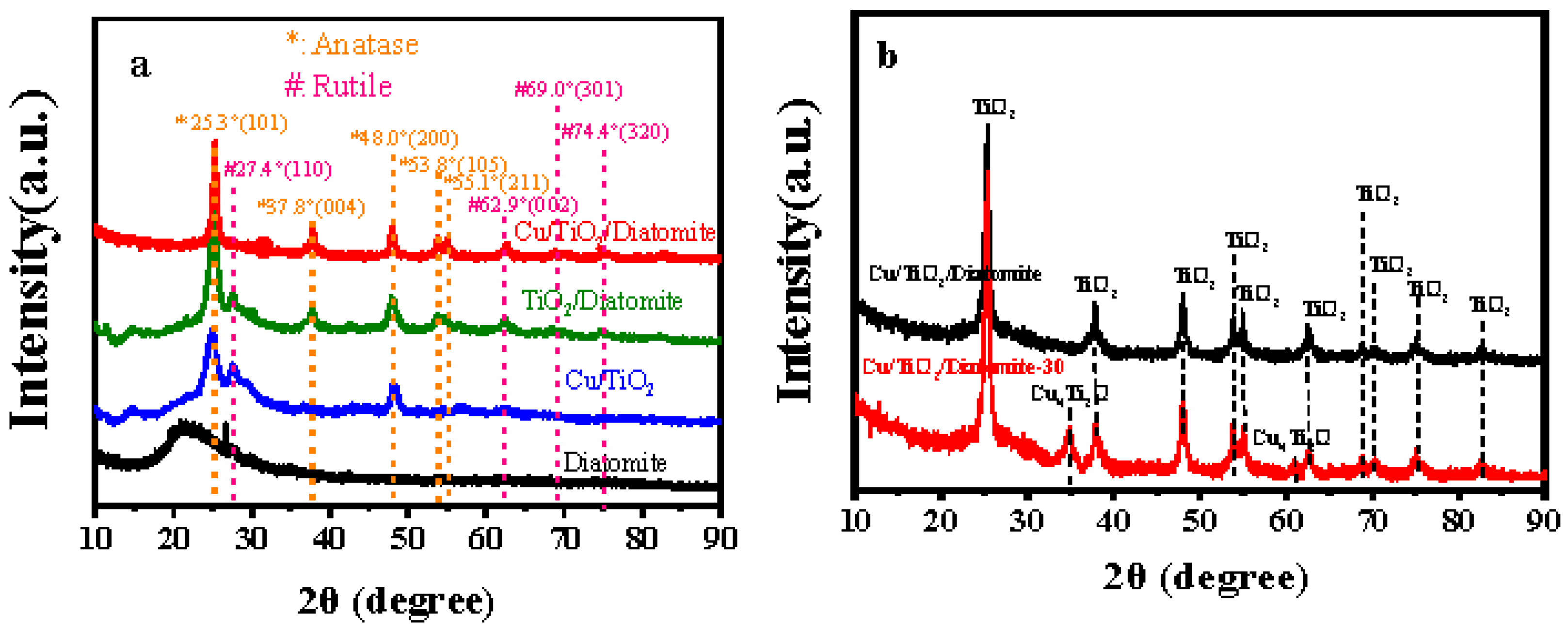
Figure 2.
(a) Nitrogen adsorption–desorption (b) Pore radius distributions of diatomite, Cu/TiO2, TiO2/diatomite, and Cu/TiO2/diatomite.
Figure 2.
(a) Nitrogen adsorption–desorption (b) Pore radius distributions of diatomite, Cu/TiO2, TiO2/diatomite, and Cu/TiO2/diatomite.
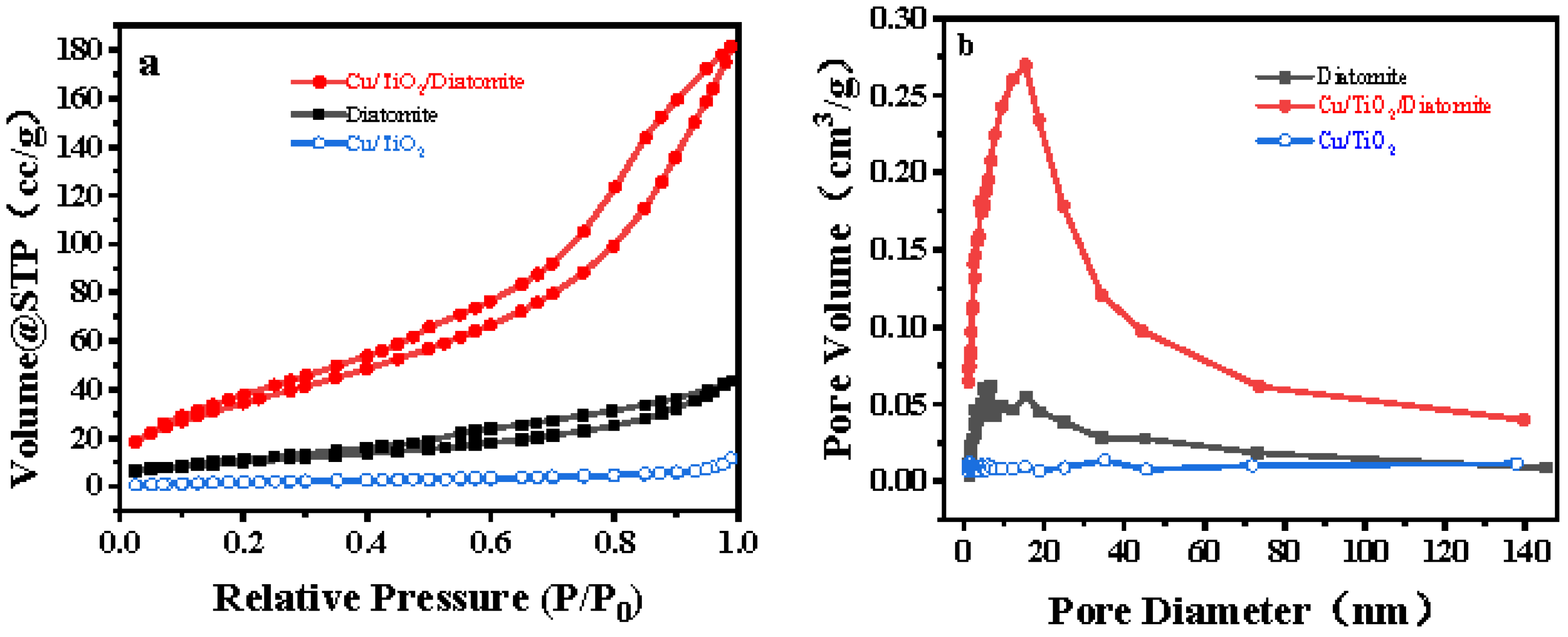
Figure 3.
(a) SEM and (b) EDS-mapping of Cu/TiO2/diatomite.

Figure 4.
(a) SEM and (b) TEM of Cu/TiO2/diatomite.
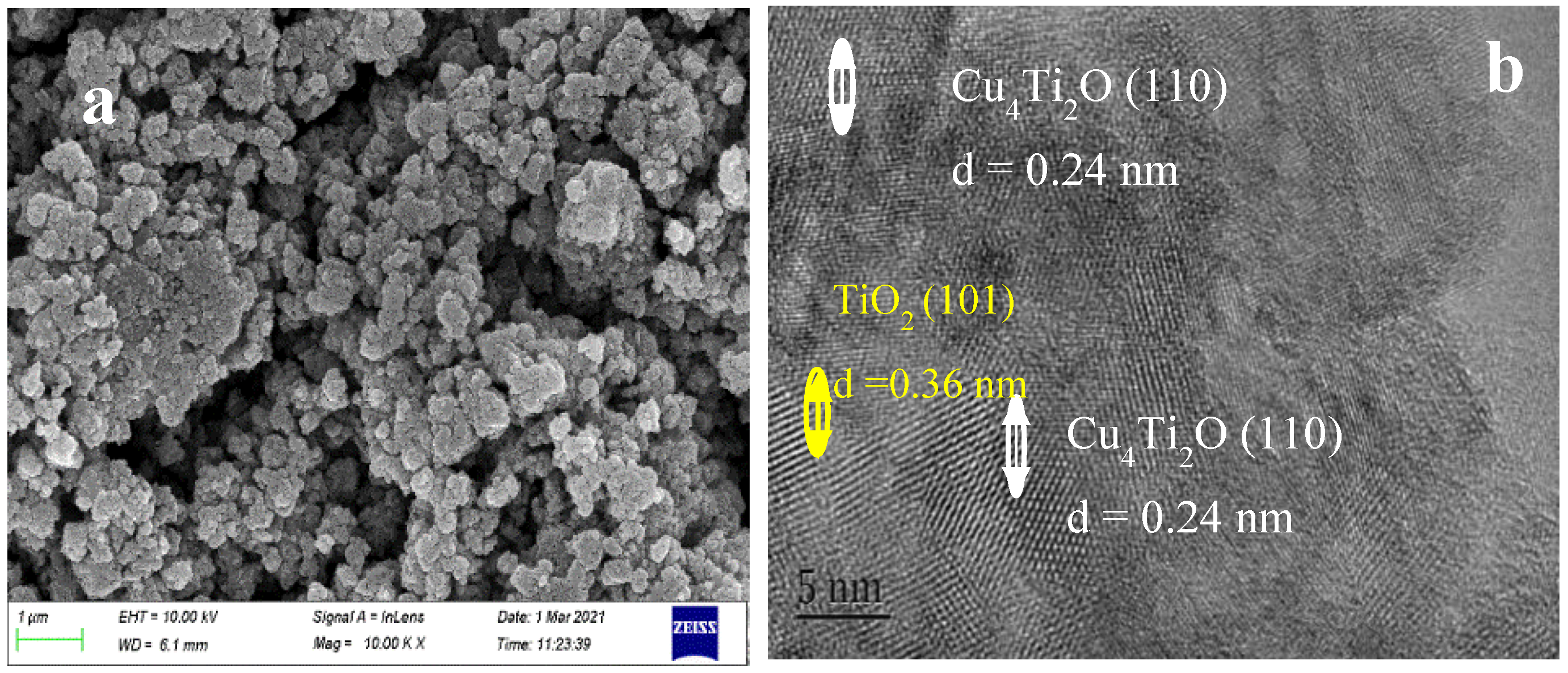
Figure 5.
FTIR of Cu/TiO2/diatomite and diatomite.
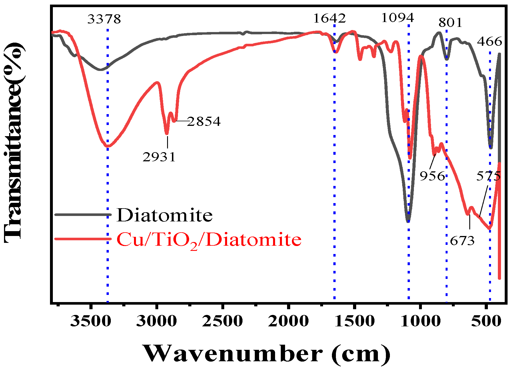
Figure 6.
XPS of (a) O 1s of diatomite and (b) O 1s (c) Cu 2p of Cu/TiO2/diatomite samples.

Figure 7.
UV-vis DRS of diatomite, Cu/TiO2, TiO2/diatomite, and Cu/TiO2/diatomite.

Figure 8.
The influence of synthesis condition on the activity of A. baumannii.

Figure 9.
The influence of synthesis temperature on the activity of A. baumannii (a), bacteriostatic rate of Cu/TiO2, TiO2/diatomite and Cu/TiO2/diatomite (b).
Figure 9.
The influence of synthesis temperature on the activity of A. baumannii (a), bacteriostatic rate of Cu/TiO2, TiO2/diatomite and Cu/TiO2/diatomite (b).
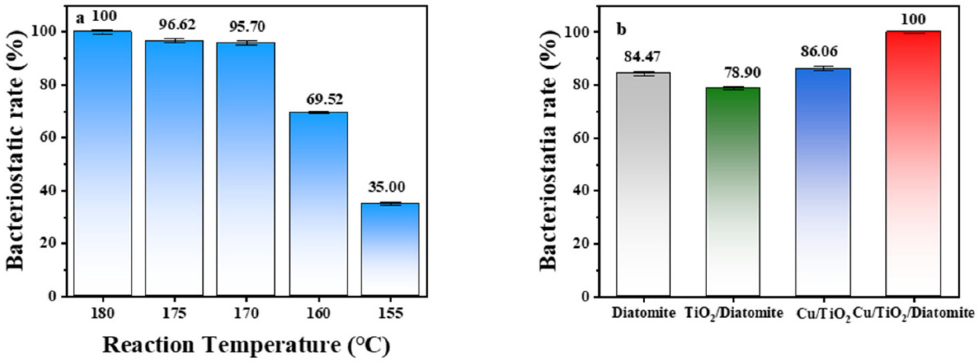
Figure 10.
The spectra of the xenon lamp.


Table 1.
BET parameters of different diatomite materials.
| Sample | BET Surface Area (m2/g) | Pore Volume (cc/g) | Pore Size (nm) |
|---|---|---|---|
| Diatomite | 42.456 | 0.069 | 2.516 |
| Cu/TiO2/Diatomite | 207.759 | 0.305 | 3.938 |
| Cu/TiO2 | 13.361 | 0.020 | 1.356 |
Publisher’s Note: MDPI stays neutral with regard to jurisdictional claims in published maps and institutional affiliations. |
© 2022 by the authors. Licensee MDPI, Basel, Switzerland. This article is an open access article distributed under the terms and conditions of the Creative Commons Attribution (CC BY) license (https://creativecommons.org/licenses/by/4.0/).
Share and Cite
MDPI and ACS Style
Xu, X.; Yang, Y.; Miao, Y.; Liu, K.; Lv, F.; Zhou, L.; Tang, X.; Liu, Y.; Guo, X. Photocatalytic Bacterial Inactivation of Acinetobacter baumannli on Cu/TiO2/Diatomite. Catalysts 2022, 12, 1217. https://doi.org/10.3390/catal12101217
AMA Style
Xu X, Yang Y, Miao Y, Liu K, Lv F, Zhou L, Tang X, Liu Y, Guo X. Photocatalytic Bacterial Inactivation of Acinetobacter baumannli on Cu/TiO2/Diatomite. Catalysts. 2022; 12(10):1217. https://doi.org/10.3390/catal12101217
Chicago/Turabian StyleXu, Xiaolin, Yacong Yang, Yingchun Miao, Kaiquan Liu, Fujian Lv, Liping Zhou, Xuqi Tang, Yanmi Liu, and Xinchun Guo. 2022. "Photocatalytic Bacterial Inactivation of Acinetobacter baumannli on Cu/TiO2/Diatomite" Catalysts 12, no. 10: 1217. https://doi.org/10.3390/catal12101217
Note that from the first issue of 2016, this journal uses article numbers instead of page numbers. See further details here.