Article

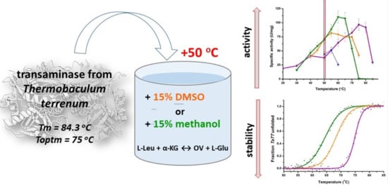
Counterbalance of Stability and Activity Observed for Thermostable Transaminase from Thermobaculum terrenum in the Presence of Organic Solvents

by
Ekaterina Yu. Bezsudnova
1,*,
Alena Yu. Nikolaeva
2,
Sergey Y. Kleymenov
1,3,
Tatiana E. Petrova
4,
Sofia A. Zavialova
1,
Kristina V. Tugaeva
1,
Nikolai N. Sluchanko
1 and
Vladimir O. Popov
1,2
1
Bach Institute of Biochemistry, Federal Research Center of Biotechnology of the Russian Academy of Sciences, Leninsky Ave. 33, bld. 2, 119071 Moscow, Russia
2
Kurchatov Complex of NBICS-technologies, National Research Centre “Kurchatov Institute”, Akad. Kurchatova sqr 1, 123182 Moscow, Russia
3
Koltzov Institute of Developmental Biology of Russian Academy of Sciences, 26 Vavilov Street, 119334 Moscow, Russia
4
Institute of Mathematical Problems of Biology, RAS, Branch of Keldysh Institute of Applied Mathematics of the Russian Academy of Sciences, 1, Professor Vitkevich St., 142290 Pushchino, Russia
*
Author to whom correspondence should be addressed.
Catalysts 2020, 10(9), 1024; https://doi.org/10.3390/catal10091024
Submission received: 11 August 2020 / Revised: 30 August 2020 / Accepted: 4 September 2020 / Published: 6 September 2020
(This article belongs to the Special Issue Biocatalytic Applications in Biotechnology)

Abstract

:
Pyridoxal-5’-phosphate-dependent transaminases catalyze stereoselective amination of organic compounds and are highly important for industrial applications. Catalysis by transaminases often requires organic solvents to increase the solubility of reactants. However, natural transaminases are prone to inactivation in the presence of water-miscible organic solvents. Here, we present the solvent tolerant thermostable transaminase from Thermobaculum terrenum (TaTT) that catalyzes transamination between L-leucine and alpha-ketoglutarate with an optimum at 75 °C and increases the activity ~1.8-fold upon addition of 15% dimethyl sulfoxide or 15% methanol at high but suboptimal temperature, 50 °C. The enhancement of the activity correlates with a decrease in the thermal denaturation midpoint temperature. The blue-shift of tryptophan fluorescence suggested that solvent molecules penetrate the hydration shell of the enzyme. Analysis of hydrogen bonds in the TaTT dimer revealed a high number of salt bridges and surface hydrogen bonds formed by backbone atoms. The latter are sensitive to the presence of organic solvents; they rearrange, conferring the relaxation of some constraints inherent to a thermostable enzyme at low temperatures. Our data support the idea that the counterbalance of stability and activity is crucial for the catalysis under given conditions; the obtained results may be useful for fine-tuning biocatalyst efficiency.

Graphical Abstract

1. Introduction

Pyridoxal-5’-phosphate (PLP)-dependent transaminases (TAs) catalyze stereoselective amination of keto acids, ketones and aldehydes and are of particular interest as industrially relevant enzymes. Catalysis by TAs requires the application of organic solvents to increase the solubility of reactants and drive the reaction equilibrium towards the amination of ketones [1]. The industrial applications of TAs require an improvement of their thermostability and solvent tolerance as well as a search for new robust enzymes in nature [1]. In the optimized sitagliptin manufacturing process, (R)-amination of the pro-sitagliptin ketone is performed by the engineered ATA-117-Rd11 transaminase that tolerates 200 g/L ketone in 50% dimethyl sulfoxide (DMSO) at 40 °C [1,2]. For industrial applications, a search for thermostable TAs is advantageous because of the proven feature of naturally thermostable enzymes to manifest stability under various harsh conditions [3,4,5]. The enzymes engineered for thermostability showed stability in the presence of organic solvents [6]. The stability of enzyme molecules is largely determined by the non-covalent interactions of different natures and, in particular, by the properties of the surface based on the amino acid composition [3,7]. Electrostatic interactions of various intensity, namely, salt bridges, hydrogen bonds, long-range ion pairs, dipole-dipole interactions, etc., are considered to be the main structural factors responsible for the thermostability of a protein globule [8,9]. A large number of salt bridges on the surface of thermostable enzymes maintain the structural integrity under “hot” water attacks due to a low temperature dependence of electrostatic interactions as well as a lack of geometrical restrictions because of the central symmetry of a charge [10,11]. A high number of charged residues promote the extensive hydrogen-bonding network and tighten both the hydration shell and the interface of the enzyme molecules, thus defining the balance between the structural integrity and the conformational flexibility at any temperatures [4,9,12]. Recent advances in the improvement of enzyme thermostability are based in particular on the insertion of salt bridges into the protein globule [13,14].
The solvent tolerance as well as the enzymatic activity changes in water-organic solvent media are still poorly understood. Somewhat counterintuitively, crystallographic studies of solvent-resistant enzymes did not reveal significant changes in structures obtained from crystals grown (or soaked) in organic solvents and showed almost indistinguishable protein conformation to that obtained in buffers [15,16,17,18,19]. However, several solvent interaction sites were identified as well as changes in the side-chain conformations [19,20]. In particular, it was observed that solvent molecules replaced water molecules on the surface and even in the active site of enzymes [17,18,19,20,21]. It is considered that water-miscible organic solvent–water mixtures destabilize the protein globule: solvent molecules penetrate the hydration shell of the enzyme molecules and strip water from the surface, thus solvating hydrophobic patches and disrupting the surface hydrogen bonds due to the interaction with backbone atoms [7,22,23,24,25,26]. A water-miscible organic solvent can interfere with the activity of an enzyme by substituting catalytic water molecules and breaking conserved hydrogen bonds in the active site [27,28]. Thus, the ability of a protein molecule to maintain the integrity of its hydration shell contributes to organic solvent tolerance [3,16,23]. The effective accumulation of water molecules by the charged residues and the strengthening of Coulomb interactions in water-miscible organic solvent–water media make an excess of charged surface residues a powerful instrument counteracting solvent penetration [3,29,30,31]. Some enzymes can increase activity in the presence of water-miscible organic solvents. The enhancement of specific activity was observed for proteases, laccases, lipases, etc. [15,22,32,33,34]. The changes in non-covalent interactions and conformational flexibility as well as the influence of a solvent on particular steps of the catalyzed reactions seem to facilitate the catalysis and underlie the enzyme activation in water–solvent media [15,22,32,35]. Recently, the increase of transaminase activity in cell-free lysates in the presence of organic solvents was demonstrated [36].
The effects of organic cosolvents depend on their physico-chemical characteristics. Non-polar water-immiscible organic solvents do not penetrate the hydration shell, and enzymes preserve structural integrity and the hydration shell in the presence of non-polar cosolvents [3,5]. The penetration potential and water-stripping capacity of water-miscible organic solvents depend on such physical parameters as hydrophobicity, polarity index, dipole moment, and the hydrogen bond donating/accepting abilities [22,37,38]. The enzymes’ responses to the addition of organic solvents are complex and depend on the concentration and properties of the organic solvents as well as the enzyme’s characteristics.
Here, we focus on the enhancement of the activity of recombinant thermostable transaminase from Thermobaculum terrenum (TaTT) in water–methanol and water–DMSO media. Both DMSO and methanol are water-miscible organic solvents and are employed in biocatalytic applications. These organic cosolvents were selected for their different physico-chemical characteristics: they differed by hydrophobicity (logP for DMSO is −1.35 and for methanol is −0.74), by dipole moment (µ for DMSO is 3.96 D and for methanol is 1.70 D) and by hydrogen bond formation ability. Methanol is a hydrogen bond donating cosolvent, whereas DMSO is a hydrogen-bond-accepting cosolvent. We found that TaTT significantly increased its activity in the presence of methanol and DMSO at the expense of the thermal stability, with the impact of methanol being more significant. The hydrogen bonding of TaTT in terms of its structural integrity and solvent tolerance is discussed.

2. Results and Discussion

TaTT is a highly thermostable enzyme [39]. The optimal temperature (Toptm) for the catalyzed transamination reaction between L-Leu and α-ketoglutarate is 75 °C [39] (Scheme 1).
The differential scanning calorimetry (DSC) profile of TaTT is represented by two calorimetric domains with melting points (Tm) of 79.4 and 84.3 °C (Figure 1a). DSC rescanning indicated the irreversibility of the thermal denaturation of TaTT. Kinetic stability assay revealed the 50% reduction of TaTT only after 40 h incubation at 70 °C and 150 h incubation at 50 °C. TaTT retained 100% of the initial activity after 24 h incubation in 50% DMSO and 70% of the activity after 24 h incubation in 50% methanol at 50 °C (Figure 1b,c). The addition of 50% DMSO or 50% methanol in the standard assay decreased the specific activity of TaTT by half. The addition of 30% methanol slightly increased the specific activity of TaTT; 30% DMSO in the standard assay reduced the specific activity of TaTT by 10–15%. By contrast, the addition of 15% methanol and 15% DMSO led to an increase in specific activity by 1.5-1.6 times; at the same time, after 24 h incubation in the presence of 15% methanol and 15% DMSO at 50 °C, TaTT retained the initial activity. The latter indicated the absence of TaTT denaturation and the reversibility of changes in the TaTT molecules at low concentrations of DMSO and methanol.
We examined the effects of 15% methanol and 15% DMSO on TaTT activity in the transamination reaction between L-Leu and α-ketoglutarate at different temperatures. The temperature dependences (Figure 2) showed the shift of Toptm to lower temperatures and, as a result, an enhancement of the TaTT activity at 50 °C. The observed decrease in thermophilicity of TaTT in the presence of the cosolvents pointed out small changes in protein molecule as well as possible effects of the co-solvent on the enzymatic reaction at 50 °C. To clarify this issue, we determined the parameters of the transamination reaction in the presence of 15% DMSO (DMSO was a better solvent for the indirect GDH assay, see Material and Method section). The addition of 15% DMSO induced a 1.5-fold increase in maximum velocity and Km value at 50 °C, with catalytic efficiency toward L-Leu remaining constant (Table 1). Minor changes of kinetic parameters indicated the similarity of substrate binding and catalytic transformations in both reaction media.
To address the organic solvent-induced structural changes, we studied the thermal unfolding of TaTT in the presence of 15% cosolvents using intrinsic fluorescence (Figure 3). T0.5 decreased from 74.9 °C in the buffer to 68.3 °C and 64.5 °C in the presence of 15% DMSO and 15% methanol, respectively. Notably, the cosolvents expanded the temperature range of the thermal unfolding of TaTT from ~10 °C in the buffer to 20.5 and 22.5 °C in the presence of DMSO and methanol, respectively, thereby significantly lowering the cooperativity of thermal transition. This is in line with the idea that water-miscible cosolvents increase structural dynamics of the enzyme, promoting its structural rearrangements in a wide range of temperatures and removing some structural constraints typical of thermostable enzymes at low temperatures.
Additionally, we observed that 0-30% methanol or DMSO caused blue-shifts of the tryptophan fluorescence spectrum of TaTT (Inset panel on Figure 3; Figure 4a) at 50 °C, implying an alteration of the environment of tryptophan residues to more hydrophobic [40,41]. The addition of 30–50% of either cosolvent caused a red-shift, which likely reflects the onset of denaturation provoked by an excessive weakening of non-covalent interactions. Among eight tryptophan residues of the TaTT subunit, all except one were exposed to the bulk solvent (Figure 4c). The blue-shift is in favor of the notion that solvent molecules penetrate the hydration shell of TaTT, causing a decrease in the water density near the hydrophobic indolyl groups and preferential solvation of tryptophan residues (and similarly other exposed hydrophobic groups) by cosolvents [29,30,33]. Sharp growth in light scattering indicated aggregation accompanying the unfolding (Figure 4b) due to the exposure of hydrophobic patches. These changes as well as changes of TaTT thermostability and activity were more pronounced in the presence of methanol. This is likely because of a higher hydrophobicity of methanol (logP = −0.74), compared to DMSO (logP = −1.35). Overall, the T0.5 decrease coincided with the decrease in Toptm of the transamination reaction, suggestive of the common reason underlying the changes. The solvent-induced structural changes can alter the conformational flexibility of the enzyme and adjust the balance between rigidity and flexibility at suboptimal temperatures, i.e., lower than 75 °C.
To interpret the effects of the organic solvents on the thermostability and thermophilicity of TaTT, we focused on the analysis of hydrogen bonding in TaTT, considering hydrogen-bonding network as a crucial structural factor of the stability of enzymes in harsh conditions. According to the recent studies, the penetration of solvent molecules into the hydration shell of an enzyme molecule disturbs the surface hydrogen bonds because of the amphiphilic nature of the cosolvents (dipole strength is 3.96 D for DMSO, 1.70 D for methanol, and 1.85 D for water) [24,26,29]. Both DMSO and methanol can interact with the backbone atoms and outcompete water molecules to form surface hydrogen bonds [24,25,26,33,39]. Present in the hydration shell, DMSO can interact with the exposed backbone NH-groups via its oxygen atoms [29]. Methanol is instead a hydrogen-bonding donor, which can form H-bonds with the backbone CO-group [25]. At the same time, both DMSO and methanol do not significantly affect side-chain hydrogen bonds and salt bridges [24,25,29]. The analysis of hydrogen bonds in the functional dimers of TaTT homologs showed that TaTT is distinguished by the excess of surface hydrogen bonds, by hydrogen bonds formed by side chains of charged residues (salt bridges) and by the number of hydrogen bonds per one amino acid residue (Figure 5, Table A1).
The profound hydrogen-bonding network of the molecule of TaTT coincides well with the thermostability and thermophilicity of the enzyme. It is noteworthy that in all TA dimers, the number of Neutral–Neutral hydrogen bonds is the highest among all types of hydrogen bonds. While salt bridges tolerate water-organic solvent media and maintain the integrity of both the protein globule and the hydration shell, surface Neutral–Neutral and Neutral–Charged hydrogen bonds are much more susceptible because of the interactions of organic solvent molecules with the exposed backbone atoms. These solvent-induced disturbances can increase the flexibility of particular surface regions and release extra tension in regions of the enzyme that are important for catalysis. We suggest that the combination of hydrophobic effects and disturbances of hydrogen bonds induced by the addition of organic solvents leads to a weakening of some constraints inherent for TaTT as a thermostable enzyme at low temperatures. These changes induce an increase in the conformational flexibility of TaTT molecules and enhance the catalysis at lower temperatures, compared with catalysis in buffer systems.

3. Materials and Methods

3.1. Enzyme Production and Activity Assays

Enzyme production was described in [39]. Briefly, the His6TEV-tagged TaTT was expressed in E. coli BL21(DE3)pLys (Stratagene, California, CA, USA). The recombinant TaTT was isolated using subtractive Ni-affinity chromatography and gel filtration. Fractions showing the activity were stored in 50 mM Tris-HCl buffer, pH 8.0, containing 100 mM NaCl, 100 μM PLP and 50% glycerol at −20 °C. The PLP form of TaTT was obtained by incubating TaTT with the excess of both PLP and α-ketoglutarate overnight, followed by the transfer into the assay buffer using a HiTrap Desalting column (GE Healthcare, Chicago, IL, USA).
In the standard assay, the activity of TaTT at 0.5-1.5 µg/mL was measured in the overall transamination reaction with 5 mM L-Leu and 1 mM α-ketoglutarate in 50 mM Tris-HCl buffer, pH 8.0, supplemented with 50 mM NaCl and 60 µM PLP at 50 °C. The amount of L-Glu product was measured by the indirect photometric glutamate dehydrogenase (GluDH (Sigma-Aldrich, St. Louis, MO, USA, cat. N G2626)) assay described in [39]. To evaluate the effects of organic solvents on the overall reaction, the standard assay was carried out in the presence of 15–50% (v/v) methanol or DMSO. The effect of solvents on the GluDH reaction was taken into account, and the corrected calibration curves were used accordingly. All measurements were performed at least in duplicate. The data were analyzed using Origin 8.0 software (Origin Lab, Northampton, MA, USA).

3.2. Analysis of TaTT Stability

To evaluate the thermal stability, 1.0 mg/mL TaTT was incubated at 50 °C or 70 °C in buffer S (50 mM Tris-HCl, pH 8.0, containing 100 mM NaCl, 60 μM PLP). After the thermal treatment, the residual activity was estimated in the standard assay. The solvent stability was estimated by measuring the residual activity after incubation of 1.0 mg/mL TaTT in buffer S, containing 15% or 50% (v/v) organic cosolvents (DMSO, methanol) at 50 °C. After 1, 3, 5 and 24 h, aliquots were taken to determine the residual activity using the standard assay.
Differential scanning calorimetry (DSC) was conducted on a MicroCal VP-Capillary calorimeter (Malvern Instruments, Northampton, MA, USA) with tantalum capillary cells at a heating rate of 1 K/min in 50 mM Na-phosphate buffer, pH 8.0, containing 50 mM NaCl and 1.0 mg/mL TaTT in the PLP form.
Steady-state fluorimetry was conducted using a Cary Eclipse spectrofluorimeter (Varian Inc., Victoria, Australia) equipped with a Peltier-controlled cuvette holder and thermosensor; λex was 297 nm, λem was 313 nm and 370 nm (all slits were 5 nm). Intensities at 313 nm and 370 nm correspond to the half of the maximum of the TaTT fluorescence spectrum at 50 °C, and were used to obtain thermal unfolding curves upon constant heating of the sample at 1 °C/min [44]. The half-transition temperatures of thermal denaturation (T0.5) were calculated using the Boltzmann model. Light scattering at 90 degrees as recorded at λex 350 nm and λem 355 nm (5 nm slits). The buffer was 50 mM Na-phosphate, pH 8.0, containing 50 mM NaCl; the final concentration of TaTT in the PLP form was 0.02 mg/mL.

3.3. Analysis of Hydrogen Bonds

Calculations of the number of hydrogen bonds in the dimers of TaTT and homologous TAs were performed using the programm HBOND [45] and classification described in Appendix A. For TaTT (PDB ID: 6GKR) and its counterparts, PLP and other ligands were removed from the models before the analysis (for details see the Appendix A).

4. Conclusions

In this work, we studied the effects of methanol and DMSO on the activity of thermostable transaminase from T. terrenum. Earlier, we characterized this PLP fold type IV transaminase as highly thermostable (Toptm = 75 °C) and distinguished by the high rates in the transamination reactions with branched-chain amino acids (maximum velocity 178 and 260 U/mg with L-leucine and L-norvaline at 50 °C, respectively) and aromatic amino acids (maximum velocity 46 and 25 U/mg at 50 °C with L-phenylalanine and L-tryptophan, respectively) and significant activity towards (R)-(+)-1-phenylethylamine (0.33 U/mg at 50 °C) [39,46]. Here, we analyzed the solvent tolerance and the activation of TaTT in water-miscible organic solvent–water mixtures: TaTT increased the activity upon addition of 15% DMSO or 15% methanol 1.5–1.7-fold in the standard assay at high but suboptimal temperature, 50 °C. The enhancement of the activity correlated with a decrease in the thermal denaturation midpoint temperature from 74.9 to 68.3 and 64.5 °C upon the addition of DMSO and methanol, respectively. The blue shift of tryptophan fluorescence suggested the penetration of solvent molecules into the hydration shell of the enzyme. The analysis of the hydrogen bonding of TaTT revealed a high number of salt bridges and surface hydrogen bonds formed by the backbone nitrogen and oxygen atoms. We suggested that salt bridges stabilize the protein globule against “hot” water attacks and organic solvent denaturation, but hydrogen bonds formed by the backbone nitrogen and oxygen atoms are susceptible to the presence of solvent molecules and rearrange, underlying a relaxation of some constraints inherent to a thermostable enzyme at low temperatures. According to Botero et al. [47], the gram-positive thermophile T. terrenum shows optimal growth at 67 °C, implying that all its metabolic processes are adopted to this temperature. At 50 °C, the balance between integrity and flexibility of the TaTT molecule is not optimal for catalysis. The addition of solvents seems to release the extra tension characteristic of thermostable enzymes at suboptimal temperatures and optimize conformational flexibility, thus improving the catalytic activity at a given temperature. At the same time, the inherent stability prevents TaTT from denaturation in water-organic solvent mixtures. Despite the challenge of the prediction of co-solvent effects, their great influence on the counterbalance of stability and activity is a useful tool for fine-tuning the efficiency of biocatalytic processes.

Author Contributions

Conceptualization, E.Y.B. and V.O.P.; methodology, N.N.S. and S.Y.K.; software, T.E.P.; validation, K.V.T., A.Y.N., T.E.P. and S.A.Z.; data curation, E.Y.B.; investigation, A.Y.N., T.E.P., S.Y.K., K.V.T. and S.A.Z.; writing—original draft preparation, E.Y.B.; writing—review and editing, E.Y.B and N.N.S.; supervision, V.O.P.; funding acquisition, E.Y.B. All authors have read and agreed to the published version of the manuscript.

Funding

This research was funded by the Russian Science Foundation, grant number 19-14-00164 (in part of fluorescence and structural analysis), and by the Ministry of Science and Higher Education of the Russian Federation.

Conflicts of Interest

The authors declare no conflict of interest.

Appendix A

Procedure for the calculation and analysis of hydrogen bonds:
1. For each analyzed transaminase, a model from the Protein Data Bank (PDB) was taken, and a file (in the pdb format) was prepared, which contained only those monomers that form functional dimer. Water molecules and ligands were removed from the functional dimer model.
2. All potential hydrogen bonds (HBonds) in the model were determined using the program HBOND (http://cib.cf.ocha.ac.jp/bitool/HBOND). The maximal distance between a donor and an acceptor atom was 3.5 Å.
3. The accessible surface area (ASA) for each atom in the model was calculated using the program AREAIMOL of CCP4 package [48]. Atoms with ASA values equal to 0.0 were considered as inner atoms, other atoms were considered as surface atoms.
4. If a protein model contained some residues in multiple conformations, both files (output of HBOND and AREAMOL) were corrected.
5. Both files (output of HBOND and AREAMOL) were merged into one single file. Depending on the ASA value and the side group of the residue, all bonds were categorized into several groups. We determined the number of hydrogen bonds between inner atoms (Inside–Inside), between atoms on the surface (Surface–Surface), between atoms one of which either belonged to a side group of a neutral residue or was N or O atom of the main chain and the other belonged to a side group of a charged residue (Charged–Neutral), between the atoms of the side group of neutral residues and/or N or O atom of the main chain (Neutral–Neutral) and between the atoms of the side groups of charged residues (Charged–Charged). The difference between the total number of hydrogen bonds and the sum of Inside–Inside and Surface–Surface hydrogen bonds is the number of Inside–Surface hydrogen bonds formed between a solvent-accessible atom and an atom buried inside the globule. Note, that Charged–Charged hydrogen bonds are nothing but the salt bridges.
Table A1. Analysis of hydrogen bonds in functional dimers of TaTT and the counterparts: transaminase (TA) from Thermoproteus uzoniensis (Toptm in the standard assay is 95 °C [42]), TA from Burkholderia pseudomallei (Toptm is unknown, the organism grows optimally at 40 °C) and TA from Escherichia coli (Toptm in the standard assay is 37 °C [43]). Two values for the TaTT dimer correspond to two dimers composed of different subunits in the 6GKR model.
Dimer of
TaTTTA from
T. uzoniensis
TA from
B. pseudomalle
TA from
E. coli
Number of residues per subunit316295307309
Hydrogen bonds
  • Total number in a dimer
  • Per one a.a. residue in a dimer
  • Number of Inside–Inside hydrogen bonds (between atoms inside the globule)
  • Number of Surface–Surface hydrogen bonds (between atoms on the globule surface)
  • Number of Neutral–Neutral bonds (between atoms of the side group of neutral residues and/or N or O atoms of the main chain)
  • number of Charged–Charged bonds (between atoms of the side group of charged residues)
  • Number of Charged–Neutral hydrogen bonds
  • Between subunits in the dimer

647/648
1.025
293/296

177/179

430/434


77/68

140/146
18/24

578
0.98
302

135

416


36

126
14

617
1.005
296

151

432


47

138
23

554
0.896
287

141

400


49

105
12
PDB ID:6GKR5CE83U0G1I1K

References

  1. Slabu, I.; Galman, J.L.; Lloyd, R.C.; Turner, N.J. Discovery, engineering, and synthetic application of transaminase biocatalysts. ACS Catal. 2017, 7, 8263–8284. [Google Scholar] [CrossRef]
  2. Savile, C.K.; Janey, J.M.; Mundorff, E.C.; Moore, J.C.; Tam, S.; Jarvis, W.R.; Colbeck, J.C.; Krebber, A.; Fleitz, F.J.; Brands, J.; et al. Biocatalytic asymmetric synthesis of chiral amines from ketones applied to sitagliptin manufacture. Science 2010, 329, 305–309. [Google Scholar] [CrossRef] [PubMed] [Green Version]
  3. Doukyu, N.; Ogino, H. Organic solvent-tolerant enzymes. Biochem. Eng. J. 2010, 48, 270–282. [Google Scholar] [CrossRef]
  4. Bezsudnova, E.Y.; Petrova, T.E.; Popinako, A.V.; Antonov, M.Y.; Stekhanova, T.N.; Popov, V.O. Intramolecular hydrogen bonding in the polyextremophilic short-chain dehydrogenase from the archaeon Thermococcus sibiricus and its close structural homologs. Biochimie 2015, 118, 82–89. [Google Scholar] [CrossRef]
  5. Kumar, A.; Dhar, K.; Kanwar, S.S.; Arora, P.K. Lipase catalysis in organic solvents: Advantages and applications. Biol. Proced. Online 2016, 18, 2. [Google Scholar] [CrossRef] [Green Version]
  6. Stepankova, V.; Bidmanova, S.; Koudelakova, T.; Prokop, Z.; Chaloupkova, R.; Damborsky, J. Strategies for stabilization of enzymes in organic solvents. ACS Catal. 2013, 3, 2823–2836. [Google Scholar] [CrossRef]
  7. Feller, G. Protein stability and enzyme activity at extreme biological temperatures. J. Phys. Condens. Matter 2010, 22, 323101. [Google Scholar] [CrossRef]
  8. Kumar, S.; Tsai, C.-J.; Ma, B.; Nussinov, R. Contribution of salt bridges toward protein thermostability. J. Biomol. Struct. Dyn. 2000, 17, 79–85. [Google Scholar] [CrossRef]
  9. Berezovsky, I.N.; Shakhnovich, E.I. Physics and evolution of thermophilic adaptation. Proc. Natl. Acad. Sci. USA 2005, 102, 12742–12747. [Google Scholar] [CrossRef] [Green Version]
  10. Matsui, I.; Harata, K. Implication for buried polar contacts and ion pairs in hyperthermostable enzymes. FEBS J. 2007, 274, 4012–4022. [Google Scholar] [CrossRef] [Green Version]
  11. Elcock, A.H. The stability of salt bridges at high temperatures: Implications for hyperthermophilic proteins 1 Edited by B. Honig. J. Mol. Biol. 1998, 284, 489–502. [Google Scholar] [CrossRef] [PubMed]
  12. Dong, Y.; Liao, M.; Meng, X.; Somero, G.N. Structural flexibility and protein adaptation to temperature: Molecular dynamics analysis of malate dehydrogenases of marine molluscs. Proc. Natl. Acad. Sci. USA 2018, 115, 1274–1279. [Google Scholar] [CrossRef] [PubMed] [Green Version]
  13. Wijma, H.J.; Floor, R.J.; Janssen, D.B. Structure- and sequence-analysis inspired engineering of proteins for enhanced thermostability. Curr. Opin. Struct. Biol. 2013, 23, 588–594. [Google Scholar] [CrossRef] [PubMed]
  14. Xu, Z.; Cen, Y.K.; Zou, S.P.; Xue, Y.P.; Zheng, Y.G. Recent advances in the improvement of enzyme thermostability by structure modification. Crit. Rev. Biotechnol. 2020, 40, 83–98. [Google Scholar] [CrossRef] [PubMed]
  15. Wu, M.H.; Lin, M.C.; Lee, C.C.; Yu, S.M.; Wang, A.H.J.; Ho, T.-H.D. Enhancement of laccase activity by pre-incubation with organic solvents. Sci. Rep. 2019, 9, 9754. [Google Scholar] [CrossRef] [PubMed]
  16. Said, Z.S.A.A.M.; Arifi, F.A.M.; Salleh, A.B.; Rahman, R.N.Z.R.A.; Leow, A.T.C.; Latip, W.; Ali, M.S.M. Unravelling protein -organic solvent interaction of organic solvent tolerant elastase from Pseudomonas aeruginosa strain K crystal structure. Int. J. Biol. Macromol. 2019, 127, 575–584. [Google Scholar] [CrossRef]
  17. Fitzpatrick, P.A.; Steinmetz, A.C.U.; Ringe, D.; Klibanov, A.M. Enzyme crystal structure in a neat organic solvent. Proc. Natl. Acad. Sci. USA 1993, 90, 8653–8657. [Google Scholar] [CrossRef] [Green Version]
  18. Cianci, M.; Tomaszewski, B.; Helliwell, J.R.; Halling, P.J. Crystallographic analysis of counterion effects on subtilisin enzymatic action in acetonitrile. J. Am. Chem. Soc. 2010, 132, 2293–2300. [Google Scholar] [CrossRef]
  19. English, A.C.; Groom, C.R.; Hubbard, R.E. Experimental and computational mapping of the binding surface of a crystalline protein. Protein Eng. Des. Sel. 2001, 14, 47–59. [Google Scholar] [CrossRef]
  20. Gupta, M.N.; Tyagi, R.; Sharma, S.; Karthikeyan, S.; Singh, T.P. Enhancement of catalytic efficiency of enzymes through exposure to anhydrous organic solvent at 70 °C. Three-dimensional structure of a treated serine proteinase at 2.2 Å resolution. Proteins Struct. Funct. Genet. 2000, 39, 226–234. [Google Scholar] [CrossRef]
  21. Schmitke, J.L.; Stern, L.J.; Klibanov, A.M. The crystal structure of subtilisin Carlsberg in anhydrous dioxane and its comparison with those in water and acetonitrile. Proc. Natl. Acad. Sci. USA 1997, 94, 4250–4255. [Google Scholar] [CrossRef] [PubMed] [Green Version]
  22. Stepankova, V.; Damborsky, J.; Chaloupkova, R. Organic co-solvents affect activity, stability and enantioselectivity of haloalkane dehalogenases. Biotechnol. J. 2013, 8, 719–729. [Google Scholar] [CrossRef] [PubMed]
  23. Polizzi, K.M.; Bommarius, A.S.; Broering, J.M.; Chaparro-Riggers, J.F. Stability of biocatalysts. Curr. Opin. Chem. Biol. 2007, 11, 220–225. [Google Scholar] [CrossRef] [PubMed]
  24. Mohtashami, M.; Fooladi, J.; Haddad-Mashadrizeh, A.; Housaindokht, M.R.; Monhemi, H. Molecular mechanism of enzyme tolerance against organic solvents: Insights from molecular dynamics simulation. Int. J. Biol. Macromol. 2019, 122, 914–923. [Google Scholar] [CrossRef]
  25. Shao, Q. Methanol concentration dependent protein denaturing ability of guanidinium/methanol mixed solution. J. Phys. Chem. B 2014, 118, 6175–6185. [Google Scholar] [CrossRef]
  26. Pazhang, M.; Mardi, N.; Mehrnejad, F.; Chaparzadeh, N. The combinatorial effects of osmolytes and alcohols on the stability of pyrazinamidase: Methanol affects the enzyme stability through hydrophobic interactions and hydrogen bonds. Int. J. Biol. Macromol. 2018, 108, 1339–1347. [Google Scholar] [CrossRef]
  27. Kuper, J.; Wong, T.S.; Roccatano, D.; Wilmanns, M.; Schwaneberg, U. Understanding a mechanism of organic cosolvent inactivation in heme monooxygenase P450 BM-3. J. Am. Chem. Soc. 2007, 129, 5786–5787. [Google Scholar] [CrossRef]
  28. Roccatano, D. Computer simulations study of biomolecules in non-aqueous or cosolvent/water mixture solutions. Curr. Protein Pept. Sci. 2008, 9, 407–426. [Google Scholar] [CrossRef]
  29. Srivastava, K.R.; Goyal, B.; Kumar, A.; Durani, S. Scrutiny of electrostatic-driven conformational ordering of polypeptide chains in DMSO: A study with a model oligopeptide. RSC Adv. 2017, 7, 27981–27991. [Google Scholar] [CrossRef]
  30. Johnson, M.E.; Malardier-Jugroot, C.; Head-Gordon, T. Effects of co-solvents on peptide hydration water structure and dynamics. Phys. Chem. Chem. Phys. 2010, 12, 393–405. [Google Scholar] [CrossRef] [Green Version]
  31. Markel, U.; Zhu, L.; Frauenkron-Machedjou, V.; Zhao, J.; Bocola, M.; Davari, M.; Jaeger, K.-E.; Schwaneberg, U. Are directed evolution approaches efficient in exploring nature’s potential to stabilize a lipase in organic cosolvents? Catalysts 2017, 7, 142. [Google Scholar] [CrossRef] [Green Version]
  32. Cao, H.; Nie, K.; Xu, H.; Xiong, X.; Krastev, R.; Wang, F.; Tan, T.; Liu, L. Insight into the mechanism behind the activation phenomenon of lipase from Thermus thermophilus HB8 in polar organic solvents. J. Mol. Catal. B Enzym. 2016, 133, S400–S409. [Google Scholar] [CrossRef]
  33. Kamal, M.Z.; Yedavalli, P.; Deshmukh, M.V.; Rao, N.M. Lipase in aqueous-polar organic solvents: Activity, structure, and stability. Protein Sci. 2013, 22, 904–915. [Google Scholar] [CrossRef] [Green Version]
  34. Batra, R.; Gupta, M.N. Enhancement of enzyme activity in aqueous-organic solvent mixtures. Biotechnol. Lett. 1994, 16, 1059–1064. [Google Scholar] [CrossRef]
  35. Kudryashova, E.V.; Gladilin, A.K.; Vakurov, A.V.; Heitz, F.; Levashov, A.V.; Mozhaev, V.V. Enzyme-polyelectrolyte complexes in water-ethanol mixtures: Negatively charged groups artificially introduced into α-chymotrypsin provide additional activation and stabilization effects. Biotechnol. Bioeng. 1997, 55, 267–277. [Google Scholar] [CrossRef]
  36. Gord Noshahri, N.; Fooladi, J.; Syldatk, C.; Engel, U.; Heravi, M.M.; Zare Mehrjerdi, M.; Rudat, J. Screening and comparative characterization of microorganisms from iranian soil samples showing ω-transaminase activity toward a plethora of substrates. Catalysts 2019, 9, 874. [Google Scholar] [CrossRef] [Green Version]
  37. Sirotkin, V.A.; Zinatullin, A.N.; Solomonov, B.N.; Faizullin, D.A.; Fedotov, V.D. Calorimetric and Fourier transform infrared spectroscopic study of solid proteins immersed in low water organic solvents. Biochim. Biophys. Acta Protein Struct. Mol. Enzymol. 2001, 1547, 359–369. [Google Scholar] [CrossRef]
  38. Sirotkin, V.A.; Kuchierskaya, A.A. α-chymotrypsin in water-acetone and water-dimethyl sulfoxide mixtures: Effect of preferential solvation and hydration. Proteins. Struct. Funct. Bioinforma. 2017, 85, 1808–1819. [Google Scholar] [CrossRef]
  39. Bezsudnova, E.Y.; Boyko, K.M.; Nikolaeva, A.Y.; Zeifman, Y.S.; Rakitina, T.V.; Suplatov, D.A.; Popov, V.O. Biochemical and structural insights into PLP fold type IV transaminase from Thermobaculum terrenum. Biochimie 2019, 158, 130–138. [Google Scholar] [CrossRef]
  40. Lakowicz, J.R. Principles of Fluorescence Spectroscopy; Lakowicz, J.R., Ed.; Springer US: Boston, MA, USA, 2006; ISBN 978-0-387-31278-1. [Google Scholar]
  41. Bushueva, T.L.; Busel, E.P.; Burstein, E.A. Relationship of thermal quenching of protein fluorescence to intramolecular structural mobility. Biochim. Biophys. Acta Protein Struct. 1978, 534, 141–152. [Google Scholar] [CrossRef]
  42. Boyko, K.M.; Stekhanova, T.N.; Nikolaeva, A.Y.; Mardanov, A.V.; Rakitin, A.L.; Ravin, N.V.; Bezsudnova, E.Y.; Popov, V.O. First structure of archaeal branched-chain amino acid aminotransferase from Thermoproteus uzoniensis specific for l-amino acids and R-amines. Extremophiles 2016, 20, 215–225. [Google Scholar] [CrossRef] [PubMed]
  43. Yu, X.; Wang, X.; Engel, P.C. The specificity and kinetic mechanism of branched-chain amino acid aminotransferase from Escherichia coli studied with a new improved coupled assay procedure and the enzyme’s potential for biocatalysis. FEBS J. 2014, 281, 391–400. [Google Scholar] [CrossRef]
  44. Permyakov, E.A.; Burstein, E.A. Some aspects of studies of thermal transitions in proteins by means of their intrinsic fluorescence. Biophys. Chem. 1984, 19, 265–271. [Google Scholar] [CrossRef]
  45. Available online: http://cib.cf.ocha.ac.jp/bitool/HBOND (accessed on 29 April 2009).
  46. Bezsudnova, E.Y.; Popov, V.O.; Boyko, K.M. Structural insight into the substrate specificity of PLP fold type IV transaminases. Appl. Microbiol. Biotechnol. 2020, 104, 2343–2357. [Google Scholar] [CrossRef] [PubMed]
  47. Botero, L.M.; Brown, K.B.; Brumefield, S.; Burr, M.; Castenholz, R.W.; Young, M.; McDermott, T.R. Thermobaculum terrenum gen. nov., sp. nov.: A non-phototrophic gram-positive thermophile representing an environmental clone group related to the Chloroflexi (green non-sulfur bacteria) and Thermomicrobia. Arch. Microbiol. 2004, 181, 269–277. [Google Scholar] [CrossRef]
  48. Collaborative Computational Project, Number 4. The CCP4 suite: Programs for protein crystallography. Acta Crystallogr. Sect. D Biol. Crystallogr. 1994, 50, 760–763. [Google Scholar] [CrossRef] [PubMed]
Scheme 1. Transamination reaction between L-Leu and α-ketoglutarate catalyzed by Thermobaculum terrenum (TaTT).
Catalysts 10 01024 sch001
Figure 1. Thermal stability of TaTT. (a) Differential scanning calorimetry (DSC) profile of 1.0 mg/mL TaTT. (b)–(d) Residual activity of TaTT after incubation (b) in buffer S (50 mM Tris-HCl, pH 8.0, containing 100 mM NaCl, 60 μM PLP) at 50 °C and 70 °C; (c) in buffer S with 50% DMSO (v/v) at 50 °C; (d) in buffer S with 50% (v/v) methanol at 50 °C. 100% corresponds to 40 ± 4 U/mg in the standard assay. Error bars represent standard deviation.
Catalysts 10 01024 g001
Figure 2. Temperature dependences of the specific activity of TaTT in the transamination reaction between L-Leu and α-ketoglutarate in 50 mM Na-phosphate buffer, pH 8.0, containing 50 mM NaCl (purple), and after the addition of 15% DMSO (orange), 15% methanol (olive) or 30% methanol (blue). All measurements were performed at least in triplicates. Error bars represent standard deviation.
Catalysts 10 01024 g002
Figure 3. Thermally-induced unfolding curves of TaTT in Na-phosphate buffer (purple) and in the presence of 15% DMSO (orange) or 15% methanol (olive). Inset: The dependence of I313/I370 for 0.1 mg/mL TaTT on percentage (v/v) of methanol (olive) and DMSO (orange) in Na-phosphate buffer at 50 °C. The data are the averages from triplicate experiments.
Catalysts 10 01024 g003
Figure 4. Thermal unfolding of TaTT studied by using its intrinsic tryptophan fluorescence. (a) Fluorescence spectra of 0.1 mg/mL TaTT in the PLP form in 50 mM phosphate buffer, pH 8.0, containing 50 mM NaCl, (black) and in the presence of 30% methanol (pink) at 50 °C. Intensities at 313 nm and 370 nm correspond to the half of the maximum of the TaTT fluorescence spectrum. (b) Light scattering accompanying thermally-induced unfolding of TaTT in Na-phosphate buffer (purple) or the presence of 15% DMSO (orange) or 15% methanol (olive). (c) Model of the functional dimer of TaTT (PDB ID: 6GKR). Two subunits forming the dimer are shown. In the right subunit, the small domain is colored blue, the large domain is colored purple; in the left subunit, tryptophan residues are shown in green. PLP molecules are colored yellow.
Catalysts 10 01024 g004
Figure 5. Bar graphs showing the number of hydrogen bonds in functional dimers of TaTT (PDB ID: 6GKR) and its homologs: TA_TU from Thermoproteus uzoniensis (Toptm in the standard assay is 95 °C [42], PDB ID: 5CE8,), TA_BP from Burkholderia pseudomallei (Toptm is unknown, the organism grows optimally at 40 °C, PDB ID 3U0G) and TA_EC from Escherichia coli (Toptm in the standard assay is 37 °C [43], PDB ID 1I1K). (a) Percentage of Surface–Surface (violet) and Inside–Inside (pink) hydrogen bonds in the total number (100%, underlined by dash line) of hydrogen bonds in the dimers; (b) Number of Charged–Charged hydrogen bonds (green, left axis), Charged–Neutral (light-green, left axis) and Neutral–Neutral (grey, right axis) hydrogen bonds in the dimers.
Catalysts 10 01024 g005
Table 1. Steady-state kinetic parameters of the transamination reaction catalyzed by TaTT at 50 °C.
SolventVmax,
U/mg
Km,
mM
kcat/Km,
s−1 M−1
Buffer178 ± 237.8 ± 2.313,700 ± 4400
15% DMSO280 ± 4012 ± 414,000 ± 5000

