Biochemical Degradation of Chitosan over Immobilized Cellulase and Supported Fenton Catalysts


1
College of Chemistry & Chemical Engineering, University of Jinan, Jinan 250022, China
2
Key Laboratory of Biofuels, Qingdao Institute of Bioenergy and Bioprocess Technology, Chinese Academy of Science, Qingdao 266101, China
*
Author to whom correspondence should be addressed.
Catalysts 2020, 10(6), 604; https://doi.org/10.3390/catal10060604
Submission received: 27 April 2020
/
Revised: 19 May 2020
/
Accepted: 27 May 2020
/
Published: 29 May 2020
Abstract
:This paper describes the application of Fe-MCM-48 (Mobil Composition of Matter No.48) and cellulase-MCM-48 catalysts for the depolymerization of chitosan. The results show that H2O2 is a good oxidant for the depolymerization of chitosan in the presence of Fe-MCM-48. The average polymerization degree of the product decreased to 6.1, and decreased to 29.2 when cellulase-MCM-48 was used as a catalyst, because the effect of the enzyme was affected by the molecular structure of chitosan. When both materials were used for depolymerization, the average degree of polymerization sharply decreased to 3.8. The results show that the two degradation methods can promote each other to obtain oligosaccharides with a lower degree of polymerization. This provides a new method for the controllable degradation of chitosan and lays a good foundation for the industrial production of chitosan oligosaccharides with a low degree of polymerization.
1. Introduction
Chitosan, also known as deacetylated chitin, is deacetylated in concentrated alkali or by an enzyme from chitin that exists widely in nature. A degree of deacetylation above 55% can be defined as chitosan [1,2,3]. Chitosan is widely used in many fields, including medicine, food, chemicals, cosmetics, water treatment, metal extraction and recovery, biochemistry, and biomedical engineering [4,5,6,7,8]. Chito-oligosaccharide is an oligosaccharide product obtained by degradation of chitosan with a polymerization degree of 2–20 and a molecular weight of ≤3200 Da. It is the only alkaline oligosaccharide in nature. Chito-oligosaccharides with good water solubility and higher bioactivity have attracted considerable interest due to their large molecular weight and difficult water solubility [9].
So far, the main methods of degrading chitosan include physical, bioenzymatic, and chemical degradation [10,11]. Physical degradation works by physical means, including heat, ultrasound, microwaves, and rays, but it tends to have problems such as insufficient reaction and low recovery [12,13]. Bioenzymatic degradation uses specific enzymes to degrade chitosan, featuring no side effects, mild degradation conditions, and easy control of molecular weight distribution of degradation products [10,14], but production costs are high because the enzymes are usually expensive. Chemical degradation is usually divided into acid and oxidative degradation. Acid degradation is rarely used, because reactions are difficult to control, the products are mostly monosaccharides, and waste acid pollutes the environment. Oxidative degradation usually uses H2O2 or ozone as the oxidant and breaks down the β-1,4-glycosidic bond of the chitosan molecule with strongly oxidative free radicals [15]; this method features low cost, short degradation reaction time, and easy realization of industrial production, but products are prone to browning because of residual antioxidants as well as increased reaction temperature and antioxidant concentration [16]. Therefore, using a combination of degradation methods can exploit the advantages of each method to achieve a better degradation effect [17,18]. Zhang et al. [19] used both physical and chemical methods to degrade chitosan by microwave-assisted H2O2 oxidation for 40 min at a reaction temperature of 70 °C and microwave at 600 W power to obtain a chito-oligosaccharide with a molecular weight of 5500 Da.
In the reaction using a biological enzyme to degrade chitosan, free enzymes in the mixture are difficult to recycle after the reaction, increasing the production cost. Therefore, more attention has been paid to the degradation reaction using immobilized enzyme technology [20]. Some researchers used molecular sieves to immobilize cellulase, and found significant increases in their catalytic and operational stability [21]. During oxidative degradation of chitosan by H2O2, adding iron salt can make a Fenton reagent to enhance the oxidation effect; meanwhile, immobilized catalysts are usually used to avoid the introduction of impurities [1,22]. Immobilized catalysts are characterized by easy separation, easy recovery, and high catalytic activity after multiple recycling. There may also be synergy between the high adsorption capacity due to high specific surface area of the support and the catalytic ability of the catalyst. Since Mobile Inc. developed the M41S series of mesoporous materials featuring high specific surface area, high thermal stability, and homogeneous and adjustable pore size in 1992, such materials have been a research focus [23].
In particular, the MCM-48 molecular sieve, a silicon-based mesoporous material, has a stable and well-organized three-dimensional spiral porous structure, and it has attracted great attention in the catalytic field due to its advantages, including high specific surface area and adjustable pore size [24]. The mesoporous material has a good protein adsorption effect and helps maintain the activity of the immobilized enzyme catalyst, because the pore size of the mesoporous material is close to the size of a protein molecule, and silanol groups on its surface form hydrogen bonds with some groups of the protein molecule [25].
With the MCM-48 molecular sieve as a support, this study investigates the degradation of chitosan by Fenton degradation (Fe-MCM-48), biological enzymolysis (cellulase-MCM-48), and the combination of both methods. A low-cost monolith was used as the carrier to verify the degradation effect of monolith catalyst. Characteristics of immobilized catalysts and degradation products were analyzed to obtain chito-oligosaccharide with a controllable polymerization degree in an efficient and low-cost manner, laying the foundation for economic production of chito-oligosaccharide.
2. Results and Discussion
2.1. Characterization of Catalysts
2.1.1. N2 Adsorption–Desorption Analysis
The Brunauer–Emmett–Teller (BET) specific areas and pore distributions of Fe-MCM-48 and cellulase-MCM-48 were measured, and are summarized in Table 1 and Figure 1 and Figure 2. The specific surface area of MCM-48 was 797.7 m2/g (Table 1, entry 1). When modified by Fe and cellulase, the BET area decreased to 691.8 and 589.5 m2/g, respectively (Table 1, entries 2 and 3), which indicates that some pores of MCM-48 were blocked by ion or cellulase. The pore volume of MCM-48 also decreased (Table 1), verifying this point. The average pore diameter of MCM-48 was 3.04 nm, which increased to 3.50 and 3.72 nm for Fe-MCM-48 and cellulase-MCM-48, respectively. This indicates that the amount of micropore was blocked. The curve in P/P0 ranging from 0 to 0.1 of the modified MCM-48 was lower than that of MCM-48, suggesting that the micropores decreased (Figure 1), which was evidenced by the decrease in the number of pores 2 nm in size (Figure 2). From the above results, the effect of cellulase on blocking pores is larger than that of iron, which can probably be attributed to the higher loading of cellulase (3.4% versus 6.4%; Table 1).
When modified by iron and cellulase, the textural structure of MCM-48 showed different characteristics. In addition to the pore properties, the functionalities of these catalysts might be changed, which needs to be further investigated with FT-IR characterization.
2.1.2. FT-IR of Catalysts
Figure 3 shows the Fourier transform infrared spectroscopy (FT-IR) spectra of MCM-48, Fe-MCM-48, and cellulase-MCM-48. For MCM-48, the broad peak at 3475 cm−1 was assigned to the stretching vibration of silicon hydroxyl groups, indicating that there were amounts of OH group existing on the surface of MCM-48. In addition, absorption at 1213 and 1066 cm–1 belonged to the Si–C and Si–O groups, respectively. All of these functionalities could be found in the Fe-MCM-48 and cellulase-MCM-48, indicating that modification of iron or cellulase remained the basic skeleton structure of MCM-48, which was not broken by the modification.
For Fe-MCM-48, no new groups were introduced into its surface, and it had similar structure and functionalities to MCM-48. In contrast, cellulase-MCM-48 changed the properties of the catalyst to some extent. The peaks at 3411 cm–1 and 3420 cm–1 belonged to the –NH2 group derived from cellulase. The absorption at 1080 cm–1, which was not seen in MCM-48 and Fe-MCM-48, belonged to the C–O group of cellulase as well. It is worth noting that the appearance of the C–O group seemed to affect the distribution of –SiOH in terms of forming an interaction between –SiOH and cellulase.
FT-IR spectra showed that the functionalities of cellulase-MCM-48 were significantly different from those of MCM-48 and Fe-MCM-48. The morphologies should be further studied.
2.1.3. High-Resolution Transmission Electron Microscopy and Scanning Electron Microscope Analysis of Catalysts
High-resolution transmission electron microscopy (TEM) images of MCM-48, Fe-MCM-48, and cellulase-MCM-48 are depicted in Figure 4a–c, respectively. In principle, MCM-48 is a synthetic molecular sieve with a three-dimensional channel. The distinct structure makes it a suitable support to load some active components with the aim of achieving good dispersion. The TEM micrographs in Figure 4a show that some uniform ordered structure was seen in MCM-48. Figure 4b reveals that Fe-MCM-48 had a well-ordered structure similar to MCM-48, while the black shaded areas in Figure 4b suggest that the iron species probably aggregated on the surface of MCM-48, which might affect the decomposition of chitosan. With respect to cellulase-MCM-48, in addition to the remaining well-ordered structure, some cellulase granules about 2–4 nm in size were well dispersed on the surface (Figure 4c). Scanning electron microscope (SEM) images show that there were no obvious differences with respect to morphology when MCM-48 was modified by iron and cellulase (Figure 5).
Modifying the iron species or cellulase not only affected the textural structure and functionalities, but also subsequently impacted the catalytic performance in the degradation of chitosan, which is shown below.
2.1.4. X-ray Diffraction of Catalysts
Figure 6 gives X-ray diffraction (XRD) patterns obtained with different catalysts. All catalysts showed a large peak at 23.8° like carrier MCM-48 but the diffraction intensity was weaker. No obvious diffraction peaks were found, possibly because the active components on the carrier were in the amorphous form. Cellulase is harder to crystallize. No iron sulfate crystals were found, possibly indicating the high dispersion of active component on the surface of the carrier MCM-48.
2.2. Catalytic Performances
2.2.1. Degradation of Chitosan Catalyzed by MCM-48, Fe-MCM-48, and Cellulase-MCM-48
The activity of the catalyst was evaluated by the effect of chitosan degradation. Table 2 shows the results. The chitosan used in this work had a relative molecular mass of 100,000–1,500,000 (polymerization degree: 600–9000), and is composed of glucosamine as the basic constituent unit linking with another glucosamine via a glycosidic bond. The functionalities on the chitosan, including hydroxyl, amino, and amide groups, participate in the formation of intermolecular and intramolecular hydrogen bonds. This feature makes chitosan swell easily, which can improve the mass transfer between the reaction medium and the feedstock. Generally, partial or total depolymerization of chitosan leads to a decrease in the relative molecular mass of the target product or the formation of oligosaccharide with polymerization degree of less than 20.
H2O2 is a good oxidant for depolymerization of chitosan; it can generate active free radicals for decomposition, especially in the presence of iron species. As expected, the average polymerization degree of the product decreased to 6.1 (Table 2, entry 1), which indicates that the chitosan was greatly depolymerized into oligosaccharide catalyzed by H2O2 in the presence of Fe-MCM-48. As for applying cellulase-MCM-48 as a catalyst, the average polymerization of the product reached 29.2 (about 5000 Da; Table 2, entry 2), which indicates that the chitosan underwent substantial depolymerization. Chitosan with lower relative molecular mass rather than oligosaccharide was obtained. The mass transfer played a significant role in the depolymerization of chitosan. Unlike the indirect action of Fe-MCM-48 on chitosan, cellulase-MCM-48 directly interacts with glycosidic bonds. Hence, the supramolecular structure of chitosan creates a steric hindrance between chitosan and cellulase-MCM-48 as the heterogeneous catalyst. This obstacle leads to worse performance of cellulase-MCM-48 than Fe-MCM-48.
Encouraged by the above results, both materials were used in the depolymerization of chitosan. The average degree of polymerization sharply decreased to 3.8 (Table 2, entry 3), which is smaller compared to using a single catalyst. This indicates that homogeneous hydrogen peroxide depolymerized chitosan a lot in the presence of Fe-MCM-48, which weakened the steric hindrance, thus cellulase-MCM-48 can decompose chitosan with low molecular mass by breaking the glycosidic bonds.
2.2.2. Activity of Fe-MCM-48 Catalysts at Different Temperatures
To explore the activity of Fe-MCM-48 catalyst at different temperatures, the reaction was conducted at 40 °C, 60 °C, and 70 °C. The product characteristics and average degree of polymerization are shown in Table 3. The product color at 60 °C was deeper than at 50 °C, and the product turned brown at 70 °C, which may have been caused by the browning of chitosan oligosaccharides at a too-high temperature. At this point, the amino group in the chitosan oligosaccharides and the carbonyl group condensed, finally forming the brown substance. It can be seen that increasing temperature can increase the activity and enhance the degradation effect, but it can easily lead to product metamorphism. The degree of polymerization cannot be measured when the product deteriorates. In addition, the effect of increasing temperature on degradation is not obvious and should be limited by the content of H2O2 in the system.
2.2.3. Distribution of Degradation Products
Chitosan was depolymerized to obtain various degradation products with different degrees of polymerization. The product distribution in the presence of combined Fe-MCM-48 and cellulase-MCM-48 was analyzed by gas permeation chromatography (GPC), and the results are listed in Table 4 and Figure 7. The reaction products were separated in the GPC column, of which the molecular mass could be calculated according to retention time and peak area. Based on the molecular mass, the components were deduced (Table 4).
Given that oligosaccharides with polymerization degree of 1–6 can be more easily absorbed and utilized by organisms than those with larger polymerization degree (PD), these degradation products were analyzed by HPLC, and the results are shown in Table 4. With hydrogen peroxide and Fe-MCM-48 as the catalyst, the order of oligosaccharides with different polymerization degrees in terms of relative content is as follows (Table 5, entry 1): PD = 1 > PD = 5 > PD = 6 > PD = 4 > PD = 3 > PD = 2. The results show that with the help of iron, H2O2/Fe produced a large number of hydroxyl radicals with degradation activity against chitosan, which were well dispersed in the solution. The high content of oligosaccharide (19.0% and 18.4%, PD = 5 and 6) shows that the activity of H2O2 together with Fe-MCM-48 in decomposing chitosan with small molecular mass is relative weaker.
Catalyzed by cellulase-MCM-48, the relative content of products with polymerization degree of 1–6 was very low (Table 4, entry 2), which was attributed to the steric hindrance of chitosan. This is consistent with the results of Table 2. When the two catalysts were combined to co-catalyze the depolymerization of chitosan, the products (PD = 1) increased to 31.8% (Table 5, entry 3), compared with adding only Fe-MCM-48. Chitosan can be depolymerized well with the co-action of these two catalysts. The content of products with different polymerization degrees was impacted by the catalysts, which might affect the properties of the products. The functionalities should be further investigated with FT-IR.
2.2.4. FT-IR Analysis of Degradation Products
Figure 8 shows the FT-IR spectra of chitosan and its degradation products. The peaks at 1085 and 900 cm–1 belonged to the skeleton structure of chitosan, while absorption at 1647 and 1320 cm–1 were assigned to the amide group. It was noted that the peak at about 3600–3500 cm–1 broadened to 3600–3400 cm–1 in the products catalyzed by Fe-MCM-48 and the combined catalysts. This indicates the formation of new hydrogen bonds, which is greatly attributed to the intermolecular interaction of the oligosaccharides. In addition, a new peak at 1750 cm–1, belonging to the aldehyde group of the formed oligosaccharides, appeared in the products catalyzed by Fe-MCM-48 and the combined catalysts, while, in the presence of cellulase-MCM-48, they did not have such a wide band for the OH or aldehyde group at 1750 cm–1. The aldehyde group might be formed from the open ring of the oligosaccharide with polymerization degree of 1–6.
3. Experimental Section
3.1. Materials and Reagents
Cellulase (optimal temperature 50 °C, optimal pH 4.5) was supplied by Ruichen Biotechnology Co., Ltd. (Weifang, China). MCM-48 was purchased from Jicang Nanotechnology Co., Ltd. (Nanjing, China). Ferric sulfate (Fe2(SO4)3, AR) was bought from Macklin Biochemical Co., Ltd. (Shanghai, China). Chitosan (100,000–1,500,000 Da, polymerization degree 600–9000) was bought from Beinuo Biochemical Co., Ltd. (Weifang, China). Acetic acid (CH3COOH, AR), sodium acetate (CH3COONa 3H2O, AR), and hydrogen peroxide (H2O2, 30 wt %) were bought from Sinopharm Chemical Reagent Co., Ltd. (Shanghai, China). Deionized water was used throughout the whole experiment.
3.2. Preparation of the Modified MCM-48 Catalyst
3.2.1. Cellulase-MCM-48
The acetic acid–sodium acetate buffer solution with pH 4.6 was prepared first (40.18 g of sodium acetate, 30.1 mL of acetic acid, and quantitative water were added to a volumetric flask with a volume of 1 L). Then 0.2 g of cellulase was added to 100 mL of this buffer solution and the mixture was stirred at room temperature for 0.5 h. After that, the mixture solution was centrifuged and the cellulase solution in the supernatant was obtained. Then 1 g of MCM-48 molecular sieve was impregnated into 50 mL of the prepared cellulase solution for 1 h and dried in vacuum at 50 °C for 12 h. The obtained yellow powder was the cellulase-MCM-48 catalyst. The exact loading of the enzyme in the catalyst was calculated with the Coomassie brilliant blue method [26].
3.2.2. Fe-MCM-48
Fe-MCM-48 was prepared with the incipient impregnation method. First, 7.5 g of ferric (Ⅲ) sulfate was dissolved into 50 g of water to obtain a uniform, transparent yellow solution. Then 2 g of MCM-48 was immersed in this solution and remained at room temperature for 12 h. The wet solid was dried at 110 °C for 12 h, and then was calcinated in a muffle furnace at 500 °C for 2 h. The exact iron loading was calculated by the o-phenanthroline spectrophotometric method [27].
3.3. Catalyst Characterization
The special surface area, pore volume, and pore size distribution of the catalysts were measured by a 2020 M accelerated surface area and porosimetry system (Micromeritics, Norcross, GA, USA). Prior to the test, the samples were dried at 105 °C (cellulase-MCM-48 was treated at 60 °C) in vacuum for 6 h to remove the moisture adsorbed in/on the pores of the catalysts. After that treatment, the specific surface area and pore size of samples was calculated by Brunauer–Emmett–Teller (BET) equations and the Barret–Joyner–Halenda (BJH) model.
The micromorphology of catalysts was measured by a transmission electron microscope (JEM-2010, JEOL, Tokyo, Japan). About 4 mg of the catalyst was dispersed in ethanol and a uniform mixture was obtained with ultrasonic treatment. Subsequently, one drop of this mixture was put on the copper grid.
Catalysts were characterized by a field emission scanning electron microscope (FEI Quanta FEG 250, FEI Inc., Valley City, ND, USA). Samples were placed in an SS-550-IC sputtering system at 15 mA for metal spraying for 40 s, and observed under electron microscope. The testing voltage was 20 kV.
The functionalities of the catalysts were measured by using a Fourier transform infrared absorption spectrometer (Vertex 70, Bruker Optics Inc., Billerica, MA, USA) using the KBr pellet method. Scanning range was 4000–450 cm–1, resolution was 4 cm–1, and scan frequency was 16 min–1.
Powder x-ray diffraction (XRD) was recorded on a Bruker diffractometer using Cu-Kα radiation (40 kV and 40 mA). Each sample was prepared by gently grinding it into powder, and then placed in a quartz glass holder for measurement.
3.4. Catalytic Test
In a typical run, 4 g of chitosan was dissolved in a certain amount of acetic acid solution (0.05 wt %) containing 5 g of H2O2, then poured into a three-neck boiling flask. The final concentration the solution was 1% H2O2 (w/v), 2.67% chitosan (w/v), and 0.05% acetic acid (w/v). Then 0.1 g of catalyst was added. The mixture was in a water bath at 50 °C with magnetic stirring for 6 h. After the reaction, the flask was cooled down in an ice-water bath, then the catalyst was recovered by filtration, followed by drying via rotary vacuum evaporator and vacuum drying oven at 50 °C to obtain the product.
The yield of product was calculated as (mass of product)/(mass of feedstock loaded) × 100%.
The average polymerization degree of products was measured by acetylacetone spectrophotometry (Elson–Morgan method), referring to the work of Xu et al. [28]. The polymerization degree of chito-oligosaccharides was analyzed by high performance liquid chromatography (HPLC; Dionex D300) with a Venusil XBP NH2 column (250 mm × 4.5 mm) and a UV detector. The detection wavelength was 210 nm. The testing conditions were as follows: mobile phase was acetonitrile:water = 70:30 (v/v), flow rate was 1 mL/min, testing temperature was 30 °C. Optimal product determination results were verified by the Agilent PL-GPC 50 gel permeation chromatography system. Standard curves were determined by the polyvinyl alcohol (PVA) standard.
4. Conclusions
Two catalysts, Fe-MCM-48 and cellulase-MCM-48, were prepared based on MCM-48 and characterized. Modified by Fe or cellulase, some pores of MCM-48 were blocked, especially by cellulase. The results of FT-IR show that after modification by iron or cellulase the basic skeleton structure of MCM-48 remained, which was not broken by the modification. SEM and TEM images show, in addition to the remaining well-ordered structure, well-dispersed active component on the surface.
Catalysts were used for degradation of chitosan. H2O2 is a good oxidant for depolymerization of chitosan in the presence of Fe-MCM-48. The results of HPLC indicate that the molecular structure of chitosan affected the effect of cellulase-MCM-48 and made its performance poor. When both materials were used in the depolymerization of chitosan, the average degree of polymerization sharply decreased to 3.8. Fe-MCM-48 produced a large amount of short chain molecules and weakened the steric hindrance, thus cellulase-MCM-48 can decompose chitosan with low molecular mass by breaking the glycosidic bonds.
Further studies should focus on adjusting the catalyst to obtain a product with a specific degree of polymerization, as well as the long-term stability of catalysts.
Author Contributions
Conceptualization, C.Y.; Data curation, H.G.; Formal analysis, H.G.; Investigation, H.G.; Methodology, H.G.; Project administration, Z.M. and F.L.; Resources, C.Y. and Z.L.; Writing—original draft, H.G.; Writing—review & editing, C.Y. All authors have read and agreed to the published version of the manuscript.
Funding
This work was supported by the Key R&D Program of Shandong Province (2019GSF111068).
Conflicts of Interest
The authors declare no conflict of interest.
References
- Chang, K.L.B.; Tai, M.-C.; Cheng, F.-H. Kinetics and products of the degradation of chitosan by hydrogen peroxide. J. Agric. Food Chem. 2001, 49, 4845–4851. [Google Scholar] [CrossRef] [PubMed]
- Józef, S.; Nadia Ali, A.K. Production, properties, and some new applications of chitin and its derivatives. Crit. Rev. Food Sci. Nutr. 2003, 43, 145–171. [Google Scholar] [CrossRef]
- Tharanathan, R.N.; Kittur, F.S. Chitin—The undisputed biomolecule of great potential. C R C Crit. Rev. Food Technol. 2003, 43, 61–87. [Google Scholar] [CrossRef] [PubMed]
- Guojane, T.; Wu, Z.Y.; Su, W.H. Antibacterial activity of a chitooligosaccharide mixture prepared by cellulase digestion of shrimp chitosan and its application to milk preservation. J. Food Prot. 2000, 63, 747–752. [Google Scholar] [CrossRef]
- Kondo, Y.; Nakatani, A.; Hayashi, K.; Ito, M. Low molecular weight chitosan prevents the progression of low dose streptozotocin-induced slowly progressive diabetes mellitus in mice. Biol. Pharm. Bull. 2000, 23, 1458. [Google Scholar] [CrossRef] [Green Version]
- Li, Y.; Wang, W.; Zhang, Y.; Wang, X.; Li, Y. Chitosan Sulfate Inhibits Angiogenesis via Blocking VEGF/VEGFR2 Pathway and Suppress Tumor Growth in Vivo. Biomater. Sci. 2019, 7, 1584–1597. [Google Scholar] [CrossRef]
- Soe, Z.C.; Poudel, B.K.; Nguyen, H.T.; Thapa, R.K.; Ou, W.; Gautam, M.; Poudel, K.; Jin, S.G.; Jeong, J.H.; Ku, S.K. Folate-targeted nanostructured chitosan/chondroitin sulfate complex carriers for enhanced delivery of bortezomib to colorectal cancer cells. Asian J. Pharm. Sci. 2019, 14, 40–51. [Google Scholar] [CrossRef]
- Xu, Q.; Chao, Y.L.; Wan, Q.B. Health benefit application of functional oligosaccharides. Carbohydr. Polym. 2009, 77, 435–441. [Google Scholar] [CrossRef]
- Xia, W.; Liu, P.; Zhang, J.; Chen, J. Biological activities of chitosan and chitooligosaccharides. Food Hydrocoll. 2011, 25, 170–179. [Google Scholar] [CrossRef]
- Kim, S.K.; Rajapakse, N. Enzymatic production and biological activities of chitosan oligosaccharides (COS): A review. Carbohydr. Polym. 2005, 62, 357–368. [Google Scholar] [CrossRef]
- Lee, M.Y.; Shinya, Y.; Kajiuchi, T.; Yang, J.W. Optimum conditions for the precipitation of chitosan oligomers with DP 5–7 in concentrated hydrochloric acid at low temperature. Process. Biochem. 1999, 34, 493–500. [Google Scholar] [CrossRef]
- Czechowska-Biskup, R.O.K.I.T.A.; Rokita, B.; Ulanski, P.; Rosiak, J.M. Radiation-induced and sonochemical degradation of chitosan as a way to increase its fat-binding capacity. Nucl. Instrum. Methods Phys. Res. Sect. B Beam Interact. Mater. At. 2005, 236, 383–390. [Google Scholar] [CrossRef]
- Wu, S. Preparation of water soluble chitosan by hydrolysis with commercial α-amylase containing chitosanase activity. Food Chem. 2011, 128, 769–772. [Google Scholar] [CrossRef]
- Popa-Nita, S.; Lucas, J.-M.; Ladavière, C.; David, L.; Domard, A. Mechanisms involved during the ultrasonically induced depolymerization of chitosan: Characterization and control. Biomacromolecules 2009, 10, 1203–1211. [Google Scholar] [CrossRef] [PubMed]
- Feng, T.; Liu, Y.; Hu, K.; Zhao, B. The depolymerization mechanism of chitosan by hydrogen peroxide. J. Mater. Sci. 2003, 38, 4709–4712. [Google Scholar] [CrossRef]
- Bu, F.; Bo, L.; Xu, G.F.; Wang, S.L. The research on progress of nonenzymic browning of chitooligomers and glucosamine. Food Ind. 2013, 30, 36–39. [Google Scholar] [CrossRef]
- Kang, B.; Dai, Y.-D.; Zhang, H.-Q.; Chen, D. Synergetic degradation of chitosan with gamma radiation and hydrogen peroxide. Polym. Degrad. Stab. 2007, 92, 359–362. [Google Scholar] [CrossRef]
- Wang, S.M.; Huang, Q.-Z.; Wang, Q.-S. Study on the synergetic degradation of chitosan with ultraviolet light and hydrogen peroxide. Carbohydr. Res. 2005, 340, 1143–1147. [Google Scholar] [CrossRef]
- Zhang, Y.; Zhang, H.; Chen, S.; Fu, H.; Zhao, Y. Microwave-assisted degradation of chitosan with hydrogen peroxide treatment using Box-Behnken design for enhanced antibacterial activity. Int. J. Food Sci. Technol. 2018, 53, 156–165. [Google Scholar] [CrossRef]
- Yiu, H.H.P.; Wright, P.A.; Botting, N.P. Enzyme immobilisation using siliceous mesoporous molecular sieves. Microporous Mesoporous Mater. 2001, 44–45, 763–768. [Google Scholar] [CrossRef]
- Liu, L.K.; Zhong-Liang, S.U.; Lu, L.I.; Shi-Tao, Y.U. Immobilization of cellulase on amino-functionalized SBA-15 molecular sieve. J. Qingdao Univ. Sci. Technol. 2013, 6, 33–38. [Google Scholar]
- Kresge, C.T.; Leonowicz, M.E.; Roth, W.J.; Vartuli, J.C.; Beck, J.S. Ordered mesoporous molecular sieves synthesized by a liquid-crystal template mechanism. Nature 1992, 359, 710–712. [Google Scholar] [CrossRef]
- Yao, Y.F.; Zhang, M.S.; Yang, Y.S. Synthesis of Nano-sized MCM-41 Mesoporous Molecular Sieve by Microwave Radiation Method. Acta Phys.-Chim. Sin. 2001, 17, 1117–1121. [Google Scholar] [CrossRef]
- Zhang, W.; Pinnavaia, T.J. Transition metal substituted derivatives of cubic MCM-48 mesoporous molecular sieves. Catal. Lett. 1996, 38, 261–265. [Google Scholar] [CrossRef]
- Liu, J.; Yang, Q. Research progress and application prospect of enzyme immobilization on mesoporous silica-based materials. Petrochem. Technol. 2014, 43, 357–363. [Google Scholar]
- Han, T.; Shi, G.; Suzhe, L.I.; Liu, J. Study of the Determination Method of Protein Content in Chitosan. Chin. J. Med. Instrum. 2016, 40, 122–124. [Google Scholar]
- Kasaai, M.R. A review of several reported procedures to determine the degree of N-acetylation for chitin and chitosan using infrared spectroscopy. Carbohydr. Polym. 2008, 71, 497–508. [Google Scholar] [CrossRef]
- Xu, Z.P.; Yang, C.L.; Zhang, X.Q.; Wang, X.Z.; Huang, B.S. Determination of Chitosan Oligosaccharide Content by Acetylacetone Spectrophotometry Method. Adv. Mat. Res. 2012, 503–504, 543–547. [Google Scholar] [CrossRef]
Figure 1.
Adsorption–desorption isotherms of MCM-48 catalysts.

Figure 2.
Pore size distribution of MCM-48 catalysts.
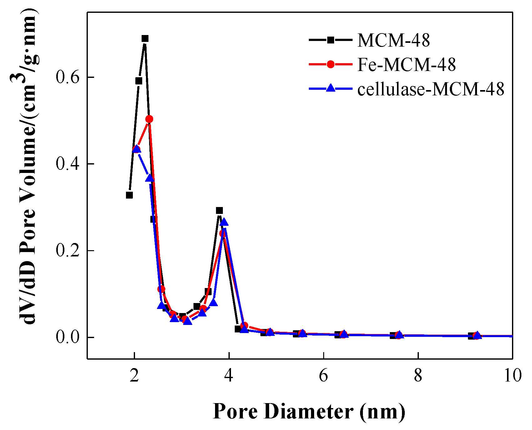
Figure 3.
FT-IR spectra of MCM-48 supported catalysts.
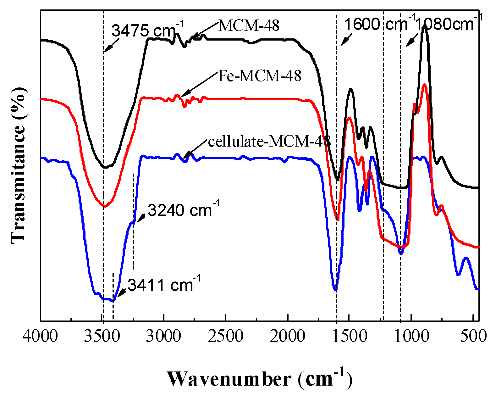
Figure 4.
TEM micrographs of MCM-48 and its supported catalysts: (a) MCM-48, (b) Fe-MCM-48, and (c) cellulase-MCM-48.
Figure 4.
TEM micrographs of MCM-48 and its supported catalysts: (a) MCM-48, (b) Fe-MCM-48, and (c) cellulase-MCM-48.
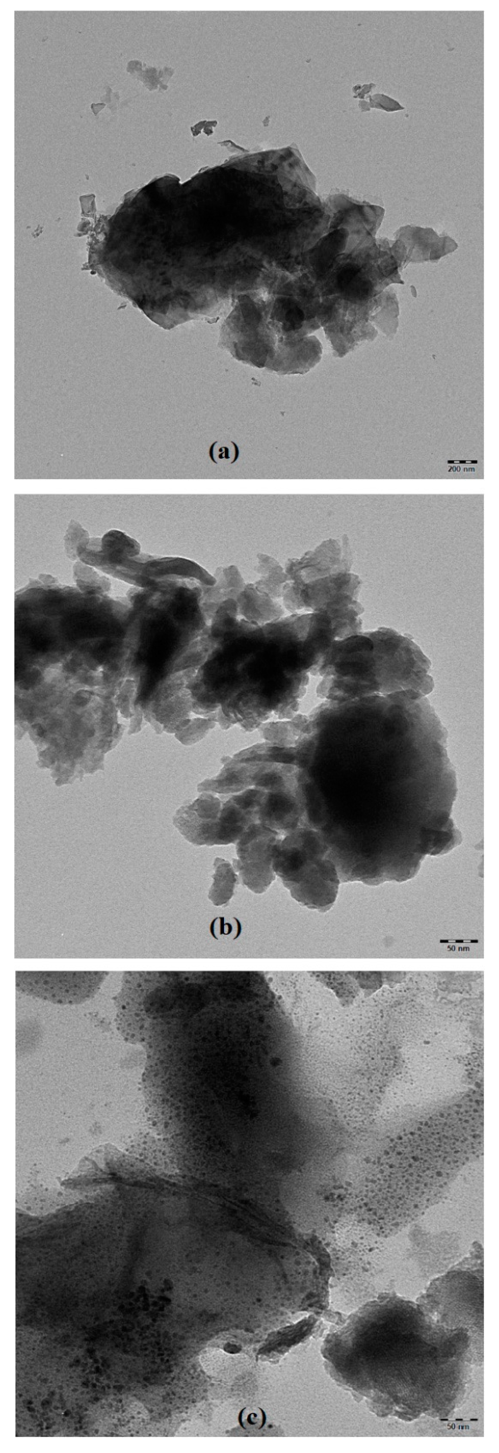
Figure 5.
SEM micrographs of catalysts: (a) MCM-48, (b) Fe-MCM-48, and (c) cellulase-MCM-48.
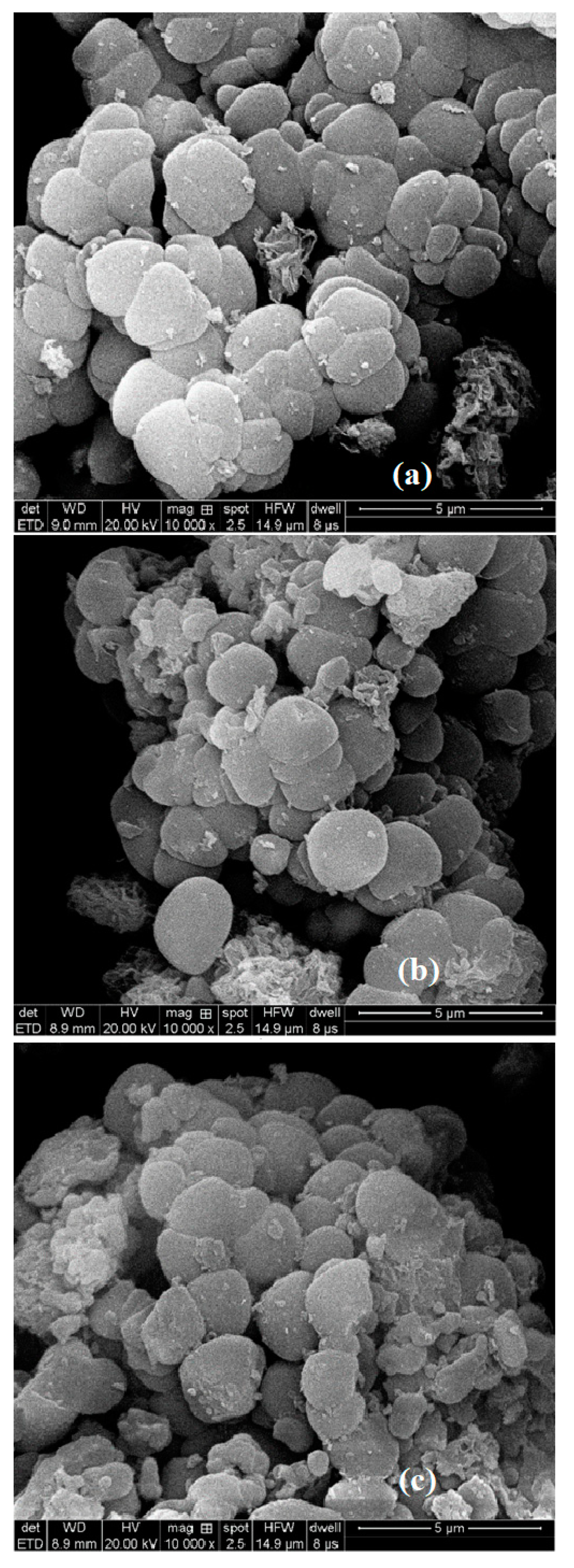
Figure 6.
XRD micrographs of catalysts.
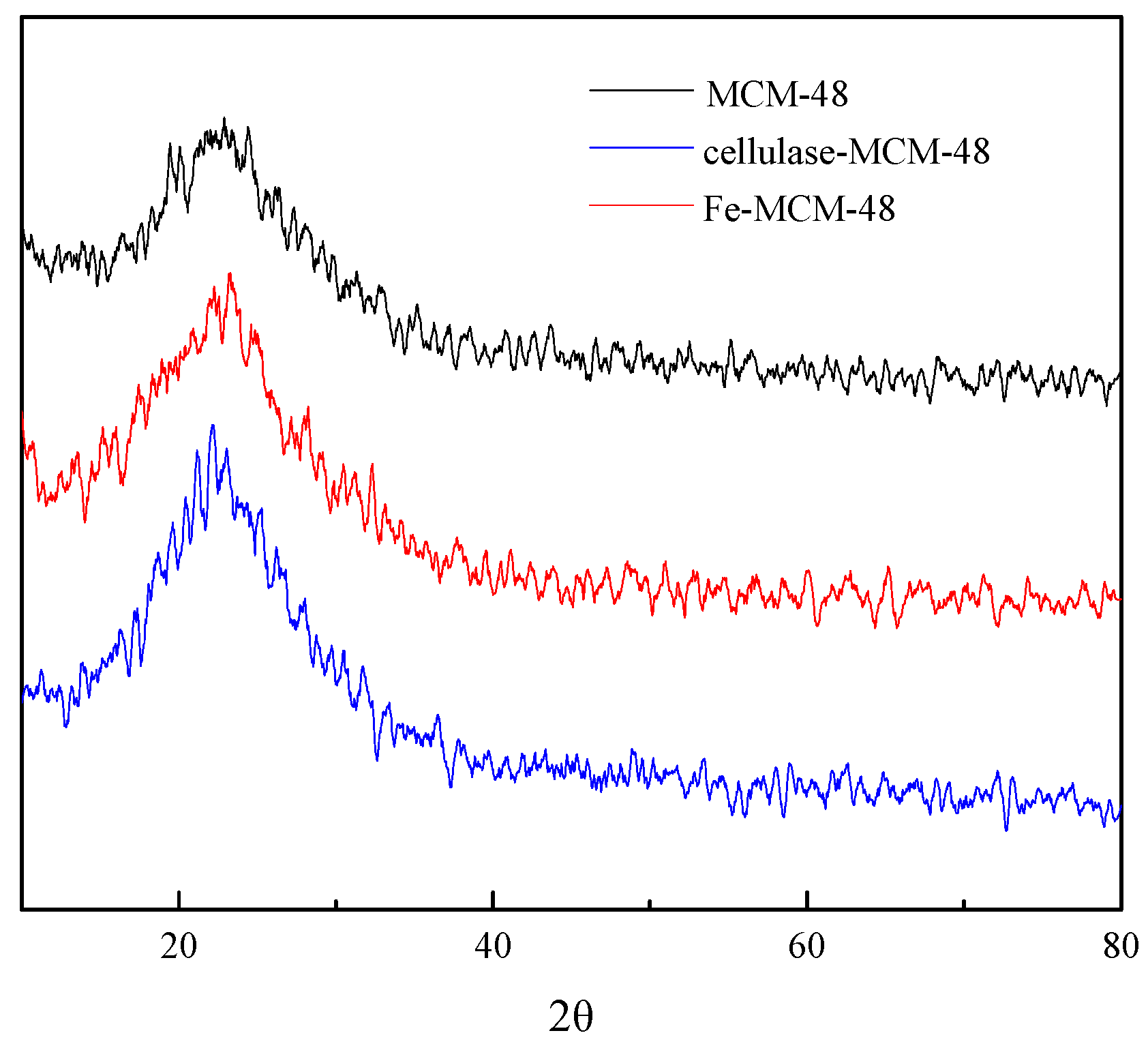
Figure 7.
Gas permeation chromatograms of products of chitosan degraded in a combined manner.

Figure 8.
FT-IR spectra of chitosan and its degradation products.


Table 1.
Textural properties of various catalysts. BET, Brunauer–Emmett–Teller.
| Entry | Samples | Fe (wt %) | Enzyme (wt %) | BET Surface Area (m2/g) | Pore Volume (cm3/g) | Average Pore Diameter (nm) |
|---|---|---|---|---|---|---|
| 1 | MCM-48 | - | - | 797.7 | 0.65 | 3.04 |
| 2 | Fe-MCM-48 | 3.4% | - | 691.8 | 0.48 | 3.50 |
| 3 | Cellulase-MCM-48 | - | 6.4% | 589.5 | 0.39 | 3.72 |
Table 2.
Results of the chitosan degradation experiment.
| Entry | Catalyst | Product Yield (%) | Average Degree of Polymerization |
|---|---|---|---|
| 1 a | Fe-MCM-48 | 92 | 6.1 |
| 2 b | Cellulase-MCM-48 b | 95 | 29.2 |
| 3 c | Fe-MCM-48 and cellulase-MCM-48 c | 93 | 3.8 |
a Reaction conditions: chitosan: 4 g; solvent: 1% H2O2 (w/v) and 0.05% acetic acid; catalyst: 0.1 g; reaction temperature: 50 °C; reaction time: 6 h; b reaction conditions: chitosan: 4 g; solvent: 0.05% acetic acid; catalyst: 0.1 g; reaction temperature: 50 °C; reaction time: 6 h; c Fe-MCM-48 was used as the catalyst for 6 h, then cellulase-MCM-48 was used for another 6 h. Other conditions were the same as entry 1.
Table 3.
Activity of catalysts at different temperatures.
| Entry a | Temperature (°C) | Product Yield (%) | Average Degree of Polymerization |
|---|---|---|---|
| 1 | 40 | 94 | 7.5 |
| 2 | 50 | 95 | 6.1 |
| 3 | 60 | 93 | 5.9 |
| 4 | 70 | 90 | – b |
a Reaction conditions: chitosan: 4 g; solvent: 1% H2O2 (w/v) and 0.05% acetic acid; catalyst: 0.1 g; reaction time: 6 h; b the degree of polymerization cannot be measured when the product deteriorates.
Table 4.
Gas permeation chromatography (GPC) molecular weight analysis a.
| Peak b | Retention Time/min | Peak Area | Molecular Mass | Proportion | Possible Components |
|---|---|---|---|---|---|
| 1 | 20.80 | 1.15 | 49 | 2.9% | - |
| 2 | 19.72 | 14.14 | 125 | 35.4% | Glucosamine |
| 3 | 19.51 | 3.20 | 170 | 8.0% | N-Acetyl-D-Glucosamine |
| 4 | 18.80 | 5.89 | 413 | 14.7% | Chitobiose Hydrochloride |
| 5 | 18.35 | 6.06 | 663 | 15.2% | Chitotriose Hydrochloride |
| 6 | 18.15 | 2.80 | 819 | 7.0% | Chitotetraose Hydrochloride |
| 7 | 17.87 | 1.21 | 1100 | 3.0% | Chitopentaose Hydrochloride |
| 8 | 17.71 | 1.39 | 1303 | 3.5% | Chitohexaose Hydrochloride |
| 9 | 17.52 | 1.49 | 1593 | 3.7% | - |
| 10 | 17.25 | 1.41 | 2120 | 3.5% | - |
| 11 | 16.89 | 0.18 | 3103 | 0.5% | - |
a Fe-MCM-48 was used as the catalyst for 6 h, then cellulase-MCM-48 was used for another 6 h; b peaks in accordance with Figure 7.
Table 5.
Relative content of degradation products obtained from depolymerization of chitosan. PD, polymerization degree.
Table 5.
Relative content of degradation products obtained from depolymerization of chitosan. PD, polymerization degree.
| Entry | Catalyst | Relative Content of Product (%) | |||||
|---|---|---|---|---|---|---|---|
| PD = 1 | PD = 2 | PD = 3 | PD = 4 | PD = 5 | PD = 6 | ||
| 1 | Fe-MCM-48 a | 22.1 | 9.7 | 10.7 | 11.5 | 19.0 | 18.4 |
| 2 | Cellulase-MCM-48 b | 0.77 | 0.60 | 0.76 | 0.71 | 0.71 | 0.66 |
| 3 | Fe-MCM-48 and cellulase-MCM-48 c | 31.8 | 12.0 | 17.7 | 16.6 | 10.4 | 7.8 |
a Reaction conditions: chitosan: 4 g; solvent: 1% H2O2 (w/v) and 0.05% acetic acid; catalyst: 0.1 g; reaction temperature: 50 °C; reaction time: 6 h; b reaction conditions: chitosan: 4 g; solvent: 0.05% acetic acid; catalyst: 0.1 g; reaction temperature: 50 °C, reaction time: 6 h; c Fe-MCM-48 was used as the catalyst for 6 h, then cellulase-MCM-48 was used for another 6 h. Other conditions were the same as entry 1.
© 2020 by the authors. Licensee MDPI, Basel, Switzerland. This article is an open access article distributed under the terms and conditions of the Creative Commons Attribution (CC BY) license (http://creativecommons.org/licenses/by/4.0/).
Share and Cite
MDPI and ACS Style
Geng, H.; Mou, Z.; Liu, Z.; Li, F.; Yang, C. Biochemical Degradation of Chitosan over Immobilized Cellulase and Supported Fenton Catalysts. Catalysts 2020, 10, 604. https://doi.org/10.3390/catal10060604
AMA Style
Geng H, Mou Z, Liu Z, Li F, Yang C. Biochemical Degradation of Chitosan over Immobilized Cellulase and Supported Fenton Catalysts. Catalysts. 2020; 10(6):604. https://doi.org/10.3390/catal10060604
Chicago/Turabian StyleGeng, Huawei, Zonggang Mou, Ziyong Liu, Fuli Li, and Cheng Yang. 2020. "Biochemical Degradation of Chitosan over Immobilized Cellulase and Supported Fenton Catalysts" Catalysts 10, no. 6: 604. https://doi.org/10.3390/catal10060604
Note that from the first issue of 2016, this journal uses article numbers instead of page numbers. See further details here.