Facile One-Pot Biogenic Synthesis of Cu-Co-Ni Trimetallic Nanoparticles for Enhanced Photocatalytic Dye Degradation

Chemistry Department, Faculty of Sciences, King Abdulaziz University, P.O. Box 80203, Jeddah 21589, Saudi Arabia
*
Author to whom correspondence should be addressed.
Catalysts 2020, 10(10), 1138; https://doi.org/10.3390/catal10101138
Submission received: 28 August 2020
/
Revised: 27 September 2020
/
Accepted: 28 September 2020
/
Published: 2 October 2020
(This article belongs to the Special Issue Photocatalytic Degradation of Organic Pollutants and Inactivation of Microorganisms Using Innovative Catalysts)
Abstract
:Biomolecules from plant extracts have gained significant interest in the synthesis of nanoparticles owing to their sustainable properties, cost efficiency, and environmental wellbeing. An eco-friendly and facile method has been developed to prepare Cu-Co-Ni trimetallic nanoparticles with simultaneous bio-reduction of Cu-Co-Ni metal precursors by aqueous extract of oregano (Origanum vulgare) leaves. Dramatic changes in physicochemical properties of trimetallic nanoparticles occur due to synergistic interactions between individual metal precursors, which in turn outclass the properties of corresponding monometallic nanoparticles in various aspects. The as biosynthesized Cu-Co-Ni trimetallic nanoparticles were initially analyzed using ultraviolet (UV)–visible spectroscopy. The morphology, structure, shape, and size of biosynthesized trimetallic nanoparticles were confirmed by transmission electron microscopy (TEM), scanning electron microscopy (SEM), and X-ray diffraction (XRD) spectroscopy. The elemental analysis was carried out by energy-dispersive X-ray (EDX) spectroscopy. Fourier transform infrared (FTIR) microscopy was carried out to explain the critical role of the biomolecules in the Origanum vulgare leaf extract as capping and stabilizing agents in the nanoparticle formation. Additionally, simultaneous thermogravimetric analysis (TGA) and differential thermogravimetry (DTG) analysis was also performed to estimate the mass evaluation and rate of the material weight changes. The photocatalytic activity of as biosynthesized trimetallic nanoparticles was investigated towards methylene blue (MB) dye degradation and was found to be an efficient photocatalyst for dye degradation. Kinetic experiments have shown that photocatalytic degradation of MB dye followed pseudo-first-order kinetics. The mechanism of the photodegradation process of biogenic Cu-Co-Ni trimetallic nanoparticles was also addressed.
1. Introduction
The explanation for the changing catalytic properties of metals lies in their synergistic doping combination of bimetallic and trimetallic nanoparticles [1]. The trimetallic nanoparticles had recently gained appealing applications in the field of catalysis and are significantly manifested in various catalytic processes [2,3]. Trimetallic nanoparticles have a greater catalytic activity [4,5], increased antimicrobial action [6], diverse morphologies [7], highly selective detection and sensitivity [8,9,10], virtuous stability [11], and chemical transformation [12], when compared with monometallic nanoparticles. The biogenic or green synthetic approach of developing metal nanoparticles is a great challenge. Polysaccharide hydrogels and sodium alginate hydrosol are used as the template in metal nanoparticle synthesis [13,14,15]. The synthesis/preparation of these multimetallic or alloy catalysts are mostly based on the traditional preparation methods viz. impregnation; mechanical alloying; melt spinning; hydrothermal preparation; microwave irradiation; co-reduction; and electro-deposition [16,17,18]. These synthesized multimetallic nanoparticles have proven excellent catalysts with more chemical reactivity than their monometallic counterparts because of enhanced electronic charge transfer; lattice strain; and geometric effects [19,20,21]. In particular trimetallic nanoparticles have better catalytic properties than their counter mono and bimetallic nanoparticles [22]. Adding other metal to bimetallic nanoparticles has improved the nature and selectivity of catalysis [23]. Thus, it has been observed as a revolution in the enhanced catalytic process by adding other metals to bimetallic nanoparticles. The catalytic nature is believed to be from the composition and nature of individual metal constituent of the trimetallic nanoparticles [24]. The amalgamation process in bimetallic and trimetallic nanoparticles through dissimilar metal additive has proven to have appreciable catalytic properties rather than the individual metal constituents [25]. Moreover; various investigations have been carried out regarding the synthesis and application of trimetallic nanoparticles. The trimetallic nanocatalysts of Pt@Fe@Ni were applied in fuel cells to enhance oxygen reduction reaction activity compared to the Pt electrode catalyst [26]. In the reduction of 4-nitrophenol, the trimetallic nanoparticles such as Al2O3@Ag/Au core-shell; Pt@Pd@Bi nanowires; Cu@Ni@Pt dendrites; and Au@Pd@Ru core-shell played a vital role [27,28,29,30]. Similarly, Zhang et al. reported trimetallic nanoparticles of Au-Pt-Pd with much better catalytic activity for the glucose oxidation than monometallic and bimetallic nanoparticles [31]. Matin et al. synthesized core-shell trimetallic nanoparticles of Pd/Co@Pt with enhanced electrocatalytic performance in oxygen reduction reaction compared to commercial Pt/C catalyst [32]. Yurderi et al. and Wang et al. reported the higher catalytic activity of Ni@Ag@Pd and Ni@Au@Pd, respectively, in the dehydrogenation of formic acid [29,33]. Moreover, Tayal et al. demonstrated that Ir/Pt/Sn electrocatalyst showed higher activity towards ethanol oxidation as compared to the corresponding bimetallic nanoparticles [34]. Very recently, the catalytic activity of the Pd synthesized trimetallic nanoparticle was tested using the Suzuki C–C coupling reaction [35]. Similarly, Fe@Ag@Pd trimetallic nanocatalyst retain its catalytic property after being used six times for formic acid degradation [36].
The photocatalytic process has gained tremendous attention for water-splitting reactions. It plays a significant role in providing cost-efficient, environmentally friendly technology to eliminate the chemical contaminants and plays a vital role in ethanol’s chemical transformation into CO2 and water [37,38,39,40,41]. Recently, photocatalysts with excellent photocatalytic performances after regeneration have been developed for photocatalytic oxidation of ethanol under visible light irradiation [42]. Zhen et al. have developed a highly stable and promising photocatalysts of Co-Cu-P/GP and Co-Ni-P/GP for H2 generation reactions under visible light irradiation [43]. Most of the organic dyes have many roles in human life, as the use of dye is increasing rapidly in different dying industries, including printing, textile, plastic, and leather industries, and most of these synthetic organic dyes are non-biodegradable and are highly toxic [44,45]. They are resistant to direct degradation by sunlight and other light sources and, therefore, they are considered unrelenting pollutants [46]. The release of contaminated water into the atmosphere poses a severe threat to marine and human life. Dyes are the primary pollutants produced from pharmaceutical, Kraft bleaching, plastic, textile, and other industries [47]. The photochemical degradation of dyes is one of the significant aspects able to alleviate the hazardous effect of dyes. The conventional treatment methods such as electrochemical, chemical, ultrafiltration, and adsorption were not found to be suitable for removing such dyes from wastewater [48]. The wastewater beside other pollutants as dyes included the methylene blue as a common azo dye [49]. The research focus is on developing bio-synthesized trimetallic photocatalysts to assist dye degradation by the process of photocatalysis. Consequently, we reported the seedless one-pot biosynthesis of Cu-Co-Ni trimetallic nanoparticles using phytochemicals/biomolecules present in the aqueous extract of Origanum vulgare leaves, which act as reducing as well as stabilizing/capping agents. A comprehensive photocatalytic degradation of methylene blue (MB) dye using as-synthesized trimetallic nanoparticles has been reported. Such trimetallic nanoparticles have advantages over mono and bimetallic nanoparticles for catalytic properties.
2. Results and Discussion
Several studies have shown that plant extracts aim to reduce metal ions to compact nanoparticles and stabilize them to prevent agglomeration, through the chelate-effect binding to the metal atom [50,51]. Origanum vulgare L. being a rich source of several bioactive phytochemicals, can productively be proven advantageously into the biosynthesis and fabrication of metallic nanoparticles [52]. The phytochemical analysis of Origanum vulgare L. accentuates the presence of rosmarinic acid, caffeic acid, protocatechuic acid, a glycoside of protocatechuic acid, and derivative of rosmarinic acid, 2-caffeoyloxy-3-[2-(4-hydroxy benzyl)-4,5-dihydroxy]phenylpropionic acid in certain amounts (Figure 1). These specific kinds of phytochemical, including polyphenols, flavonoids, and phenolic acids, function as a bio-reducing and stabilizing agent without external agents or surfactants.
Among these phytochemicals, water-soluble glycosides, rosmarinic acid, and rosmarinic acid congeners are the main compounds responsible for bio-reduction and stabilization of Cu, Ni, and Co ions according to their corresponding reduction potential into Cu-Co-Ni trimetallic nanoparticles. The hydroxyl groups of these water-soluble biomolecules tend to interact with metal ions in solution to form chelates due to the loss of hydrogen from hydroxyl groups. The probable formation of H+ radical is attributed to the electron losing property of biomolecules and results in the reduction of Cu2+, Co2+, and Ni2+ metal ions into Cu-Co-Ni hybrid trimetallic nanoparticles. The possible reduction potential of the metals involved in the biosynthesis of trimetallic Cu-Co-Ni hybrid from Origanum vulgare L. extract can be summarized by anticipating the mechanism as depicted in Figure 2. However, the formation of Cu-Co-Ni trimetallic nanoparticles using Origanum vulgare L. extract was physically observed from the color change of precursor solution from dark brown to the dark greenish-brown solution, which indicated the formation of stable trimetallic nanoparticles.
Under similar circumstances, no color change was observed on mixing three different metal ions in the absence of the aqueous extract of Origanum vulgare leaves. Biosynthesized Cu-Co-Ni trimetallic nanoparticles were observed at wavelengths of 200 to 800 nm in the ultraviolet (UV)–visible absorption spectrum. The UV–visible spectrum of Origanum vulgar L. extract reveals absorption bands at 326nm and 280nm distinguished by the absorbance of benzoyl-related phyto-biomolecules. The UV–visible absorption spectrum of Origanum vulgare L. extract and biosynthesized Cu-Co-Ni trimetallic nanoparticles are revealed in Figure 3 The UV–visible absorption spectrum of the biosynthesized trimetallic nanoparticles displays two prominent small shoulder peaks at 270 nm, and 320 nm attributed to the presence of phytochemicals and act as capping/stabilizing agents in the biosynthesis of Cu-Co-Ni trimetallic nanoparticles. The presence of peaks in Origanum vulgare extract and the steepness of peaks in the bio-synthesized Cu-Co-Ni trimetallic nanoparticles demonstrated the presence of phytochemicals performing the role of bio-reducing and stabilization along with the functionalization of the surface of the biosynthesized trimetallic nanoparticles. Using the Tauc plot (αhv)2 versus (hv) and extrapolating the linear portions of curves into the energy axis in accordance to αhv = B(hv-Eg)1/2, where α is the absorption coefficient, hv is the photon energy, Eg is the direct bandgap energy, and B is a constant, the bandgap energy (Eg = 3.06 eV) of the biogenic Cu-Co-Ni trimetallic nanoparticles was measured from the UV–visible spectrums [53].
Fourier transform infrared (FTIR) spectra have been scrutinized to interpret different functional groups in reaction mixtures of metal particles and biomolecules. To determine the presence of bioactive molecules involved in the bio-reduction and stabilization of Co2+, Ni2+, and Cu2+ metal ions in trimetallic hybrid nanoparticles, FTIR spectroscopic analysis of plant extract and the synthesized hybrid metallic nanoparticles was conducted in 4000-400 cm−1 range. The plant extract’s character as reducing, stabilizing/capping agent was inferred by comparing the FTIR spectra of Origanum vulgare L. extract and biosynthesized Cu-Co-Ni trimetallic nanoparticles, as shown in Figure 4. The FTIR of Cu-Co-Ni trimetallic nanoparticles and the phyto-synthesized extract showed (Figure 4) typical peaks at 3456 cm−1, 2945 cm−1, 1640 cm−1, 1573 cm−1, 1423 cm−1, 1360 cm−1, 1253 cm−1, 1175 cm−1, 1081 cm−1, 992 cm−1, 873 cm−1 and 622 cm−1, respectively. The obtained peaks at 3456 cm−1 of trimetallic nanoparticles and extract were from –OH stretching vibration of phenolic compounds and –OH stretching vibration due to the presence of water molecules, respectively. The observed peak at the absorption bands 2945 cm−1 is from the –C-H symmetrical vibration because of the saturated hydrocarbons, while the peak at 622 cm-1 was from the –C–H bending vibrations. The presence of bands at 1573 cm−1 and 1423 cm−1 are expected from the symmetric stretching of –C–C=C– from aromatic compounds onto the surface of nanoparticles, and an additional absorption band at 1640 cm−1 is due to the α,β-unsaturated carbonyl CH=CH–C=O functional group, which confirms the presence of rosmarinic acid in the reaction mixture. The presence of peaks as analyzed from FTIR spectroscopy confirms the occurrence of phenolic, aromatic, and alcoholic compounds in leaf extract predominantly responsible for stabilizing and forming Cu-Co-Ni trimetallic nanoparticles.
2.1. Surface Morphological Characterization of Cu-Co-Ni Trimetallic Nanoparticles
The surface morphology and the particle size of the biosynthesized Cu-Co-Ni trimetallic nanoparticles were analyzed by scanning electron microscopy (SEM) and transmission electron microscopy (TEM) (Figure 5). TEM observation clearly demonstrates the formation of nanoflakes and confirms the 2-D flake-like morphology (Figure 5a,b). It is clear from the TEM images that the random nanoflakes are lightly packed together without any apparent origin or center. SEM images of as-synthesized trimetallic nanoparticles show they possess nanoflake-like morphology and exhibit assemblage and agglomeration of the particles (Figure 5e). It is clear from the SEM micrographs of biosynthesized Cu-Co-Ni trimetallic nanoparticles that the agglomeration of the nanoflakes results in structural porosity (Figure 5e). The interpenetrated growth of nanoflakes on each other and the structural porosity is clearly visible from the gaps between the nanoflakes. The agglomeration of biosynthesized Cu-Co-Ni trimetallic nanoparticles increases the particle size. The observed results suggested that the individual nanoflakes instantaneously formed were associated as the growth of the flakes continued. The particle size distribution and average particle size (28.25 nm) were estimated by using a particle size analyzer (ImageJ software) by plotting the diameter histogram, and the average particle size was calculated by using Gaussian fit (Figure 5f).
The elemental composition analysis and targeted elemental mapping of the biosynthesized Cu-Co-Ni trimetallic nanoparticles using Origanum vulgare L. extract as reducing and stabilizing agent were elucidated by energy-dispersive X-ray (EDX) spectroscopy analysis shown in Figure 6a,b). The individual elemental composition (in weight %) as determined by the EDX spectrum was 17.34, 10.37, and 34.01 for Co, Ni, and Cu, respectively, with an approximate weight ratio of 2:1:3 (Figure 6b). Thus, Co, Ni, and Cu elemental peaks conform to the formation of Cu-Co-Ni trimetallic nanoalloy structures. The presence of C and O weak signals may be due to the presence of surface-bound biomolecules or phytochemicals, which act as capping agents on the surface of the Cu-Co-Ni trimetallic nanoalloy particles. The selected area elemental mapping of Cu-Co-Ni trimetallic nanoparticles is represented by red, green, and white for Cu, Co, and Ni, respectively (Figure 6a).
The phase structure and crystallinity of as-synthesized trimetallic nanoparticles were scrutinized by means of X-ray powder diffraction (XRD) pattern analysis. It is well documented that biosynthesized CuNPs’ diffraction peaks (2θ) correspond to Miller indices (111), (200), and (220) planes following the standard Cu (JCPDS No. 04-014-0265) [54] and NiNPs exhibit XRD diffractions peaks (2θ) indexed as (111), (200), (220), and (311) crystal planes of Ni (JCPDS No. 04-0850) [55]. Similarly, in the case of CoNPs, the XRD pattern illustrates that diffraction peaks (2θ) are assigned to (111), (200), and (220) planes that match with the Co (JCPDS No. 89-7093) [56]. Moreover, the XRD analysis of as-synthesized biogenic Cu-Co-Ni trimetallic nanoparticles showed typical diffraction peaks located at 2θ value of 42.85°, 49.85°, 74.21°, and 89.65° are assigned to (111), (200), (220), and (311) crystal planes, respectively (Figure 7). The typical perusal of biosynthesized Cu-Co-Ni trimetallic nanoparticles’ diffractogram describes numerous sharp, intense peaks that determine the crystallinity and formation of Cu-Cu-Ni trimetallic nanoparticles. The slight shift in the 2θ peak values confirms the formation of trimetallic nanoalloy particles and confirms the presence of all constituents in the synthesized material [57,58,59]. We did not observe any additional metal oxide peaks in the XRD spectra, an indication of the formation of pure hybrid Cu-Co-Ni trimetallic nanoparticles. Scherrer’s equation has been used to estimate the particle size of the biosynthesized trimetallic nanoparticle.
where β indicates maximum width at half-length maximum (FWHM) in radians, λ refers to the X-ray wavelength (1.4506Å), θ refers to the Bragg diffraction angle, and k is a constant (0.9), and d refers to the crystal-size of the biosynthesized Cu-Co-Ni nanoparticles. The average particle size of the biosynthesized Cu-Co-Ni nanoparticles was estimated to be 15 nm using Scherrer’s equation (Equation (1)).
The presence of the phytochemicals as stabilizing/capping agents on the surface of the biosynthesized Cu-Co-Ni trimetallic nanoparticles was further analyzed by thermogravimetric analysis (TGA) and differential thermogravimetry (DTG) analysis [60]. Thermogravimetric experiments with a heating rate of 10 °C per minute were conducted under a nitrogen atmosphere. The thermal stability of Cu-Co-Ni trimetallic nanoparticles along the thermal degradation pattern was obtained are shown in Figure 8. The thermal degradation pattern of the synthesized Cu-Co-Ni nanoparticles was stable up to 800 °C with an approximate weight loss of 40%. The apparent weight losses as observed in Figure 8 are from 60 °C to 260 °C, 260 °C to 470 °C, and 470 °C to 680 °C respective. The synthesized trimetallic Cu-Co-Ni nanoparticles, as per the obtained DTG curve (black line), are thermally stable below 200 °C. The appeared DTG peak at 169.99 °C in a region of TGA 60–260 °C (weight loss 7.35%) was attributed to the evaporation of adsorbed moisture from surroundings by phytochemicals capped onto the surface of Cu-Co-Ni trimetallic nanoparticles. The pragmatic intensity peak at 317.74 °C by DTG analysis and the region of TGA between 260–470 °C (weight loss 18.58%) was behind the thermal decomposition of biomolecules, including amino acids as reducing agents in stabilizing Cu-Co-Ni trimetallic nanoparticles. However, the presence of phenolic acids and flavonoids of leaf extract as stabilizing and capping agents in the biosynthesis of Cu-Co-Ni trimetallic nanoparticles was determined by the thermal decomposition peak at 550.34 °C of DTG analysis further reinforced by TGA region between 470–680 °C (weight loss 14.31%), respectively. The results obtained from the DTG and TGA analysis showed the biosynthesized Cu-Co-Ni trimetallic nanoparticles from Origanum vulgare leaves are thermally stable with a small size. The as-obtained results elicited these trimetallic nanoparticles can be used as a photocatalyst upon their obtained biogenic surfaces, thermal stability, and smaller size traits.
2.2. Photochemical Activities Cu-Co-Ni Nanoparticles
The photocatalytic capability of biosynthesized Cu-Co-Ni trimetallic nanoparticles was explored on target toxic dye methylene blue under dark and UV light conditions. Methylene blue is a stable dye with a characteristic absorption band λmax at 663 nm. Under dark conditions, the decoloration of MB by biosynthesized Cu-Co-Ni nanohybrid photocatalyst was negligible. The photocatalytic efficiency of as-synthesized nanoparticles was monitored by recording the UV–visible spectra of the experimental solution at a 10 min time interval. Before the irradiation of UV light, the experimental solution containing MB dye (100 ppm) and Cu-Co-Ni photocatalyst (30 mg) at 25 °C was kept in the dark with constant stirring to achieve significant equilibrium between MB dye and Cu-Co-Ni nanoparticles. On the addition of the photocatalyst under optimal experimental conditions under UV light irradiation, the peak intensity of the MB starts decreasing, and its gradual disappearance with a raised peak at 663 nm incites the degradation of MB dye as shown in Figure 9a. It was accentuated more than 50% MB dye degradation was completed at 50 min of reaction time. Dye degradation percentage under the optimal experimental condition for a fixed time interval of 10 minutes was calculated as per the following equation (Equation (2)):
where Ao is the initial absorbance, and At is the absorbance at time t of the dye solution. Further, Co is the initial dye concentration, and Ct is the concentration of the dye solution at the time t.
During the said experimental condition, 92.67% photodegradation of MB was achieved in 100 min (Figure 9b). For the dye MB by Cu-Co-Ni nanoparticles, the values of concentration with time graphs were used to study the kinetics behind the reaction. As per the obtained standard deviations, the pseudo-first order kinetics equation was selected as follows:
where Kapp is the pseudo-first order constant in min−1. The linear correlation was obtained with the degradation pseudo-fist order rate constant of 0.02588 min−1 with high precision and linear fit R2 values of 0.935.
The catalytic degradation efficiency of Cu-Co-Ni nanoparticles was studied by observing the effect of initial MB dye concentration on the decolorization process under UV light irradiation by changing the initial dye concentrations 100ppm to 500ppm and photocatalyst (Cu-Co-Ni nanoparticles) concentration of 30 mg. The degradation depended very heavily on their concentration, as the dye molecules sorbed on the photocatalyst surface affect the formation of hydroxyl radicals. The photocatalytic process shows the highest activity with 30 mg of Cu-Co-Ni photocatalyst load, and in this manner ln(Ct/Co) versus time (min) was plotted with varying the initial MB dye concentrations to investigate the effect of the MB dye on the degradation process as shown in Figure 10a. The data analysis offers a linear relationship (R2) of 0.935. The photocatalytic degradation rate of dye quenches with an increase in the dye concentration in a range of 100 ppm to 500 ppm was observed. The apparent rate constant (kapp) values of the photocatalytic degradation were estimated to be 0.02588, 0.01138, 0.00523, 0.00339 and 0.00178 min−1 for 100, 200, 300, 400 and 500 ppm, respectively. The rate constants, degradation percentages, and the corresponding correlation coefficients (R2) have been determined from the plots and recorded in Table 1. The highest dye degradation achieved is because of the maximum available •OH radicals on the photocatalyst’s surface, and the decrease in the dye degradation percentage with an increase in dye concentration might be accredited to the inhibition of hole/electron production. This impact may be because the initial concentration of MB dye molecules was increased, and more MB molecules could be adsorbed on the photocatalyst surface and occupy the active sites of Cu-Co-Ni nano catalytic material. This process results in fewer hydroxyl radicals (•OH) on the surface of the as-synthesized photocatalyst. Therefore, the oxidation cycle requires higher photocatalyst dosage and reaction time for higher concentrations of MB dye molecules. Consequently, the above results indicate that nanocomposite Cu-Co-Ni may serve as an effectual photocatalyst for even higher MB dye concentrations.
The photocatalyst loading was optimized in the presence of different quantities of photocatalyst Cu-Co-Ni nano-hybrid (10 mg, 15 mg, 20 mg, 25 mg, and 30 mg) (Figure 10b). With the increased concentration of the photocatalyst, the efficiency of photodegradation was improved, which can be a result of an increase in the adsorbed photons and a large number of reactive sites on the photocatalyst surface were measured and the results are shown in Figure 10b. While the concentration of the photocatalyst was augmented from 10 to 30 mg, the degradation effectiveness of MB dye increased linearly and degraded the MB dye molecules and the apparent photocatalytic degradation percentage for various amounts of the photocatalyst dosages were established in Figure 10b. More efficiency of degradation was observed with the increase in the photocatalyst dosage, which increased active sites on the photocatalyst surface and generated free hydroxy radicals (•OH). The further rise in photocatalyst has no noticeable impact on the efficiency of degradation. This may be caused by the aggregation of nanocomposite particles of Cu-Co-Ni. Nanoparticles are applied to the number of active sites on the photocatalyst. These aggregated nanoparticles also serve as hydroxyl radical scavengers. Hence, the optimal degradation efficiency for the 30 mg photocatalyst dose was achieved.
2.3. The Mechanism behind the Photocatalytic-Degradation of MB
A mechanistic diagram has been proposed based on our control experiments and observations (Figure 11). The photocatalyst’s electron storage behavior can promote the faster formation of electron-hole pairs under UV light irradiation. When the UV light illuminates an aqueous solution of synthesized materials in the presence of water containing dissolved oxygen and organic dye molecules, the dye molecules can degrade. The decrease in the absorption peak at 663 nm from azo bonds of MB by bio-synthesized Cu-Co-Ni nanoparticles was observed physically from diminishing the blue color of dye to colorless after 100 min of reaction time. The electron-hole-pair surface transmission process over nanocomposites makes them more acceptable for the formation of reactive oxygen species (ROS). These generated ROS-like hydroxyl radicals and superoxide radicals, are accountable for MB dye degradation. The hybrid nanoparticles of Cu-Co-Ni provide a large surface area to the available MB dye molecules. As a result, additional MB dye molecules will interact with the Cu-Co-Ni photocatalyst, increasing photodegradation efficiency. Because of induced transition from valence band (VB) electrons to the conduction band (CB) leaving a hole in the VB, electron (e-)-hole (h+) pairs are produced when light is incidental to the MB dye solution. Such holes are permitted to react with OH ions and produce •OH radicals in the aqueous MB dye solution while electrons can react with dissolved O2 and generate O2•− radicals. Those radicals eventually degrade MB dye molecules into tiny non-toxic degraded products.
It is well known that the hydroxyl radicals (•OH), superoxide radical anions (O2•™), hole (h+), and electrons (ē) play a vital role in the photocatalytic degradation of organic dyes [61,62]. A series of scavenging experiments were carried out to investigate the role of active species responsible for the degradation of MB dye using Cu-Co-Ni trimetallic nanoparticles as photocatalysts. Radical trapping experiments were performed by using isopropyl alcohol (IPA), benzoquinone (BQ), and ammonium oxalate (AO) as radical scavengers for •OH radical, O2•− radical, and hole (h+), respectively [61,62]. In the absence of radical scavengers, the degradation efficiency of the Cu-Co-Ni trimetallic nanoparticles was 92.67%. The results indicate that the scavenger-free photocatalytic process shows the maximum degradation rate under optimal experimental conditions compared to the scavenger-assisted system. When IPA (•OH radical scavenger), BQ (O2•− radical scavenger), and AO (hole (h+) scavenger) were added into the reaction solution during the MB degradation, the degradation ratios of MB dropped to 17.23%, 58.52%, and 88.63% within 100 min, respectively (Figure 12a). The results revealed that the addition of IPA showed a significant decrease in the degradation of MB dye by quenching hydroxyl radical (•OH) produced in the reaction mixture, implying the dominant role of •OH radicals in the process of MB dye degradation. Furthermore, with the addition of BQ and AO, the photodegradation of MB is slightly reduced, providing further evidence of the presence of hydroxyl radicals as the main reactive species [63,64]. The BQ captures photogenerated electrons and reduces the possibility of double-electron reduction, while AO captures the hole during the photocatalytic reaction process. Thus, the possible mechanism behind the degradation of MB by bio-synthesized Cu-Co-Ni nanoparticles can be summarized as:
2.4. Stability and Reuse of the Photocatalyst
In the photocatalytic degradation process, functional applicability, stability, and recyclability play an essential role. The catalytic system’s stability and reusability were evaluated by recycling the MB degradation reactions under visible light through the Cu-Co-Ni photocatalyst. In each of the samples, the photocatalyst was isolated by centrifugation from the suspension and washed over deionized water and dried for 1 hour at 100 °C. After washing and drying, the photocatalyst maintained its initial dark greenish-brown color. As seen in the Figure 12b, Cu-Co-Ni trimetallic nanoparticle’s photocatalytic performance shows only a slight decrease in recycling experiments even after five consecutive cycles under the same environment, which can be due to the minor loss and deactivation of the photocatalyst in the cycling experiment, suggesting its high stability and durability. A comparative analysis of the photocatalytic dye degradation efficiencies of the reported photocatalysts in the literature with as biogenic Cu-Co-Ni trimetallic photocatalyst is important and is exemplified in Table 2 It is clear from the results that the biogenic Cu-Co-Ni trimetallic nanoparticles have efficient photocatalytic efficiency. Therefore, Cu-Co-Ni trimetallic nanoparticles could be excellent photocatalysts for dye degradation and have excellent reusability results. The synthesis procedure of Cu-Co-Ni trimetallic nanoparticles reported in our study is straightforward, green, and cost-efficient compared to the other reported methods.
3. Experimental
Copper(II) nitrate trihydrate (Cu(NO3)2·3H2O) molecular weight = 241.60 g/mol, cobalt(II) nitrate hexahydrate (Co(NO3)2·6H2O) molecular weight = 291.03 g/mol, nickel(II) nitrate hexahydrate (Ni(NO3)2·6H2O) molecular weight = 290.79 g/mol were purchased from Sigma Aldrich (Bangalore, India)with the purity of 99%. Ethanol (99.8%) was purchased from Sigma Aldrich (Bangalore, India). The scavenger reagents, including isopropyl alcohol (IPA, 99.5%), benzoquinone (BQ, 98%), and ammonium oxalate (AO,99.5%) were also purchased from Sigma Aldrich. Ultrapure water (Millipore, 18.2MΩ cm, Jeddah, Saudi Arabia) was used in all experiments. Fresh leaves of Origanum vulgare were acquired from the local market. All the glassware used for the synthesis of trimetallic nanoparticles was washed by aqua regia followed by rinsing with double-distilled water and then oven-dried.
3.1. Preparation of Origanum Vulgare Leaves Extract
The aqueous extract of the Origanum vulgare leaves was prepared after thorough washing of the leaves under running tap water to remove all dust particles, followed by several piles of washing with double-distilled water. These Origanum vulgare leaves were allowed to dry in room temperature (30 °C), and 5 g of the leaves were crushed into a fine powder. The powder was poured into a 250 mL beaker containing double distilled water and heated at 50 °C for 30 min followed by filtration with filter paper (Whatman No. 1) to gain a clear aqueous extract of Origanum vulgare. This extract was kept in a cold and dark place for further experimental use for the preparation of trimetallic nanoparticles.
3.2. Biogenic Synthesis of Trimetallic Nanoparticles
In a typical experiment, all metal-salt precursors, including Cu (0.01 M, 10 mL, Co (0.01 M, 10 mL), Ni (0.01 M, 10 mL), were mixed in a 250 mL beaker and heated at 50 °C under continuous constant stirring on a magnetic stirrer heating plate. A 20 mL of Origanum vulgare leaf extract was added to the trimetallic salt solution under constant stirring at 40 °C until the formation of dark greenish-brown color was achieved, resulting in the formation of trimetallic nanoparticles. Initially, the formation of the nanoparticles was observed by recording the UV–visible spectra as the color change in the reaction mixture was observed. The reaction was allowed to complete and the reaction mixture to cool down, followed by centrifugation of the solution for 20 min at 20,000 rpm to collect pure and stable trimetallic nanoparticles. The acquired Cu-Co-Ni trimetallic nanoparticles were washed several times with distilled water followed by ethanol to remove impurities from the surface of the as-synthesized nanoparticles. Cu-Co-Ni trimetallic nanoparticles were dried in an oven at 100 °C for 12 h and finally stored in a dry place under vacuum conditions for further physicochemical characterization and application as photocatalysts.
3.3. Characterization
The biogenic Cu-Co-Ni trimetallic nanoparticles prepared by using Origanum vulgare leaf extract as a reducing and stabilizing/capping agent were analyzed using a UV–vis double beam spectrophotometer (UV-1601, Shimadzu, Kyoto, Japan) between the range 200–800 nm. The UV–visible absorption spectrum was recorded using quartz cuvette in comparison with reference to deionized double distilled water. An advanced X-ray diffractometer (Bruker AXS D8 advance, Madison, USA) with Cu Ka (k = 1.5418 Å) radiation was operated to analyze XRD patterns of the samples. The morphology of biogenic trimetallic nanoparticles was observed under SEM (JSM-6380 LA, JEOL, Tokyo, Japan) operated at 30 kV. The SEM samples were prepared by placing a thin layer of fine powder of synthesized nanoparticles on a sample holder and placed in the specimen chamber to record the SEM images. The as-synthesized trimetallic nanoparticles were examined for their chemical/elemental composition by an EDX spectroscopy equipped SEM (JSM-6380 LA, JEOL, Tokyo, Japan) at room temperature. TEM observation was performed on a JEOL-2010 Microscope (JEOL, Tokyo, Japan), with an acerbating voltage of 80 kV. The TEM image was observed to interpret the particle size, predominantly exposed crystal plane, and nanoalloys distribution. All TEM samples were prepared by placing a droplet of the biosynthesized trimetallic nanoparticle solution on a copper grid that was completely dried at room temperature. TGA-DTG curves of trimetallic nanoparticles were examined using Perkin-Elmer Pyris Diamond (Perkin-Elmer, Waltham, MA, USA) under the nitrogen atmosphere at a heating rate of 10 °C min−1 in an open alumina pan. FTIR spectral analysis was accompanied to interpret the functional groups of bio-synthesized trimetallic nanoparticles using a Bruker FTIR spectrophotometer (Model: ALPHA II, Bruker, Billerica, MA, USA) within the range of 400–4000 cm−1.
3.4. Photocatalytic Degradation
The photocatalytic activity of as-synthesized Cu-Cu-Ni trimetallic nanoparticles was evaluated by determining the dye degradation efficiency under photocatalytic experimental conditions. The target dye used was methylene blue (MB), which is an anionic dye. All experiments with degradation reactions were conducted at 25 °C, with the use of a UV–visible spectrophotometer to track the progress of all reactions. MB shows maximum absorption at a wavelength of 663nm, and thus the photodegradation of the dye was controlled by observing changes in the overall absorption, followed by measurement of the first order constant. A Xenon (Xe) arc lamp (150 W) with irradiation intensity of 50 mW/cm2 was used to test the photocatalytic activity of the prepared photocatalysts for the degradation of MB under UV light radiation. In a quartz reactor at room temperature, the photocatalyst (30 mg) was dispersed into 30 mL of 100 ppm aqueous MB dye solution. The reaction solution was continuously stirred in the dark for 30 min before the light was switched on to ensure an adsorption-desorption equilibrium was formed. Before and during the illumination, the suspension was magnetically stirred. During the experiment, a 3 mL sample solution was drawn from the device at some time interval. Following the irradiation and removal by centrifugation of the photocatalyst nanoparticles, the catalyst-free MB dye concentration in the solution was analyzed based on its characteristic optical absorption at 663 nm using the UV–visible spectrometer (Shimadzu, Kyoto, Japan) to calculate the decline in MB dye concentration with irradiation time based on the Lambert–Beer law (A = ɛ × l × C), where A is the absorbance of the solution at 663 nm, ɛ is the molar extinction coefficient, l is the width of the cuvette, and C is the concentration of the MB dye [74]. The results indicated that trimetallic nanoparticles degraded MB completely in 100 min, with 92.67% conversion efficiency.
Experiments for photocatalyst recycling have also been conducted under optimal reaction conditions. To avoid catalyst loss, the samples collected after centrifugation were washed three times with deionized water, dried at 100 °C for 5 h, and used directly without any additional treatment for MB dye degradation experiments in the subsequent recycling process. The radical trapping experiments were performed to detect the role of active species responsible for photocatalytic degradation of MB dye. A 1.0 mM concentration of IPA, BQ, and AO was added to 30 mL (100 ppm) MB dye solution containing 30 mg of Cu-Co-Ni nanoparticles and irradiated by UV light under optimal experimental conditions to trap hydroxyl radicals (•OH), superoxide radical anions (O2•−), and holes (h+), respectively.
4. Conclusions
We established an eco-friendly, facile, seedless, one-pot approach to synthesize flake-shaped trimetallic Cu-Co-Ni nanoparticles using phyto-biomolecules present in the aqueous extract of oregano (Origanum vulgare) leaves as reducing and stabilizing/capping agents. The formation of biogenic trimetallic Cu-Co-Ni nanoparticle formation was confirmed by various spectroscopic techniques, including UV–visible spectroscopy, FTIR, XRD, and EDX analysis. The biogenic Cu-Co-Ni trimetallic nanoparticles were found to have irregular morphology with an average particle size of 28.25 nm. The bio-synthesized Cu-Co-Ni nanoparticles, as such, were used as a photocatalyst for the photocatalytic degradation of MB under UV light irradiation. The results from the UV–visible spectrum showed that the photocatalytic degradation was significant at 663 nm and the lowering peak intensity for MB by Cu-Co-Ni nanoparticles conforms to the complete degradation of MB dye upon photodegradation within 100 minutes. The result shows that the photocatalytic degradation of MB by Cu-Co-Ni nanoparticles followed pseudo-first-order kinetics as interpreted from the obtained linear correlations. Five cycles of photocatalytic degradation runs were performed to check the biogenic Cu-Co-Ni trimetallic nanoparticles’ stability and reusability. The possible photocatalytic degradation mechanism over Cu-Co-Ni nanoparticles was proposed, and the role of •OH radicals was dominant in the photodegradation process. This study could provide an insight into developing novel photocatalysts for large-scale degradation of toxic materials.
Author Contributions
A.A.A. and M.A.M. designed and conducted the experiments. Both authors analyzed the data and wrote the article. All authors have read and agreed to the published version of the manuscript.
Funding
Only funding received from DSR.
Acknowledgments
This project was funded by the Deanship of Scientific Research (DSR) at King Abdulaziz University, Jeddah, under Grant No. (G:373-130-1439). The authors, therefore, acknowledge with thanks DSR for technical and financial support.
Conflicts of Interest
The authors declare no conflict of interest.
References
- Khanal, S.; Bhattarai, N.; Velázquez-Salazar, J.J.; Bahena, D.; Soldano, G.; Ponce, A.; Mariscal, M.M.; Mejía-Rosales, S.; José-Yacamán, M. Trimetallic nanostructures: The case of AgPd–Pt multiply twinned nanoparticles. Nanoscale 2013, 5, 12456–12463. [Google Scholar] [CrossRef] [PubMed]
- Jiang, H.-L.; Xu, Q. Recent progress in synergistic catalysis over heterometallic nanoparticles. J. Mater. Chem. 2011, 21, 13705–13725. [Google Scholar] [CrossRef]
- Aranishi, K.; Jiang, H.-L.; Akita, T.; Haruta, M.; Xu, Q. One-step synthesis of magnetically recyclable Au/Co/Fe triple-layered core-shell nanoparticles as highly efficient catalysts for the hydrolytic dehydrogenation of ammonia borane. Nano Res. 2011, 4, 1233–1241. [Google Scholar] [CrossRef]
- Pandey, P.; Pandey, G. One-pot two-step rapid synthesis of 3-aminopropyltrimethoxysilane-mediated highly catalytic Ag@(PdAu) trimetallic nanoparticles. Catal. Sci. Technol. 2016, 6, 3911–3917. [Google Scholar] [CrossRef]
- Feng, Y.; Zhang, J.; Ye, H.; Li, L.; Wang, H.; Li, X.; Zhang, X.; Li, H. Ni0.5Cu0.5Co2O4 Nanocomposites, Morphology, Controlled Synthesis, and Catalytic Performance in the Hydrolysis of Ammonia Borane for Hydrogen Production. Nanomaterials 2019, 9, 1334. [Google Scholar] [CrossRef] [Green Version]
- Yadav, N.; Jaiswal, A.K.; Dey, K.K.; Yadav, V.B.; Nath, G.; Srivastava, A.K.; Yadav, R.R. Trimetallic Au/Pt/Ag based nanofluid for enhanced antibacterial response. Mater. Chem. Phys. 2018, 218, 10–17. [Google Scholar] [CrossRef]
- Akbarzadeh, H.; Abbaspour, M.; Mehrjouei, E.; Kamrani, M. AgPd@ Pt nanoparticles with different morphologies of cuboctahedron, icosahedron, decahedron, octahedron, and Marks-decahedron: Insights from atomistic simulations. Inorg. Chem. Front. 2018, 5, 870–878. [Google Scholar] [CrossRef]
- Ge, S.; Zhang, Y.; Zhang, L.; Liang, L.; Liu, H.; Yan, M.; Huang, J.; Yu, J. Ultrasensitive electrochemical cancer cells sensor based on trimetallic dendritic Au@ PtPd nanoparticles for signal amplification on lab-on-paper device. Sens. Actuators B Chem. 2015, 220, 665–672. [Google Scholar] [CrossRef]
- Wu, P.; Ding, P.; Ye, X.; Li, L.; He, X.; Wang, K. One-pot synthesized Cu/Au/Pt trimetallic nanoparticles as a novel enzyme mimic for biosensing applications. RSC Adv. 2019, 9, 14982–14989. [Google Scholar] [CrossRef] [Green Version]
- Nie, F.; Ga, L.; Ai, J.; Wang, Y. Trimetallic PdCuAu Nanoparticles for Temperature Sensing and Fluorescence Detection of H2O2 and Glucose. Front. Chem. 2020, 8, 244. [Google Scholar] [CrossRef] [Green Version]
- Khan, Z. Chitosan capped Au@ Pd@ Ag trimetallic nanoparticles: Synthesis, stability, capping action and adsorbing activities. Int. J. Biol. Macromol. 2020, 153, 545–560. [Google Scholar] [CrossRef]
- Tang, M.; Luo, S.; Wang, K.; Du, H.; Sriphathoorat, R.; Shen, P. Simultaneous formation of trimetallic Pt-Ni-Cu excavated rhombic dodecahedrons with enhanced catalytic performance for the methanol oxidation reaction. Nano Res. 2018, 11, 4786–4795. [Google Scholar] [CrossRef]
- dos Santos Araújo, P.; Belini, G.B.; Mambrini, G.P.; Yamaji, F.M.; Waldman, W.R. Thermal degradation of calcium and sodium alginate: A greener synthesis towards calcium oxide micro/nanoparticles. Int. J. Biol. Macromol. 2019, 140, 749–760. [Google Scholar] [CrossRef] [PubMed]
- Morales, M.; Finotelli, P.; Coaquira, J.; Rocha-Leão, M.; Diaz-Aguila, C.; Baggio-Saitovitch, E.; Rossi, A. In situ synthesis and magnetic studies of iron oxide nanoparticles in calcium-alginate matrix for biomedical applications. Mater. Sci. Eng. C 2008, 28, 253–257. [Google Scholar] [CrossRef]
- Kawano, T.; Niidome, Y.; Mori, T.; Katayama, Y.; Niidome, T. PNIPAM gel-coated gold nanorods for targeted delivery responding to a near-infrared laser. Bioconjugate Chem. 2009, 20, 209–212. [Google Scholar] [CrossRef] [PubMed]
- Allaedini, G.; Tasirin, S.M.; Aminayi, P. Synthesis of Fe–Ni–Ce trimetallic catalyst nanoparticles via impregnation and co-precipitation and their application to dye degradation. Chem. Pap. 2016, 70, 231–242. [Google Scholar] [CrossRef]
- Loganathan, B.; Karthikeyan, B. Au core Pd/Pt shell in trimetallic Au/Pd/Pt colloidal nanocomposites–physicochemical characterization study. Colloids Surf. A Physicochem. Eng. Asp. 2013, 436, 944–952. [Google Scholar] [CrossRef]
- Ding, K.; Li, Y.; Zhao, Y.; Zhao, J.; Chen, Y.; Wang, Q. Preparation of a trimetallic Pd1NiyAl0.5 composite nanoparticle by a hydrothermal method for ethanol oxidation reaction. Int. J. Electrochem. Sci. 2015, 10, 8844–8857. [Google Scholar]
- Zhang, H.; Watanabe, T.; Okumura, M.; Haruta, M.; Toshima, N. Catalytically highly active top gold atom on palladium nanocluster. Nat. Mater. 2012, 11, 49–52. [Google Scholar] [CrossRef]
- Zhang, H.; Watanabe, T.; Okumura, M.; Haruta, M.; Toshima, N. Crown Jewel catalyst: How neighboring atoms affect the catalytic activity of top Au atoms? J. Catal. 2013, 305, 7–18. [Google Scholar] [CrossRef]
- Du, J.; Cheng, F.; Si, M.; Liang, J.; Tao, Z.; Chen, J. Nanoporous Ni-based catalysts for hydrogen generation from hydrolysis of ammonia borane. Int. J. Hydrogen Energy 2013, 38, 5768–5774. [Google Scholar] [CrossRef]
- Cai, X.-L.; Liu, C.-H.; Liu, J.; Lu, Y.; Zhong, Y.-N.; Nie, K.-Q.; Xu, J.-L.; Gao, X.; Sun, X.-H.; Wang, S.-D. Synergistic effects in CNTs-PdAu/Pt trimetallic nanoparticles with high electrocatalytic activity and stability. Nano Micro Lett. 2017, 9, 48. [Google Scholar] [CrossRef] [PubMed]
- Ralston, W.T.; Liu, W.-C.; Alayoglu, S.; Melaet, G. Bimetallic Cobalt Nanoparticles (Co–M): Synthesis, Characterization, and Application in the Fischer–Tropsch Process. Top. Catal. 2018, 61, 1002–1015. [Google Scholar] [CrossRef]
- Bhunia, K.; Khilari, S.; Pradhan, D. Trimetallic PtAuNi alloy nanoparticles as an efficient electrocatalyst for the methanol electrooxidation reaction. Dalton Trans. 2017, 46, 15558–15566. [Google Scholar] [CrossRef] [PubMed]
- Sharma, G.; Bhogal, S.; Naushad, M.; Kumar, A.; Stadler, F.J. Microwave assisted fabrication of La/Cu/Zr/carbon dots trimetallic nanocomposites with their adsorptional vs. photocatalytic efficiency for remediation of persistent organic pollutants. J. Photochem. Photobiol. A Chem. 2017, 347, 235–243. [Google Scholar] [CrossRef]
- Li, B.; Chan, S.H. PtFeNi tri-metallic alloy nanoparticles as electrocatalyst for oxygen reduction reaction in proton exchange membrane fuel cells with ultra-low Pt loading. Int. J. Hydrogen Energy 2013, 38, 3338–3345. [Google Scholar] [CrossRef]
- Shen, Y.-Y.; Sun, Y.; Zhou, L.-N.; Li, Y.-J.; Yeung, E.S. Synthesis of ultrathin PtPdBi nanowire and its enhanced catalytic activity towards p-nitrophenol reduction. J. Mater. Chem. A 2014, 2, 2977–2984. [Google Scholar] [CrossRef]
- Singh, R.; Soni, R. Improved catalytic activity of laser generated bimetallic and trimetallic nanoparticles. J. Nanosci. Nanotechnol. 2014, 14, 6872–6879. [Google Scholar] [CrossRef]
- Yang, P.; Xu, A.-D.; Xia, J.; He, J.; Xing, H.-L.; Zhang, X.-M.; Wei, S.-Y.; Wang, N.-N. Facile synthesis of highly catalytic activity Ni–Co–Pd–P composite for reduction of the p-Nitrophenol. Appl. Catal. A Gen. 2014, 470, 89–96. [Google Scholar] [CrossRef]
- Sahoo, A.; Tripathy, S.K.; Dehury, N.; Patra, S. A porous trimetallic Au@Pd@Ru nanoparticle system: Synthesis, characterisation and efficient dye degradation and removal. J. Mater. Chem. A 2015, 3, 19376–19383. [Google Scholar] [CrossRef]
- Zhang, H.; Lu, L.; Cao, Y.; Du, S.; Cheng, Z.; Zhang, S. Fabrication of catalytically active Au/Pt/Pd trimetallic nanoparticles by rapid injection of NaBH4. Mater. Res. Bull. 2014, 49, 393–398. [Google Scholar] [CrossRef]
- Matin, M.A.; Jang, J.-H.; Kwon, Y.-U. One-pot sonication-assisted polyol synthesis of trimetallic core–shell (Pd, Co)@ Pt nanoparticles for enhanced electrocatalysis. Int. J. Hydrogen Energy 2014, 39, 3710–3718. [Google Scholar] [CrossRef]
- Wang, Z.-L.; Ping, Y.; Yan, J.-M.; Wang, H.-L.; Jiang, Q. Hydrogen generation from formic acid decomposition at room temperature using a NiAuPd alloy nanocatalyst. Int. J. Hydrogen Energy 2014, 39, 4850–4856. [Google Scholar] [CrossRef]
- Tayal, J.; Rawat, B.; Basu, S. Bi-metallic and tri-metallic Pt–Sn/C, Pt–Ir/C, Pt–Ir–Sn/C catalysts for electro-oxidation of ethanol in direct ethanol fuel cell. Int. J. Hydrogen Energy 2011, 36, 14884–14897. [Google Scholar] [CrossRef]
- Venkatesan, P.; Santhanalakshmi, J. Synthesis, characterization and catalytic activity of trimetallic nanoparticles in the Suzuki C–C coupling reaction. J. Mol. Catal. A Chem. 2010, 326, 99–106. [Google Scholar] [CrossRef]
- Khan, Z. Trimetallic nanoparticles: Synthesis, characterization and catalytic degradation of formic acid for hydrogen generation. Int. J. Hydrogen Energy 2019, 44, 11503–11513. [Google Scholar] [CrossRef]
- Albeladi, S.S.R.; Malik, M.A.; Al-thabaiti, S.A. Facile biofabrication of silver nanoparticles using Salvia officinalis leaf extract and its catalytic activity towards Congo red dye degradation. J. Mater. Res. Technol. 2020, 9, 10031–10044. [Google Scholar] [CrossRef]
- Anpo, M.; Dohshi, S.; Kitano, M.; Hu, Y.; Takeuchi, M.; Matsuoka, M. The preparation and characterization of highly efficient titanium oxide–based photofunctional materials. Annu. Rev. Mater. Res. 2005, 35, 1–27. [Google Scholar] [CrossRef]
- Ghosh, M.; Lohrasbi, M.; Chuang, S.S.; Jana, S.C. Mesoporous titanium dioxide nanofibers with a significantly enhanced photocatalytic activity. ChemCatChem 2016, 8, 2525–2535. [Google Scholar] [CrossRef]
- Ibhadon, A.O.; Fitzpatrick, P. Heterogeneous photocatalysis: Recent advances and applications. Catalysts 2013, 3, 189–218. [Google Scholar] [CrossRef] [Green Version]
- Schubert, J.S.; Popovic, J.; Haselmann, G.M.; Nandan, S.P.; Wang, J.; Giesriegl, A.; Cherevan, A.S.; Eder, D. Immobilization of Co, Mn, Ni and Fe oxide co-catalysts on TiO2 for photocatalytic water splitting reactions. J. Mater. Chem. A 2019, 7, 18568–18579. [Google Scholar] [CrossRef] [Green Version]
- Ghosh, M.; Liu, J.; Chuang, S.S.; Jana, S.C. Fabrication of hierarchical V2O5 nanorods on TiO2 nanofibers and their enhanced photocatalytic activity under visible light. ChemCatChem 2018, 10, 3305–3318. [Google Scholar] [CrossRef]
- Zhen, W.; Guo, Y.; Wu, Y.; Lu, G. Co–P/graphene alloy catalysts doped with Cu and Ni for efficient photocatalytic hydrogen generation. New J. Chem. 2017, 41, 13804–13811. [Google Scholar] [CrossRef]
- Zhou, Y.; Lu, J.; Zhou, Y.; Liu, Y. Recent advances for dyes removal using novel adsorbents: A review. Environ. Pollut. 2019, 252, 352–365. [Google Scholar] [CrossRef]
- Hassaan, M.A.; El Nemr, A. Health and environmental impacts of dyes: Mini review. Am. J. Environ. Sci. Eng. 2017, 1, 64–67. [Google Scholar]
- Herath, A.C.; Rajapakse, R.; Wicramasinghe, A.; Karunaratne, V. Photodegradation of triphenylamino methane (magenta) by photosensitizer in oxygenated solutions. Environ. Sci. Technol. 2009, 43, 176–180. [Google Scholar] [CrossRef]
- Carmen, Z.; Daniela, S. Textile organic dyes–characteristics, polluting effects and separation/elimination procedures from industrial effluents–a critical overview. In Organic Pollutants Ten Years after the Stockholm Convention-Environmental and Analytical Update, 2012; IntechOpen: London, UK, 2012; p. 32373. [Google Scholar]
- Tang, W.Z.; An, H. UV/TiO2 photocatalytic oxidation of commercial dyes in aqueous solutions. Chemosphere 1995, 31, 4157–4170. [Google Scholar] [CrossRef]
- Panakoulias, T.; Kalatzis, P.; Kalderis, D.; Katsaounis, A. Electrochemical degradation of Reactive Red 120 using DSA and BDD anodes. J. Appl. Electrochem. 2010, 40, 1759–1765. [Google Scholar] [CrossRef]
- Valenti, L.E.; Giacomelli, C.E. Stability of silver nanoparticles: Agglomeration and oxidation in biological relevant conditions. J. Nanopart. Res. 2017, 19, 156. [Google Scholar] [CrossRef]
- Roy, A.; Bulut, O.; Some, S.; Mandal, A.K.; Yilmaz, M.D. Green synthesis of silver nanoparticles: Biomolecule-nanoparticle organizations targeting antimicrobial activity. RSC Adv. 2019, 9, 2673–2702. [Google Scholar] [CrossRef] [Green Version]
- Sankar, R.; Karthik, A.; Prabu, A.; Karthik, S.; Shivashangari, K.S.; Ravikumar, V. Origanum vulgare mediated biosynthesis of silver nanoparticles for its antibacterial and anticancer activity. Colloids Surf. B Biointerfaces 2013, 108, 80–84. [Google Scholar] [CrossRef]
- Alshehri, A.A.; Malik, M.A. Biogenic fabrication of ZnO nanoparticles using Trigonella foenum-graecum (Fenugreek) for proficient photocatalytic degradation of methylene blue under UV irradiation. J. Mater. Sci. Mater. Electron. 2019, 30, 16156–16173. [Google Scholar] [CrossRef]
- Jayarambabu, N.; Akshaykranth, A.; Rao, T.V.; Rao, K.V.; Kumar, R.R. Green synthesis of Cu nanoparticles using Curcuma longa extract and their application in antimicrobial activity. Mater. Lett. 2020, 259, 126813. [Google Scholar] [CrossRef]
- Pandian, C.J.; Palanivel, R.; Dhananasekaran, S. Green synthesis of nickel nanoparticles using Ocimum sanctum and their application in dye and pollutant adsorption. Chin. J. Chem. Eng. 2015, 23, 1307–1315. [Google Scholar] [CrossRef]
- Guo, K.; Hansen, V.F.; Li, H.; Yu, Z. Monodispersed nickel and cobalt nanoparticles in desulfurization of thiophene for in-situ upgrading of heavy crude oil. Fuel 2018, 211, 697–703. [Google Scholar] [CrossRef]
- Karpuz, A.; Kockar, H.; Alper, M. Study of electrolyte pH in production of Cu–Co–Ni ternary alloys and its effect on microstructural and magnetic properties. IEEE Trans. Magn. 2014, 50, 1–4. [Google Scholar] [CrossRef]
- Singh, S.; Srivastava, P.; Singh, G. Synthesis, characterization of Co–Ni–Cu trimetallic alloy nanocrystals and their catalytic properties, Part–91. J. Alloys Compd. 2013, 562, 150–155. [Google Scholar] [CrossRef]
- Mondal, B.; Basumallick, A.; Chattopadhyay, P. Correlation of microstructure and magnetic properties in Cu–Co–Ni alloys. Mater. Sci. Eng. B 2010, 166, 174–179. [Google Scholar] [CrossRef]
- David, L.; Moldovan, B. Green Synthesis of Biogenic Silver Nanoparticles for Efficient Catalytic Removal of Harmful Organic Dyes. Nanomaterials 2020, 10, 202. [Google Scholar] [CrossRef] [Green Version]
- Liu, W.; Wang, M.; Xu, C.; Chen, S.; Fu, X. Significantly enhanced visible-light photocatalytic activity of g-C3N4 via ZnO modification and the mechanism study. J. Mol. Catal. A Chem. 2013, 368, 9–15. [Google Scholar] [CrossRef]
- Gupta, N.K.; Ghaffari, Y.; Kim, S.; Bae, J.; Kim, K.S.; Saifuddin, M. Photocatalytic Degradation of Organic Pollutants over MFe2O4 (M = Co, Ni, Cu, Zn) Nanoparticles at Neutral pH. Sci. Rep. 2020, 10, 1–11. [Google Scholar] [CrossRef] [PubMed]
- Alam, U.; Khan, A.; Ali, D.; Bahnemann, D.; Muneer, M. Comparative photocatalytic activity of sol–gel derived rare earth metal (La, Nd, Sm and Dy)-doped ZnO photocatalysts for degradation of dyes. RSC Adv. 2018, 8, 17582–17594. [Google Scholar] [CrossRef] [Green Version]
- Kong, J.-Z.; Zhai, H.-F.; Zhang, W.; Wang, S.-S.; Zhao, X.-R.; Li, M.; Li, H.; Li, A.-D.; Wu, D. Visible light-driven photocatalytic performance of N-doped ZnO/gC3N4 nanocomposites. Nanoscale Res. Lett. 2017, 12, 1–10. [Google Scholar] [CrossRef] [PubMed] [Green Version]
- Tammina, S.K.; Mandal, B.K.; Kadiyala, N.K. Photocatalytic degradation of methylene blue dye by nonconventional synthesized SnO2 nanoparticles. Environ. Nanotechnol. Monit. Manag. 2018, 10, 339–350. [Google Scholar] [CrossRef]
- Li, S.; Lin, Q.; Liu, X.; Yang, L.; Ding, J.; Dong, F.; Li, Y.; Irfan, M.; Zhang, P. Fast photocatalytic degradation of dyes using low-power laser-fabricated Cu2O–Cu nanocomposites. RSC Adv. 2018, 8, 20277–20286. [Google Scholar] [CrossRef] [Green Version]
- Suresh, M.; Sivasamy, A. Fabrication of graphene nanosheets decorated by nitrogen-doped ZnO nanoparticles with enhanced visible photocatalytic activity for the degradation of Methylene Blue dye. J. Mol. Liq. 2020, 317, 114112. [Google Scholar] [CrossRef]
- Xu, L.; Srinivasakannan, C.; Peng, J.; Yan, M.; Zhang, D.; Zhang, L. Microfluidic reactor synthesis and photocatalytic behavior of Cu@ Cu2O nanocomposite. Appl. Surf. Sci. 2015, 331, 449–454. [Google Scholar] [CrossRef]
- Sharma, G.; Kumar, A.; Sharma, S.; Naushad, M.; Ahamad, T.; Al-Saeedi, S.I.; Al-Senani, G.M.; Al-Kadhi, N.S.; Stadler, F.J. Facile fabrication of Zr2Ni1Cu7 trimetallic nanoalloy and its composite with Si3N4 for visible light assisted photodegradation of methylene blue. J. Mol. Liq. 2018, 272, 170–179. [Google Scholar] [CrossRef]
- Gorduk, S.; Avciata, O.; Avciata, U. Photocatalytic degradation of methylene blue under visible light irradiation by non-peripherally tetra substituted phthalocyanine-TiO2 nanocomposites. Inorg. Chim. Acta 2018, 471, 137–147. [Google Scholar] [CrossRef]
- Chen, J.; Xing, Z.; Han, J.; Su, M.; Li, Y.; Lu, A. Enhanced degradation of dyes by Cu-Co-Ni nanoparticles loaded on amino-modified octahedral metal–organic framework. J. Alloys Compd. 2020, 834, 155106. [Google Scholar] [CrossRef]
- Lin, J.; Luo, Z.; Liu, J.; Li, P. Photocatalytic degradation of methylene blue in aqueous solution by using ZnO-SnO2 nanocomposites. Mater. Sci. Semicond. Process. 2018, 87, 24–31. [Google Scholar] [CrossRef]
- Kazemi, F.; Mohamadnia, Z.; Kaboudin, B.; Karimi, Z. Photodegradation of methylene blue with a titanium dioxide/polyacrylamide photocatalyst under sunlight. J. Appl. Polym. Sci. 2016, 133, 43386. [Google Scholar] [CrossRef]
- Hou, C.; Hu, B.; Zhu, J. Photocatalytic degradation of methylene blue over TiO2 pretreated with varying concentrations of NaOH. Catalysts 2018, 8, 575. [Google Scholar] [CrossRef] [Green Version]
Figure 1.
Phytochemicals present in the Origanum vulgare L. Extract.
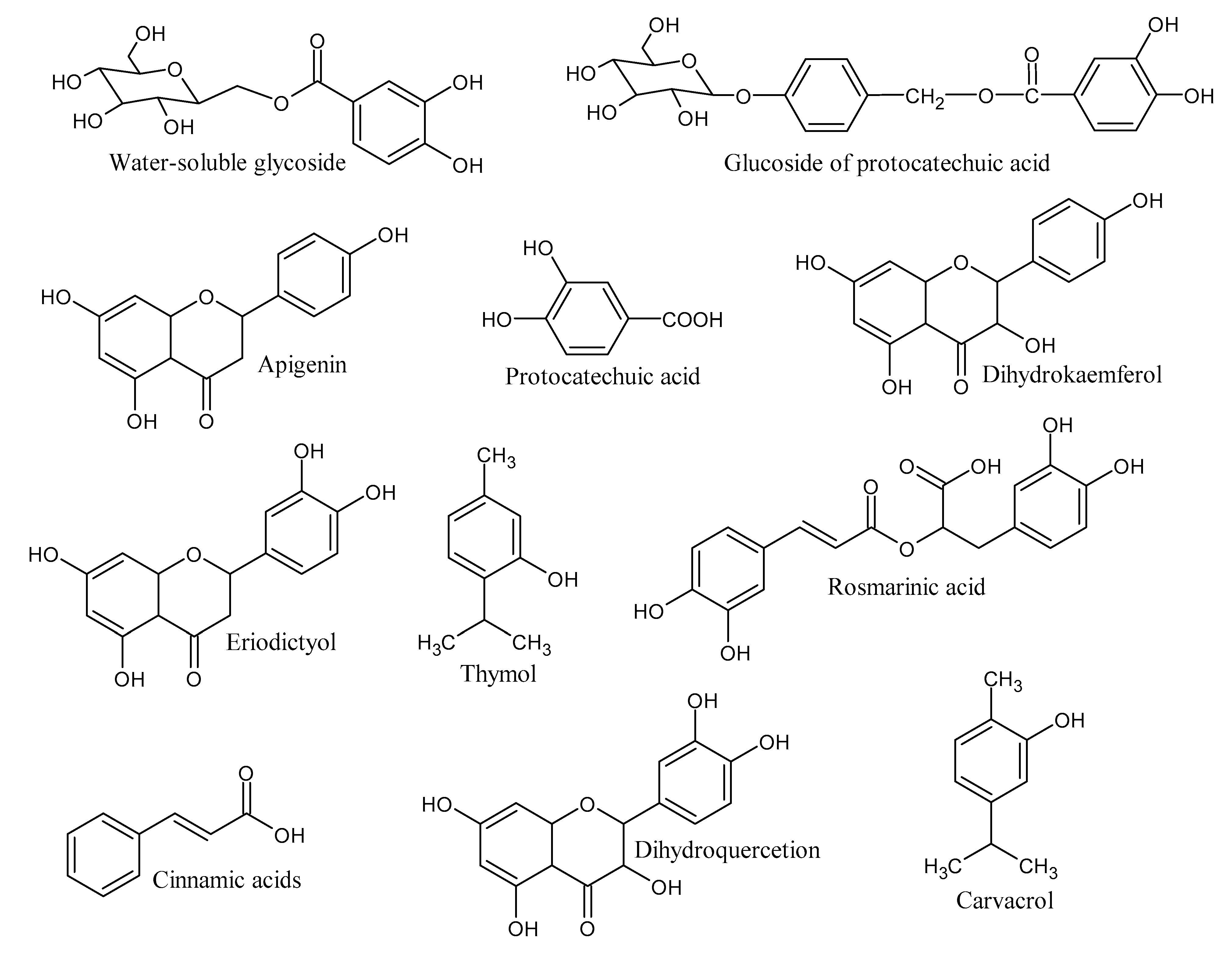
Figure 2.
The systematic illustration for the synthesis of tri-metallic Co-Cu-Ni nanoparticle composite reduces rosmarinic acid and water-soluble glycosides.
Figure 2.
The systematic illustration for the synthesis of tri-metallic Co-Cu-Ni nanoparticle composite reduces rosmarinic acid and water-soluble glycosides.
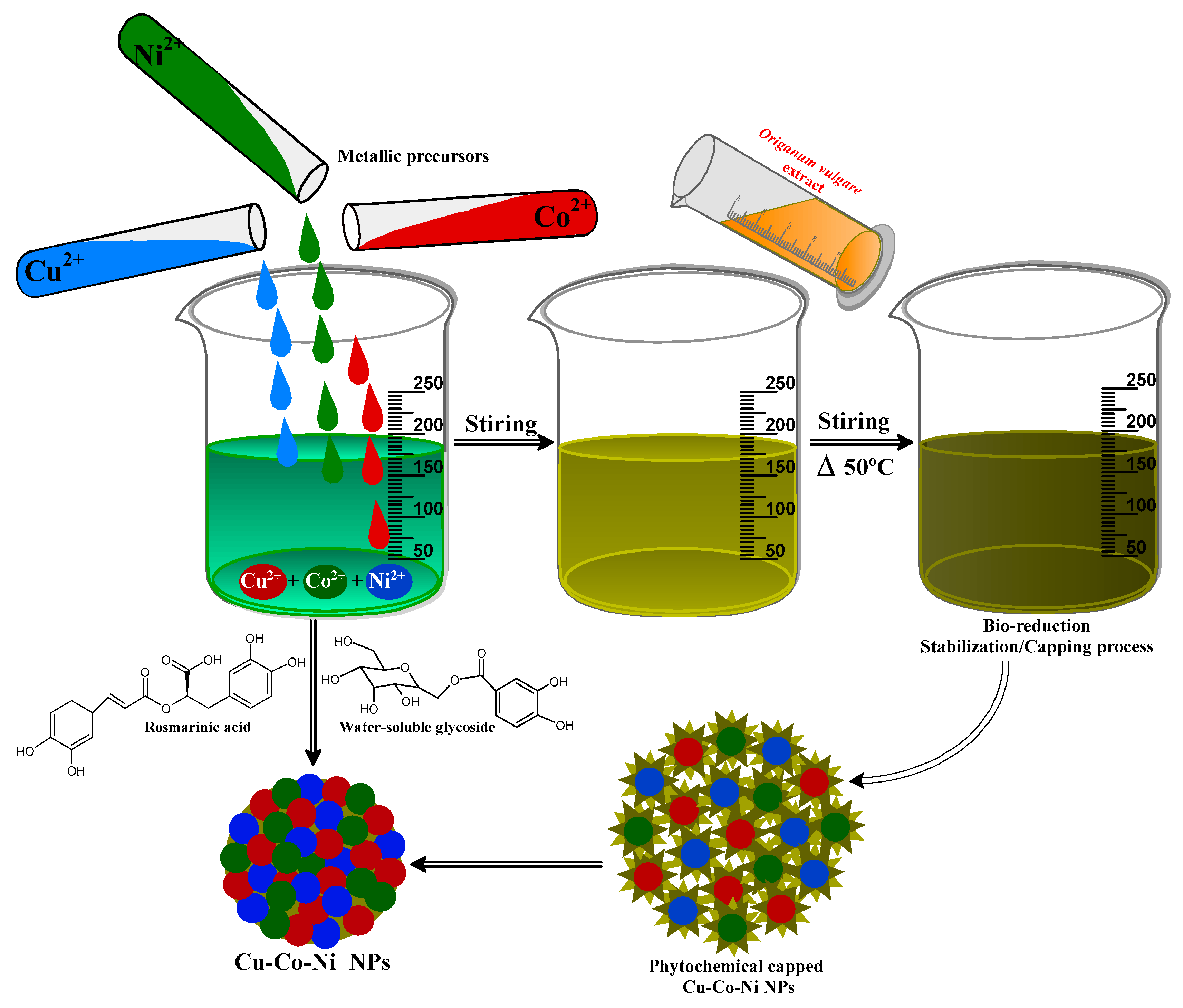
Figure 3.
Ultraviolet (UV)–visible spectra of Origanum vulgare L. extract and biosynthesized Cu-Co-Ni trimetallic nanoparticles.
Figure 3.
Ultraviolet (UV)–visible spectra of Origanum vulgare L. extract and biosynthesized Cu-Co-Ni trimetallic nanoparticles.
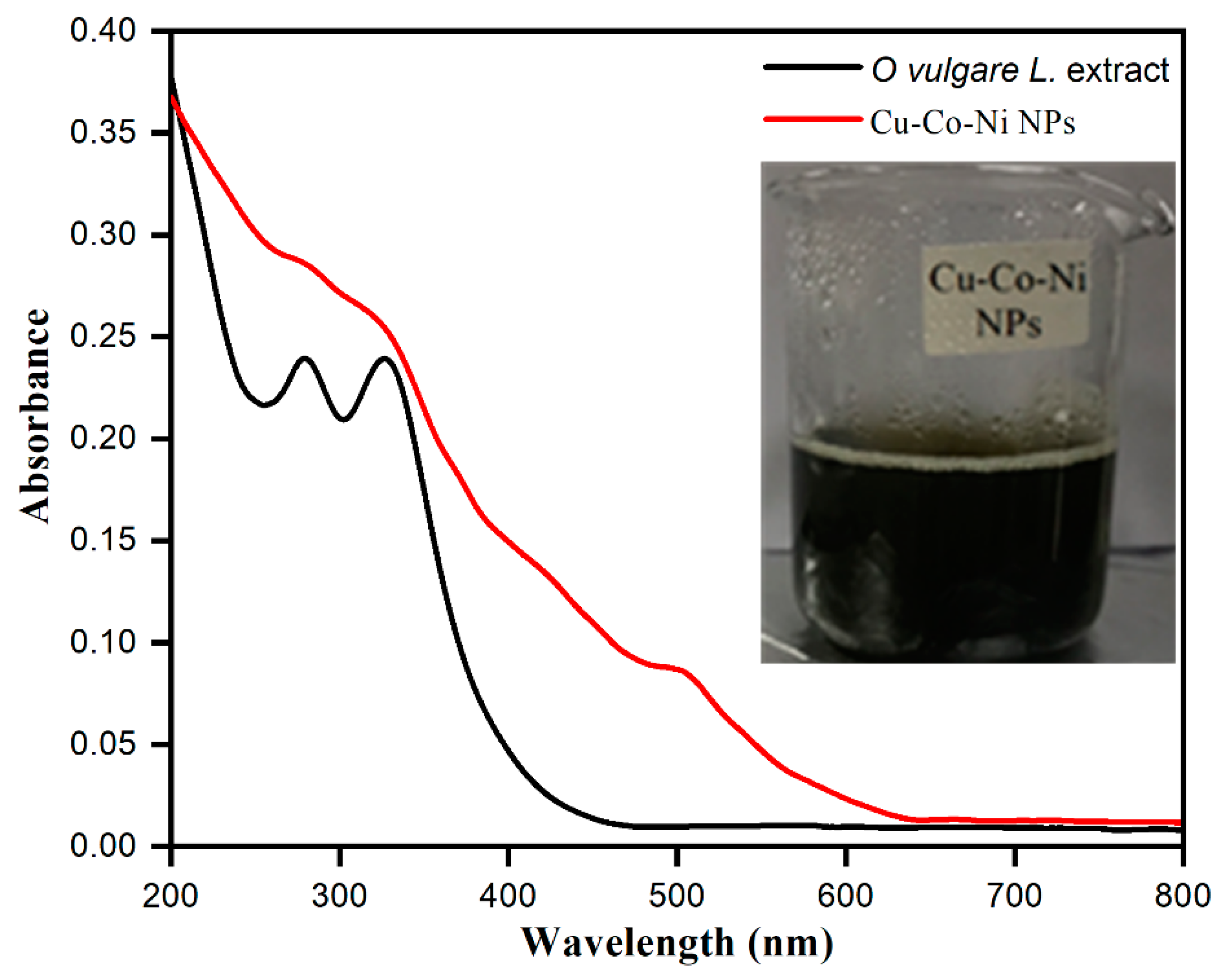
Figure 4.
Fourier transform infrared (FTIR) spectra of Origanum vulgare extract and biosynthesized Cu-Co-Ni trimetallic nanoparticles.
Figure 4.
Fourier transform infrared (FTIR) spectra of Origanum vulgare extract and biosynthesized Cu-Co-Ni trimetallic nanoparticles.
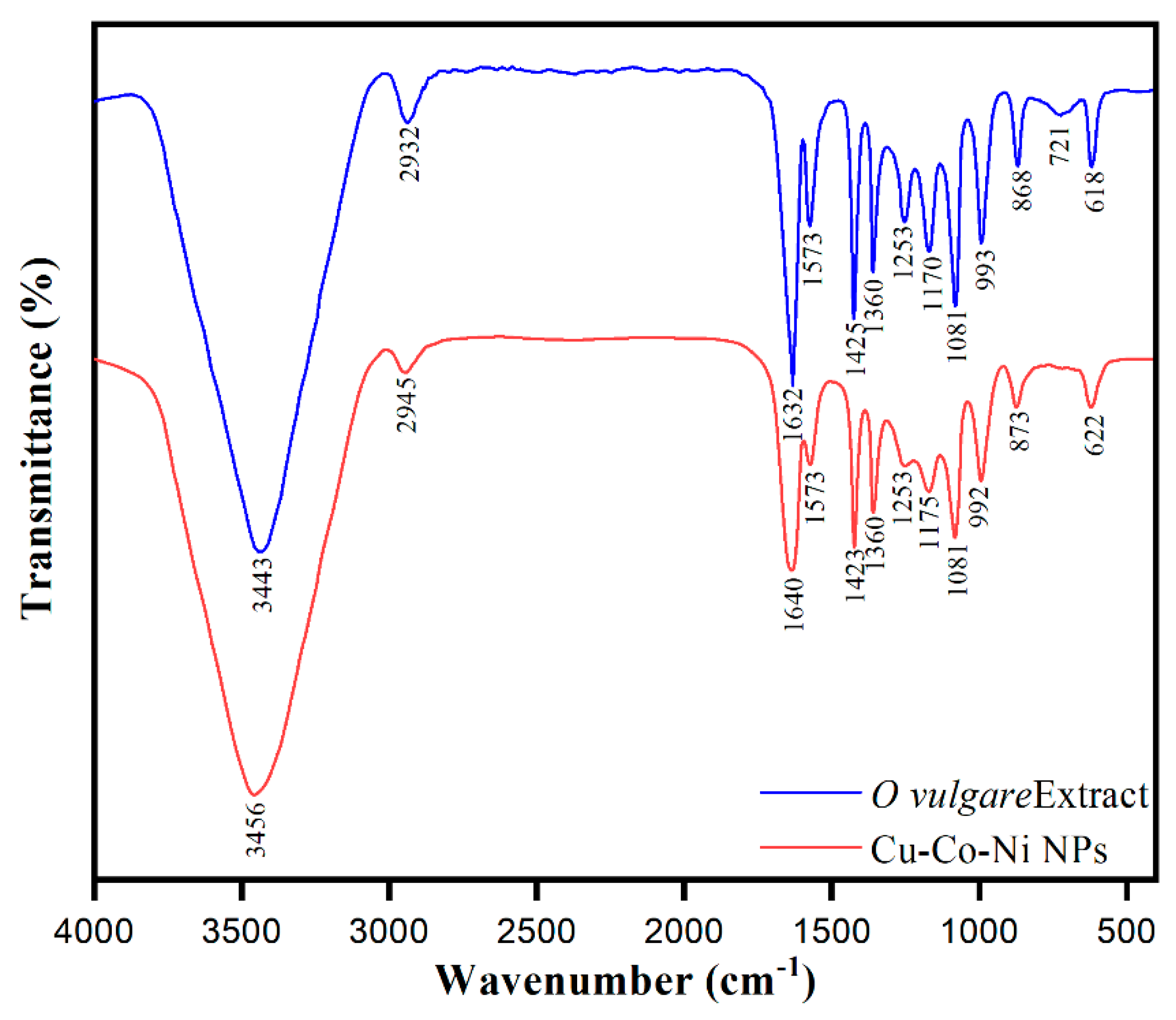
Figure 5.
Transmission electron microscopy (TEM) (a,b), scanning electron microscopy (SEM) (c–e) micrographs of biosynthesized Cu-Co-Ni trimetallic nanoparticles, and (f) particle size distribution histogram of Cu-Co-Ni trimetallic nanoparticles.
Figure 5.
Transmission electron microscopy (TEM) (a,b), scanning electron microscopy (SEM) (c–e) micrographs of biosynthesized Cu-Co-Ni trimetallic nanoparticles, and (f) particle size distribution histogram of Cu-Co-Ni trimetallic nanoparticles.
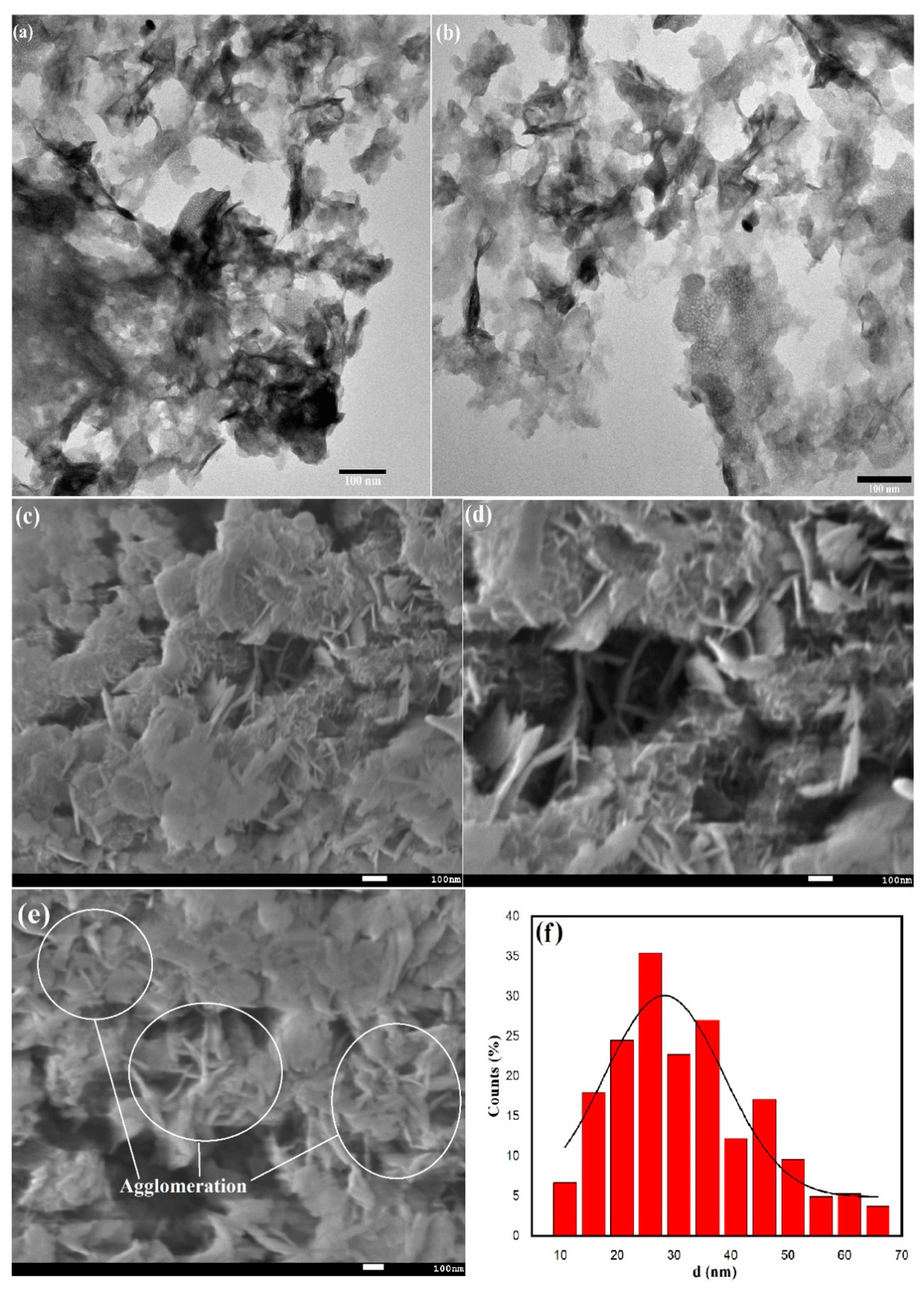
Figure 6.
Energy-dispersive X-ray (EDX) elemental mapping of Cu-Co-Ni trimetallic nanoparticles (a) and EDX spectrum of biosynthesized Cu-Co-Ni trimetallic nanoparticles (b).
Figure 6.
Energy-dispersive X-ray (EDX) elemental mapping of Cu-Co-Ni trimetallic nanoparticles (a) and EDX spectrum of biosynthesized Cu-Co-Ni trimetallic nanoparticles (b).

Figure 7.
X-ray diffraction (XRD) patterns of biosynthesized Cu-Co-Ni trimetallic nanoparticles.
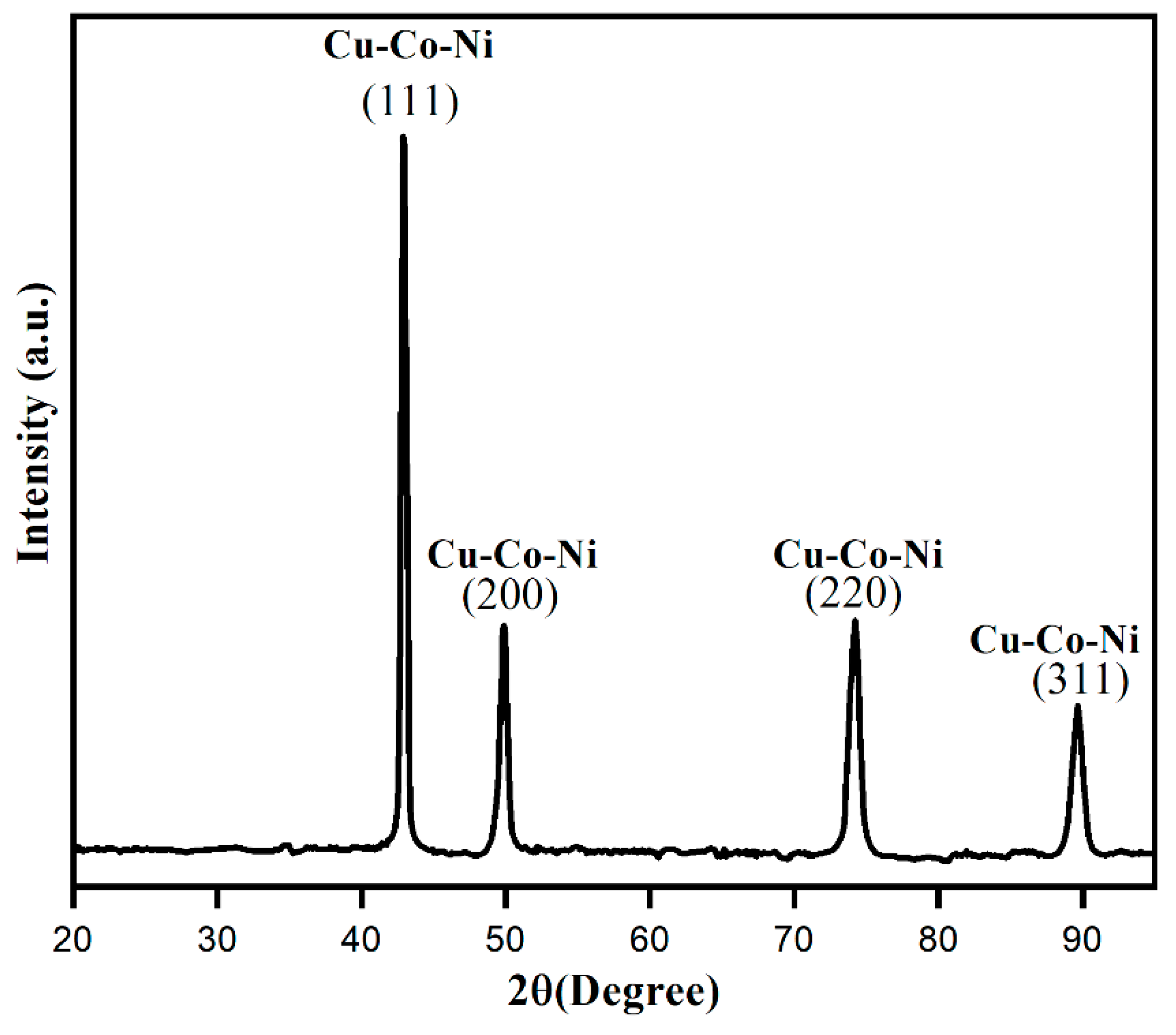
Figure 8.
Thermogravimetric analysis and differential thermogravimetry (TGA-DTG) curves of the biosynthesized of Cu-Co-Ni trimetallic nanoparticles.
Figure 8.
Thermogravimetric analysis and differential thermogravimetry (TGA-DTG) curves of the biosynthesized of Cu-Co-Ni trimetallic nanoparticles.

Figure 9.
(a) Ultraviolet–visible (UV–vis) spectra of the degradation of methylene blue (MB), (b) % degradation of MB dye with respect to irradiation time.
Figure 9.
(a) Ultraviolet–visible (UV–vis) spectra of the degradation of methylene blue (MB), (b) % degradation of MB dye with respect to irradiation time.
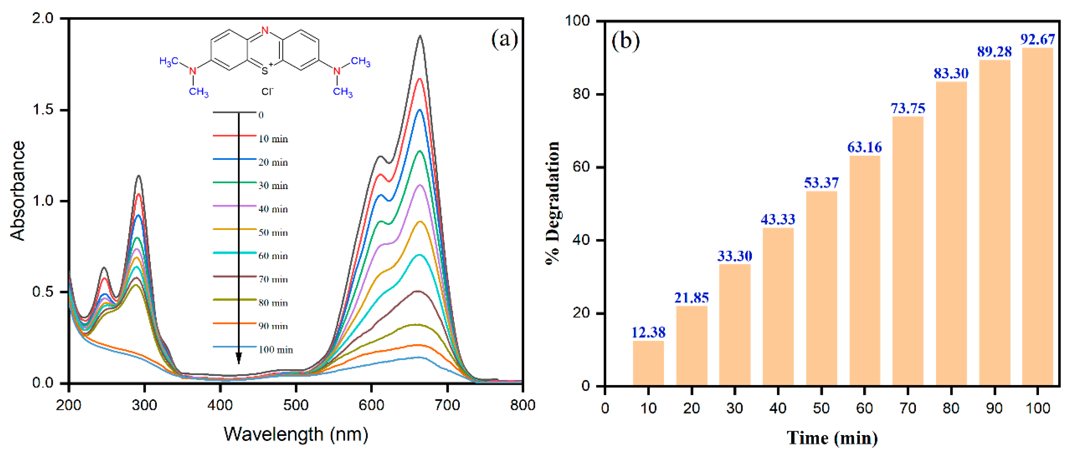
Figure 10.
(a) The plot of ln(Ct/Co) vs. time (min) as a function of initial MB dye concentration on the degradation efficiency, (b) plot of ln(Ct/Co) vs. time (min) as a function of photocatalyst dosages on the degradation of MB.
Figure 10.
(a) The plot of ln(Ct/Co) vs. time (min) as a function of initial MB dye concentration on the degradation efficiency, (b) plot of ln(Ct/Co) vs. time (min) as a function of photocatalyst dosages on the degradation of MB.
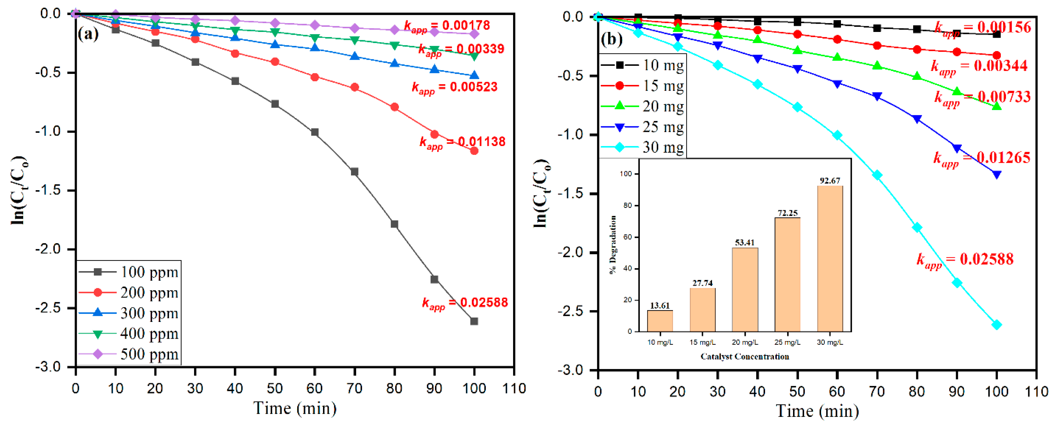
Figure 11.
Possible mechanism of photodegradation of MB by Cu-Co-Ni nanoparticles.
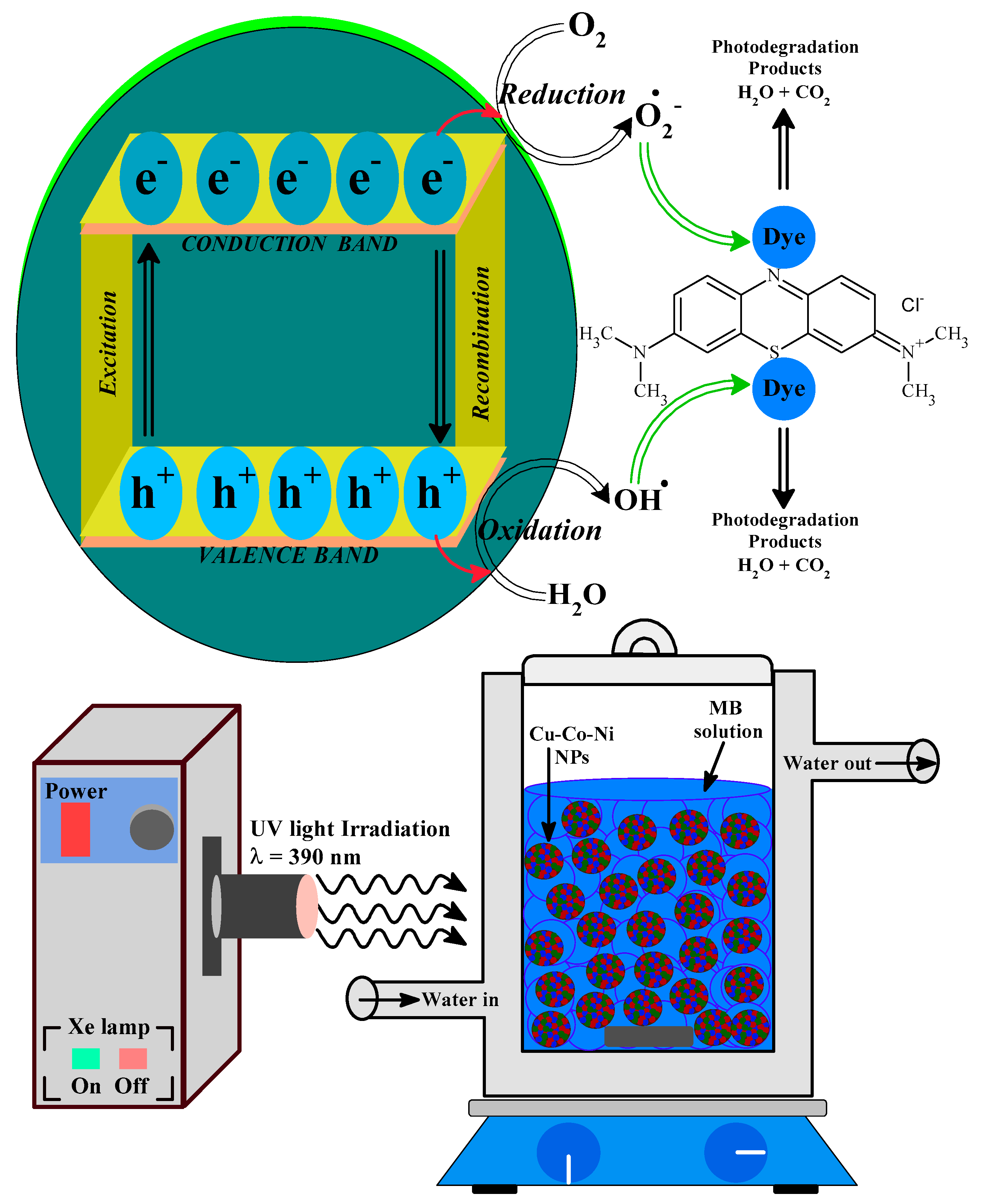
Figure 12.
(a) Photocatalytic % degradation of MB in the presence of different scavengers over Cu-Co-Ni nanoparticles. (b) Stability and reusability of the photocatalyst.
Figure 12.
(a) Photocatalytic % degradation of MB in the presence of different scavengers over Cu-Co-Ni nanoparticles. (b) Stability and reusability of the photocatalyst.


Table 1.
Rate constants, % degradation of MB dye, and correlation coefficients (R2) of the MB photocatalytic degradation.
Table 1.
Rate constants, % degradation of MB dye, and correlation coefficients (R2) of the MB photocatalytic degradation.
| [MB] (ppm) | Rate Constant (min−1) | % Degradation | R2 | [Cu-Co-Ni] (mg) | % Degradation | Rate Constant (min−1) | R2 |
|---|---|---|---|---|---|---|---|
| 100 | 0.02588 | 92.67 | 0.935 | 10 | 13.61 | 0.00156 | 0.942 |
| 200 | 0.01138 | 68.75 | 0.961 | 15 | 27.74 | 0.00344 | 0.990 |
| 300 | 0.00523 | 40.93 | 0.997 | 20 | 53.41 | 0.00733 | 0.996 |
| 400 | 0.00339 | 29.89 | 0.991 | 25 | 72.25 | 0.01265 | 0.947 |
| 500 | 0.00178 | 15.73 | 0.993 | 30 | 92.67 | 0.02588 | 0.935 |
Table 2.
Photocatalytic performance comparison of different photocatalysts for degradation of methylene blue.
Table 2.
Photocatalytic performance comparison of different photocatalysts for degradation of methylene blue.
| Photocatalyst | Irradiation Time (min) | % Degradation | Irradiation Source | References |
|---|---|---|---|---|
| ZnO | 90 | 88.37 | UV Light | [53] |
| SnO2 | 30 | 90 | UV Light | [65] |
| Cu2O–Cu | 50 | 90.1 | Visible light | [66] |
| RGO-N-ZnO | 120 | 98.5 | Visible light | [67] |
| Cu@Cu2O | 50 | 96.5 | UV Light | [68] |
| Zr2Ni1Cu7/Zr2Ni1Cu7/Si3N4 | 90 | 92 | Visible light | [69] |
| Pc-TiO2 | 100–140 | 100 | Visible light | [70] |
| NH2-MIL-101(Fe)@CuCoNi | 120 | 99 | Visible light | [71] |
| ZnO-SnO2 | 60 | 96.53 | UV Light | [72] |
| TiO2/PAAm | 300 | 95 | Sunlight | [73] |
| Cu-Co-Ni | 100 | 92.67 | UV Light | Present work |
© 2020 by the authors. Licensee MDPI, Basel, Switzerland. This article is an open access article distributed under the terms and conditions of the Creative Commons Attribution (CC BY) license (http://creativecommons.org/licenses/by/4.0/).
Share and Cite
MDPI and ACS Style
Alshehri, A.A.; Malik, M.A. Facile One-Pot Biogenic Synthesis of Cu-Co-Ni Trimetallic Nanoparticles for Enhanced Photocatalytic Dye Degradation. Catalysts 2020, 10, 1138. https://doi.org/10.3390/catal10101138
AMA Style
Alshehri AA, Malik MA. Facile One-Pot Biogenic Synthesis of Cu-Co-Ni Trimetallic Nanoparticles for Enhanced Photocatalytic Dye Degradation. Catalysts. 2020; 10(10):1138. https://doi.org/10.3390/catal10101138
Chicago/Turabian StyleAlshehri, Abdulmohsen Ali, and Maqsood Ahmad Malik. 2020. "Facile One-Pot Biogenic Synthesis of Cu-Co-Ni Trimetallic Nanoparticles for Enhanced Photocatalytic Dye Degradation" Catalysts 10, no. 10: 1138. https://doi.org/10.3390/catal10101138
Note that from the first issue of 2016, this journal uses article numbers instead of page numbers. See further details here.