Article

Fine Characterization of Natural SiO2-Doped Catalyst Derived from Mussel Shell with Potential Photocatalytic Performance for Organic Dyes

1
Marine Science and Technology College, Zhejiang Ocean University, Zhoushan 316022, China
2
National Engineering Research Center for Marine Aquaculture, Zhejiang Ocean University, Zhoushan 316022, China
3
Petrochemical and Energy Engineering College, Zhejiang Ocean University, Zhoushan 316022, China
4
Institute of Innovation & Application, Zhejiang Ocean University, Zhoushan 316022, China
5
Ocean College, Zhejiang University, Zhoushan 316021, China
6
Environmental and Science Technology College, Donghua University, Shanghai 201620, China
7
Food and Medicine College, Zhejiang Ocean University, Zhoushan 316022, China
*
Authors to whom correspondence should be addressed.
Catalysts 2020, 10(10), 1130; https://doi.org/10.3390/catal10101130
Submission received: 9 July 2020 / Revised: 6 August 2020 / Accepted: 21 August 2020 / Published: 1 October 2020

Abstract

:
In this work, a SiO2-doped natural photocatalyst derived from waste mussel shell (HAS) was prepared by acidification. The as-prepared sample was characterized by scanning electron microscopy (SEM), transmission electron microscopy (TEM), inductively coupled plasma-optical emission spectroscopy (ICP-OES), field emission scanning electron microscope (FE-SEM), X-ray diffraction (XRD), Fourier transform infrared (FTIR), UV-visible diffuse-reflectance spectrum (UV-vis DRS), and Differential scanning and thermogravimetric analyses (DTA/TGA). The results exhibited that HAS was mesopores nanomaterial consisting of uneven arranged rod-like structure, the dominant component of HAS was SiO2 with a large number of hydroxyl groups, and a variety of transition metals uniformly distributed in HAS. Rhodamine B (RhB) and methylene blue (MB) removal efficiencies (equal to 92.59% and 99.14%, respectively) were observed under the HAS presence when exposed to the visible light. The degradation products were analyzed using liquid Chromatograph Mass Spectrometer (LC-MS) and Total Organic Carbon (TOC), among which, MB was degraded by demethylation and deamination, and RhB was degraded by N-deethylation and conjugate structure destruction. After four successive recycles, the removal efficiency of RhB and MB are still reach 86.103% and 75.844%. This study indicated that the mussel shells might be suggested as a novel natural photocatalyst in the application of dye wastewater treatment.

Graphical Abstract

1. Introduction

Photocatalysis-based technologies are considered eco-friendly and economically feasible and are widely studied for their applications in environment decontamination, water splitting, clean energy production, CO2 conversion, etc. [1,2,3,4,5]. Various semiconductors were employed as photocatalysts, among which, TiO2 is the most widely investigated mostly because of its affordability, chemical stability, excellent durability, and environmental friendliness [6,7]. However, mono-component semiconductor photocatalyst has been constrained in the practical application, owing to its insufficient sunlight utilization, inefficient photogenerated charge separation, and low quantum yield [8,9]. In recent years, a composite photocatalyst, consisting of semiconductor and other components, has attracted extensive attention, which could improve the utilization efficiency of solar energy and significantly enhance photocatalytic performances [10,11,12]. Thus, various composite photocatalysts have been designed, such as nanocomposite configuration, Z-scheme, cocatalyst, metal–organic framework, etc. [13,14,15,16], which could be prepared by sol-gel, hydrothermal, co-precipitation, solvent-thermal, emulsion method, etc. [17,18,19,20]. Although composite photocatalysts have achieved significant improvements compared to the mono-component photocatalysts, industrial applications of these photocatalysts are still challenging, especially in terms of their mass production, cost, and stability that need to be addressed. Thus, in recent years, researchers have been focusing on cheap and efficient photocatalysts, especially those from natural biomaterials with multiple scales and hierarchical morphologies, such as pollen [21], rice straw [22], kelp [23], seashells [24], etc.
Marine bivalves are essential components of aquacultural industries, and over 15 million tons per year are produced globally [25], with China producing 85% of this amount [26]. Shells, a by-product of the bivalves processing, contributing 75% to the total bivalve weight [27], are discarded during processing. These shells contain 95–99.9% CaCO3 and only some amount of organics [26]. Outstanding structural and mechanical properties of these shelves attracted a significant amount of the scientists worldwide with the goal to utilize them and use for wastewater treatment applications [28,29], for soil remediation [30,31], and as bio-fillers to polymeric matrices [32,33]. Recently, the previous studies demonstrated that waste mussel shells could be prepared into carriers by pyrolysis to support photocatalyst (Bi2MoO6) in the application of dyes degradation [34,35]. However, the mussel shells were treated by carbonization at high temperatures to render its large specific surface area, which is confronted with the challenges of high energy consumption and high cost.
In the study, a facile and low-cost acidizing method was employed to prepare a novel natural photocatalyst derived from mussel shells for the first time, which was characterized by FE-SEM, X-ray diffraction (XRD), transmission electron microscopy (TEM), Fourier transform infrared (FTIR), UV-vis DRS, inductively coupled plasma-optical emission spectroscopy (ICP-OES), and Differential scanning and thermogravimetric analyses (DTA/TGA). Its photocatalytic performances towards the decomposition of organic dyes were investigated. The reactive oxidative species and degradation products were identified, respectively, and the probable degradation pathways were established.

2. Results

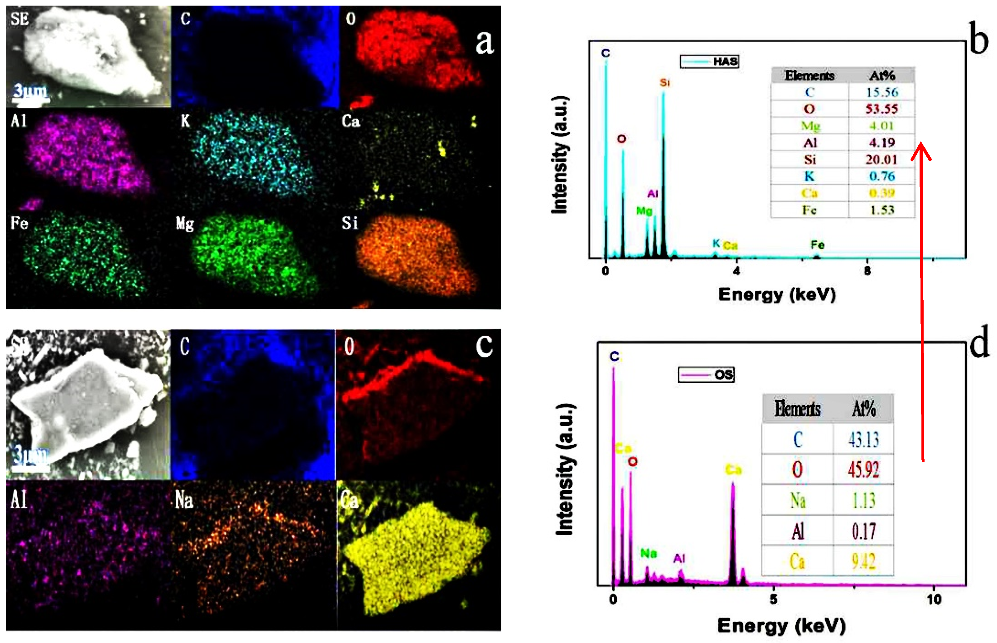
2.1. Morphology Analysis

Scanning electron microscopy (SEM) of the Original shell (OS) exhibits blocky textures arranged unevenly (see Figure S1). After acidification, there are many depressions and cracks in the surface of waste mussel shell (HAS), forming nonuniformly arranged mesopores. Besides, TEM images of HAS further verify the rod-like structure of HAS, and there is no lattice fringe in the High Resolution Transmission Electron Microscope (HRTEM) images, indicating that HAS is an amorphous substance.
N2 adsorption isotherms of HAS and OS belonged to type V [36] with a hysteresis loop at P/P0 > 0.5 (see Figure 1a), which is indicative of the mesopore presence [37]. Capillary condensation is typical for mesoporous materials. However, capillary condensation and evaporation do not occur at the same pressure, which causes hysteresis loops [38]. The pore sizes of HAS and OS were in the mesopore range (2–35 nm) (see Figure 1b). The specific surface areas and pore volumes of HAS and OS were 147.5 and 2.6 m2/g and 0.3538 and 0.2208 cm3/g, respectively. After acidification, the surface area and pore volume of mussel shells increased 57.5 and 1.7 times, respectively.

2.2. Chemical Composition Analysis

The chemical compositions of as-prepared samples were investigated by using field emission scanning electron microscope (FE-SEM), inductively coupled plasma-optical emission spectroscopy (ICP-OES), FTIR, XRD, and DTA/TGA. The SEM mapping results of HAS and OS are shown in Figure 2, analyzing the element distribution of a selected area in as-prepared sample with 5000× magnification. The image indicates that OS was formed by the elements C, O, Al, Na, and Ca, and the dominating elements are C, O, and Ca in OS, demonstrating that OS is mainly composed of CaCO3. While HAS was formed by C, O, Al, K, Ca, Fe, Mg, and Si, and the dominating elements are Si, O, and C, indicating that the acidification has destroyed the skeletal structure of calcium carbonate, removed most of the calcium carbonate. Instead, silicon dioxide turns into the main skeleton substrate in HAS. In addition, metal elements were further detected by ICP-OES and listed in Table 1, which is consistent with FE-SEM-energy dispersive spectroscopy (EDS) analysis. And some transition elements like Fe, Ti, Zr, and Mn have been detected in HAS, which might synergistically construct the active catalytic sites, being beneficial to the production of reactive oxygen species [39,40].
The FTIR indicates that OS exhibited peaks (at 705, 867, 1381, and 1800 cm−1) characteristic for calcium carbonate. After acidification, HAS presents characteristic peaks of silicon dioxide located at 470, 1024, 1650, 2355, and 3400 cm−1. The absorption peak at 3560 cm−1 was attributed to the axial O-H stretching mode, indicating an abundance of active hydroxyl groups on the HAS surface, which is very beneficial for its adsorption capacity [23]. XRD pattern of OS showed peaks located at 30.96, 41.14, 44.94, and 50.12°, which correspond to (104), (113), (202), and (110) crystal planes of CaCO3. However, the diffraction peaks in HAS located at 20.826, 26.593, 42.383, 50.046 are ascribed to the (100), (101), (200), and (112) crystal planes of SiO2, which agrees with the FR-IR analysis.
Weight loss during DTA/TGA of HAS and OS in the 100–200 °C range was 2.49%, which was attributed to physically adsorbed water. Weight loss in the 200–800 °C range was 9.29%, which was attributed to Si-OH transformation to Si-O-Si [41], which can result in a collapse of mesopores of HAS. No significant weight loss was detected after 800 °C. Weight loss of OS in the 200–600 °C range was 8.6% due to the organic impurities decomposition. Endothermic OS weight loss in the 600–800 °C region was ascribed to CaCO3 decomposition. Thus, DTA/TGA analysis suggested the presence of abundant OH surface groups, which play a critical role in the adsorption and photocatalytic degradation of organic dyes, which agrees with FTIR analysis.

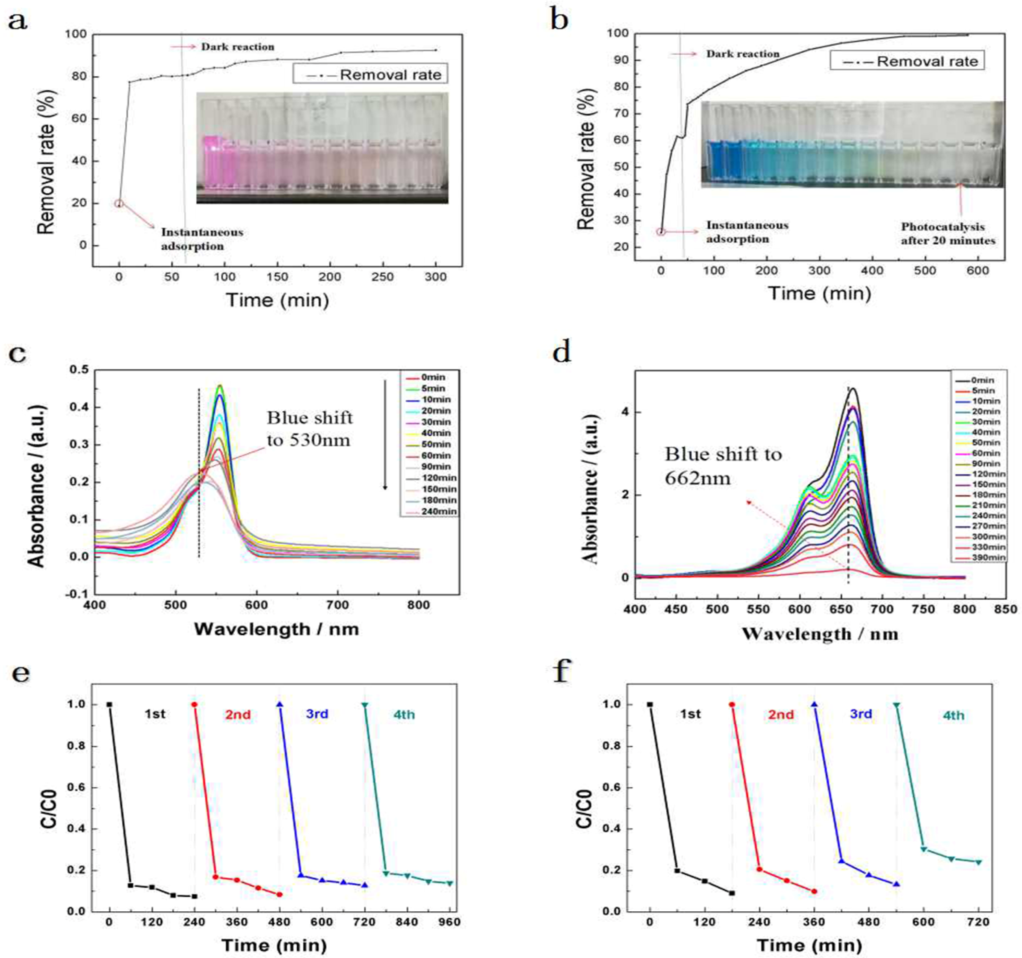
2.3. Optical Absorption Tests

The optical absorption performance of material strongly correlates with its photocatalytic efficiency. From the UV-Vis DRS of HAS analysis, it can be showed that there is a sharp peak located at 250 nm and a broad peak in the region of 300–500 nm. The absorption edge of HAS (at 415 nm) was used to calculate its bandgap (Eg), which was equal to 2.98 eV. Thus, HAS possessed photocatalytic activity, potentially even better than that of TiO2 (with the bandgap equal to 3.2 eV). The bandgap can also be calculated from the UV-Vis spectroscopy absorption peak by the following Equation (1):
Eg = h C/λ
where h is the Plank’s constant (equal to 6.626 × 10⁻³⁴ J·s), C is the speed of light (equal to 2.99 × 10⁸ m/s), and λ is absorption peak value (in nm).

2.4. Photocatalytic Performance

Photocatalytic activities of catalysts prepared in this work were assessed using RhB and MB organic dyes as model substances and visible light. The adsorption/desorption equilibria were established before irradiation to avoid the adsorption process affecting the photocatalytic activity of HAS 9 (as Figure 3a,b shown). It can be demonstrated that the adsorption capacity of MB and RhB on HAS are 72 and 38.4 mg/g at the adsorption equilibrium, and the removal rates of MB and RhB were 60.92% and 80.75%, respectively, due to the special rod-like skeleton structure of as-prepared sample, which provided these materials with abundant porosity and high surface area. After 2.5 h irradiation, the introduction of HAS leads to RhB degradation percentages of 11.84%, and the total removal rate of RhB is 92.59%. While the introduction of HAS leads to MB degradation percentages of 38.22% after 6.5 h irradiation, and the total removal rate of MB is 99.14%. Moreover, the Kapp value of HAS (as shown in Table 2) is slightly less than other single photocatalyst such as pure Bi2MoO6 (BKBC) with K = 0.0079 min−1 [23] as listed in Table 2. This phenomenon may be caused by the fact that the natural mussel shell photocatalyst were not all pure components, resulting in a slightly smaller Kapp (apparent rate constant) value, while it also proved that the photocatalyst prepared in this experiment did have photocatalytic activity. RhB and MB absorption spectra at different degradation times under the HAS presence, and visible light irradiation are shown in Figure 3c,d. RhB adsorption peak shifted from 554 to 530 nm, while that of MB has slightly shifted from 665 to 662 nm, which all have a blue-shift with irradiation time. In addition, RhB and MB absorption peaks became weaker upon prolonged irradiation, confirming their degradation by HAS. RhB solution changed its color from pink to light pink, then to pale yellow and then the solution became colorless, while that of MB solution changed from blue to light blue, pale yellow and colorless over the photodegradation process, indicating that the chromophoric groups of RhB and MB were gradually destroyed and some intermediates were formed through N-deethylation and N-demethylation, respectively.
To further prove the stability and durability of HAS, the recycle experiments were carried out under identical conditions (see Figure 3e,f). The adsorption and photocatalytic activity of HAS did not decrease significantly. After four successive recycles, the removal efficiency of RhB and MB are still reach 86.103% and 75.844%, respectively, which prove that HAS possesses higher stability and reusability.

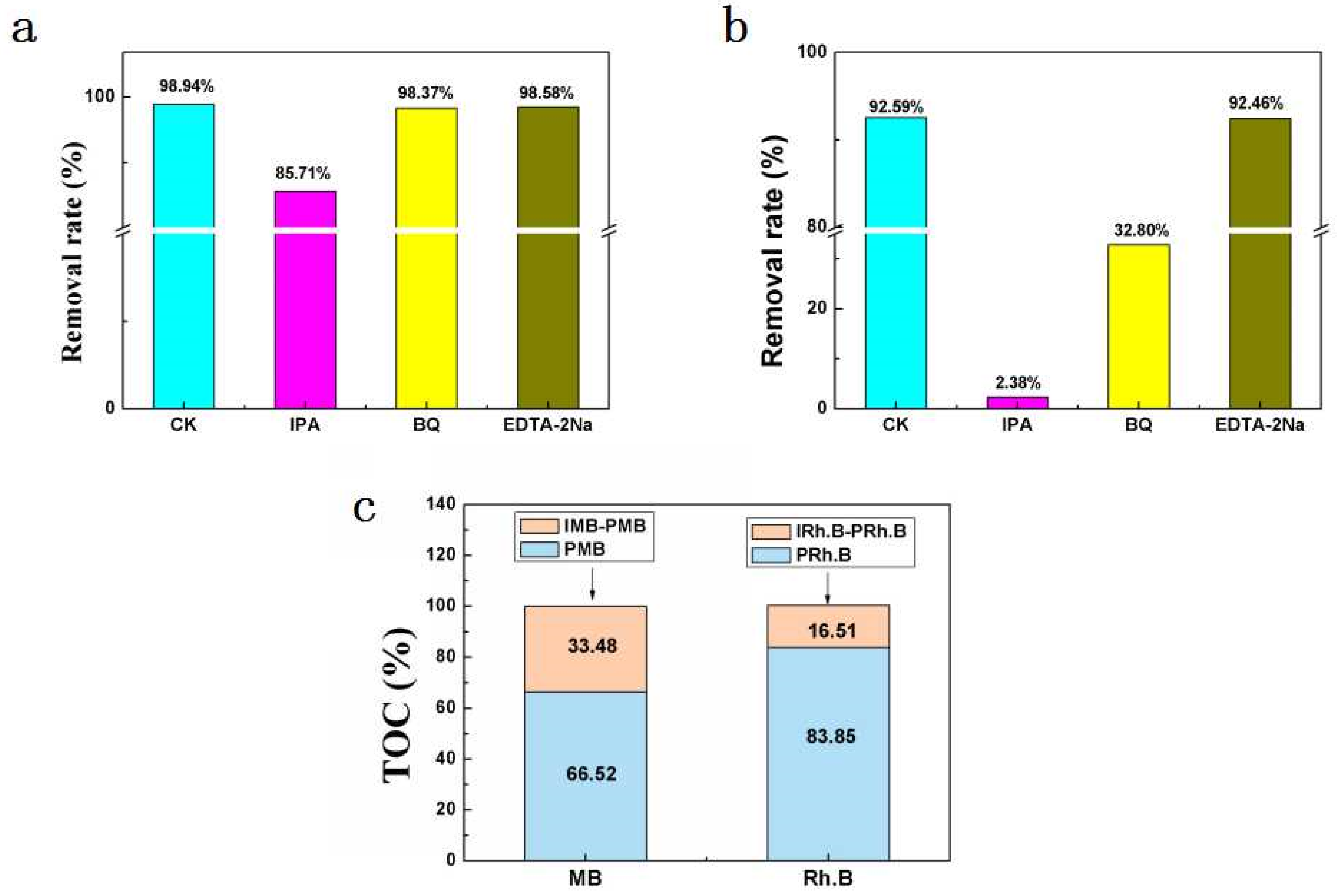
2.5. Active Species Detection

Specifics of the RhB and MB photocatalytic degradation mechanisms were studied using scavenger trapping experiments using ethylenediamine tetraacetic acid disodium solution (EDTA-2Na), isopropanol (IPA), and p-benzoquinone (BQ,) as scavengers for H+, •OH, and •O2, respectively (see Figure 4a,b). The presence of IPA reduced MB degradation, while EDTA-2Na and BQ did not affect RhB and MB degradation at all. Thus, •OH was the dominant reactive species during the photocatalytic degradation of MB under the presence of HAS [45,46,47]. While in the photocatalytic process of RhB on HAS, a sharp decrease of degradation rate was observed via the introduction of IPA. Similarly, there is a dramatic decline in the degradation of RhB in the presence of BO. Thus, it is worth mentioning that •OH and •O2 play a key role in the effective degradation of RhB using HAS. Therefore, •OH is the dominant active species of HAS during the photocatalytic degradation of dyes, which can effectively decompose organic compounds due to its higher oxidation potential (E0 = 2.8 eV) [48].

2.6. Total Organic Carbon (TOC)Analysis of Degradation Products

TOC analysis was used to study RhB and MB mineralization. TOC removal efficiencies with respect to MB and RhB degradation by the HAS under visible light irradiation are shown in Figure 4c. After the photocatalytic reaction, the TOC of MB and RhB solutions were 36.0 and 35.7 mg/L. These values correspond to mineralization rates equal to 33.5 and 16.5%. Thus, MB and RhB were decomposed and partially mineralized under the HAS presence upon exposure to the visible light irradiation.

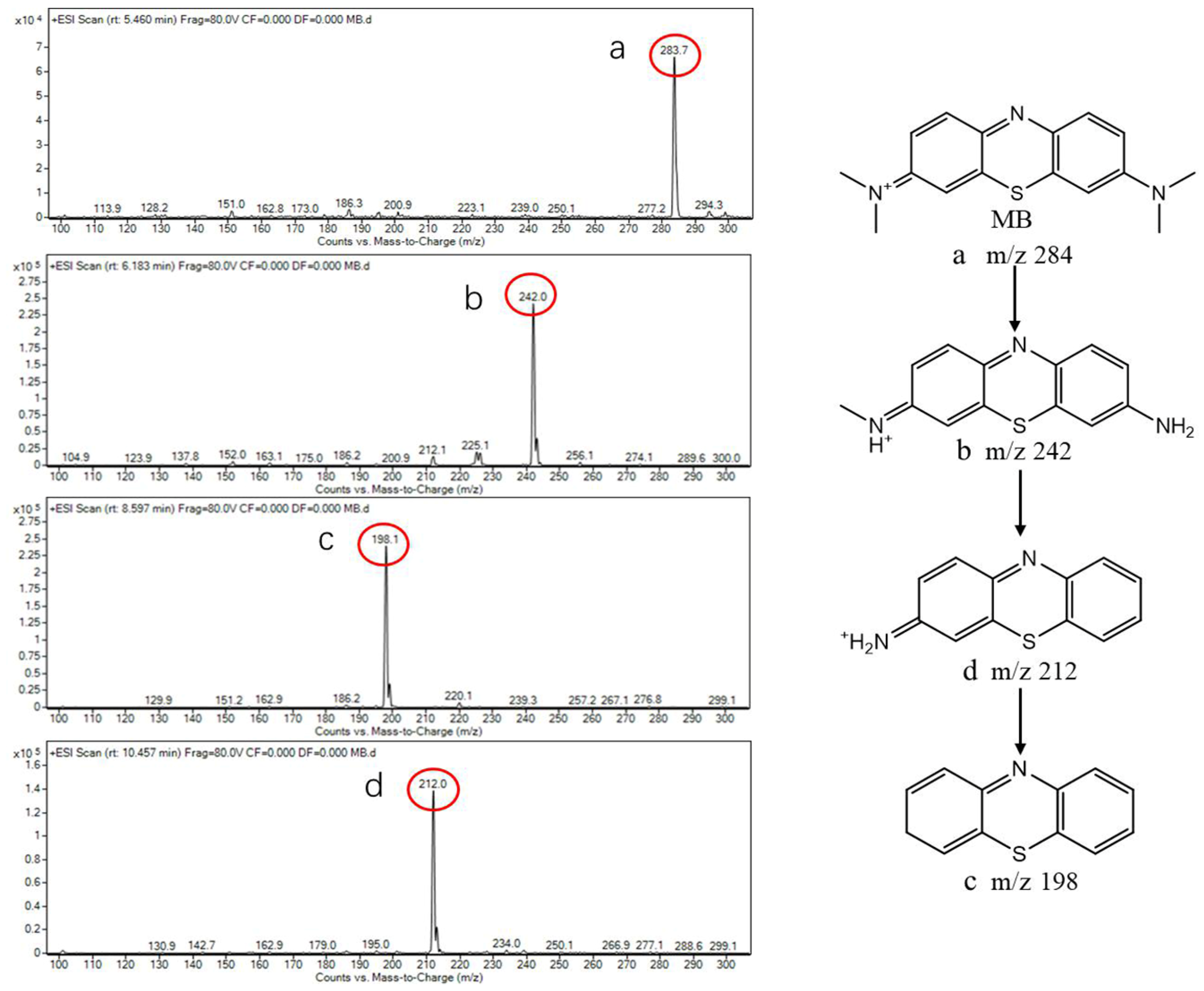
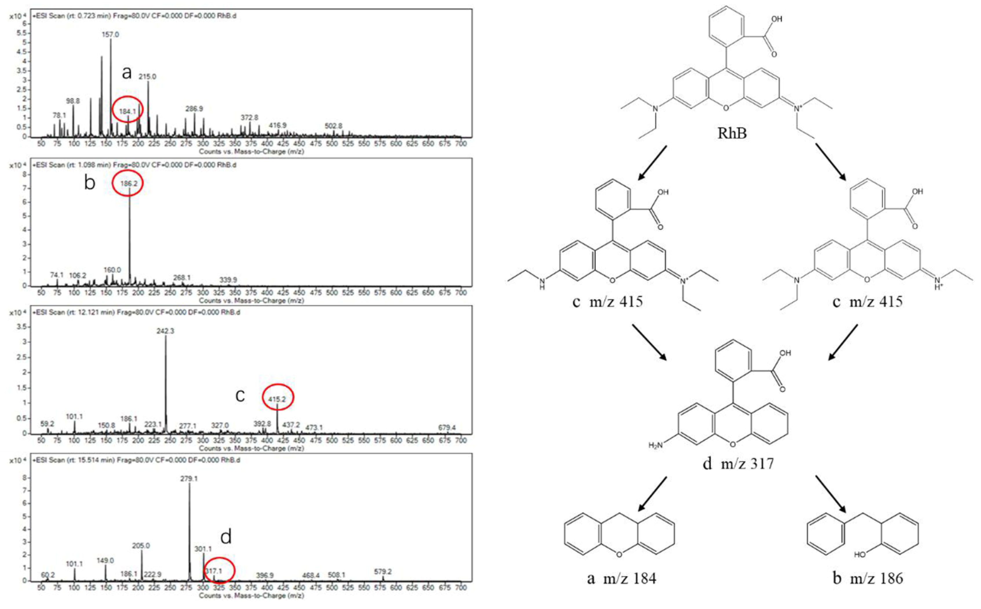
2.7. Photocatalytic Degradation Products Analysis

We used liquid chromatography- electrospray ionization-mass spectrometry (LC-ESI-MS) to analyze intermediate products of MB and RhB degradation. Intermediate products of MB marked as “a”, “b”, “c” and “d” were detected at the m/z values equal to 283.7, 242 (corresponding to the MB molecule without three CH3 groups), 212 (MB without one amino group), and 98.1 (further removal of amino groups from the intermediate degradation products), respectively (see Figure 5). Based on these results, we concluded that photocatalytic degradation of MB under the HAS presence started with a demethylation reaction, the product of which further reacted with the radicals forming product “b” and then, after consecutive deamination process, to “c” and “d”.
RhB molecule showed an intermediate product at m/z value equal to 415.2 (product “c”, see Figure 6), which corresponds to N-diethyl-N’-ethyl-rhodamine formed as a result of ethyl group removal from the RhB xanthene ring. N-deethylation intermediate (product d) is observed at the m/z value of 317.1, and N-deethylated intermediate is degraded into the possible products “a” and “b”, corresponding to the m/z values of 184.1 and 186.2, respectively. As is known that RhB photodegradation occurred by two processes: N-deethylation and conjugate structure destruction [49]. Thus, during photocatalytic RhB degradation under the HAS presence, the reactive species very likely first attacked the central carbon atom (which is responsible for the RhB coloration), after which N-deethylation process [50] and oxidation of the conjugated RhB structure completed the degradation.
It can be demonstrated that the main component of HAS is SiO2 from ICP-OES and FE-SEM-EDS analysis, endowing abundant porous structures and larger specific surface areas of HAS, which can effectively absorb MB and RhB, being beneficial to the photocatalytic degradation performances. In addition, FTIR and DTA/TGA analysis showed that HAS contained numerous hydroxyl groups capable of preventing electron-hole pair recombinations and improving photocatalytic activity. Yet, pure SiO2 does not possess photocatalytic activity due to its larger bandgap value (Eg > 5 eV) [48]. Thus, the photocatalytic activity of HAS is mainly derived from various transition metal elements, which synergistically construct the active catalytic sites [1,51,52,53]. Therefore, we believe that such efficient organic dye degradation occurred because of the synergistic processes occurring at the metal/SiO2 interface.
Typical photocatalytic processes undergo a series of surface redox reactions involving photogenerated holes and electrons. These reactive species quickly react with O2, and H2O adsorbed on the photocatalyst, forming very reactive •OH and •O2, which then react with dye molecules [11]. During the MB degradation, •OH was the dominant reactive species, which could decolorize and degrade the dye by demethylation and deamination reactions, while during RhB degradation, •OH and •O2 are the most prominent active species damaging dye by N-deethylation and conjugate structure destruction [54].

3. Materials and Methods

3.1. Initial Materials

Mussel (Mytilus coruscus) shells were collected from Zhoushan, Zhejiang, China repeatedly washed with deionized (DI) water to eliminate the surface impurities, soaked in 1% NaOH solution for 6 h to remove surface lipids, then washed again with DI water until neutral pH and dried at 50 °C for 24 h. The shells were then crushed into powders with a micro-nano pulverizer (Yuki, Japan), and sieved 100 mesh.
HCl, NaOH, rhodamine B (RhB), and methylene blue (MB) (all of the analytical grade) were acquired from Sinopharm Chemical Reagent Co and used as received, As shown in Figure 7 below.

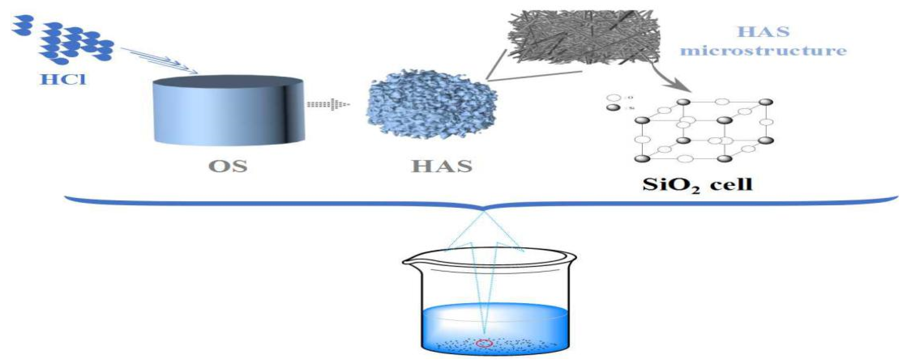
3.2. Mussel Shell Photocatalyst Preparation

Crushed shells were immersed in 1 mol/L HCl solution for 18 h (M/V = 1:1) until no bubbles appeared, and washed with deionized water until neutral, which was donated as HAS, and those without HCl treatment were as the control group, donated as OS.

3.3. Characterization

The N2 adsorption/desorption isotherms were recorded using a static volumetric adsorption analyzer (NOVA 2200e, Quantachrome, FL, USA) and analyzed by the Brunner−Emmet−Teller (BET) method. Microstructural and morphological analyses were performed by scanning electron microscopy (SEM) using the S4800 (Hitachi, Japan) instrument and by transmission electron microscopy (TEM) using TecnaiG2F20S-TWIN instrument (FEI, USA). Elemental analysis was performed using by inductively coupled plasma-optical emission spectroscopy (ICP-OES) using PerkinElmer 5300 DV (USA) analyzer. Energy dispersive spectroscopy (EDS) coupled with a field emission scanning electron Nova Nano SEM 450 microscope (FE-SEM) fabricated by FEI (USA) was employed to detect chemical elements content and distribution. Ultraviolet-visible (UV-Vis) measurements were performed using UV-2600 on a UV-Vis spectrophotometer fabricated by Shimadzu Corporation (Shimadzu, Japan). Differential scanning and thermogravimetric analyses (DTA/TGA) were performed by a synchronous measurement DTG 60 system fabricated by Shimadzu (Japan). Repayments were performed under nitrogen flow (50 mL/min) operating in the 40–1000 °C temperature range with a heating rate of 10 °C/min. Fourier transform infrared (FTIR) spectroscopy performed using IRAffinity-1S instrument (Tokyo, Japan) was applied to analyze surface functional groups. X-ray diffraction (XRD) was conducted using the Ultima IV instrument fabricated by DX-2700 corporation (China) in the 20–80° 2θ range.

3.4. Photodegradation of Organic Dyes

The degradation experiments were conducted using the XPA-7 photochemical reactor fabricated by the Xujiang Electromechanical Plant (Xujiang, China). For this purpose, 0.02 g of the prepared material was added to 40 mL of 60 mg/L MB solution and 40 mL of 24 mg/L RhB solution, respectively. First, the suspensions were stirred in the dark to reach an adsorption-desorption equilibrium, after which they were exposed to the visible light irradiation using a Xe FSX-300 lamp (fabricated by NBeT Group, Hong Kong, China) with a 390 nm cut-off filter with the average light intensity was 31.2 mW/cm2. The mixture was aliquoted (1 mL) at specific time intervals and centrifuged at 4000 rpm for 5 min. The resulting liquid was analyzed for MB and/or RhB presence using UV-vis spectrophotometry. 665 nm and 554 nm wavelengths were used to detect MB and RhB, respectively. Changes in TOC values reflect the degree of mineralization of dyes, and degradation efficiency was also measured by TOC (Vario TOC cube, Elementar, Germany). Catalyst stability was tested using a series of sequential experiments.
The photocatalytic degradation efficiency D (in%) was calculated from Equation (2):
D = [(A0 − At)/A0] × 100%
where A0 and At are initial absorbance and absorbance at time t.

3.5. Active Species Detection

To understand what reactive species were the most active during the photocatalytic dye degradation, 2 mL of 0.1 mol/L ethylenediamine tetraacetic acid disodium solution (EDTA-2Na, used as a h+ scavenger), 2 mL of 0.1 mol/L isopropanol (IPA, used as a •OH scavenger,) and 2 mL of 0.05 mol/L p-benzoquinone (BQ, used as a •O2 scavenger) were added to the system prior to the exposure to the X lamp.

3.6. Analysis of the Degradation Products

Analysis of the degradation products was performed using LC–MS (1290–6460) fabricated by Agilent (Santa Clara, CA, USA) coupled with an electrospray ionization (ESI) positive ion mode. Measurement conditions are listed in Table 3.

4. Conclusions

In the present work, a novel SiO2-doped natural photocatalyst derived from waste mussel shells (HAS) was obtained using a simple acidification synthesis technique. The as-prepared catalysts possessed large surface area and abundant porosity, both of which contributed to the high adsorption capacity of this material. And HAS exhibits remarkable photocatalytic efficiencies and stabilities for MB and RhB under visible light irradiation. LC-MS analysis of the degradation products demonstrated that MB degradation occurred mostly by demethylation and deamination, while RhB was damaged by N-deethylation and conjugate structure destruction. This study demonstrates mussel shell has enormous potential for the application of wastewater treatment.

Supplementary Materials

The following are available online at https://www.mdpi.com/2073-4344/10/10/1130/s1, Figure S1: (a) SEM images of OS, (b) SEM images of HAS, (c) TEM images of HAS, (d) HRTEM images of HAS. Figure S2: (a), (b) XRD patterns and FTIR of as-prepared samples; (c) DTA/TGA of HAS; (d) DTA/TGA of OS. Figure S3: UV-Vis diffuse reflectance spectra of HAS.

Author Contributions

Conceptualization, Z.W., W.S., L.X., and L.J.; data curation, Z.W., and L.X.; formal analysis, J.C. and Y.Z.; funding acquisition, J.C. and Y.Z.; methodology, Y.Z., L.C., and J.G.; writing—original draft, Y.W. and W.S.; resources, Z.W., J.C., and Y.Z.; Writing, Review, Editing, Z.W., L.X., and L.J. All authors have read and agreed to the published version of the manuscript.

Funding

This research was funded by the Fundamental Research Funds for Zhejiang Provincial Universities and Research Institutes (No. 2019J00045), State Scholarship Fund of China, and Key Research and Development Projects of Zhejiang Province of China (No. 2018C02043).

Acknowledgments

This study was supported by the Natural Science Foundation of Zhejiang Province of China (No. LQ16D060004), Key Research and Development Projects of Zhejiang Province of China (No. 2018C02043), Demonstration.

Conflicts of Interest

The authors declare no conflict of interest.

References

  1. Zheng, Y.; Lin, L.H.; Wang, B.; Wang, X.C. Graphitic carbon nitride polymers toward sustainable photoredox catalysis. Angew. Chem. Int. Ed. 2015, 54, 12868–12884. [Google Scholar] [CrossRef] [PubMed]
  2. Sharma, S.; Basu, S. Highly reusable visible light active hierarchical porous WO3 /SiO2 monolith in centimeter length scale for enhanced photocatalytic degradation of toxic pollutants. Sep. Purif. Technol. 2020, 231, 115916. [Google Scholar] [CrossRef]
  3. Vieillard, J.; Bouazizi, N.; Mohammad, N.M. CuO nanosheets modified with amine and thiol grafting for high catalytic and antibacterial activities. Ind. Eng. Chem. Res. 2019, 58, 10179–10189. [Google Scholar] [CrossRef]
  4. Qin, N.; Wei, W.; Huang, C. An efficient strategy for the fabrication of cus as a highly excellent and recyclable photocatalyst for the degradation of organic dyes. Catalysts 2019, 10, 40. [Google Scholar] [CrossRef] [Green Version]
  5. Sapkota, K.P.; Lee, I.; Hanif, M.A.; Islam, M.A.; Akter, J.; Hahn, J.R. Enhanced visible-light photocatalysis of nanocomposites of copper oxide and single-walled carbon nanotubes for the degradation of methylene blue. Catalysts 2020, 10, 297. [Google Scholar] [CrossRef] [Green Version]
  6. Zucker, I.; Lester, Y.; Avisar, D.; Hübner, M.; Jekel, U.; Weinberger, Y. Influence of wastewater particles on ozone degradation of trace organic contaminants. Environ. Sci. Technol. 2015, 49, 301–308. [Google Scholar] [CrossRef]
  7. Wang, W.; Tade, M.O.; Shao, Z. Research progress of perovskite materials in photocatalysis-and photovoltaics-related energy conversion and environmental treatment. Chem. Soc. Rev. 2015, 44, 5371–5408. [Google Scholar] [CrossRef]
  8. Wang, F.; Li, Q.; Xu, D. Recent progress in semiconductor-based nanocomposite photocatalysts for solar-to-chemical energy conversion. Adv. Energy. Mater. 2017, 7, 1700529. [Google Scholar] [CrossRef] [Green Version]
  9. Cho, S.; Lee, M.J.; Kim, M.S.; Lee, S.; Kim, Y.K.; Lee, D.H.; Chung, J.H. Infrared plus visible light and heat from natural sunlight participate in the expression of MMPs and type I procollagen as well as infiltration of inflammatory cell in human skin in vivo. J. Dermatol. Sci. 2008, 50, 123–133. [Google Scholar] [CrossRef]
  10. He, K.; Chen, G.; Zeng, G.; Chen, A.; Huang, Z.; Shi, J.; Huang, T.; Peng, M.; Hu, L. Three-dimensional graphene supported catalysts for organic dyes degradation. Appl. Catal. B-Environ. 2018, 228, 19–28. [Google Scholar] [CrossRef]
  11. Wang, M.; Tan, G.; Zhang, D.; Li, B.; Lv, L.; Wang, Y.; Ren, H.; Zhang, X.L.; Xia, A.; Liu, Y. Defect-mediated Z-scheme BiO2-x/Bi2O2.75 photocatalyst for full spectrum solar-driven organic dyes degradation. Appl. Catal. B-Environ. 2019, 254, 98–112. [Google Scholar] [CrossRef]
  12. Alansi, A.M.; Qunaibit, M.A.; Alde, L.O.; Qahtan, T.F.; Saleh, T.A. Visible-light responsive BiOBr nanoparticles loaded on reduced graphene oxide for photocatalytic degradation of dye. J. Mol. Liq. 2018, 253, 297–304. [Google Scholar] [CrossRef]
  13. Asahi, R.; Morikawa, T.; Irie, H.; Ohwaki, T. Nitrogen-doped titanium dioxide as visible-light-sensitive photocatalyst: Designs, developments, and prospects. Chem. Rev. 2014, 114, 9824–9852. [Google Scholar] [CrossRef] [PubMed]
  14. Ani, I.J.; Akpan, U.G.; Olutoye, M.A.; Hameed, B.H. Photocatalytic degradation of pollutants in petroleum refinery wastewater by TiO2- and ZnO-based photocatalysts: Recent development. J. Clean. Prod. 2018, 205, 930–954. [Google Scholar] [CrossRef]
  15. Li, H.J.; Tu, W.G.; Zhou, Y.; Zou, Z.G. Z-scheme photocatalytic systems for promoting photocatalytic performance: Recent progress and future challenges. Adv. Sci. 2016, 3, 12. [Google Scholar] [CrossRef] [Green Version]
  16. Yang, H.; Wang, J.; Ma, J.; Yang, H.; Zhang, J.; Lv, K.; Peng, T. A novel bodipy-based mof photocatalyst for efficient visible-light-driven hydrogen evolution. J. Mater. Chem. A 2019, 7, 10439–10445. [Google Scholar] [CrossRef]
  17. Malengreaux, C.M.; Pirard, S.L.; Mahy, J.G.; Herlistschke, M.; Klobes, B.; Hermann, R.; Heinrichs, B.; Bartlett, J.R. Study of the photocatalytic activity of Fe3+, Cr3+, La3+ and Eu3+ single-doped and co-doped TiO2 catalysts produced by aqueous sol-gel processing. J. Alloys Compd. 2017, 691, 726–738. [Google Scholar] [CrossRef] [Green Version]
  18. Ohno, T.; Akiyoshi, M.; Umebayashi, T.; Asai, K.; Mitsui, T.; Matsumura, M. Preparation of S-doped TiO2 photocatalysts and their photocatalyticactivities under visible light. Appl. Catal. A 2004, 265, 115–121. [Google Scholar] [CrossRef]
  19. Khan, M.; Cao, W. Preparation of Y-doped TiO2 by hydrothermal method and investigation of its visible light photocatalytic activity by the degradation of methylene blue. J. Mol. Catal. A Chem. 2013, 376, 71–77. [Google Scholar] [CrossRef]
  20. Zuas, O.; Abimanyu, H.; Wibowo, W. Synthesis and characterization of nanostructured CeO2with dyes adsorption property. Process. Appl. Ceram. 2014, 8, 39–46. [Google Scholar] [CrossRef] [Green Version]
  21. Jiang, D.; Cai, L.; Ji, L.; Zhang, H.; Song, W. Nano-Bi2MoO6/calcined mussel shell composites with enhanced photocatalytic performance under visible-light irradiation. Micro Nano Lett. 2018, 13, 1021–1025. [Google Scholar] [CrossRef]
  22. Liu, C.; Chen, L.; Ding, D.; Cai, T. From rice straw to magnetically recoverable nitrogen doped biochar: Efficient activation of peroxymonosulfate for the degradation of metolachlor. Appl. Catal. B-Environ. 2019, 254, 312–320. [Google Scholar] [CrossRef]
  23. Zhou, Y.; Zhang, H.; Cai, L.; Guo, J.; Wang, Y.; Ji, L.; Song, W. Preparation and characterization of macroalgae biochar nanomaterials with highly efficient adsorption and photodegradation ability. Materials 2018, 11, 1709. [Google Scholar] [CrossRef] [PubMed] [Green Version]
  24. Gao, Z.; Yang, H.; Liu, Q. Natural Seashell Waste as an Efficient and Low-Cost Catalyst for the Synthesis of Arylmethylenemalonitriles. CLEAN–Soil Air Water 2019, 47, 1900129. [Google Scholar] [CrossRef]
  25. Morris, J.P.; Backeljau, T.; Chapelle, G. Shells from aquaculture: A valuable biomaterial. Not a nuisance waste product. Rev. Aquac. 2019, 11, 42–57. [Google Scholar] [CrossRef] [Green Version]
  26. Wijsman, J.W.M.; Troost, K.; Fang, J.; Roncarati, A. Global production of marine bivalves. Trends and challenges. In Goods and Services of Marine Bivalves; Springer: New York, NY, USA; Cham, Switzerland, 2019; pp. 7–26. [Google Scholar]
  27. Tokeshi, M.; Ota, N.; Kawai, T. A comparative study of morphometry in shell-bearing molluscs. J. Zool. 2000, 251, 31–38. [Google Scholar] [CrossRef]
  28. Paradelo, R.; Conde-Cid, M.; Cutillas-Barreiro, L.; Arias-Estévez, M.; Nóvoa-Muñoz, J.C.; Álvarez-Rodríguez, E.; Fernández-Sanjurjo, M.J.; Núñez-Delgado, A. Phosphorus removal from wastewater using mussel shell: Investigation on retention mechanisms. Ecol. Eng. 2016, 97, 558–566. [Google Scholar] [CrossRef]
  29. Peinemann, J.C.; Krenz, L.M.M.; Pleissner, D. Is seashell powder suitable for phosphate recovery from fermentation broth? New. Biotechnol. 2019, 49, 43–47. [Google Scholar] [CrossRef]
  30. Osorio-López, C.; Seco-Reigosa, N.; Garrido-Rodríguez, B.; Cutillas-Barreiro, L.; Arias-Estévez, M.; Fernández-Sanjurjo, M.J.; Núñez-Delgado, A. As (V) adsorption on forest and vineyard soils and pyritic material with or without mussel shell: Kinetics and fractionation. J. Taiwan Inst. Chem. Eng. 2014, 45, 1007–1014. [Google Scholar] [CrossRef]
  31. Otero, M.; Cutillas-Barreiro, L.; Nóvoa-Muñoz, J.C.; Arias-Estévez, M.; Fernández-Sanjurjo, M.J.; Álvarez-Rodríguez, E.; Núñez-Delgado, A. Cr (VI) sorption/desorption on untreated and mussel-shell-treated soil materials: Fractionation and effects of pH and chromium concentration. Solid Earth 2015, 6, 373–382. [Google Scholar] [CrossRef] [Green Version]
  32. Wu, C.S.; Wu, D.Y.; Wang, S.S. Antibacterial properties of bio-based polyester composites achieved through modification with thermally treated waste scallop shell. ACS Appl. Bio Mater. 2019, 25, 2262–2270. [Google Scholar] [CrossRef]
  33. Melo, P.M.A.; Macêdo, O.B.; Barbosa, G.P.; Ueki, M.M.; Silva, L.B. High-density polyethylene/mollusk shell-waste composites: Effects of particle size and coupling agent on morphology, mechanical and thermal properties. J. Mater. Res. Technol. 2019, 8, 1915–1925. [Google Scholar] [CrossRef]
  34. Jiang, Z.; Sun, H.; Wang, T.; Wang, B.; Wei, W.; Li, H.; Yuan, S.; An, T.; Zhao, H.; Yu, J.; et al. Nature-based catalyst for visible-light-driven photocatalytic CO2 reduction. Energ. Environ. Sci. 2018, 11, 2382–2389. [Google Scholar] [CrossRef] [Green Version]
  35. Cai, L.; Zhou, Y.; Wang, Z.; Chen, J.; Ji, L.; Guo, J.; Liu, J. Preparation and evaluation of a hierarchical Bi2MoO6/MSB composite for visible-light-driven photocatalytic performance. RSC. Adv. 2019, 9, 38280–38288. [Google Scholar] [CrossRef] [Green Version]
  36. Sing, K.S.W. Reporting physisorption data for gas/solid systems with special reference to the determination of surface area and porosity (Recommendations 1984). Pure Appl. Chem. 1985, 57, 603–619. [Google Scholar] [CrossRef]
  37. Shen, Y.; Li, H.; Zhu, W.; Ho, S.H.; Yuan, W.; Chen, J.; Xie, Y. Microalgal-biochar immobilized complex: A novel efficient biosorbent for cadmium removal from aqueous solution. Bioresour. Technol. 2017, 244, 1031–1038. [Google Scholar] [CrossRef]
  38. Kruk, M.; Jaroniec, M. Gas adsorption characterization of ordered organic- inorganic nanocomposite materials. Chem. Mater. 2001, 13, 3169–3183. [Google Scholar] [CrossRef]
  39. Zhang, Q.Z.; Deng, J.J.; Xu, Z.H.; Chaker, M.; Ma, D.L. High-efficiency broadband C3N4 photocatalysts: Synergistic effects from upconversion and plasmons. ACS Catal. 2017, 7, 6225–6234. [Google Scholar] [CrossRef]
  40. Gangaraju, D.; Sridhar, V.; Lee, I.; Park, H. Graphene–carbon nanotube–Mn3O4, mesoporous nano-alloys as high capacity anodes for lithium-ion batteries. J. Alloys Compd. 2016, 699, 106–111. [Google Scholar] [CrossRef]
  41. Mueller, R.; Kammler, H.K.; Wegner, K.; Pratsinis, S.E. OH surface density of SiO2 and TiO2 by thermogravimetric analysis. Langmuir 2003, 19, 160–165. [Google Scholar] [CrossRef]
  42. Zhou, F.; Shi, R.; Zhu, Y. Significant enhancement of the visible photocatalytic degradation performances of γ-Bi2MoO6, nanoplate by graphene hybridization. J. Mol. Catal. A Chem. 2011, 340, 77–82. [Google Scholar] [CrossRef]
  43. Cai, L.; Gong, J.; Liu, J.; Zhang, H.; Song, W.; Ji, L. Facile preparation of nano-Bi2MoO6/diatomite composite for enhancing photocatalytic performance under visible light irradiation. Materials 2018, 11, 267. [Google Scholar] [CrossRef] [PubMed] [Green Version]
  44. Liu, H.; Cao, W.; Su, Y.; Wang, Y.; Wang, X. Synthesis, characterization and photocatalytic performance of novel visible-light-induced ag/bioi. Appl. Catal. B Environ. 2012, 111–112, 271–279. [Google Scholar] [CrossRef]
  45. Li, S.; Hu, S.; Zhang, J.; Jiang, W.; Liu, J. Facile synthesis of Fe2O3 nanoparticles anchored on Bi2MoO6 microflowers with improved visible light photocatalytic activity. J. Colloid Interface Sci. 2017, 497, 93–101. [Google Scholar] [CrossRef] [PubMed]
  46. Liang, Q.; Zhang, M.; Yao, C.; Liu, C.; Xu, S.; Li, Z. High performance visible-light driven photocatalysts of Bi2MoO6-g-C3N4 with controllable solvothermal fabrication. J. Photochem. Photobiol. A 2017, 332, 357–363. [Google Scholar] [CrossRef]
  47. Fu, H.; Pan, C.; Yao, W.; Zhu, Y. Visible-light-induced degradation of rhodamine b by nanosized Bi2Wo6. J. Phys. Chem. B 2005, 109, 22432. [Google Scholar] [CrossRef] [PubMed]
  48. Sun, R.; Chen, Z.; Yang, Y.; Peng, J.; Zheng, T. Effects and mechanism of SiO2 on photocatalysis and super hydrophilicity of TiO2 films prepared by sol-gel method. Mater. Res. Express. 2019, 64, 046409. [Google Scholar] [CrossRef]
  49. Xu, H.; Li, H.; Sun, G.; Xia, J.; Wu, C.; Ye, Z.; Zhang, Q. Photocatalytic activity of La2O3-modified silver vanadates catalyst for Rhodamine B dye degradation under visible light irradiation. Chem. Eng. J. 2010, 160, 33–41. [Google Scholar] [CrossRef]
  50. Natarajan, T.S.; Thomas, M.; Natarajan, K.; Bajaj, H.C.; Tayade, R.J. Study on UV-LED/TiO2 process for degradation of Rhodamine B dye. Chem. Eng. J. 2011, 169, 126–134. [Google Scholar] [CrossRef]
  51. Kasinathan, K.; Kennedy, J.; Elayaperumal, M.; Henini, M.; Malik, M. Photodegradation of organic pollutants RhB dye using UV simulated sunlight on ceria based TiO2 nanomaterials for antibacterial applications. Sci. Rep. UK 2016, 6, 1–12. [Google Scholar] [CrossRef] [Green Version]
  52. Laura, M.; Darinka, F.; Matej, B.; Selestina, G. Efficiency of differently processed membranes based on cellulose as cationic dye adsorbents. Nanomaterials 2020, 10, 642. [Google Scholar]
  53. Poudel, M.B.; Yu, C.; Kim, H.J. Synthesis of Conducting Bifunctional Polyaniline@Mn-TiO2 Nanocomposites for Supercapacitor Electrode and Visible Light Driven Photocatalysis. Catalysts 2020, 10, 546. [Google Scholar] [CrossRef]
  54. Tummino, M.; Laurenti, E.; Deganello, F.; Prevot, A.; Magnacca, G. Revisiting the catalytic activity of a doped SrFeO3 for water pollutants removal: Effect of light and temperature. Appl. Catal. B Environ. 2017, 207, 174–181. [Google Scholar] [CrossRef]
Figure 1. N2 adsorption-desorption isotherms of waste mussel shell (HAS) (a), N2 adsorption-desorption isotherms of OS (b).
Catalysts 10 01130 g001
Figure 2. Field emission scanning electron microscope (FE-SEM) image and corresponding energy dispersive X-ray spectrum (EDX-SEM) mapping images of C, O, Al, K, Ca, Fe, Mg and Si in HAS (a). The corresponding elemental contents in HAS measured by Energy dispersive spectroscopy (EDS) (b). FE-SEM image and corresponding EDX-SEM mapping images of C, O. Al, Na, Ca in OS (c). The corresponding elemental contents in OS measured by EDS (d).
Catalysts 10 01130 g002
Figure 3. Absorption and photocatalytic performance regarding RhB (a) and MB (b) dye degradation using HAS; UV-vis spectral changes of RhB (c) and MB (d); photocatalytic recyclability of HAS for the degradation of RhB (e) and MB (f) under visible light irradiation.
Catalysts 10 01130 g003
Figure 4. Effects of, isopropanol (IPA), p-benzoquinone (BQ,), and ethylenediamine tetraacetic acid disodium solution (EDTA-2Na) on the photocatalytic degradation rates of MB (a) and RhB (b) on HAS under visible light irradiation. Total Organic Carbon (TOC) removal rate of the MB and RhB degradation by the HAS (c).
Catalysts 10 01130 g004
Figure 5. Liquid Chromatograph Mass Spectrometer (LC-MS) analysis of degradation pathway of MB by HAS.
Catalysts 10 01130 g005
Figure 6. LC-MS analysis of degradation pathway of RhB by HAS.
Catalysts 10 01130 g006
Figure 7. Preparation flow chart.
Catalysts 10 01130 g007
Table 1. Main metal elements of as-prepared samples obtained by inductively coupled plasma-optical emission spectroscopy (ICP-OES) (mg/kg).
Metal ElementCaAlSiFeKMgTiZrMn
HAS1621.533,437.8233,580.027,243.67512.822,660.63622.1779.7127.4
OS385,000.0125.4642.010.444.9967.0--2.8
Table 2. Kapp values of different photocatalysts in literatures and this work.
CatalystDyeDye (mL)Dye Conc. (mg/L)Catalyst Content (g)Reaction Time (min)K (min−1)Degradation Rate (%)Ref.
Bi2MoO6/GrapheneMB1000.010.051200.01480[42]
TiO2@SiO2MO15200.02180-97.5[43]
BKBCMB50800.01600.007961.39[23]
AKBMB50800.01600.007394.12[23]
BiOIMO50100.052400.084331.3[44]
BiOIRB50100.052400.050217.8[44]
HASMB40600.025400.005999.14This study
HASRhB40240.022400.004092.59This study
Table 3. Conditions employed during LC-MS measurement.
ParameterCondition
ColumnAgilent ZORBAX SB-C18
Eluent typesmethanol-water (80:20, V/V)
Flow rate (mL/min)0.3
Injection Volume (μL)5
Nebulizer pressure (psi)70
Column temperature (°C)25
Capillary voltage (kV)
mass range
3.5
50–700

