Article

A Comparison between Local and Global Spaceborne Chlorophyll Indices in the St. Lawrence Estuary

1
Institut de Sciences de la Mer de Rimouski, Université du Québec a Rimouski, 310 Alleé des Ursulines, Office P-216, Rimouski, QC G5L 3A1, Canada
2
Diagnostics and Metrology Laboratory (UTAPRAD-DIM), Italian National Agency for New Technologies, Energy and Sustainable Economic Development (ENEA), 00044 Frascati (RM), Italy
*
Author to whom correspondence should be addressed.
Remote Sens. 2012, 4(12), 3666-3688; https://doi.org/10.3390/rs4123666
Submission received: 3 September 2012 / Revised: 11 November 2012 / Accepted: 12 November 2012 / Published: 22 November 2012

Abstract

:
Spaceborne chlorophyll indices based on red fluorescence (wavelength = 680 nm) and water leaving radiance (Lw) in the visible spectrum (i.e., 400–700 nm) were evaluated in the St Lawrence Estuary (SLE) during September of 2011. Relationships between chlorophyll concentration (chl) and fluorescence were constructed based on fluorescence line height (FLH) measurements derived from a compact laser-based spectrofluorometer developed by ENEA (CASPER) and using spectral bands corresponding to the satellite sensor MERIS (MEdium Resolution Imaging Spectrometer). Chlorophyll concentration as estimated from CASPER (chlCASPER) was relatively high NE of the MTZ (upper Estuary), and nearby areas influenced by fronts or freshwater plumes derived from secondary rivers (lower estuary). These findings agree with historical shipboard measurements. In general, global chl products calculated from Lw had large biases (up to 27-fold overestimation and 50-fold underestimation) with respect to chlCASPER values. This was attributed to the smaller interference of detritus (mineral + organic non-living particulates) and chromophoric dissolved organic matter on chlCASPER estimates. We encourage the use of spectrofluorometry for developing and validating remote sensing models of chl in SLE waters and other coastal environments characterized by relatively low to moderate (<10 g·m−3) concentrations of detritus.

1. Introduction

The analysis of phytoplankton distribution in estuarine systems is of fundamental interest for planning human activities linked to recreation and managing ecological services such as shellfish farms. In fact, spatial and temporal patterns of phytoplankton can be used for identifying biological productive zones or detecting harmful microalgae blooms [1,2]. Chlorophyll concentration (chl) determinations are commonly used in oceanography for inferring temporal and spatial patterns of phytoplankton abundance. Unfortunately, mapping of chl may be challenging in large coastal environments due to the limitations associated with shipboard sampling (e.g., cruising cost, incomplete area coverage, low time-space resolution) and the lack of accurate remote sensing models for estimating phytoplankton pigments in waters having a terrestrial influence and optical properties dominated by chromophoric dissolved organic matter (CDOM) and/or detritus (organic + mineral) [3,4].
In general, there are two main types of ocean color models for estimating chl: (I). aph-based [5], where aph is the light absorption coefficient of phytoplankton (Table 1), and (II). fluorescence-based [6]. In both cases, the contribution of phytoplankton to the total water leaving radiance reaching the remote sensing sensor is directly associated to variations in chl. In general, the use of model type I is inaccurate and sometimes useless in optically complex waters due to the spectral overlap between phytoplankton and other dominant constituents affecting light attenuation (e.g., sediments, CDOM) [3,4,7]. Conversely, ocean color algorithms relying on chlorophyll-derived fluorescence signatures are less influenced by the presence of optical components having a strong light absorption (e.g., CDOM) or scattering (e.g., detritus) at the Soret bands [810].
The Saint Lawrence Estuary (SLE) is one the largest estuarine systems of the world (surface area = 12,850 km2), and is characterized by waters having low visibility due to the presence of CDOM and particles in suspension [7,11]. In general, the inorganic fraction of SPM in the SLE is above 50% (range = 45 to 95%) [7] and presents the highest values upstream (e.g., over the MTZ) [12].
These characteristics make difficult the mapping of chl in this environment due to the high cost of field surveys and the large uncertainty of existing spaceborne models of chlorophyll [13]. Therefore, the main objective of the present study is to develop a low cost methodology that can be used to obtain synoptic and more accurate chl estimates in the SLE. To achieve this goal we propose a satellite-based approach using red fluorescence (wavelength = 680 nm) information and an optical model validated with CASPER (Compact and Advanced laser SPEctrometeR) measurements. In the first part of the study, we performed experiments using phytoplankton cultures and field samples in order to construct an empirical relationship between CASPER-derived fluorescence line height (FLH) and chl determinations. In the second part, we combined the CASPER-based parameterization and satellite ocean color observations for describing spatial patterns of chl in the SLE as a function of latitude and longitude. In addition, we compared these distributions with those derived from shipboard surveys, and three standard chl products proposed by ESA (European Space Agency) and NASA (National Aeronautics and Space Administration). Lastly, we quantified the relative bias on current NASA and ESA spaceborne chl algorithms based on water leaving radiance (Lw) with respect to those estimates obtained from the CASPER-derived model.

2. Materials and Methods
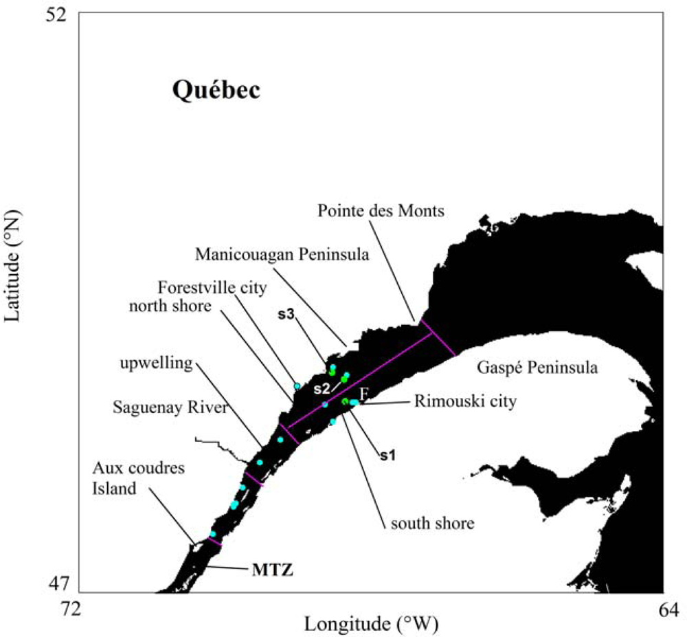
2.1. Field Surveys

Two experiments were designed in the SLE for relating in situ chl determinations with satellite- and CASPER-derived fluorescence measurements. In the first experiment, water samples for chl were obtained from surface waters (0–20 m depth) of different sections of the Estuary as part of an oceanographic cruise on the Coriolis II vessel and organized by ISMER during 17–22 September 2011 (Figure 1). The chosen sampling depth interval is larger than the maximum depth range contributing to the fluorescence signal detected at the surface of SLE waters. However, due to the strong vertical mixing of SLE waters [14], magnitude of fluorescence or chl is quite homogeneous in the top 20 m of the water column. Thus, no fluorescence variations due to vertical layering of chl are anticipated in this study.
Discrete sample volumes (0.5 to 1 L) for chl measurements were collected at three depths (5, 10 and 20 m) using Niskin bottles attached to an oceanographic rosette. Vertical profiles were performed in 23 locations encompassing waters west of Rimouski city (48°26′N, 68°31′W) and east of Aux-Coudres Island (47°24′N, 70°22′W). Since some sampling stations were revisited on different days, a smaller number of sampling positions are shown in Figure 1. In the second experiment, we performed a time series of chl and CASPER fluorescence measurements in the lower estuary (hereafter ‘Frascati’ station, 48.483°N, 68.533°W) (Figure 1). This site was chosen near (∼5 km) the port of Rimouski city where a temporary lab was assembled to facilitate processing of samples. We used a small boat (7 m length) for collecting surface water samples (i.e., 3 m depth) during the morning (9:00 am and 11:00 am), noon (12:00 pm) and afternoon (13:30 pm and 14:45 pm) of 9 September 2011.

2.2. Lab Analysis of Optical Variables

Discrete fluorometric determinations of chl were performed following the acidification technique [15]. Briefly, each volume of sample (100 and 500 mL) was concentrated using a fiber glass filter (GF/F, Whatmann, mean pore diameter = 0.7 microns). Then, organic-soluble pigments were extracted with 90% acetone during 24 h. The fluorescence before and after the addition of diluted acid (HCl 5%) was measured at room temperature (25 °C) and using a single excitation-emission wavelength (λexc = 443 nm, λemiss = 680 nm) fluorometer RD-10AU (Turner designs).
The fluorescence emission spectra of water samples were measured within the size range 0.2–30 microns using the laser-based fluorometer CASPER [16]. This instrument was originally designed to study desertification and includes the following innovations: (1) it is a portable optical system that can be operated by only one person, (2) it has two laser sources one in the UV (wavelength = λ = 266 nm, 2 mW) and one in the visible spectrum (λ = 405 nm, power = 25 mW), that make possible simultaneous measurements of multiple optical components (e.g., CDOM and chl), (3) it allows discrimination between dissolved and particulate phases due to two sets of filters, one to eliminate particulates above 30 μm (Millipore optiscale capsule, polycarbonate, pore size = 30 μm, diameter = 69 mm), and another one to separate fractions below and above 0.2 μm (Sartorius, Sartobran P, mixed cellulose esters, pore size = 0.2 μm, diameter = 47 mm), (4) it provides fast determinations since each sample (particulate + dissolved) can be analyzed in less than one minute, and (5) it has a miniature detector consisting of a fiber optic spectrometer (USB2000+, 2,048 pixels, 200–1,100 nm, Ocean Optics).
Each measurement requires five steps: (1) purge and fill up of system components (tygon tubing and quartz cuvettes) with water samples, (2) background light measurement for each excitation wavelength by alternatively turning off the lasers, (3) high spectral resolution (0.3 nm) fluorescence spectra measurements at UV excitation, (4) idem as (3) but using the visible wavelength, and (5) rinsing of system components with deionized water (Milli-Q). The minimum detection level of CASPER for measuring chlorophyll is 0.1 μg·L−1 at an emission wavelength of 680 nm.

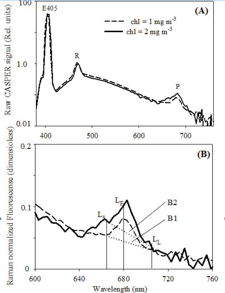
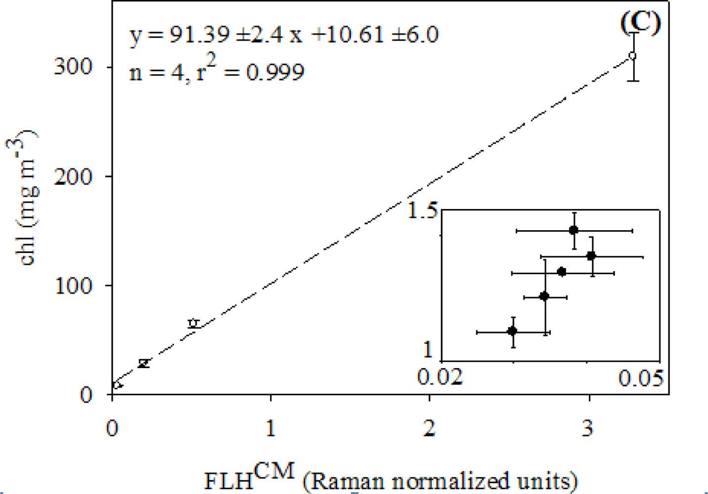
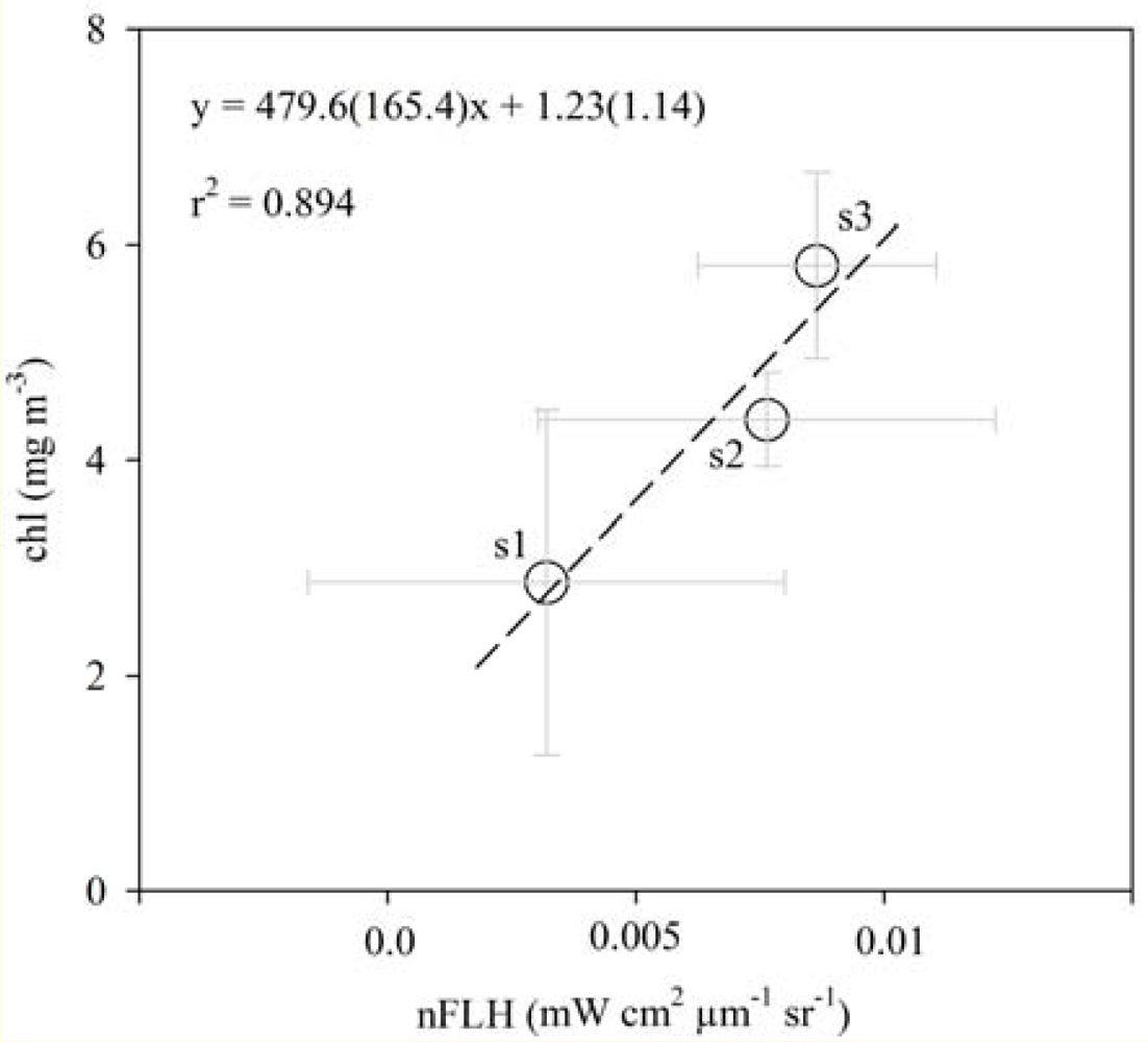
2.3. Relationships between FLH and Chlorophyll

Chlorophyll-fluorescence is a major pathway for dissipating energy in excess going to photosystem II or PS II (i.e., the main protein complex of photosynthesis). The ratio fluorescence/chlorophyll is variable and depends on light harvesting pigments or antenna characteristics and rate of electron flow between PS II and PS I [17]. Fluorescence/chlorophyll increases with light as reaction centers become saturated and starts decreasing beyond saturating light intensities as absorbed photons are transformed into heat due to the xanthophyll cycle. In Canadian coastal waters, Neville and Gower [18] showed for the first time a positive correlation between chlorophyll a concentration and the magnitude of sun-stimulated fluorescence at 685 nm as estimated from airborne reflectance spectra. Based on these preliminary results, Borstad et al.[19] suggested the use of FLH for estimating chl even though there is a large variability of FLH/chl (0.01 to 0.08 mW·cm−2·sr−1·μm−1·mg·Chlorophyll−1) between different water types. In simple terms, FLH is defined as follows:
FLH = L F kL S ( 1 k ) L L
k = ( λ L λ F ) ( λ L λ S ) 1
where LF, LS and LL are the radiance measurements coinciding with the chlorophyll fluorescence peak (LF) and spectral limits (lower, LS and higher, LL) defining FLH, and λS, λF and λL are the corresponding wavelengths. The linear baseline between LS and LL is weakly influenced by atmospheric components such as oxygen and water vapor [10,20], and other optical components when waters are classified as case I (i.e., phytoplankton covaries with other optical components) [9,10]. Thus, most of the variability of FLH in oceanic waters can be attributed to phytoplankton abundance and physiology [9,10,20]. However, in case II waters, the use of FLH for estimating chl may be questionable or useless depending on the contribution of CDOM and detritus to the light attenuation in the fluorescence excitation and emission spectral ranges [9].
Response of FLH to different chl values was analyzed based on Raman-normalized (wavelength = 468 nm) CASPER spectra (hereafter FLHCM). Raman normalization is intended to eliminate CASPER fluorescence variability due to changes in light source intensity or water temperature. FLHCM was computed based on MERIS (MEdium Resolution Imaging Spectrometer) radiometric channels along with their respective bandwidths (LS = 665 ± 10 nm, LF = 681 ± 7.5 nm, and LL = 709 ± 10 nm). Unlike MODIS (Moderate Resolution Imaging Spectroradiometer), spectral location of radiometric channels in MERIS is more suitable for chlorophyll fluorescence in terms of signal/noise ratio (1.4 fold higher) [20]. In addition, the use of FLH instead of single wavelength (e.g., centered at 680 nm) chl indices is justified by the fact that remotely sensed (airborne or spaceborne) FLH calculation minimizes elastic scattering contribution originated from water and atmospheric components [10,20].
Samples for FLH-chl experiments were obtained during a time series performed at the Frascati station (see Section 2.1), and from phytoplankton lab cultures. For the second comparison, we prepared four chl dilutions (8.2, 28.7, 64.8 and 309.3 mg·m−3) from the same culture of microalgae Nannochloropsis oculata. This Eustigmatophyceae species was cultivated using f/2 medium and samples were collected during the exponential phase of growth.

2.4. Satellite Ocean Color Measurements

The analysis of spaceborne ocean color information allowed us to investigate three main questions: (1) How do satellite chl estimates based on CASPER-chl relationships compare with global ocean color products or local parameterizations developed using concurrent ship and satellite-derived fluorescence data?, (2) What is the influence of CDOM and detritus on chl estimates computed from FLH measurements?, and (3) How alike are regional spatial patterns of fluorescence- vs. Lw-based chl in the SLE?

2.4.1. Datasets

Global ocean color products derived from SeaWiFS (Sea-viewing Wide Field-of-view Sensor), MODIS-Aqua, MERIS sensors were browsed from NASA [21] and ESA [22] data distribution servers, and with a spatial resolution of 4.5 and 5.6 km, respectively. The analysis of satellite measurements was performed using two images (1 MERIS and 1 MODIS-Aqua) obtained during 28 September 2011. Redundant information was avoided by selecting complementary ocean color products from NASA (normalized FLH or nFLH, chlorophyll band-ratio model oc3 or chloc3) and ESA (CDOM absorption coefficient at 443 nm or CDM, total suspended matter concentration or TSM, nLw or normalized Lw, chlorophyll neural network model or chlNN, and chlorophyll band-ratio model Garver-Siegel-Maritorena or chlGSM).
All the online chlorophyll products used in this study are based on Lw even though they differ with respect to: (1) the mathematical model (chloc3 is empirical, chlGSM is semi-empirical, chlNN is statistical), and (2) the spectral information (3 wavelengths in chloc3vs. 4 or more in chlGSM or chlNN). The inversion model chloc3 is based on a polynomial of order four that depends on the maximum remote sensing reflectance (Rrs) blue-green band ratio (i.e., Rrs(443)/Rrs(551) vs. Rrs(488)/Rrs(551)) [23]. The chlGSM model computes chl by minimizing differences between spectral nLw measurements at four or more wavelengths in the visible range and simulated nLw derived from a quadratic function [24]. The solution is found by optimizing the specific aph (i.e., aph* = aph/chl), the spectral slope of particulate backscattering and absorption coefficient of CDOM using a nonlinear least squares technique. Notice that the optimization of the spectral slope of particulate backscattering may have implicit a major error due to the inherent uncertainties associated with the backscattering measurements from the space.
Lastly, the chlNN, originally named chl2, is obtained from aph at 443 nm after training a neural network using spectral nLw corresponding to 8 MERIS bands in the visible spectrum [25]. Briefly, nLw, downwelling irradiance, and Rrs are simulated for different solar and viewing angles with a Monte Carlo radiative transfer model that is initialized with chl (1–50 mg·m−3), CDM (0.1–2 m−1), and TSM (1–50 g m−3) values obtained from the North Sea and Baltic Sea waters [26]. The neural network is a feed-forward back-propagation optimization technique composed by two hidden layers of neurons and four output variables (chlNN, CDM, TSM, and aerosol path radiance at 670 nm).

2.4.2. Analysis of Imagery

The selection of images was based on the following criteria: (1) each swath should have at least an area covered with 25% of valid pixels, (2) match-ups between satellite-derived nFLH measurements and shipboard samples should occur within 3-h of satellite overpass, and (3) the time window for the satellite overpass should correspond with September 2011, a period of the year characterized by minimum cloudiness. Quality control for each pixel bin was based on latest flags suggested by NASA and ESA and mainly related to land, bottom, turbidity and glint. In addition, NASA L2 flags (N = 32) are more with respect to ESA flags (N = 16) because the latest summarizes many of the former in bits 13 to 15. For each ocean color product, we defined a valid pixel as a pixel that has not been flagged by the previous constraints and has no negative values since they may reflect inversion model issues or very low signal/noise ratios caused by dominance of water constituents such as CDOM.
Two types of satellite-derived chl indices based on fluorescence were used, one derived from MODIS-Aqua nFLH values (hereafter, chlSM), and another one based on CASPER model and nFLH computed from MERIS nLw measurements (hereafter chlCASPER). In all cases, negative nFLH or nLw values were excluded from the analysis, and unlike other studies [27] no offsets were used to initialize nFLH when chl is 0 mg·m−3 since these corrections are small compared the detection limit of LF (±0.002 mW·cm−2·sr−1·μm−1) (Equation (7) in [10]), and the fact that our chl proxies are not totally absolute because of the lack of field validation. Unlike Lw, nLw is corrected by the sun-sensor geometry as well as for atmospheric scattering and absorption [28].
Calculation of chlCASPER was performed using FLHCM-chl functionality derived in the lab and consisting in field and phytoplankton culture samples. Unfortunately, no quasi-simultaneous comparisons between in situ FLHCM and MERIS imagery data were available in this study. Thus, we propose an algorithm that only relies on lab optical measurements. We recognize two main differences for applying this chl proxy: (1) the spectral characteristics of the excitation light source in CASPER (i.e., laser-based and monochromatic) with respect to MERIS (sun-induced and multiple wavelengths), and (2) variations between CASPER and MERIS detector response functions. These variations, however, may be disregarded here if the main purpose is to perform relative comparisons in terms of chl spatial trends or inter-algorithm comparisons.
Processing of satellite ocean color products derived from NASA and ESA were performed with the development platform BEAM 4.10.3 [29] and the Windows Image Manager [30] software, respectively. Image data was extracted using a filter of 3 × 3 pixels and was intended to relate ship and spaceborne information, compare nFLH-based variables with global ocean color products of chl, and evaluate interference of CDOM and detritus on nFLH in different regions of the SLE.

2.4.3. Statistical Analysis

Variation of chl as a function of FLHCM was quantified for in situ and cultivated phytoplankton samples using a linear regression model type II (i.e., random error for dependent and independent variables) based on least squares differences [31]. Similarity between chlCASPER (independent variable x), chlSM, and global ocean color products of chl (dependent variable y) was evaluated based on magnitude of coefficient of determination (r2) corresponding to a polynomial regression function: y = mx + I, where m and I are the slope and the intercept, respectively. For pigment concentrations above 0.5 mg·m−3, the phytoplankton fluorescence has a non-linear behavior as a function of chl due to a greater re-absorption of photons [20]. Thus, we also examined the response of chlSM and global chl products with respect to chlCASPER variations by fitting the data using a hyperbolic model: y = (k1 x)/(k2 + x), where x is chlCASPER and k1 and k2 are constants that are computed by the Levenberg-Marquardt non-linear least square differences method. Discrimination and removal of outliers affecting the aforementioned functionalities was conducted by applying a Hampel filter [32].
Ratios between chloc3, chlGSM or chlNN (numerator), and chlCASPER (denominator) were computed for each pixel bin and used to examine the relative bias between the fluorescence-based and the Lw-based chl indices. The lower the magnitude of r2 and the greater the (m−1) difference the larger the relative error of chl estimates as computed using the respective global ocean color algorithm. The potential interference of CDOM and detritus on nFLH and consequently chlCASPER was explored for each SLE sub-region (Figure 1) based on linear regression models type II between nFLH and CDM, and nFLH and TSM, respectively. Notice that TSM is used here as a proxy for detritus concentration since more than 50% of the TSM mass in the SLE is composed by inorganic matter [7], however this parameter also includes a variable contribution of microalgae that may have a substantial contribution to the fluorescence signal. CDOM will mainly compete with light absorption by chlorophyll at relatively short wavelengths (i.e., <400 nm) [9,20], thus less red fluorescence is anticipated in those cases where CDOM is a dominant water optical component. This effect is expected to occur if the slope of the nFLH-CDM regression is less than zero. Moreover and over detritus-rich waters, most of the reflected energy in the spectral range 670–690 nm is associated to elastic scattering contributions coming from sediments and/or organic non-living particulates. Thus, as TSM becomes enriched in detritus, nFLH is not longer a good indicator of chl [33] and an uncoupling between nFLH and TSM is anticipated resulting in m’s that are near zero or negative.

3. Results

3.1. Relationships between chl and FLH as Derived from Field and Lab Samples

The raw and processed fluorescence signal as measured by the CASPER spectrofluorometer and corresponding to samples obtained during the Frascati time series is shown in Figure 2(A,B). As expected, variability of the raw CASPER emission spectra (i.e., non-corrected by elastic contributions from the excitation source and inelastic water contributions from Raman) in the spectral range 380–760 was mainly attributed to changes in phytoplankton pigments when chl values varied between 1 and 2 mg·m−3 (Figure 2(A)). In fact, signal modifications due to the laser source performance (18.2% at λ = 405 nm) or water temperature variability (13.8% at λ = 468 nm) was small with respect to phytoplankton-related fluorescence (41.5% at λ = 680 nm). In addition, it is worthy to emphasize the spectral shift of the emission peak of chlorophyll (∼4 nm) to longer wavelengths when chl was doubled (Figure 2(B)). The relationship between chl and FLHCM measurements based on in situ measurements or phytoplankton culture samples was linear for a chlorophyll concentration range between 1.1 and 309.3 mg·m−3 (Figure 2(C)). Regression slope coefficients were different from zero for both functionalities; however the magnitude of m for CASPER and Turner comparisons based on phytoplankton samples growing in controlled conditions was 2.7-fold greater (i.e., the fluorescence per unit of chl was 2.7-fold smaller) with respect to that computed using field samples (chl = FLHCM (33.4 ± 9.3) + 0.13 ± 0.34, r2 = 0.81, N = 5).
Since our intention here is to propose a fluorescence-based index and not an absolute metrics of chl that can be applied to optical satellite measurements over the SLE, and given the limited range of pigment values used when comparing field samples, we decided to use the chl-FLHCM function obtained from phytoplankton cultures for modeling spaceborne chl based on fluorescence line height estimates.

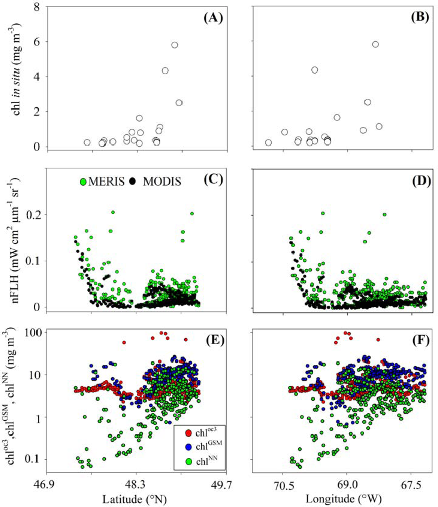
3.2. Chlorophyll Proxies Based on Satellite-Derived FLH and Lw

The direct relationship between in situ chl and satellite fluorescence was supported by shipboard surveys performed during 21 September 2011 (Figure 3). Given the range of pigment concentrations measured during the cruise (0.2 to 5.8 mg·m−3), a non-linear variation should be expected between averaged surface (0–20 m depth) chl determinations and MODIS-Aqua nFLH values. However, this effect could not be detected since only 3 of 23 cruise stations coincided in time and space with satellite measurements. The resulting linear regression function was developed with samples obtained across the lower estuary, had a regression slope greater than 1 (P < 0.05), and was characterized by a regression intercept not different from zero (P > 0.05). Despite the small time difference between MODIS-Aqua (17:10 GMT) and MERIS (16:53 GMT) overpasses during 28 September 2011, the linear covariation between chlSM and chlCASPER was relatively weak (r2 = 0.335) (Figure 4(A)). Nevertheless, the co-dependency between chlSM and chlCASPER values was greater with respect to that seen between CASPER and Lw-based chl indices (r2 up to 0.255, Figure 4(C)). Notice that the number of valid data points differ between regression curves due to geographic differences in algorithm performance. In general and based on the regression slope, chlCASPER was underestimated and overestimated with respect to chlSM (m > 1, P < 0.05, Figure 4(A)) and chlNN (m < 1, P < 0.05, Figure 4(D)), respectively. The regression intercept was in all cases greater than one but for chlCASPER − chlSM and chlCASPER − chloc3 where I was not different from zero (P < 0.05). The hyperbolic regression model only converged when chlCASPER was related with chlGSM and chlNN (Figure 4(C,D); Table 2), and presented values of r2 that reflected a slight improvement on explaining residual variability (up to 7%) with respect to the linear regression functions.

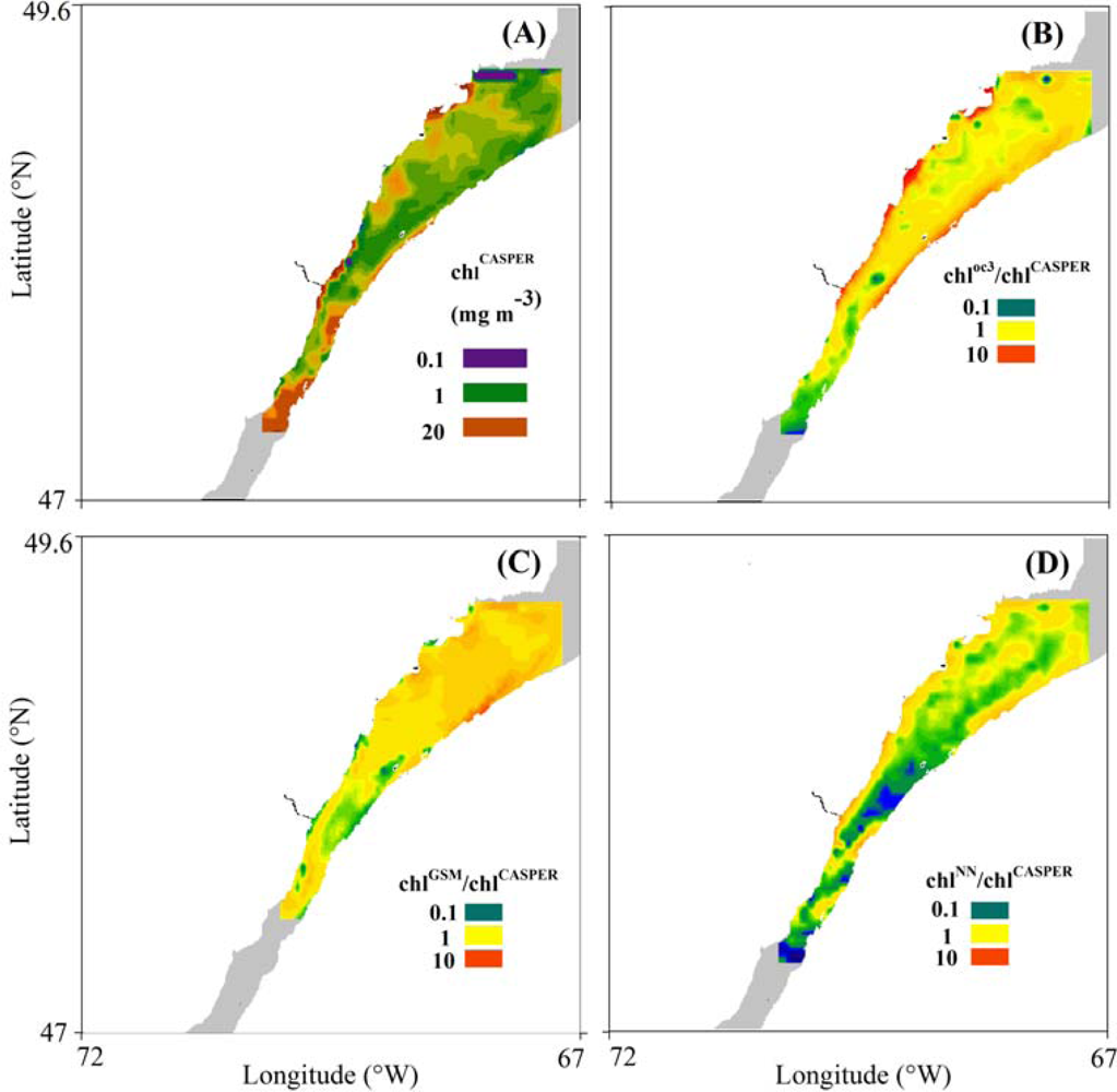
3.3. Spatial Coherence between Fluorescence and Lw-Based Indices of chl

The spatial distribution chlCASPER values throughout the SLE and during noon of September 28, 2011, was very irregular with values changing up to one order of magnitude (2.27–21.5 mg·m−3) (Figure 5(A)). Highest pigment concentrations (>20 mg·m−3) were found north of the MTZ and near the mouth of the most important north shore rivers draining in the lower part of the St Lawrence Estuary (Saguenay, Manicougan and Outardes). Intermediate to high values (1–10 mg·m−3) were observed south of the Outardes Delta and over the main channel, and around the archipelago extending along the south shore of the lower estuary (Figure 1). These spatial patterns were also observed in satellite images collected 15 days before or after September 28 (data not shown). In general and consistent with Figure 4, the ratio between chlCASPER and chloc3, chlGSM and chlNN over the whole SLE varied 134, 48 and 860-fold, respectively (Figure 5(B–D)). Lower chloc3 values with respect to CASPER estimates were mainly computed in the upper estuary (ratio < 0.5) (Figure 5(B)). Conversely, oc3 pigment predictions overestimated CASPER (ratio > 20) over waters near the coast of the north and south shores of the lower estuary. In general, chlGSM and chlCASPER estimates followed comparable spatial patterns even though large differences were observed in specific locations of the south shore (ratios < 0.2) and waters north-east of Rimouski city (ratios > 5) (Figure 5(C)). Lastly, the spatial distribution of chlNN showed relatively low values (ratio up to 0.005) with respect to chlCASPER in a large portion of the Estuary including upper and lower sections, and mostly coinciding with deeper waters along the Laurentian Channel (Figure 5(D)).
These remarkable differences in chl indices may lead to ambiguous spatial patterns of phytoplankton along and across the Estuary. In fact, latitudinal and longitudinal changes in chlorophyll during the September 2011 cruise showed no increase toward the southwest, a trend clearly defined by MODIS-Aqua or MERIS fluorescence measurements (Figure 6(A–D)). Notice that these discrepancies were also present even after analyzing satellite scenes corresponding to other days during September. Differences in large scale spatial patterns were also striking between satellite fluorescence and global products of chl. In general, latitudinal and longitudinal distribution of chloc3 magnitude was quite constant compared to those associated with satellite fluorescence (Figure 6(C–F)). Geographic variability of chlGSM and satellite fluorescence was comparable even though this correspondence tended to disappear over the westernmost locations of the upper estuary (Figure 6(E)). Conversely, the chlNN showed an inverse spatial variation with respect to nFLH, and consequently was a very misleading index for mapping phytoplankton distributions in the SLE (Figure 6(F)).

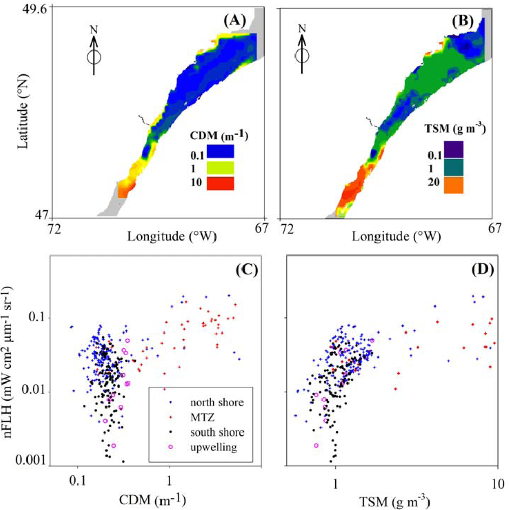
3.4. Effects of CDOM and Detritus on FLH

Relationships between satellite-derived nFLH, CDM, and TSM values during 28 September 2001 are shown in Figure 7. In general, CDM and TSM presented higher values over areas influenced by river discharge and turbidity plumes (e.g., NE of the MTZ in the upper estuary and along the north shore in the lower estuary) (Figure 7(A–B)). The highest concentrations of CDM (up to 5.7 m−1, north shore) and TSM (up to 20 g·m−3, MTZ) were found in the same sub-regions where maximum values of chlCASPER (>20 mg·m−3) and nFLH (up to 0.20 mW·cm2·μm−1·sr−1) were obtained (Figure 5(A) and Figure 7(C,D)). Likewise, the lowest CDM (<0.1 m−1) and TSM (<1 g·m−3) values were detected in those locations characterized by the lowest chlCASPER (<3 mg·m−3) and nFLH (<0.005 mW·cm2·μm−1·sr−1) values (e.g., central portion of lower estuary) (Figures 5(A) and 7(A,B)). In general, the magnitude of nFLH varied independently from CDOM variability when CDM magnitude was less than 0.3 m−1 (e.g., all sub-regions). Also, nFLH appeared to be positively associated with CDOM at CDM values greater than 0.5 m−1 (e.g., north shore and MTZ) even though the slope of this trend was not different from zero (P > 0.05) (Figure 7(C)). Conversely, nFLH was positively related with TSM when TSM concentrations were relatively low (<2 g·m−3) (all sub-regions, r2 = 0.20, nFLH = 0.028 TSM (8·10−4, 1 standard error) + 0.006 (9·10−4), N = 4719, Figure 7(D)). Likewise, this correspondence was weaker at greater TSM values (2–10 g·m−3), and disappeared at TSM greater than 10 g·m−3 when nFLH reached a relatively constant value around 0.1 mW·cm2·μm−1·sr−1.

4. Discussion

4.1. Relationships between chl and CASPER Fluorescence

Lab measurements using CASPER demonstrated that fluorescence emission at a wavelength of 680 nm varied less with respect to chlorophyll changes. Indeed at chl >0.5 mg·m−3, a variation in chlorophyll of 100% only corresponded with a 42% relative variation of normalized fluorescence line height. At higher pigment concentrations (i.e., chl > 0.5 mg·m−3), light excitation is less efficient due to partial absorption of emitted fluorescence and a smaller phytoplankton optical cross section (i.e., aph/chl smaller) associated with a greater ‘packaging effect’ [20]. Another interesting feature of the CASPER fluorescence spectra was the spectral shift of the phytoplankton fluorescence peak at higher chl values. This phenomenon was previously reported based on field measurements [9,34] and is related with a spectral shift to longer wavelengths on Lw and driven by a greater contribution of elastic scattering with respect to chlorophyll fluorescence [9]. Certainly, this spectral shift can not be detected using conventional fluorometers based on a single-wavelength excitation. Thus, we suggest that CASPER and other comparable spectrofluotrometers are a better choice to calibrate in-water and satellite fluorescence-based models of chl.
The relationships found between CASPER-based nFLH and chl samples (field and derived from cultures) are not expected to be substantially influenced by the fluorometric method (i.e., with acidification) or type of fluorometer (excitation wavelength = 436 nm) used in this study. This statement is supported by previous experiments in the SLE during spring 2000 and 2001, and comparing chl measurements based on non-acidification method (chlWEL) [35] or High-Pressure liquid Chromatography (chlHPLC) with fluorometric determinations (chlacid = (0.74 ± 0.05, 1 standard error) chlHPLC +(0.09 ± 0.25), r2 = 0.80, N = 70; chlacid = 0.88 chlWEL ± 0.03+(0.06 ± 0.15), r2 = 0.93 N = 70) performed using the same technique and optical instrument (chlacid) [36].
Although a direct relationship was always observed between FLHCM and chl, the fluorescence yield per unit chlorophyll was lower when experiments were performed with phytoplankton cultures. The discrepancy was likely attributed to the greater light levels used in the incubated samples with respect to those characteristic of field measurements. PAR (i.e., photosynthetic available radiation, wavelength = 400–700 nm) above a threshold of 1.0·10−4 mol·quanta·m−2·s−1 can be photo-inhibitory and tends to lower fluorescence/chl [27]. Averaged PAR of incubated samples was 3.5·10−4 mol·quanta·m−2·s−1 or more than 3-fold greater than theoretical in situ estimates computed as <PAR> = PAR(0+)/(Hmix KPAR) [37], where PAR(0+) is the PAR at noon and incident at the sea surface during 28 September 2011 (1.04·10−3 mol·quanta·m−2·s−1), Hmix is the averaged mixed layer depth (10 m), and KPAR is the lowest vertical light attenuation coefficient for PAR measured in the SLE (1.1 m−1). Therefore, the above calculations suggest potential modifications on fluorescence/chl in phytoplankton cultures due to excessive light.
Variations in fluorescence between experiments due to changes in micronutrients concentration, water temperature, taxonomic differences, and CDOM are anticipated to be secondary due to the following facts. As specified by Turner designs, the variation of acetone-extracted chlorophyll fluorescence due to temperature is 0.3%/°C. This effect may introduce a negligible error on FLH calculation (up to 3%) if we consider a maximum temperature shift of 10 °C between field and lab measurements.
Based on published reports, the phytoplankton culture used in this study (N. oculata) has a fluorescence emission spectrum that is comparable to those associated with bacillariophytes (i.e., diatoms) [38]. Thus, since diatoms dominate over the lower St Lawrence Estuary (i.e., where field samples were collected for CASPER comparisons) [39], uncertainties on CASPER fluorescence curves due to phytoplankton composition are likely minor. In general, phytoplankton growth is not nutrient-limited in the SLE [40], thus nutrients should not have a major influence on phytoplankton fluorescence in our experiments.
The linear behavior between chl and FLHCM variations seemed to contradict empirical and theoretical parameterizations reported by other studies [9,20,33]. However, we attribute this deviation to the lack of ‘packaging effect’ and re-absorption of photons as a function of chl in the phytoplankton culture experiments. As chl increases, the aph* tends to decrease and re-absorption of photons tends to increase creating a saturation plateau on fluorescence values [20, 27]. This phenomenon did not occur in our phytoplankton cultures since we worked with diluted samples (i.e., same abundance per volume) and phytoplankton cells were exposed to similar light conditions during the incubation.

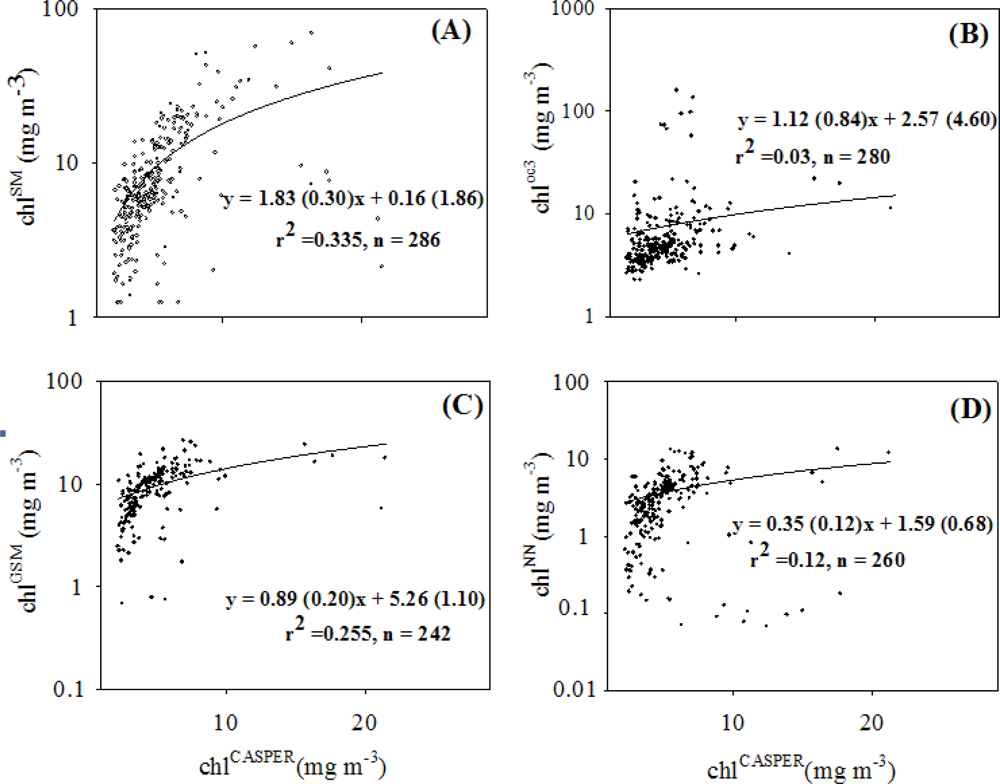
4.2. Coherence between chlCASPER and other Chlorophyll Indices

Although chlCASPER and chlSM were both based on fluorescence line height changes, the empirical model developed with MODIS-Aqua and shipboard measurements only corresponded with CASPER estimates in 34% of the cases (Figure 4(A)). In addition, both variables did not follow a 1:1 relationship. This is not surprising given three main reasons: (1) the increase in fluorescence variability in field samples with respect to laboratory measurements, due mainly to changes on aph* (Equation (2) in [20]), (2) differences between spectral bands used in MODIS-Aqua and CASPER-MODIS models to compute FLH (Figure 1 in [20]), and (3) bias attributed to spectral changes in excitation light source (i.e., laser in CASPER vs. sun-induced in MODIS or MERIS) and receiver’s differences (i.e., detector response of CASPER vs. satellite sensors). As highlighted in Figure 4(C), a variation in chlCASPER was followed by a comparable rate of change of chlGSM even though the regression intercept suggested a ‘residual’ pigment as computed by the GSM model when chlCASPER was 0 mg·m−3. This pigment offset was attributed to a greater CDOM interference on chlGSM with respect to chlCASPER. CDOM is a dominant optical component that is responsible for absorbing most of the blue light in the SLE [7]. Thus, in CDOM-rich waters, the total light absorption at 443 nm (i.e., maximum absorption peak for chlorophyll) increases, resulting in an overestimation of chl when this pigment is measured using remote sensing reflectance ratios. To illustrate this effect we have that chl is inversely proportional to R1 = Rrs(443)/Rrs(550) = (a(550) bb(443))/(a(443) bb(550)), where a and bb are total absorption (i.e., water + particulates + CDOM) and backscattering coefficients at the corresponding wavelengths. Therefore, if chl and bb(443)/bb(550) are constant, and magnitude of aCDOM(443) is larger, 1/R1 becomes smaller and chl is higher.
Why did chloc3 and chlNN not have a good correspondence with chlCASPER? Unlike the other global products of chl, the performance of chloc3 is highly influenced by CDOM interference since it uses less spectral information than the other models to tease out the optical contribution of phytoplankton from that associated with CDOM. The lack of covariability between chlNN and chlCASPER in the SLE is not surprising since chlNN was originally developed with specific neural network coefficients obtained from European coastal waters. In fact, this neural network is initialized with TSM measurements made over the North Sea and the Baltic Sea. Thus, a large uncertainty on chlNN can be expected between the SLE and European coastal environments due to changes on SPM optical properties between different water bodies and associated to variations on organic content and size distribution of particulates. As shown in Table 2, a hyperbolic function was a more accurate regression model for relating chlCASPER with global chl products. These results support the non-linear behavior between chl and fluorescence, and corroborate semi-empirical relationships suggested by Gower’s and Gilerson’s studies [9,20].

4.3. Spatial Distribution of Spaceborne Chl Indices

The use of discrete shipboard measurements for characterizing large scale patterns of phytoplankton in surface waters of the Saint Lawrence Estuary may be misleading if oceanographic surveys are inadequate in terms of spatial coverage or density of sampling locations. Likewise, satellite measurements based on Lw may not be useful for mapping horizontal variations of chlorophyll in this environment if global ocean color products (e.g., chlNN) are not locally validated. In that regard, we demonstrated the superior ability of spaceborne FLH measurements for deriving more realistic distributions of chl in SLE waters.
Consistent with ‘in situ’ historical fluorescence measurements [11], chlCASPER presented maximum values (i.e., >10 mg·m−3) in three areas of the Estuary: north of Aux-Coudres Island in the upper Estuary (47.7°N, 69.9°W, I), nearby Forestville city (48.55°N, 69.145°W, II), and the Manicougan Peninsula (49.07°N, 68.19°W, III) in the north shore of the lower Estuary. These high chl spots are likely created by downstream advection and resuspension of benthic diatoms (I) [41], divergence fronts (II) [42], and river plumes (III) [43]. Unlike other studies, the analysis of chlCASPER fields during September 28 also suggested relatively high pigment concentrations in the proximity of the Saguenay Fjord entrance and along the opposite coast extending to 48.49°N, 68.55°W. These sites correspond with major tidal flats environments (e.g., Bay Sainte-Anne, Berte Island, Figure 1) and an active transport of sediments including attached microalgae [41].
Although chlCASPER and chlSM are more suitable metrics for describing variations of chlorophyll in the SLE with respect to global ocean color products, the interpretation of geographic variability of chlorophyll based on satellite fluorescence may be compromised if one or more of the following factors play an important role altering FLH: (1) inaccurate atmospheric correction, (2) modified phytoplankton physiological parameters affecting quantum yield of fluorescence (ΦF) and/or aph*, and (3) influence of high concentration of detritus and/or CDOM on fluorescence excitation/emission spectra. Regarding the first factor, we compared MERIS top-of-the atmosphere (TOA) reflectance measurements over the SLE with simulated TOA values computed using a radiative transfer model (6SV, [44]) and local meteorological information obtained from AERONET and airport weather stations located at Mont Joli and Quebec City. During 28 September 2011, measured TOA radiance due to aerosols accounted for 20 and 40% of total radiance at λS and λL, respectively. Satellite-derived optical thickness of aerosols at 869 nm, the most important uncertainty in atmospheric models, was relatively small and invariable (0.03 to 0.08) suggesting relatively low concentration of aerosols over the study area. Preliminary results show a maximum bias of 25% (29.9% at λS, 20.8% at λL) between simulated and measured TOA reflectance. Thus, we conclude that atmospheric correction of ocean color products used in this study was satisfactory and its uncertainty is relatively small with respect to the nFLH variation observed in the SLE (several orders of magnitude, see Figures 6(C,D) and 7(C,D)). However, satellite measurements of nFLH under turbid atmospheres and high solar zenith angles are expected to be more inaccurate by increasing the detection limit of chl retrievals up to 2.6-fold [10].
With reference to the second factor, nFLH is expected to change in a direct way with respect ΦF and aph* (Equation (2) in [27]). Previous investigations have shown that the magnitude of aph* during April–May 2000 and 2001 cruises in the SLE varied between 2.8 and 6.4-fold, respectively [45]. Therefore, in our study area, the impact of this variability for satellite chlorophyll fluorescence should be secondary. The influence of ΦF on nFLH is more difficult to assess over the SLE since there are no direct in situ measurements of ΦF (e.g., based on fast rate pulse fluorometers). However, no major changes on ΦF are expected in our study area given the lack of nutrient-limiting conditions for phytoplankton growth [43]. Based on radiative transfer simulations, Gilerson et al.[9] concluded that ΦF is relatively constant and equal to 1% in coastal waters having chl values ranging from 1 to 50 mg·m−3.
Reduction in nFLH due to non-photochemical quenching may occur if phytoplankton cells are exposed to damaging solar radiation levels [27]. However, mid-day fluorescence depression due to photo-inhibition is unlikely in our satellite-derived nFLH values (see Section 4.1) unless waters are relatively shallow (bottom depth <10 m) and clear (KPAR < 1 m−1). These environments are spatially constrained as they only represent less than 10% of the SLE surface area [46]. The third factor affecting nFLH mainly relates to the interference exerted by dominant optical water constituents other than pigmented particulates. The analysis of relationships between the CDM and nFLH for different regions of the SLE did not support a hypothetical suppression of phytoplankton fluorescence due to CDOM-mediated spectral overlap of chlorophyll excitation wavelengths as proposed by other studies [9]. This apparent discrepancy was probably attributed to the larger effect of detritus-derived elastic scattering with respect to CDOM light attenuation on nFLH variability.
In general, nFLH and TSM were directly related over different waters of the SLE (Figure 7(D)). However, this correspondence varied for different TSM intervals (m = 0.028 for TSM = 0.5 and 2 g·m−3 and m = 0 for TSM = 2.5 to 20 g·m−3). TSM includes a variable contribution of algal particulates, thus we suggest that nFLH variability is dominated by microalgae at lower TSM values (e.g., south shore and upwelling regions). Conversely, the leveling off of nFLH at higher TSM values could be interpreted as an increase in detritus mass with respect to microalgae (e.g., MTZ and north shore). The aforementioned impact of detritus on chlorophyll fluorescence peak and FLH has been investigated by Gilerson et al.[9,33] who showed that chl explains one-third of FLH variability when concentrations of non-algal particulates (NAP) are relatively low (<2 g·m−3). However, FLH may not be an accurate proxy for chl when NAP concentrations are above 2 g·m−3 and becomes an useless index at NAP concentrations above 10 g·m−3 due to the overwhelming contribution of elastic scattering (>95%) with respect to fluorescence. In this study, we estimate a NAP range between 0.5 (e.g., upwelling and south shore) and 20 g·m−3 (e.g., MTZ and north shore) [7], thus our chlCASPER index may be used in the SLE only in those locations characterized by relatively low to moderate turbidity and having concentrations of total suspended particulate matter below 13 g·m−3.

5. Conclusions

Comparisons between satellite and field data showed that nFLH is a very good indicator of chl in surface waters of the SLE (e.g., r2 = 0.89 in Figure 3) characterized by relatively low to moderate detritus concentrations (i.e., ≤10 g·m−3). In addition, we found that alternative remote sensing proxies of chl based on elastic scattering and light absorption coefficient variations may present large uncertainties (up to 48, 134 and 860-fold for chlGSM, chloc3 and chlNN, respectively) with respect to fluorescence-based estimates. Global NASA and ESA chl products examined here mainly failed due to the high interference of CDOM at relatively short wavelengths (<400 nm) and the local variability of water optical properties.
Although more comparisons between CASPER-derived fluorescense, Lw, and chl are needed to make chlCASPER an absolute proxy for chlorophyll, we suggest the use of spectrofluorometry to validate remote sensing models of chl in dynamic and optically complex waters such as those corresponding to the SLE. Mini-spectrofluorometers like CASPER are ideal instruments for validating satellite-derived chl models since they allow cheap (i.e., <1 US dollar per sample), fast (i.e., <1 s) and accurate (i.e., ±0.1%) chl measurements at very high temporal (i.e., seconds) and spatial (i.e., cm) resolution, and over a wide dynamic range (i.e., 0.1 to 400 mg·m−3).

Acknowledgments

We really appreciate the constructive comments from two anonymous reviewers. We thank Michel Gosselin at ISMER-UQAR for sharing chlorophyll calibration data and Larisa Poryvkina at Laser Diagnostics Instruments AS, Estonia for making available fluorescence phytoplankton spectra measurements. We thank Roberta Fantoni (ENEA) and Massimo Iannetta (ENEA) for constant encouragement, Maria Sighicelli (ENEA), Ileana Iocola (Intrega Srl) and Daniele Pittalis (Intrega Srl) for the fundamental contribution in CASPER operation and upgrading. Also, we really appreciate the logistic support of ISMER (Gilles Desmeules, Bruno Cayouette) and ENEA (Claudio Falchi) technicians who helped to make possible the field experiments during September 2011. This study is part of the Québecois-Italian collaborative project CLIMAT (Complimentary use of Lidar for IMproving sATellite-derived bio-optical models in the St Lawrence System) supported by the Ministère des Relations Internationales du Québec.

References

  1. Legendre, L. The significance of microalgal blooms for fisheries and for the export of particulate organic carbon in oceans. J. Plankton Res 1990, 12, 681–699. [Google Scholar]
  2. Fauchot, J.; Saucier, F.J.; Levasseur, M; Roy, S; Zakardjian, B. Wind-driven river plume dynamics and toxic Alexandrium tamarense blooms in the St. Lawrence estuary (Canada): A modeling study. Harmful Algae 2008, 7, 214–227. [Google Scholar]
  3. Montes-Hugo, M.; Carder, K.; Foy, R.; Cannizzaro, J.; Brown, E.; Pegau, S. Estimating phytoplankton biomass in coastal waters of Alaska using airborne remote sensing. Remote Sens. Environ 2005, 98, 481–493. [Google Scholar]
  4. McKee, D.; Cunningham, A.; Dudek, A. Optical water type discrimination and tuning remote sensing band-ratio algortihms: Application to retrieval of chlorophyll and Kd(490) in the Irish and Celtic Seas. Estuar. Coast. Shelf Sci 2007, 73, 827–834. [Google Scholar]
  5. Carder, K.; Chen, F.R.; Lee, Z.P.; Hawes, S.K.; Kamykowski, D. Semianalytic Moderate-Resolution Imaging Spectrometer Algorithms for Chlorophyll A and Absorption with bio-optical domains based on nitrate-depletion temperatures. J. Geoph. Res.-Oceans 1999, 104, 5403–5421. [Google Scholar]
  6. Letelier, R.M.; Abbott, M.R. An analysis of chlorophyll fluorescence algorithms for the Moderate Resolution Imaging Spectrometer (MODIS). Remote Sens. Environ 1996, 58, 215–223. [Google Scholar]
  7. Montes-Hugo, M.A.; Mohammadpour, G. Biogeo-optical modeling of SPM in the St Lawrence Estuary. Can. J. Remote Sens 2012, 38, 1–13. [Google Scholar]
  8. Gower, J.F.R. Study of Fluorescence-Based Chlorophyll a Concentration Algorithms for Case 1 and Case 2 Waters; European Space Agency (ESA), European Space Research and Technology Centre (ESTEC): Noordwijk, The Netherlands, 1999; Contract 12295/97/NL/RE. [Google Scholar]
  9. Gilerson, A.; Zhou, J.; Hlaing, S.; Iannou, I.; Schalles, J.; Gross, B.; Moshary, F.; Ahmed, S. Fluorescense component in the reflectance spectra from coastal waters. Dependence on water composition. Opt. Exp 2007, 15, 15702–15721. [Google Scholar]
  10. Abbott, M.R.; Letelier, R.M. Algorithm Theoretical Basis Document. Chlorophyll Fluorescence (MODIS Product Number 20); NASA: Pasadena, MD, USA, 1999; p. 42. [Google Scholar]
  11. Nieke, B.; Reuter, R.; Heuermann, R.; Wang, H.; Babin, M.; Therriault, J.C. Light absorption and fluorescence properties of chromophoric dissolved organic matter (CDOM) in the St. Lawrence Estuary (Case 2 waters). Cont. Shelf Res 1997, 17, 235–252. [Google Scholar]
  12. D’Anglejan, B.F.; Smith, E.C. Distribution, transport and composition of suspended matter in the St. Lawrence Estuary. Can. J. Earth Sci 1973, 10, 1380–1396. [Google Scholar]
  13. Fuentes-Yaco, C.; Vezina, A.F.; Larouche, P; Vigneau, C; Gosselin, M; Levasseur, M. Phytoplankton pigment in the Gulf of St. Lawrence, Canada, as determined by the Coastal Zone Color Scanner. Part 1. Spatio-temporal variability. Cont. Shelf Res 1997, 17, 1421–1439. [Google Scholar]
  14. Mucci, A.; Starr, M.; Gilbert, D.; Sundby, B. Acidification of the lower St Lawrence Estuary bottom waters. Atmosphere-Ocean 2011, 49, 206–218. [Google Scholar]
  15. Trees, C.C.; Bidigare, R.R.; Karl, D.M.; Van Heukelem, L. Fluorometric Chlorophyll a: Sampling, Laboratory, Methods and Data Analysis Protocols. In Ocean Optics Protocols for Satellite Ocean Color Sensor Validation; Chapter 14, Revision 2; NASA Report; Goddard Space Flight Space Center: Greenbelt, MD, USA, 2000. [Google Scholar]
  16. Colao, F.; Demetrio, E.; Drozdowska, V.; Fiorani, L.; Lazi, V.; Okladnikov, I.; Palucci, A. Seawater Dual Fluorescence Analysis during the Arctic Summer 2006 Polish Oceanographic Campaign. Proceedings of 3rd EARSel Workshop Remote Sensing of the Coastal Zone, Bolzano, Italy, 7–9 June 2007.
  17. Falkowski, P.G.; Kiefer, D.A. Chlorophyll a fluorescence in phytoplankton: relationship to photosynthesis and biomass. J. Plankton Res 1985, 7, 715–731. [Google Scholar]
  18. Neville, R.A.; Gower, J.F.R. Passive remote sensing of phytoplankton via chlorophyll fluorescence. J. Geophys. Res 1977, 82, 3487–3493. [Google Scholar]
  19. Borstad, G.A.; Edel, H.R.; Gower, J.F.R.; Hollinger, A.B. Analysis of Test and Flight Data from the Fluorescence Line Imager. In Can. Spec. Publ. Fish. Aquat. Sci.; Canada Dept. of Fisheries and Oceans: Ottawa, ON, Canada, 1985; Volume 83, p. 48. [Google Scholar]
  20. Gower, J.F.R; Brown, L.; Borstad, G.A. Observation of chlorophyll fluorescence in west coast waters of Canada using the MODIS satellite sensor. Can. J. Remote Sens 2004, 30, 17–25. [Google Scholar]
  21. Ocean Color Web. Available online: http://oceancolor.gsfc.nasa.gov/ (accessed on 1 January 2012).
  22. GlobColour. Available online: http://hermes.acri.fr/ (accessed on 1 January 2012).
  23. O’Reilly, J.E.; Maritorena, S.; Mitchell, B.G.; Siegel, D.A.; Carder, K.L.; Garver, S.A.; Karhu, M.; Garver, S.A.; MCClain, S.A. Ocean color algorithms for SeaWiFS. J. Geoph. Res 1998, 103, 24937–24953. [Google Scholar]
  24. Maritorena, S.; Siegel, D.A. Consistent merging of satellite ocean color data sets using a bio-optical model. Remote Sens. Environ 2005, 94, 429–440. [Google Scholar]
  25. Doerffer, R.; Schiller, H. The MERIS Case 2 algorithm. Int. J. Remote Sens 2007, 28, 517–535. [Google Scholar]
  26. Doerffer, R.; Schiller, H. MAPP Data Products Definition and Algorithm Specification. Algorithm Theoretical Basis Document (ATBD). Regionalized Case II Water Products and Algorithms; ATBD 1, Report; GKSS Research Center: Hamburg, Germany, 2000; p. 87. [Google Scholar]
  27. Behrenfeld, M.; Westberry, T.K.; Boss, E.S.; O’Malley, R.T.; Siegel, D.A.; Wiggert, J.D.; Franz, B.A.; McClain, C.R.; Feldman, G.C.; Doney, S.C.; Moore, J.K.; Dall’Olmo, G.; Milligan, A.G.; Lima, I; Mahowald, N. Satellite-detected fluorescence reveals global physiology of ocean phytoplankton. Biogeosciences 2009, 6, 779–794. [Google Scholar]
  28. Gordon, H.R.; Clark, D.K. Clear water radiances for atmospheric correction of coastal zone color scanner imagery. Appl. Opt 1981, 20, 4175–4180. [Google Scholar]
  29. GlobProject. Available online: http://www.brockmann-consult.de (accessed on 1 January 2012).
  30. Windows Image Manager Software. Available online: http://wimsoft.com/ (accessed on 1 January 2012).
  31. Sokal, R.R.; Rohlf, F.J. Biometry; W.H. Freeman: New York, NY, USA, 1995; p. 498. [Google Scholar]
  32. Pearson, R.K. Mining Imperfect Data. Dealing with Contamination and Incomplete Records; Society for Industrial and Applied Mathematics: Philadelphia, PA, USA, 2005; p. 305. [Google Scholar]
  33. Gilerson, A.; Zhou, J.; Hlaing, S.; Iannou, I.; Gross, B.; Moshary, F.; Ahmed, S. Fluorescence component in the reflectance spectra from coastal waters. II. Performance of retrieval algorithms. Opt. Exp 2007, 16, 2446–2460. [Google Scholar]
  34. Ahn, Y-H; Shanmugam, P. Derivation and analysis of the fluorescence algorithms to estimate phytoplankton pigment concentration in optically complex coastal waters. J. Opt. A-Pure Appl. Opt 2007, 9, 352–362. [Google Scholar]
  35. Welschmeyer, N.A. Fluorometric analysis of chlorophyll a in the presence of chlorophyll b and phaeopigments. Limnol. Oceanogr 1994, 39, 1985–1992. [Google Scholar]
  36. Mas, S. Le Rôle du Phytoplancton de petite Taille (<20 μm) dans les Variations des Propriétés Optiques des Eaux du Saint-Laurent. Université du Québec a Rimouski, Rimouski, QC, Canada, 2008; p. 286. [Google Scholar]
  37. Cloern, J.E.; Grenz, C.; Vidergar-Lucas, L. An empirical model of the phytoplankton chlorophyll: Carbon ratio-the conversion factor between productivity and growth rate. Limnol. Oceanogr 1995, 40, 1313–1321. [Google Scholar]
  38. Poryvkina, L.; Babichenko, S.; Leeben, A. Analysis of Phytoplankton Pigments by Excitation Spectra of Fluorescence. Proceedings of EARsel-SIG Workshop, Lidar Remote Sensing of Land and Sea, Dresden, Germany, 16–17 June 2000.
  39. Roy, S.; Chanut, J.P.; Gosselin, M.; Sime-Ngando, T. Characterization of phytoplankton communities in the lower St Lawrence Estuary using HPLC-detected pigments and cell microscopy. Mar. Ecol. Progr. Ser 1996, 142, 55–73. [Google Scholar]
  40. Levasseur, M.; Therriault, J.C.; Legendre, L. Hiercharchid controls of phytoplankton succession by physical factors. Mar. Ecolo. Progr.Ser 1984, 19, 211–222. [Google Scholar]
  41. Lucotte, M. Organic carbon isotope ratios and implications for the maximum turbidity zone of the St Lawrence Estuary. Est. Coast. Shelf Sci 1989, 29, 293–304. [Google Scholar]
  42. Fortier, L.; Leggett, W.C. A drift study of larval fish survival. Mar. Ecol. Progr. Ser 1985, 25, 245–257. [Google Scholar]
  43. Therriault, J.-C.; Levasseur, M. Control of phytoplankton production in the lower St. Lawrence Estuary: Light and freshwater run-off. Le Nat. Canad 1985, 112, 77–96. [Google Scholar]
  44. Vermote, E.F.; El Saleous, N.Z.; Justice, C.O. Atmospheric correction of MODIS data in the visible to middle infrared: first results. Remote Sens. Environ 2002, 83, 97–111. [Google Scholar]
  45. Roy, S.; Blouin, F.; Jacques, A.; Therriault, J.C. Absorption properties of phytoplankton in the lower Estuary and Gulf of St Lawrence. Can. J. Fish. Aquat. Sci 2008, 65, 1721–1737. [Google Scholar]
  46. Montes-Hugo, M.; Mohammadpour, G. Uncertainties on satellite-derived SPM estimates due to bottom–reflected light: A case study in the Saint Lawrence Estuary. Int. J. App. Earth Obs. Geoinf. 2012. submitted.. [Google Scholar]
Figure 1. Study area. Ship stations (solid circles), time series at Frascati station (F). The mouth of the estuary begins at Pointe des Monts. The upper estuary extends SW of Saguenay River entrance. Boundaries between SLE regions (magenta lines), MTZ is maximum turbidity zone, s1, s2, and s3 correspond with satellite match-ups during the September 2011 cruise.
Remotesensing 04 03666f1
Figure 2. CASPER fluorescence emission spectra and relationships with chl. (A) Raw spectra examples for 1 (broken line) and 2 (solid line) mg·m−3 of chlorophyll, Raman emission (R), chlorophyll excitation (E405) and emission (P) peaks, B1 and B2 are the fluorescence baselines for chl of 1 and 2 mg·m−3, respectively; (B) idem as a) but Raman-normalized spectra, LF, LS and LL are explained in Section 2.3, (C) chl as a function of FLHCM based on phytoplankton cultures samples (empty circles), error bars correspond to two standard errors (se), linear regression curve (--), coefficients ± 2 se (left-upper corner). Relationship between chl (mg·m−3, vertical axis) and FLHCM (dimensionless, horizontal axis) based on field samples (lower-right inset).
Remotesensing 04 03666f2aRemotesensing 04 03666f2b
Figure 3. Correspondence between shipboard chlorophyll and satellite fluorescence. nFLH is computed based on MODIS-Aqua channels, the linear regression type II model (dash line, upper left corner), between parentheses one standard error, error bars for data points correspond to 2 standard errors. Location of s1, s2 and s3 sampling stations is indicated in Figure 1.
Remotesensing 04 03666f3
Figure 4. Relative bias of satellite-derived chl indices with respect to chlCASPER. (A) shipboard empirical algorithm (chlSM), (B) oc3, (C) Garver-Siegel-Maritorena, and (D) neural network model. Linear fitting (solid line) and model equation (right corner), two standard errors between parentheses, all plots in semi-log10 scale.
Remotesensing 04 03666f4
Figure 5. Spatial distribution of relative bias between global ocean color products and CASPER-related proxy of chl. (A) CASPER-derived chl, (B) ratio between oc3 and CASPER models, (C) idem as (B) but for the Garver-Siegel-Maritorena model and (D) idem as (B) but for the neural network model. No data (grey), land (white), plots are in log10 scale.
Remotesensing 04 03666f5
Figure 6. Spatial gradients of satellite-derived proxies of chl. (A,B) shipboard measurements, (C,D) satellite-based normalized fluorescence line height, and (E,F) Lw-based satellite products as a function of latitude (left panels) and longitude (right panels). Different symbols are used to discriminate sensors and chl models.
Remotesensing 04 03666f6
Figure 7. Response of MERIS-derived nFLH to CDOM and TSM changes. (A) light absorption coefficient of CDOM at 443 nm, (B) concentration of total suspended particulates, (C) normalized fluorescence line height as a function of CDM, and (D) idem as (C) but as a function of TSM. No data (grey), land (white), data points for each region of the SLE (Figure 1) are plotted in log10 scale and indicated with different symbols.
Remotesensing 04 03666f7
Table 1. List of Acronyms.
AbbreviationDefinitionUnits
Lwwater leaving radiancemW·cm−2·μm−1·sr−1
nLwNormalized Lw or Lw assuming sun at zenith and no atmospheremW·cm−2·μm−1·sr−1
RrsRemote sensing reflectancesr−1
FLHFluorescence line heightmW·cm−2·μm−1·sr−1
FLHCMRaman-normalized FLH as derived from CASPERmW·cm−2·μm−1·sr−1
nFLHNormalized FLH or FLH computed using nLwmW·cm−2·μm−1·sr−1
aphLight absorption coefficient of phytoplanktonm−1
ΦFQuantum yield of fluorescencedimensionless
chlSMChlorophyll concentration as derived from ship and MODIS measurementsmg·m−3
chlCASPERChlorophyll concentration as derived from MERIS data and CASPER-derived FLH relationshipmg·m−3
chloc3Chlorophyll concentration computed using the oc3 modelmg·m−3
chlGSMChlorophyll concentration computed using the Garver-Siegel-Maritorena modelmg·m−3
chlNNChlorophyll concentration computed using the neural network modelmg·m−3
CDOMChromophoric dissolved organic matterm−1
CDMCDOM absorption coefficient at 443 nmm−1
TSMtotal suspended matter concentrationg·m−3
MTZMaximum turbidity zone
Table 2. Statistics summary of hyperbolic regressions between chlCASPER and global products of chl. For each chl model, regression parameters k1 and k2 (first line) and their standard error (second line) are given. N is the number of observations, *statistically different from 0 at 95% confidence level (P < 0.05).
k1k2r2N
chlSM109.351.810.35286
55.530.46
chloc333.5114.570.03280
28.8017.50
chlGSM27.85*8.58*0.37242
3.631.82
chlNN9.99*9.15*0.18260
2.363.52

