Article

In Vitro Propagation of Several Valuable Selections of Robinia pseudoacacia L. as a Fast and Sustainable Source for Wood Production

Faculty of Environmental Protection, University of Oradea, 26 Gen. Magheru Street, 410048 Oradea, Romania
*
Authors to whom correspondence should be addressed.
These authors contributed equally to this work.
Sustainability 2023, 15(21), 15243; https://doi.org/10.3390/su152115243
Submission received: 3 October 2023 / Revised: 21 October 2023 / Accepted: 22 October 2023 / Published: 25 October 2023
(This article belongs to the Special Issue Advances in Sustainability Research from the University of Oradea)

Abstract

:
There is an increasing trend in forest production towards planting rapid-growing trees as attractive, environmentally friendly energy sources. This study aimed to establish an alternative to the traditional propagation of a number of selections of Robinia pseudoacacia L. by developing an in vitro culture protocol. This study’s topic is of great importance, and it reflects an ongoing concern at the University of Oradea’s Faculty of Environmental Protection’s sustainable research program. The explants from four forms (called S1, S2, S3, and S4), selected for their phenotypic characteristics, were inoculated on four culture media (Murashige–Skoog (MS), Anderson, Chée–Pool, and Driver and Kuniyuki Woody (DKW)) with the same phytohormonal balance. DKW medium proved to be the better support of morphogenic activity, and it was further tested under different phytohormonal balances. Different results were observed depending on the hormone content in the DKW environment. In the presence of 0.5 mg/L benzylaminopurine (BAP) and 0.04 mg/L aminoisobutyric acid (AIB), 91.5% of the explants developed an average of 4.45 ± 0.18 shoots, whereas the average upper shoot height (3.82 cm) was recorded on DKW medium with 0.5 mg/L BAP and 0.04 mg/L α-naphthaleneacetic acid (NAA). Auxin, 0.05 mg/L AIB, promoted root production (5.27 ± 0.15 roots/explant), while 0.1 mg/L NAA promoted root length. In conclusion, the S4 selection produced the greatest outcomes of all environmental variables in terms of both the number of shoots and their heights.

1. Introduction

Woody plants are usually resistant to in vitro propagation. However, studies to establish in vitro propagation protocols are needed to obtain high-quality micropropagated plants in a short amount of time. Micropropagation techniques are also important when the preservation of genetic resources is desired, when somaclonal variation is exploited, or when genetic engineering techniques are used.
Robinia pseudoacacia L. is a fast-growing, multi-purpose nitrogen-fixing tree that is used for timber and firewood as an important melliferous species and with very good results in the conservation and fight against soil erosion [1,2,3]. This species is mainly propagated via seeds. This method of propagation has the disadvantage that the plants obtained in this way are significantly uneven. Additionally, some Robinia plants produce few seeds. With a low yield for propagation, root or stem cuttings can also be used, but these methods are difficult and require tremendous efforts and a long time to grow, seeing as, in the case of root cuttings, the radicular biological material is limited, as it is collected in small quantities during the tree’s rest period to avoid injury. The propagation of valuable biotypes in culture involves their identification and selection followed by in vitro micropropagation that ensures a very good uniformity of propagules. Several papers have been published that addressed the problem of in vitro multiplication of this species, with emphasis on the type of explants and the type and concentration of plant growth regulators [4,5,6]. Plants that are regenerated in vitro form roots more easily than cuttings taken directly from mature trees and can also be produced in large numbers [7,8]. The results that have been reported so far have described a series of difficulties related to the in vitro micropropagation of some Robinia selections [5,9,10]. As approaches to the in vitro regeneration of this species, the literature has outlined propagation from nodal explants and axillary buds [11,12,13], the formation of adventitious shoots from the callus, leaves, and hypocotyl fragments, cotyledons [14,15], and embryogenesis [16,17].
Optimizing mineral nutrients in the culture medium represents an important solution for enriching the micro-propagation protocol of plants, as these have a substantial influence on morphogenesis and organogenesis [18,19]. The ingredients which comprise culture mediums (organic and non-organic nutrients, such as carbohydrates, vitamins, and plant growth regulators) are determining factors for the quality of the final product obtained in any culturing protocol of plant cells [20]. Multiple studies have reported physiological disturbances and/or toxicity due to their lacking or excessive presence in the un-organic composition of the culture medium [21,22]. The most widely used culture medium for plant tissues is the MS medium [23,24]. Its composition makes it apt for elaborating new medium compositions, although it is often inadequate as it generates physiological disturbances, such as the necrosis of shoot tips and/or vitrification [25,26,27]. The first study relating to this topic, which focused on the in vitro propagation of acacia, was initially conducted in 1985 by Enescu and Jucan [10] in Romania. In the same year, Balla and Vértesy (1985) [9] reported the successful micropropagation of acacia in Hungary. In subsequent years, these studies have reported that the efficiency of regeneration is influenced both by the genotype and the composition and concentration of micro- and macro-elements, growth regulators, and other components of the culture medium. Initially, the MS culture medium was used, with other compositions being subsequently tested, with the predominant goal of obtaining well-rooted cuttings [28,29] In Romania, research was not focused on multiplication for the purposes of production of certain cultivars or selected acacia clones, with studies focusing foremost on establishing micropropagation protocols [2].
This study aimed to perfect the in vitro culture techniques used to multiply several valuable forms of acacia, emphasizing, as a novelty element, the influence that the composition of the culture medium has on the morphogenesis processes.

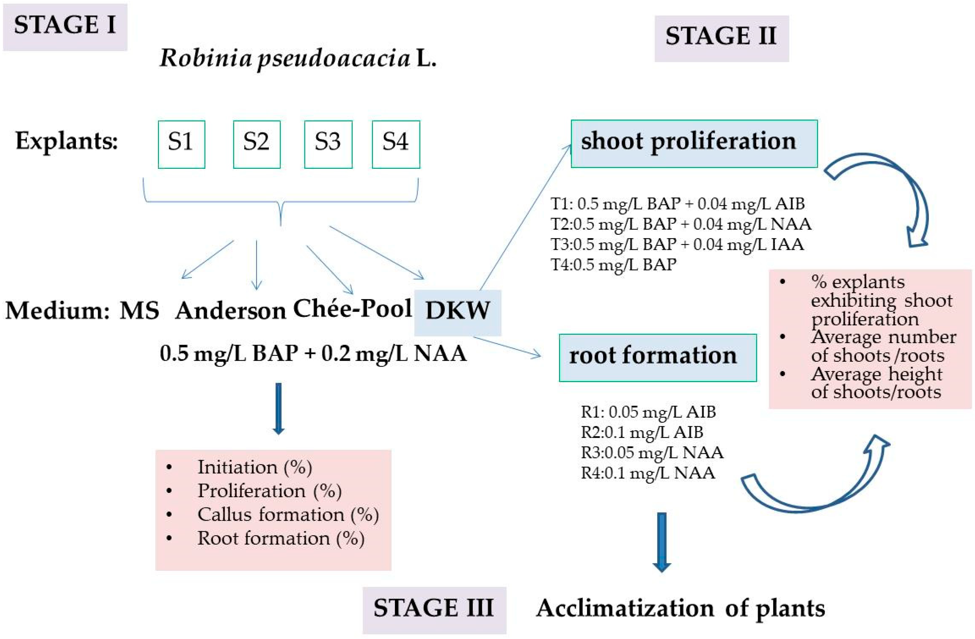
2. Materials and Methods

2.1. Acacia Selections

The four acacia selections were chosen in the O.S. Săcueni of the Bihor Forestry Directorate (latitude: 47°32′38′′ north; longitude: 22°10′23′′ east; altitude 140 m). Each selection was chosen by comparing it to at least four other trees with particular qualities and situations in its immediate area (on or within 25–30 m), with the condition that the age difference between them does not exceed ten years.
The main aim of biotype selection is productivity [30]; however, this is directly related to a complex of characteristics, such as tree height, diameter, branching tendency, adaptability to winter frosts, and resistance to diseases and pests.
The trunk/spindle shape in the biotypes investigated was defined via remarkable dimensions, the productivity of woody mass, straightness, and a height of at least 10 m from the ground level.
The phenotypic differences among the acacia selections (labeled as S1, S2, S3, and S4) are shown in Table 1.

2.2. Culture Conditions

The biological material consisted of nodal explants obtained from the selected specimens (labeled as S1, S2, S3, and S4). After the shoots were collected from the selected specimens, they were placed in the growth chamber with the bases in water until the commencement of active growth. We collected the tips of the growing shoots from which the explants were shaped for inoculation.
Nodal explants were sterilized by washing using a tap water jet for 15 min, following which they were disinfected with 70% ethyl alcohol for 30 s and with 0.1% mercuric chloride for 5 min. Treatments were followed by 5 rinses with sterile distilled water, each lasting 5 min. The inoculation was carried out in a sterile hood (Microflow, Tamil Nadu, India) in 30 mL tubes with 8 mL of medium. Following inoculation, the tubes were placed in the Sanyo growth chamber, which was set to 20 °C at night and 25 °C during the day, with a photoperiod of 8 h in the dark and 16 h of light (3000 lx). We made 14 repetitions for each variant. The following aspects were evaluated after 28 days: shoot regeneration, callus formation, number of adventitious roots, and number of shoots formed per explant. The experimental design from which the stages that were completed in this investigation emerge is presented in Figure 1.

2.3. Shoot Proliferation and Root Formation

The influence of the composition in macro- and micro-elements of some culture media in supporting morphogenesis processes was analyzed using the culture media developed by Murashige Skoog (1962), Anderson (1975), Chée and Pool (1987), and DKW (Driver and Kuniyuki, 1984) (Table 2). All culture media used in this study were purchased from Duchefa Biochemie (Haarlem, the Netherlands).
Culture media were brought to pH 5.6 before autoclaving at a pressure of 1.2 kgf/cm2 for 20 min. After sterilization, 0.5 mg/L BAP and 0.2 mg/L NAA were added to each medium recipe through a 0.22 µm sterile filter (ISOLAB, Laborgeräte GmbH, Eschau, Germany).

2.4. Acclimatization

In order to acclimatize, the tubes with rooted plants were opened gradually, over several days, with the duration of opening increasing in order to reduce the relative humidity. This acclimatization stage lasted 14 days, after which the plants were removed from the culture medium and the agar on the roots was removed under running tap water. The culture substrate, consisting of a mixture of sand, peat, and perlite (1:1:1), was previously disinfected by spraying with 0.3% KMnO4 [2]. The moisture provided to the substrate is a critical factor for plant survival. High humidity results in low aeration; the roots can be infected by mold and other pathogens; a lower substrate humidity dehydrates the leaves, and the seedlings die due to drought stress. During this stage, the relative humidity was maintained in the range of 70–80%.

2.5. Statistical Analysis

The results represent the means and standard deviation (SD). For each acacia selection, 14 repetitions were made in triplicate. Statistical significance between groups was determined with the one-way ANOVA test followed by Tukey’s multiple comparison test, using GraphPad Prism. A value of p < 0.05 was considered statistically significant. Different letters for each sample indicate statistically significant differences. Data obtained on the proliferation and rhizogenesis of acacia selections were subjected to principal component analysis (PCA) using the statistical analysis software PAST, version 4.09, in order to establish the optimal in vitro culture medium for the development of acacia selections.

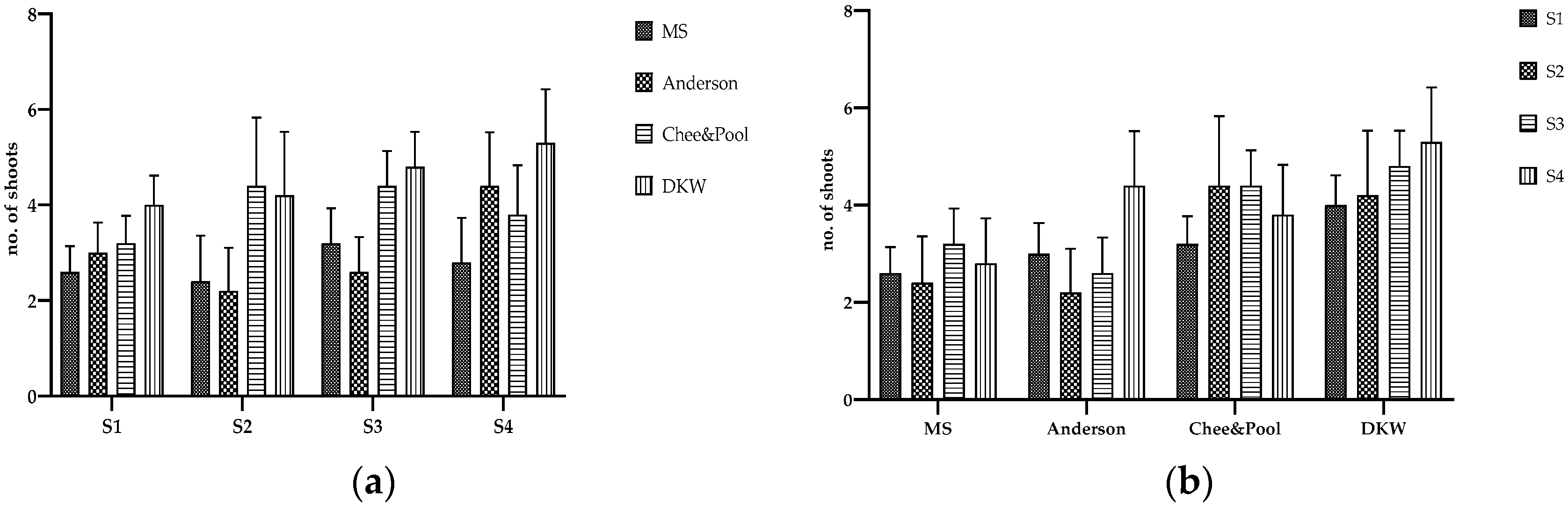
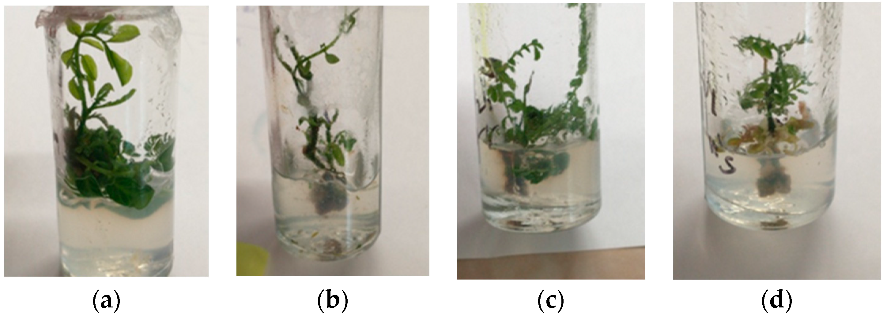
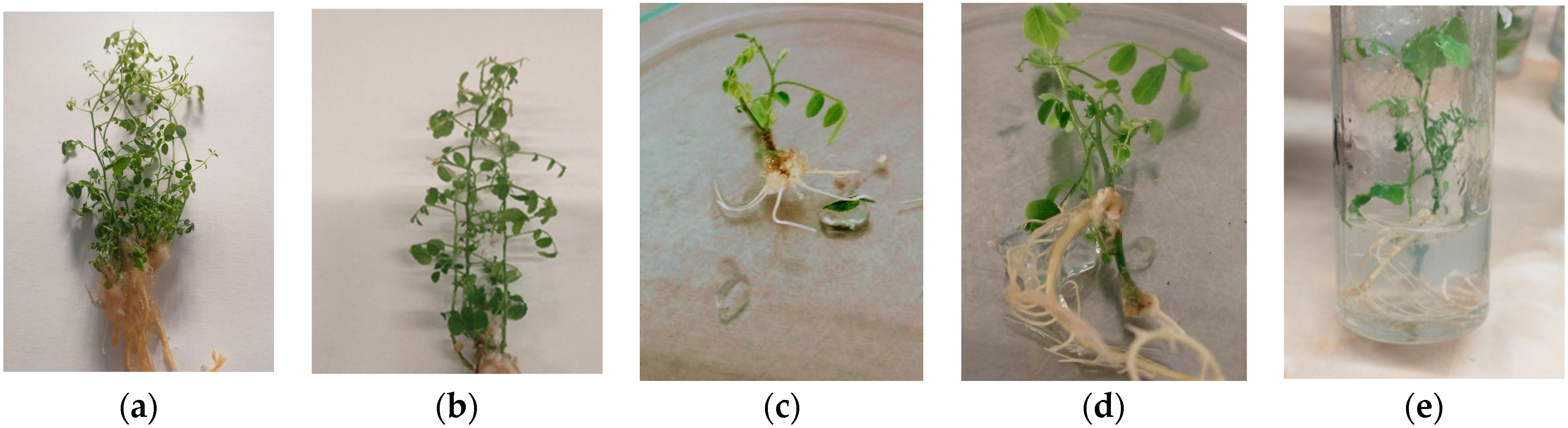
3. Results and Discussion

The type of inoculum, the culture conditions, and the ingredients of the culture media (inorganic and organic nutrients, vitamins, and plant growth regulators) are determining factors in any plant cell culture protocol [20].
Murashige–Skoog (MS) medium is the culture medium widely used in tissue culture laboratories [18], although relatively recent studies have revealed that it generates physiological disturbances, such as necrosis of the tip of the shoot and/or hyperhydricity [25,31]. It was hypothesized that the deaths of explants of some species inoculated on MS medium is due to the high concentrations of mineral salts, especially NH4NO3. For many plants, the high content of ammonium nitrogen (NH4+) in the MS medium can increase the stress level in explant tissues, with a toxic effect on tissue proliferation. As a result, vitrified micro-shoots appear, and the regeneration potential of the culture is reduced [18]. Decreasing the concentration of NH4NO3 in the MS medium by 2–3 times lowers hyperhydricity in Prunus avium [32], Phoenix dactylifera [33], and Aloe polyphylla [34].
Compared to the Murashige–Skoog medium, in the composition of the Anderson medium, we have a reduction to about 1/4 of the concentrations of NH4NO3 and KNO3. In the medium defined by Chée and Pool, the concentrations of chlorine and manganese are lower, and calcium chloride is replaced with calcium nitrate. Shoot multiplication was improved by excluding iodine and decreasing the manganese concentration. Compared to the Murashige–Skoog medium, the DKW medium lacks the micro-elements CoCl2·6H2O and KI, and Ca(NO3)2·2H2O is added to the macro-elements as a source of calcium and nitrogen, while K2SO4 is replaced with KNO3. When compared to MS basal salts, DKW media contains a similar ammonium/nitrate ratio but less total nitrogen [35].
Recent studies have revealed the potential of the DKW medium and that it may represent an alternative to MS in terms of multiple shoot induction from axillary buds and callus-mediated morphogenesis [36,37].
The composition of the culture medium is one of the recognized factors, with a significant influence on the evolution of the morphogenesis processes in the in vitro cultures of plants [35,38]. Under the conditions of the same phytohormonal balance, the morphogenesis processes evolved in different directions depending on the genotype and the chemical composition of the tested medium. The initiation of organogenesis was triggered in all environmental variants and in all selected genotypes, with values between 78–92% and with insignificant differences between variants (Table 3).
On DKW medium, 85–92% of explants produced between 4 and 5.6 shoots, while 50–64% of explants produced between 3.2 and 4.4 shoots on Chée–Pool medium. As a result, the culture medium x genotype interaction had a significant effect on the number of shoots produced. We did not record significant differences between the number of proliferated shoots from the explants of the selections inoculated on MS and Anderson media (p = 0.89935) (Figure 2a). The differences, determined with Tukey’s multiple comparison test, were significant in terms of the average number of proliferated shoots/explant between MS and Chée–Pool (p = 0.00151), MS and DKW (p = 0.00000), Anderson and Chée–Pool (p = 0.00024), Anderson and DKW (p = 0.00000), and Chée–Pool and DKW (p = 0.00896).
The Chée–Pool medium, with an average of 3.9 shoots/explant, and DKW, with 4.5 shoots/explant, are the most advantageous to support the multiplication process (Figure 2b). The Tukey’s multiple comparison test revealed statistically significant differences between MS and Chée–Pool (p = 0.00248), MS and DKW (p = 0.00001), Anderson and Chée–Pool (p = 0.02901), and Anderson and DKW (p = 0.00014).
The biotype had significant effects on the rate of initiation. There were significant differences between selections S3 and S1 (p = 0.00433), S4 and S1 (p = 0.0004), and S4 and S2 (p = 0.0433). Significant changes in proliferation capacity were found between S2 and S1 (p = 0.0016), S3 and S1 (p = 0.0007), and S4 and S1 (p = 0.0002). Several investigations have demonstrated that there are significant differences in the ability to initiate morphogenesis processes both between the different formulations of culture media and between the biotypes studied [39,40,41,42]. The biotype had a significant impact on the rate of initiation. Similarly, Juncker and Favre [43] showed that the genotype had a significant effect on micropropagation capabilities in a study on Q. robur, as some individuals perished during the initial culture phase, while others showed a gradual decline in vitality, and most of the youths displayed rapid growth over time.
The best callus initiation, with an average of 62% on the four inoculated genotypes, was recorded on the MS and Anderson media. MgSO4, CaCl2, and MnSO4 are the essential nutrient macro-elements for explant growth in tissue culture and for callus formation [44,45]. Calcium chloride is the form of calcium commonly used in the composition of in vitro culture media, with roles in cellular pH, carbohydrate translocation, and callus induction [46]. The concentration of CaCl2 is 332 mg/L in both MS and Anderson culture media. A significant reduction in the induction of calluses formed when Mg2+ is missing from the medium has been observed [47,48]. On the DKW medium, 34% of the inocula formed an organogenic callus (after 21 days of inoculation) at the level of the node immersed in the medium. After 5 weeks, calli were formed on this callus from which the shoots were later generated. The presence in the composition of the DKW medium of larger amounts of MnSO4 (33.80 mg/L) and MgSO4 (361.49 mg/L) than in the other tested environments favored the formation of this morphogenic callus.
With the exception of the S1 selection, between 7% and 14% of the inocula on the MS formed roots. On the Anderson medium, only 7% of the S4 explants initiated the rhizogenesis process, and in the other selections this process did not commence. On the Chée–Pool and DKW media, all selections generated roots. The average percentage of rooting in all four selections on the Chée–Pool medium was 22.75%, and on the DKW medium the average on the selections was 31.5%. The following selections with a rooting percentage above the average were observed: S3 on Chée–Pool (28%) and S4 on DKW (42%). Regarding the number of roots formed (Figure 3a), no significant differences between the number of roots formed on the explants inoculated between MS and Anderson (p = 0.11923) and between Chée–Pool and DKW (p = 0.45856) were recorded, as well as significant differences between the other media (MS and Chée–Pool, MS and DKW, Anderson and Chée–Pool, and Anderson and DKW). The average number of roots on explants inoculated on MS and Anderson was 1.4 ± 0.19, on Chée–Pool media 3.0 ± 0.54, and on DKW media 3.4 ± 0.47.
In S1 inoculums, the rhizogenesis process was only initiated on the Chée–Pool and DKW mediums, with an average of 2.7 ± 0.69 and 2.4 ± 0.64 roots/explant, respectively (Figure 3b). S2 and S3 did not form roots on Anderson medium. DKW medium is the only medium tested that stimulates the initiation of rhizogenesis in all four selections, ensuring an average number of 3.8 ± 0.56 roots/explant. Suggestive aspects regarding organogenesis on the culture media taken in the study are presented in Figure 4.
Based on the results obtained at this stage, we continued to pursue the possibility of modulating the morphogenesis processes using only the DKW medium with different phytohormonal balances. We carried out several experimental variants with the additions of BAP, NAA, AIB, and IAA. (Table 4). The effect of these combinations on shoot proliferation and root formation is presented in Table 5 and Table 6.
The explants consisted of nodal segments of shoots, obtained from the previous experiment. This type of explant exhibits juvenile characteristics, are poor in endogenous contaminants, and give the possibility of ensuring a high homogeneity of the explants. The inoculation was performed in 30 mL tubes with 8 mL of medium. The tubes that were inoculated were incubated at a temperature of 24 ± 1 °C, with a photoperiod of 16/8 h using a white, cold, fluorescent light of 3000 lx. Eighteen replicas of each variant were made. After 5 weeks, the culture was evaluated. The results are presented in Table 5.
The highest percentages of regeneration were obtained in the T1 and T3 environmental variants, with statistically assured differences for p < 0.05 compared to the T2 and T4 variants. The largest number of shoots was generated by explants placed on DKW medium with the combination of the phytohormones BAP (0.5 mg/L) and AIB (0.04 mg/L). The most intense shoot growth was ensured by the combination of the phytohormones BAP (0.5 mg/L) and NAA (0.04 mg/L), with statistically ensured differences compared to the other environmental combinations. Our results confirm the observations of Barghchi, M. (1987) and Salem et al. (2022) [1,49] regarding the positive role of NAA in the elongation of shoots grown in vitro in Robinia pseudoacacia [1]. The best results, in all environmental variants, in terms of the number of stems and their heights, were recorded in the S4 selection. On average, this selection produced between 10–35% more shoots and 9–14% more roots than the other selections (Table 5). The better morphogenic responses of S4 is consistent with the studies that have stated that the genotype has a large impact on micropropagation ability [43]. The organogenic responses of inoculums to in vitro cultures are determined via the interaction between endogenous phytohormones and those added to the culture medium [50]. Exogenous cytokinins are essential for shoot formation during in vitro culture, but the use of synthetic cytokinins, such as 6-benzylaminopurine (BAP), can have a long-term residual effect, interfering with subsequent subcultures [51]. Auxin has a decisive role in regulating the spatial and temporal aspects of plant growth and development, being involved in the mechanism of the orientation of the cell division plane, before lateral root initiation, and in the formation of meristems [52,53].
The shoots obtained were shaped to lengths of 2.5–3.0 cm and placed on the variants of the culture medium for rooting. After 4 weeks, the culture was evaluated. The results are presented in Table 6.
For the significance threshold p < 0.05, the Tukey’s HSD test did not reveal significant differences between the variants in terms of the percentage of explants that generated roots (Table 6).
The medium variants T1 and T3, with 0.05 mg/L AIB and NAA, ensured a higher number of roots per explant (Figure 5), statistically assured, than the variants T2 and T4 with 0.1 mg/L. It is worth noting that the variant T1, with 0.05 mg/L AIB, provided 35.1% more roots/explant than T2 (statistically assured p = 0.0001), and 17.8% more roots than T4 (p = 0.02715). The lengths of the roots were stimulated by their presence in the culture medium of NAA (0.05–0.1 mg/L). The selection S4 on the medium with 0.05 mg/L AIB provided the highest number of roots (5.8 ± 0.11) and the longest length of roots on the medium with 0.1 mg/L NAA.

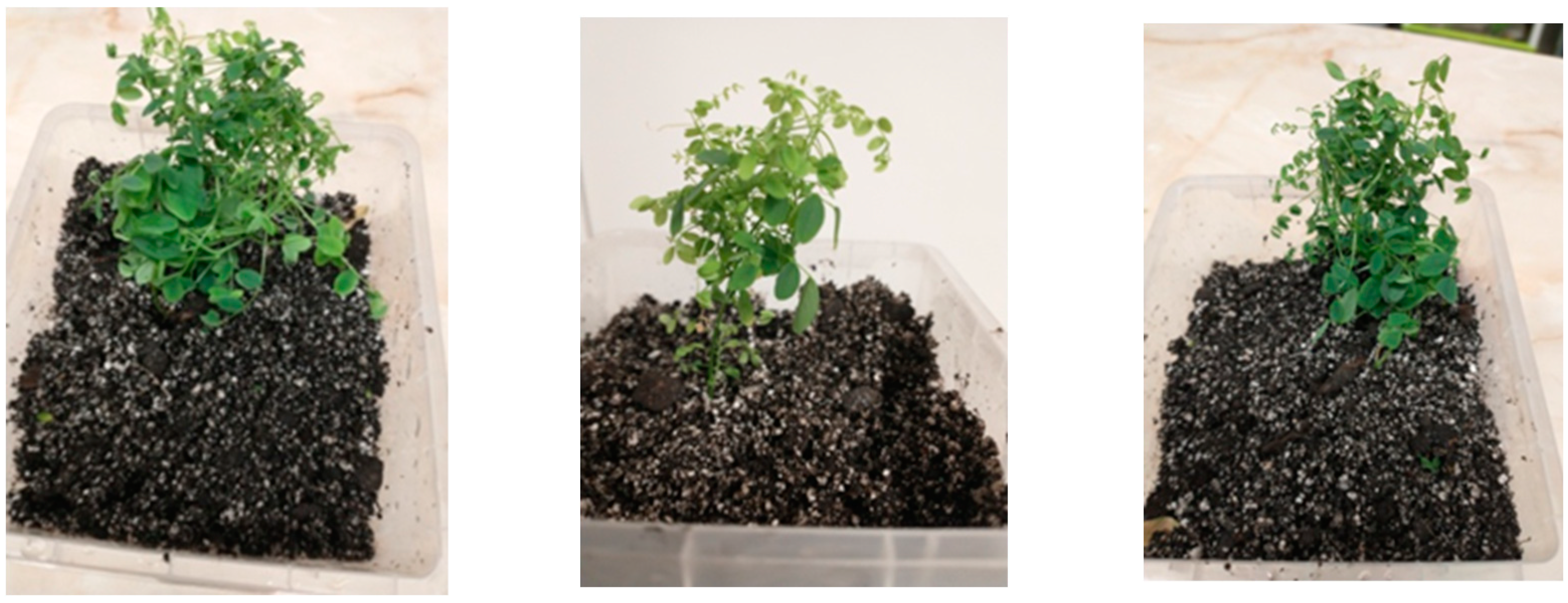
3.1. Acclimatization of Plants Obtained In Vitro

An in vitro micropropagation system can only be considered successful after establishing the conditions of transfer and acclimatization to field conditions. For plants obtained in vitro, under rigorously controlled conditions, transfer to the greenhouse or field environment represents a great challenge due to a lower relative humidity level, higher light level, and septic environment [54]. Plants grown in vitro often show a low rate of photosynthesis and incomplete autotrophy, and these may be the reasons for the low survival rates of plants during the acclimation stage [55]. In addition, the nutrients in the culture media cause deviations in development, and repress or modulate several metabolic pathways differently than soil conditions [56]. As a result, plants grown in vitro develop small juvenile leaves, with a weak cuticular layer, defective stomata, and reduced photosynthetic capacity, while the roots have no or very few absorbent hairs [57]. The acclimatization of Robinia pseudoacacia plants obtained in vitro to an ex vitro environment by gradually exposing them to the relative humidity of the environment and to different light levels better facilitate the survival of young and physiologically sensitive plants when transferred to the soil. The conditions that are ensured at this stage help the plants to develop a fully functional root system and to better control their stomatal and cuticular transpiration [56].
It has been demonstrated by the authors of [57,58,59,60,61,62] that this approach improves stomatal physiology and increases the production of epicuticular wax. The same authors recommended that the level of relative humidity should not drop below 80% in order to permit good aeration of the culture. Robinia pseudoacacia L. adapted to ex vitro conditions is shown in Figure 6. In our study, the average percentage of survival after 4 weeks was 68%.

3.2. Principal Component Analysis

Principal component analysis is a useful statistical technique for determining the correlations between variables. The PCA plot was used to analyze the following in vitro culture medium variables: MS, Anderson, Chée–Pool, and DKW. A 2D plot PCA (Figure 7) revealed the differences in the acacia selections based on the proliferation and rhizogenesis processes. The covariance matrix eigenvalues revealed that the set of the two principal components (PC1 and PC2) accounted for 88.919% of the total variance in the dataset in terms of the organogenesis process (Table 7). PC1 accounted 77.867% of the variance, with PC2 explaining the remainder of the variance, 11.052%. PC1 had an eigenvalue of 3.115, while PC2 had an eigenvalue of 0.442.

4. Conclusions

The goal of this research was to test media, with specifications for woody plants, appropriate for large-scale cloning of acacia genotypes reluctant to traditional methods of vegetative multiplication. The vegetative response of the plants varied between the four selections, with a preference for the DKW basal salts. This tendency was manifested both in terms of callogenesis and rhizogenesis and led us to the conclusion that the DKW medium is more suitable for the in vitro multiplication of the Robinia pseudoacacia species L. than the other tested media. Considering the current climate context, the Robinia pseudoacacia L. species may become more significant due to its strong adaptation to poor and eroded soils, fluctuating temperatures, and positive economic impacts. In addition to providing a starting point for future research on the selection and evolution of this species, the current study can yield important insights for forestry practice and science.

Author Contributions

Conceptualization, R.B. and V.L.; methodology, V.L.; validation, A.I.T., O.S.M. and C.O.; formal analysis, V.L.; investigation, E.A., C.A.R. and M.B.; resources, M.B., R.B. and A.I.T.; writing—original draft preparation, V.L.; writing—review and editing, V.L., S.I.V.; supervision, S.I.V.; project administration, R.B. and V.L.; funding acquisition, R.B. and O.S.M. All authors have read and agreed to the published version of the manuscript.

Funding

The APC was funded by the University of Oradea.

Institutional Review Board Statement

Not applicable.

Informed Consent Statement

Not applicable.

Data Availability Statement

Not applicable.

Acknowledgments

The authors acknowledge the support provided by the University of Oradea through the grant “Excellence scientific research related to priority fields with capitalization through technology transfer: INO-TRANSFER-UO-2nd edition”, project no. 260/11.11.2022.

Conflicts of Interest

The authors declare no conflict of interest.

References

  1. Barghchi, M. Mass clonal propagation in vitro of Robinia pseudoacacia L. (Black locust) cv. ‘Jaszkiseri’. Plant Sci. 1987, 53, 183–189. [Google Scholar] [CrossRef]
  2. Nicolescu, V.-N.; Hernea, C.; Bakti, B.; Keserű, Z.; Antal, B.; Rédei, K. Black locust (Robinia pseudoacacia L.) as a multi-purpose tree species in Hungary and Romania: A review. J. For. Res. 2018, 29, 1449–1463. [Google Scholar] [CrossRef]
  3. Du, B.; Pang, J.; Hu, B.; Allen, D.E.; Bell, T.L.; Pfautsch, S.; Netzer, F.; Dannenmann, M.; Zhang, S.; Rennenberg, H. N2-fixing black locust intercropping improves ecosystem nutrition at the vulnerable semi-arid Loess Plateau region, China. Sci. Total Environ. 2019, 688, 333–345. [Google Scholar] [CrossRef] [PubMed]
  4. Zhang, J.; Liu, Y.; Wang, H. Micropropagation of Black Locust (Robinia pseudoacacia L.). In Protocols for Micropropagation of Woody Trees and Fruits; Jain, S.M., Häggman, H., Eds.; Springer: Dordrecht, The Netherlands, 2007; pp. 193–199. ISBN 978-1-4020-6351-0. [Google Scholar]
  5. Szyp-Borowska, I.; Banha, C.; Wojda, T.; Szczygieł, K. Micropropagation of black locust (Robinia pseudoacacia L.) and genetic stability of long term cultivated plants. Folia For. Pol. 2016, 58, 13–19. [Google Scholar] [CrossRef]
  6. Rédei, K.; Keserű, Z.; Rásó, J. Early evaluation of micropropagated black locust (Robinia pseudoacacia L.) clones in Hungary. For. Sci. Pract. 2013, 15, 81–84. [Google Scholar] [CrossRef]
  7. Lyrene, P.M. Juvenility and Production of Fast-rooting Cuttings from Blueberry Shoot Cultures1. J. Am. Soc. Hortic. Sci. 1981, 106, 396–398. [Google Scholar] [CrossRef]
  8. James, D.J. Adventitious root formation ‘in vitro’ in apple rootstocks (Malus pumila) I. Factors affecting the length of the auxin-sensitive phase in M.9. Physiol. Plant. 1983, 57, 149–153. [Google Scholar] [CrossRef]
  9. Balla, I.; Vértesy, J. Experiences and problems related to the micropropagation of black locust. Acta Hortic. 1987, 212, 552. [Google Scholar] [CrossRef]
  10. Enescu, V.; Jucan, A. Problems of the “In Vitro” Micropropagation of Black Locust (Robinia pseudoacacia L.); Book of Abstracts I; International Society for Horticultural Science: Gembloux, Belgium, 1985. [Google Scholar]
  11. Davis, J.M.; Keathley, D.E. Differential responses to in vitro bud culture in mature Robinia pseudoacacia L. (black locust). Plant Cell Rep. 1987, 6, 431–434. [Google Scholar] [CrossRef]
  12. Davis, J.M. In vitro propagation of a black locust tree with an unusual phenotype. Nitrogen Fixing Tree Res. Rep. 1988, 6, 65–67. [Google Scholar]
  13. Gatti, E.; Sgarbi, E. Micropropagation of Quercus robur: Explant sources and cultural conditions affect in vitro responses differently. Acta Hortic. 2015, 1083, 303–310. [Google Scholar] [CrossRef]
  14. Arrillaga, I.; Merkle, S.A. Regenerating Plants from in Vitro Culture of Black Locust Cotyledon and Leaf Explants. HortScience 1993, 28, 942–945. [Google Scholar] [CrossRef]
  15. Han, H.K.; Keathley, D.E. Regeneration of whole plants from seedling-derived callus of black locust (Robinia pseudoacacia L.). Nitrogen Fixing Tree Res. Rep. 1989, 7, 129–131. [Google Scholar]
  16. Merkle, S.A.; Wiecko, A.T. Regeneration of Robiniapseudoacacia via somatic embryogenesis. Can. J. For. Res. 1989, 19, 285–288. [Google Scholar] [CrossRef]
  17. In Park Chungbuk, J. Plant biotechnology research at forest fields in South Korea. BMC Proc. 2011, 5, 184. [Google Scholar] [CrossRef]
  18. George, E.F.; Hall, M.A.; Klerk, G.-J.D. (Eds.) Plant Propagation by Tissue Culture: Volume 1. The Background, 3rd ed.; Springer: Dordrecht, The Netherlands, 2007; ISBN 978-1-4020-5004-6. [Google Scholar]
  19. Reed, B.M.; Wada, S.; DeNoma, J.; Niedz, R.P. Mineral nutrition influences physiological responses of pear in vitro. In Vitro Cell. Dev. Biol.-Plant 2013, 49, 699–709. [Google Scholar] [CrossRef]
  20. Hameg, R.; Arteta, T.A.; Landin, M.; Gallego, P.P.; Barreal, M.E. Modeling and Optimizing Culture Medium Mineral Composition for in vitro Propagation of Actinidia arguta. Front. Plant Sci. 2020, 11, 554905. [Google Scholar] [CrossRef]
  21. Bresinsky, A.; Körner, C.; Kadereit, J.W.; Neuhaus, G.; Sonnewald, U. Strasburger’s Plant Sciences: Including Prokaryotes and Fungi; Springer: Berlin, Heidelberg, 2013; ISBN 978-3-642-15517-8. [Google Scholar]
  22. Nezami-Alanagh, E.; Garoosi, G.-A.; Landín, M.; Gallego, P.P. Computer-based tools provide new insight into the key factors that cause physiological disorders of pistachio rootstocks cultured in vitro. Sci. Rep. 2019, 9, 9740. [Google Scholar] [CrossRef]
  23. Murashige, T.; Skoog, F. A Revised Medium for Rapid Growth and Bio Assays with Tobacco Tissue Cultures. Physiol. Plant. 1962, 15, 473–497. [Google Scholar] [CrossRef]
  24. Bhojwani, S.S.; Dantu, P.K. Micropropagation. In Plant Tissue Culture: An Introductory Text; Springer: Uttar Pradesh, India, 2013; pp. 245–274. ISBN 978-81-322-1025-2. [Google Scholar]
  25. Nezami-Alanagh, E.; Garoosi, G.-A.; Landín, M.; Gallego, P.P. Combining DOE With Neurofuzzy Logic for Healthy Mineral Nutrition of Pistachio Rootstocks in vitro Culture. Front. Plant Sci. 2018, 9, 1474. [Google Scholar] [CrossRef]
  26. Nikam, T.D.; Mulye, K.V.; Chambhare, M.R.; Nikule, H.A.; Ahire, M.L. Reduction in hyperhydricity and improvement in in vitro propagation of commercial hard fibre and medicinal glycoside yielding Agave sisalana Perr. ex Engelm by NaCl and polyethylene glycol. Plant Cell Tissue Organ Cult. 2019, 138, 67–78. [Google Scholar] [CrossRef]
  27. Gao, L.; Wang, L.; Sun, Z.; Li, H.; Wang, Q.; Yi, C.; Wang, X. Morusin shows potent antitumor activity for human hepatocellular carcinoma in vitro and in vivo through apoptosis induction and angiogenesis inhibition. Drug Des. Dev. Ther. 2017, 11, 1789–1802. [Google Scholar] [CrossRef] [PubMed]
  28. Benková, E.; Bielach, A. Lateral root organogenesis—From cell to organ. Curr. Opin. Plant Biol. 2010, 13, 677–683. [Google Scholar] [CrossRef] [PubMed]
  29. Rédei, K.; Csiha, I.; Keseru, Z.; Rásó, J.; Kamandiné Végh, Á.; Antal, B. Growth and Yield of Black Locust (Robinia pseudoacacia L.) Stands in Nyírség Growing Region (North-East Hungary). Southeast Eur. For. SEEFOR 2014, 5, 13–22. [Google Scholar] [CrossRef]
  30. Károly, R.; Beatrix, B.; Kiss, T.; Takács, M.; Keserű, Z. Yield and crown structure characteristics in a black locust (Robinia pseudoacacia L.) stand: A case study—Short Communication. J. For. Sci. 2018, 64, 96–100. [Google Scholar] [CrossRef]
  31. Mereti, M.; Grigoriadou, K.; Nanos, G.D. Micropropagation of the strawberry tree, Arbutus unedo L. Sci. Hortic. 2002, 93, 143–148. [Google Scholar] [CrossRef]
  32. Daguin, F.; Letouze, R. Ammonium-induced vitrification in cultured tissues. Physiol. Plant 1986, 66, 94–98. [Google Scholar] [CrossRef]
  33. El-Dawayati, M.M.; Zayed, Z.E. Controlling Hyperhydricity in Date Palm In Vitro Culture by Reduced Concentration of Nitrate Nutrients. In Date Palm Biotechnology Protocols Volume I; Al-Khayri, J.M., Jain, S.M., Johnson, D.V., Eds.; Methods in Molecular Biology; Springer: New York, NY, USA, 2017; Volume 1637, pp. 175–183. ISBN 978-1-4939-7155-8. [Google Scholar]
  34. Ivanova, M.; Van Staden, J. Effect of ammonium ions and cytokinins on hyperhydricity and multiplication rate of in vitro regenerated shoots of Aloe polyphylla. Plant Cell Tissue Organ Cult. 2008, 92, 227–231. [Google Scholar] [CrossRef]
  35. Phillips, G.C.; Garda, M. Plant tissue culture media and practices: An overview. In Vitro Cell. Dev. Biol.-Plant 2019, 55, 242–257. [Google Scholar] [CrossRef]
  36. Driver, J.A.; Kuniyuki, A.H. In Vitro Propagation of Paradox Walnut Rootstock. HortScience 1984, 19, 507–509. [Google Scholar] [CrossRef]
  37. Rahman, S.S. Establishment of In vitro micropropagation and callus formation protocols for large scale production of Clitoria ternatea L. J. Med. Spice Plants 2017, 22, 136–144. [Google Scholar]
  38. Polivanova, O.B.; Bedarev, V.A. Hyperhydricity in Plant Tissue Culture. Plants 2022, 11, 3313. [Google Scholar] [CrossRef] [PubMed]
  39. Puddephat, I.J.; Alderson, P.G.; Wright, N.A. Influence of explant source, plant growth regulators and culture environment on culture initiation and establishment of Quercus robur L. in vitro. J. Exp. Bot. 1997, 48, 951–962. [Google Scholar] [CrossRef]
  40. Vieitez, A.M.; Corredoira, E.; Ballester, A.; Muñoz, F.; Durán, J.; Ibarra, M. In vitro regeneration of the important North American oak species Quercus alba, Quercus bicolor and Quercus rubra. Plant Cell Tissue Organ Cult. 2009, 98, 135–145. [Google Scholar] [CrossRef]
  41. Kumar, M.; Sirohi, U.; Malik, S.; Kumar, S.; Ahirwar, G.K.; Chaudhary, V.; Yadav, M.K.; Singh, J.; Kumar, A.; Pal, V.; et al. Methods and Factors Influencing In Vitro Propagation Efficiency of Ornamental Tuberose (Polianthes Species): A Systematic Review of Recent Developments and Future Prospects. Horticulturae 2022, 8, 998. [Google Scholar] [CrossRef]
  42. Abdelghaffar, A.M.; Soliman, S.S.; Ismail, T.A.; Alzohairy, A.M.; Latef, A.A.H.A.; Alharbi, K.; Al-Khayri, J.M.; Aljuwayzi, N.I.M.; El-Moneim, D.A.; Hassanin, A.A. In Vitro Propagation of Three Date Palm (Phoenix dactylifera L.) Varieties Using Immature Female Inflorescences. Plants 2023, 12, 644. [Google Scholar] [CrossRef] [PubMed]
  43. Juncker, B.; Favre, J.M. Clonal effects in propagating oak trees via in vitro culture. Plant Cell Tissue Organ Cult. 1989, 19, 267–276. [Google Scholar] [CrossRef]
  44. Alanagh, E.N.; Garoosi, G.; Haddad, R.; Maleki, S.; Landín, M.; Gallego, P.P. Design of tissue culture media for efficient Prunus rootstock micropropagation using artificial intelligence models. Plant Cell Tissue Organ Cult. 2014, 117, 349–359. [Google Scholar] [CrossRef]
  45. Fallah Ziarani, M.; Tohidfar, M.; Navvabi, M. Modeling and optimizing in vitro percentage and speed callus induction of carrot via Multilayer Perceptron-Single point discrete GA and radial basis function. BMC Biotechnol. 2022, 22, 34. [Google Scholar] [CrossRef]
  46. Amin, N.; Khattak, M.; Ahmad, I.; Ara, N.; Alam, A.; ALI, M. Corm and cormel size of gladiolus greatly influenced growth and development of subsequent corm production. Pak. J. Bot. 2013, 2013, 1407–1409. [Google Scholar]
  47. Walker, K.A.; Sato, S.J. Morphogenesis in callus tissue of Medicago sativa: The role of ammonium ion in somatic embryogenesis. Plant Cell Tissue Organ Cult. 1981, 1, 109–121. [Google Scholar] [CrossRef]
  48. Sarropoulou, E.; Sundaram, A.Y.M.; Kaitetzidou, E.; Kotoulas, G.; Gilfillan, G.D.; Papandroulakis, N.; Mylonas, C.C.; Magoulas, A. Full genome survey and dynamics of gene expression in the greater amberjack Seriola dumerili. GigaScience 2017, 6, gix108. [Google Scholar] [CrossRef] [PubMed]
  49. Salem, J.; Hassanein, A.; El-Wakil, D.A.; Loutfy, N. Interaction between Growth Regulators Controls In Vitro Shoot Multiplication in Paulownia and Selection of NaCl-Tolerant Variants. Plants 2022, 11, 498. [Google Scholar] [CrossRef] [PubMed]
  50. Chmielarz, P.; Kotlarski, S.; Kalemba, E.M.; Martins, J.P.R.; Michalak, M. Successful In Vitro Shoot Multiplication of Quercus robur L. Trees Aged up to 800 Years. Plants 2023, 12, 2230. [Google Scholar] [CrossRef] [PubMed]
  51. Martins, J.P.R.; Santos, E.R.; Rodrigues, L.C.A.; Gontijo, A.B.P.L.; Falqueto, A.R. Effects of 6-benzylaminopurine on photosystem II functionality and leaf anatomy of in vitro cultivated Aechmea blanchetiana. Biol. Plant. 2018, 62, 793–800. [Google Scholar] [CrossRef]
  52. Blakesley, D.; Weston, G.D.; Hall, J.F. The role of endogenous auxin in root initiation: Part I: Evidence from studies on auxin application, and analysis of endogenous levels. Plant Growth Regul. 1991, 10, 341–353. [Google Scholar] [CrossRef]
  53. Marhavý, P.; Montesinos, J.C.; Abuzeineh, A.; Van Damme, D.; Vermeer, J.E.M.; Duclercq, J.; Rakusová, H.; Nováková, P.; Friml, J.; Geldner, N.; et al. Targeted cell elimination reveals an auxin-guided biphasic mode of lateral root initiation. Genes Dev. 2016, 30, 471–483. [Google Scholar] [CrossRef]
  54. Hazarika, B.N. Acclimatization of tissue-cultured plants. Curr. Sci. 2003, 85, 1704–1712. [Google Scholar]
  55. Faria, R.T.D.; Rodrigues, F.N.; Oliveira, L.D.V.R.; Müller, C. In vitro Dendrobium nobile plant growth and rooting in different sucrose concentrations. Hortic. Bras. 2004, 22, 780–783. [Google Scholar] [CrossRef]
  56. Teixeira Da Silva, J.A.; Hossain, M.M.; Sharma, M.; Dobránszki, J.; Cardoso, J.C.; Zeng, S. Acclimatization of in Vitro-derived Dendrobium. Hortic. Plant J. 2017, 3, 110–124. [Google Scholar] [CrossRef]
  57. Ziv, M. In vitro hardening and acclimatization of tissue culture plants. In Plant Tissue Culture and its Agricultural Applications; Withers, L.A., Alderson, P.G., Eds.; Butterworths: London, UK, 1986; pp. 187–203. [Google Scholar]
  58. Miller, D. Weaning and growing-on of micropropagated plants. Proc. Intern. Plant Prop. Soc. 1983, 33, 253–256. [Google Scholar]
  59. Pocock, S. Procedures and problems associated with the transfer of tissue-cultured plants. Proc. Intern. Plant Prop. Soc. 1983, 33, 316–320. [Google Scholar]
  60. Short, K.C.; Wardle, K.; Grout, B.W.W.; Simpkins, I. In vitro physiology and acclimatization of aseptically cultured plantlets. In Plant Tissue and Cell Culture Application to Crop Improvement; Novák, F.J., Havel, L., Doležel, J., Eds.; Institute of Experimental Botany of the Czech Academy of Sciences: Prague, Czech Republic, 1984; pp. 475–486. [Google Scholar]
  61. Wardle, K.; Dobbs, E.B.; Short, K.C. In Vitro Acclimatization of Aseptically Cultured Plantlets to Humidity. J. Am. Soc. Hort. Sci. 1983, 108, 386–389. [Google Scholar] [CrossRef]
  62. Iliev, I. Techniques for Acclimatization of In Vitro Propagated Plants. Bogdanov, B., Denkova, S., Alexandrov, P., Zhelev, P., Eds.; Propagation of Ornamental Plants; Ministry of Education and Science Publishing House: Sofia, Bulgaria, 1994. [Google Scholar]
Figure 1. Experimental design. S1, S2, S3, and S4 acacia selections described in Table 1. MS—Murashige–Skoog medium, DKW—Driver and Kuniyuki Woody medium, BAP-6—benzylaminopurine, NAA-α—naphthaleneacetic acid, AIB-α—aminoisobutyric acid, and IAA—indole-3-acetic acid.
Sustainability 15 15243 g001
Figure 2. The average number of shoots formed on the tested media after 21 days after inoculation (a) genotype influence; (b) the influence of the culture environment.
Sustainability 15 15243 g002
Figure 3. Average number of roots formed on the media tested after 21 days after inoculation (a) genotype influence; (b) the influence of the culture environment.
Sustainability 15 15243 g003
Figure 4. Morphogenesis on the tested media. (a) Multiplication on DKW medium; (b) regeneration of shoots with basal callus on Chée–Pool medium; (c) formation of adventitious shoots with basal callus on Anderson medium; (d) the formation of shoots with friable, green basal callus on MS medium.
Sustainability 15 15243 g004
Figure 5. Micromultiplication and rhizogenesis (a,b) and rooting (ce) of Robinia pseudoacacia L. S4 inoculums on DKW medium.
Sustainability 15 15243 g005
Figure 6. Plants of Robinia pseudoacacia L. adapted to ex vitro conditions.
Sustainability 15 15243 g006
Figure 7. Principal component analysis (PCA) of the organogenesis process of four acacia selections. The red dots represent the percentage of S1 acacia selections’ root production (samples with R as the first letter) and proliferation (samples with S from shoots), while the yellow dots represent S2, the blue dots represent S3, and the green dots represent S4.
Sustainability 15 15243 g007
Table 1. Phenotypic characterization of the acacia selections.
Acacia SelectionHeight (m)Diameter at a Height of 1.3 m Volume (m3)Height at Which the First Lateral Branch Appears (m)
S124522.10317
S222501.78015
S319461.31013
S420481.49414
Table 2. The chemical composition of the culture media used in the micro-multiplication stage of the species Robinia pseudoacacia L.
Culture Media Composition
Micro-Elements (mg/L)Murashige–SkoogChée–PoolAndersonDKW
CoCl2·6H2O0.0250.0250.025
CuSO4·5H2O0.0250.0250.0250.25
FeNaEDTA36.7036.7073.4044.63
H3BO36.206.206.20-
KI0.83-0.304.80
MnSO4·H2O16.900.8516.9033.80
Na2MoO4·2H2O0.250.250.250.39
ZnSO4·7H2O8.608.608.6017.0
Macro-Elements (mg/L)
Ca(NO3)2-492.30--
KH2PO4170.00170.00-265.0
KNO31900.001900.00480.0-
MgSO4180.54180.54180.54361.49
NH4NO31650.001650.00400.01416.0
CaCl2332.02-332.02112.50
NaH2PO4--330.60-
Ca(NO3)2·2H2O---1664.64
K2SO4---1559.0
Table 3. The effect of different types of culture media, under the conditions of the same phytohormonal balance, on the percentage of initiation in organogenesis, proliferation, callus formation, and rhizogenesis.
MediumAcacia SelectionInitiation
(%)
Proliferation
(%)
Callus Formation (%)Root Formation (%)
Murashige–SkoogS185 ± 1.84 a28 ± 2.73 bc71 ± 3.26 a-
S285 ± 2.34 a21 ± 1.23 b71 ± 2.24 a7 ± 0.23 bc
S385 ± 3.42 a21 ± 1.24 b64 ± 1.82 a14 ± 1.12 ac
S492 ± 2.24 a42 ± 2.36 b42 ± 2.24 a7 ± 0.14 bc
AndersonS178 ± 2.11 a28 ± 3.09 b50 ± 2.38 a-
S285 ± 3.12 a28 ± 2.12 b64 ± 2.12 a-
S392 ± 2.16 a21 ± 2.02 b71 ± 3.24 a-
S492 ± 4.14 a42 ± 3.46 b64 ± 2.46 a7 ± 0.48 bc
Chée–PoolS178 ± 2.53 a50 ± 2.97 b35 ± 2.77 b21 ± 2.12 ac
S285 ± 3.18 a57 ± 1.46 b28 ± 1.86 b21 ± 1.60 ac
S385 ± 1.26 a42 ± 3.21 b35 ± 2.44 bc28 ± 1.60 ac
S492 ± 4.42 a64 ± 2.48 a42 ± 3.12 ac21 ± 1.22 ac
DKWS185 ± 3.45 a78 ± 2.11 a42 ± 3.45 b21 ± 2.08 ac
S285 ± 2.62 a85 ± 2.08 a21 ± 3.42 b28 ± 0.68 ac
S392 ± 4.20 a78 ± 1.68 a64 ± 4.12 a35 ± 1.68 ac
S492 ± 3.26 a92 ± 2.82 a21 ± 2.42 b42 ± 2.24 a
Results are expressed as the mean ± SD (n = 14). Different lowercase letters indicate significant differences within the same column (p < 0.05).
Table 4. The phytohormonal balances of the species Robinia pseudoacacia L. investigated in vitro on DKW medium.
Variant DKWAP(mg/L) 1AIB (mg/L) 2 NAA(mg/L) 3IAA (mg/L) 4
MultiplicationT10.50.04--
T20.5-0.04-
T30.5--0.04
T40.5---
RootingR1-0.05--
R2-0.1--
R3--0.05-
R4--0.1-
1 BAP-6—benzylaminopurine; 2 AIB—indole-3-butyric acid; 3 NAA—1-naphthaleneacetic acid; 4 IAA—indole acetic acid.
Table 5. Shoot proliferation from stem nodal explants under the influence of different concentrations of phytohormones in DKW medium.
Phytohormonal Variant
(mg/L)
Selection% of Explants Exhibiting Shoot ProliferationAverage Number of Shoots Average Height of Shoots
(cm)
T1
BAP (0.5 mg/mL)
+
AIB (0.04 mg/mL)
S194 ± 3.30 3.8 ± 0.122.6 ± 0.8
S289 ± 2.424.7± 0.282.8 ± 0.2
S392 ± 2.024.1 ± 0.132.4± 0.2
S491 ± 3.325.2 ± 0.183.5 ±0.12
Mean T1 91.5 ± 2.24 a4.45 ± 0.18 a2.82 ± 0.42 b
T2
BAP (0.5 mg/mL) +
NAA (0.04 mg/mL)
S180 ± 2.402.8 ± 0.343.8 ± 0.08
S286 ± 2.373.4 ± 0.323.8 ± 0.3
S388 ± 3.823.2 ± 0.223.5 ± 0.22
S490 ± 2.023.6 ± 0.214.2 ± 0.13
Mean T2 86 ± 1.86 b3.25 ± 0.06 b3.82 ± 0.58 a
T3
BAP (0.5 mg/mL) +
IAA (0.04 mg/mL)
S192 ± 2.32 1.5 ± 0.082.7 ± 0.08
S290 ± 3.122.2 ±0.222.4 ± 0.16
S391 ± 2.321.8 ± 0.162.6 ± 0.2
S490 ± 1.392.8 ± 0.143.2 ± 0.03
Mean T3 90.75 ± 1.92 a2.07 ± 0.12 c2.72 ± 0.08 bc
T4
BAP (0.5 mg/mL)
S178 ± 2.221.5 ± 0.151.8 ± 0.2
S274 ± 2.801.2 ± 0.162.1 ± 0.12
S380 ± 2.121.4 ± 0.142.4 ±0.23
S482 ± 2.791.6 ± 0.042.4 ± 0.14
Mean T4 78.5 ± 2.02 c1.42 ± 0.02 d2.17 ± 0.12 d
Results are represented as means ± SD (n = 14); different lowercase letters indicate significant differences within the same column (p < 0.05).
Table 6. Plant rooting in vitro under the influence of different combinations of auxins in DKW medium.
Phytohormonal Variant
(mg/L)
Selection% of Explants Exhibiting Shoot ProliferationAverage Number of Roots/ExplantAverage Length of Roots
(cm)
R1
AIB
(0.05 mg/L)
S170 ± 2.02 4.5 ± 0.163.8 ± 0.16
S268 ± 3.125.0 ± 0.154.6 ± 0.14
S382 ± 2.225.8 ± 0.183.8 ± 0.05
S490 ± 2.805.8 ± 0.114.2 ± 0.25
Mean R1 77.5 ± 2.54 a 5.27 ± 0.15 a4.1 ± 0.15 b
R2
AIB
(0.1 mg/mL)
S167 ± 2.024.6 ± 0.162.2 ± 0.06
S260 ± 3.323.8 ± 0.152.8 ± 0.17
S386 ± 1.323.4 ± 0.282.5 ± 0.14
S472 ± 3.623.8 ± 0.142.9 ± 0.25
Mean R2 71.25 ± 2.57 a 3.9 ± 0.18 b2.6 ± 0.15 c
R3
NAA
(0.05 mg/mL)
S168 ± 2.38 4.7 ± 0.264.5 ± 0.05
S271 ± 2.524.6 ± 0.054.7 ± 0.15
S367 ± 2.825.8 ± 0.194.2 ± 0.2
S476 ± 1.625.6 ± 0.094.8 ± 0.24
Mean R3 70.5 ± 2.33 a 5.17 ± 0.14 a4.55 ± 0.12 a
R4
NAA
0.1 (mg/mL)
S168 ± 2.024.4 ± 0.184.5 ± 0.14
S266 ± 3.123.8 ± 0.044.9 ± 0.25
S369 ± 1.024.6 ± 0.124.4 ± 0.05
S468 ± 1.825.1 ± 0.135.0 ± 0.15
Mean R4 67.75 ± 2.32 a 4.47 ± 0.11 b4.7 ± 0.14 a
Results are represented as means ± SD (n = 14); different lowercase letters indicate significant differences within the same column (p < 0.05).
Table 7. The percentage of variance explained by each successive principal component.
PCEigenvalueCumulative Eigenvalues Explained by Each PC Percentage of VariancePercentage of Variance Explained by Each PC (%)Cumulative Percentage of Variance (%)
13.1153.11577.86777.867
20.4423.55711.05288.919
30.3323.8898.295997.215
40.1114.0002.7848100.00
Disclaimer/Publisher’s Note: The statements, opinions and data contained in all publications are solely those of the individual author(s) and contributor(s) and not of MDPI and/or the editor(s). MDPI and/or the editor(s) disclaim responsibility for any injury to people or property resulting from any ideas, methods, instructions or products referred to in the content.

