Article

Genome Sequence and Characterisation of Peribacillus sp. Strain AS_2, a Bacterial Endophyte Isolated from Alectra sessiliflora

by
Mehabo Penistacia Maela
and
Mahloro Hope Serepa-Dlamini
*
Department of Biotechnology and Food Technology, Faculty of Science, University of Johannesburg, Doornfontein Campus, P.O. Box 17011, Johannesburg 2028, South Africa
*
Author to whom correspondence should be addressed.
Microbiol. Res. 2024, 15(1), 50-65; https://doi.org/10.3390/microbiolres15010004
Submission received: 6 November 2023 / Revised: 12 December 2023 / Accepted: 19 December 2023 / Published: 21 December 2023

Abstract

:
Peribacillus sp. AS_2, a leaf endophytic bacterium isolated from the medicinal plant Alectra sessiliflora, was previously identified using the 16S rRNA gene sequence. The draft genome of AS_2 had a 5,482,853 bp draft circular chromosome, 43 contigs, N50 of 360,633 bp and an average G + C% content of 40.5%. Whole genome sequencing and phenotypic analysis showed that AS_2 was Gram-positive, endospore-forming, motile, and rod-shaped and it showed a high sequence similarity with P. frigoritolerans DSM 8801T. Genomic digital DNA–DNA hybridisation (dDDH) between strain AS_2 and Peribacillus frigoritolerans DSM 8801T and P. castrilensis N3T was 84.8% and 79.2%, respectively, and the average nucleotide identity (ANI) of strain AS_2 with P. frigoritolerans DSM 8801T and P. castrilensis N3T was 97.0% and 96.7%, respectively. The antiSMASH software predicted a total of eight secondary metabolite gene clusters comprising non-ribosomal peptide synthetase (NRPS) type koranimine, terpenes, and siderophore clusters. Strain AS_2 also displayed genes involved in endophytic lifestyle and antibiotic resistance gene clusters such as small multidrug resistance antibiotic efflux pumps (qacJs). Using the multilocus sequence analysis (MLSA), together with the phenotypic data and genomic analysis, we demonstrated that strain AS_2 is a subspecies of P. frigoritolerans DSM 8801T. Genome sequencing of Peribacillus sp. AS_2 from medicinal plants provides valuable genomic information and allows us to further explore its biotechnological applications.

1. Introduction

Bacterial endophytes are symbiotic microorganisms that colonise the internal tissues of plants without posing a threat to the plants’ health [1]. Bacterial endophytes are present in all plants and have been identified as a promising source of biological raw materials for the agricultural, pharmaceutical, cosmetic, and food sectors [2]. Moreover, research has been focused mostly on bacterial endophytes isolated from medicinal plants due to their therapeutic importance, yet little is known about the mechanisms through which endophytes interact with their host plants. Numerous studies have demonstrated that bacterial endophytes synthesise bioactive compounds that assist plants by enabling them to withstand harsh conditions like high temperatures, low pH levels, salt, and heavy metals [3,4]. Additionally, their secondary metabolites offer resistance against biotic and abiotic stressors by stimulating the formation of immunological or antimicrobial compounds [5]. Due to their lower toxicity towards humans, animals, and the environment, these endophyte-produced secondary metabolites have attracted more attention globally [6].
The genus Peribacillus was first grouped with over 100 species and subspecies of the genus Bacillus, which belonged to the Firmicutes phylum [7]. However, based on phenotypic, chemotaxonomic, and genetic differences, some of the species of Bacillus have recently been reclassified into new genera, Alkalihalobacillus, Cytobacillus, Metabacillus, Mesobacillus, Neobacillus, Peribacillus [8,9,10].
Members of the genus Peribacillus have been isolated from various sources including soil [11], cow faeces [12], plant tissues [13,14], and river water [15]. Currently, within the genus Peribacillus there are 17 species which have been validly published [16,17]. From the 17 published species, only 11 have whole-genome sequences available, which vary in size from 4.1 to 5.7 Mbp. The G + C composition of genomic DNA ranges from 37.5 to 43.0% [15]. According to [9], Peribacillus species are motile, rod-shaped, Gram-positive, aerobic, or facultatively anaerobic and can grow in different temperatures ranging from 3 to 45 °C, with their optimum temperature ranging between 25 and 37 °C.
Some Peribacillus species such as Peribacillus butanolivorans are used industrially for the remediation of butanol [9]. Similarly, other Peribacillus and Bacillus strains, such as Bacillus thuringiensis SG4 and Bacillus sp. SG2, have been shown to have biodegradation abilities for a variety of pesticides, including cypermethrin [18]. The present study aims to characterise Peribacillus sp. strain AS_2, a bacterial endophyte isolated from the medicinal plant, Alectra sessiliflora [19]. To the best of our knowledge, this is the first study to characterise a bacterial endophyte from the genus Peribacillus isolated from a medicinal plant. Strain AS_2 was previously isolated and initially identified using the 16S rRNA sequence by [19] and it showed promising antibacterial and antitumour activity against human clinical pathogens and three cancer cells. Therefore, further studies of strain AS_2 may be required to better understand and shed light on its possible applications in the industrial, medicinal, and agricultural sectors.

2. Materials and Methods

2.1. Isolation of the Bacterial Peribacillus sp. Strain AS_2

Peribacillus sp. strain AS_2 was isolated from sterilised leaves of A. sessiliflora, obtained from Eisleben, Botlokwa (23°31′49.5″ S 29°49′27.1″ E) in the Limpopo province, South Africa, as described by [19]. The 16S ribosomal RNA gene (GenBank accession number MZ976847) sequencing was used to initially identify the strain. Glycerol stocks (30%, v/v) of AS_2 were inoculated on nutrient agar (NA) plates and incubated at 28 °C for 24–48 h. For maintenance, strain AS_2 was sub-cultured and incubated at 28 °C continuously and preserved in 30% glycerol stocks at −80 °C for long-term storage.

2.2. Genome Extraction, Library Preparation, and Sequencing

The Zymo Research Fungal/Bacterial DNA MiniPrep Kit (Zymo Research, Irvine, CA, USA) was used to extract whole genomic DNA from solid colonies of strain AS_2. The Implen Nanophotometer N60 (Implen GmbH, Munich, Germany) was used to quantify the concentration of strain AS_2 DNA. The full genome sequence of strain AS_2 was completed by a commercial service provider, the Agricultural Research Council (ARC) Biotechnology platform, Onderstepoort, Pretoria, South Africa, using the HiSeq 2500 platform. Paired-end libraries (2 × 150 bp) were prepared using the Illumina HiSeq instrument v3 and the NextEra DNA library preparation kit (Illumina, San Diego, CA, USA).

2.3. Genome De Novo Assembly and Annotation

The raw sequence reads of strain AS_2 were uploaded to the Galaxy (www.usegalaxy.org) platform [20] accessed on 12 July 2023, where all pre-annotation analysis was carried out. FastQC (version 0.72.0) was used to examine the quality of the raw reads [21]. This was followed by de novo assembly with Unicycler version 0.4.8.0 [22], and the assembled genome was evaluated using the Quality Assessment Tool for Genome Assemblies (QUAST) version 5.0.2 [23]. The draft genome of strain AS_2 was annotated using the Prokaryotic Genome Annotation Pipeline (PGAP) [24] and Rapid Annotations utilising Subsystems Technology [25]. The genome completeness of strain AS_2 was assessed using BUSCO (Benchmarking Universal Single-Copy Orthologs) version 5.0 [26], whereas contamination was assessed using CheckM on Kbase (https://www.kbase.us/) [27], accessed on 28 November 2023.

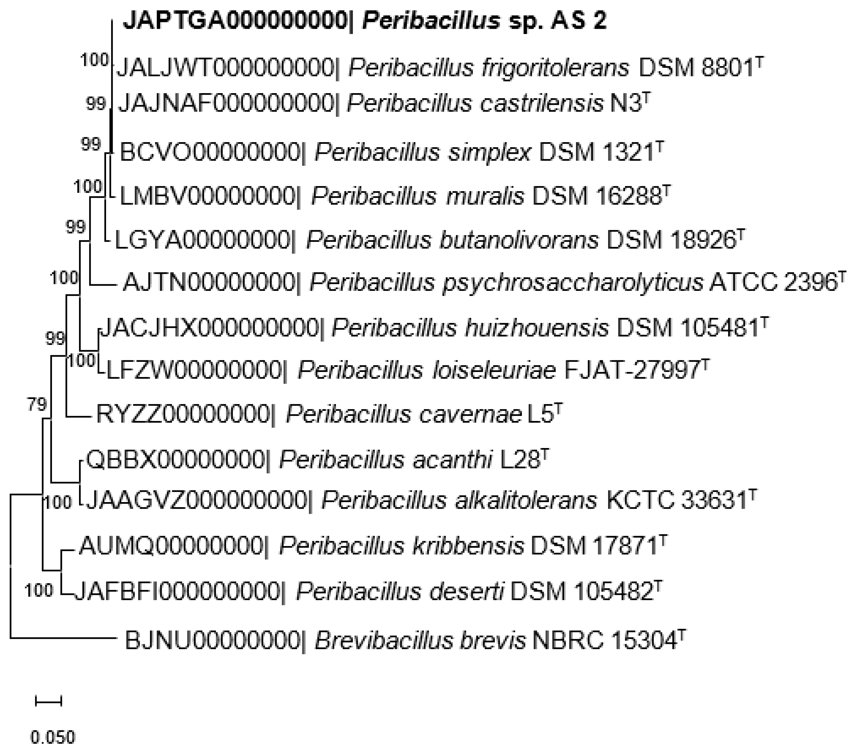
2.4. Phylogenome Analysis

The draft genome of strain AS_2 was assembled into contigs and uploaded to the Type Strain Genome Server (TYGS) (available at https://tygs.dsmz.se, accessed on 12 August 2023) for whole genome-based taxonomic analysis with other type strain species of the genus Peribacillus [28]. The Orthologous Average Nucleotide Identity (OrthoANI) software tool (version 0.93.1) [29] was used to compute the ANI values between strain AS_2 and other closely related Peribacillus species. Multilocus sequencing analysis (MLSA) was performed using four housekeeping genes, gyrA (DNA gyrase subunit A), gyrB (DNA topoisomerase ATP-hydrolysing), rpoB (DNA-directed RNA polymerase subunit beta), and rpoC (DNA-directed RNA polymerase subunit beta). The sequences of the four housekeeping genes were aligned using the ClustalW logarithm on MEGA X [30], after which a protein concatenated phylogenetic tree was constructed using the maximum likelihood method with 1000 bootstrap repetitions. The CRISPRCas finding software (available at https://crisprcas.i2bc./CrisprCasFinder/Viewing/, accessed on 15 August 2023) was used to predict the Clustered Regularly Interspaced Short Palindromic Repeats (CRISPR) [31,32,33]. Genomic islands (GIs) were identified and analysed using the IslandViewer 4 online database (version 4.0) at http://www.pathogenomics.sfu.ca/islandviewer/ [34], accessed on 15 August 2023. Furthermore, additional annotation was performed on strain AS_2 using the following databases: The antiSMASH (version 5.0) tool (available at https://antismash.secondarymetabolites.org, accessed on 15 August 2023) [35] and PRISM 4 (Prediction Informatics for Secondary Metabolomes) (available at https://magarveylab.ca/prism, accessed on 15 August 2023) were used to predict gene clusters for secondary metabolites. The Comprehensive Antibiotic Resistance Database (CARD), (https://arpcard.mcmaster.ca, accessed on 15 August 2023) was used to identify antibiotic resistance genes [36].

2.5. Phenotypic Characterisation

To differentiate Peribacillus sp. strain AS_2 from other closely related Peribacillus species, characteristics recommended by [37] were applied. Cells of strain AS_2 were stained with a Gram stain kit (Sigma-Aldrich, Eschenstr, Taufkirchen, Germany) according to the manufacturer’s protocol. Scanning electron microscopy (TESCAN VEGA SEM) was used to examine cell morphology, while light microscopy was used for the observation of spore production. The motility of the cells was observed with light microscopy using the hanging drop method after growing the cells for 24 h. Strain AS_2 was inoculated on tryptone soy agar (TSA) and incubated at 30 °C for 48 h, after which the form, size, and color of colonies were studied. Anaerobic growth on TSA was investigated for 48 h in an anaerobic chamber at 30 °C. The optimum growth and growth range of Peribacillus sp. strain AS_2 were determined in a tryptone soy broth (TSB) medium at varied NaCl concentrations ranging from 0 to 15% (w/v) with 1% intervals at 30 °C for 72 h. Growth at several temperature ranges (4, 15, 20, 25, 30, 37, 40, 45, 50 and 55 °C) was studied in TSB for 72 h. The pH growth range and optimum were determined in TSB media at 30 °C for 72 h, at pH 3 to 11 with increments of 1 pH units. Catalase activity was evaluated by observing bubble formation after adding a 3% (v/v) hydrogen peroxide (H2O2) solution. The presence of bubbles indicated a positive result. The oxidase test reagent (Thermo Scientific, Waltham, MA, USA) was used to determine oxidase activity. Hydrolysis of starch and protein were examined on modified NA enriched with 1% skimmed milk and 1% soluble starch. Other biochemical characteristics were validated using API 20E and API 50CHB strips (bioMérieux, Midrand, South Africa) according to the manufacturer’s protocol.

2.6. Statistical Analysis

All genetic studies were carried out in accordance with the manufacturer’s instructions. The experiments were conducted in triplicates, and the findings were expressed as mean standard deviations. The data was analysed in Microsoft Excel version 2016, using one-way analysis of variance with post-hoc t-test (Bonferroni Correction), and the p ≤ was declared statistically significant.

3. Results and Discussion

3.1. Genome Characteristics of Peribacillus sp. Strain AS_2

A total of 2,646,598 paired-end reads at 72× coverage were obtained from this workflow. Sequencing and de novo assembly of Peribacillus sp. strain AS_2 resulted in a draft genome sequence with 43 contigs. The genome size of strain AS_2 was 5,482,853 bp with a G + C content of 40.5%. The size of the genome and G + C% content of strain AS_2 falls within the range of most reported Peribacillus species genomes [15]. The QUAST software was used to assess the quality of the assembly. The N50 value of strain AS_2 was 360,633, whereas the L50 value was six. The PGAP annotation identified a total of 5290 genes, of which 5072 are protein coding sequences (CDS) and 130 are pseudogenes. A total of 88 RNAs were predicted from the draft genome and 79 of these coded for tRNA genes, three coded for rRNA genes comprising three operons (5S, 16S, 23S), and six genes were non-coding RNA (ncRNA). The BUSCO analysis showed a completeness of 99.8% (445 completed and single-copy genes, and four duplicated genes). CheckM showed a genome completeness of 98.91% and a contamination of 2.21%. We identified four CRISPR repeats and one Cas cluster in strain AS_2 (Table S1). The genomic characteristics of strain AS_2 are summarised in Table 1. The draft genome sequence of strain AS_2 was deposited in the GenBank/EMBL/DDBJ database under the accession number JAPTGA000000000.

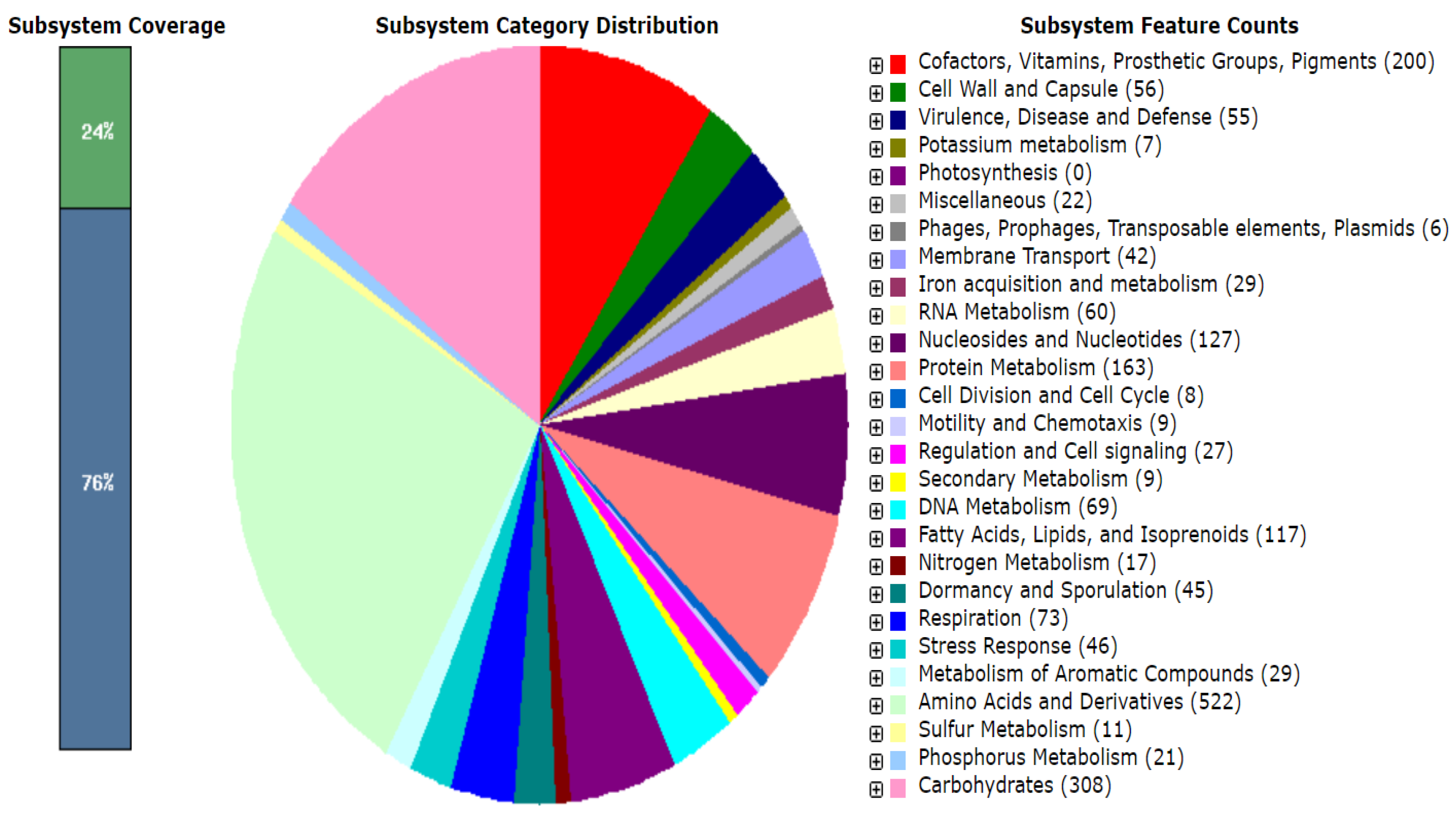
3.2. Functional Annotation

The TYGS (https://tygs.dsmz, accessed on 12 August 2023), a bioinformatics platform which is used for whole genome taxonomy analysis [28], was used for phylogenomic classification of strain AS_2 with closely related Peribacillus species. Furthermore, the OrthoANI tool was used to calculate the ANI value of Peribacillus AS_2 with other closely related species [29]. Strain AS_2 was found to be closely related to P. frigoritolerans DSM 8801T with a dDDH value of 84.8% and a 0.13% difference in G + C% content, and to P. castrilensis N3T with a dDDH value of 79.2% and a difference of 0.17 G + C% content (Table S2). The dDDH values of both Peribacillus species exceeded the dDDH > 70% threshold recognised as the cut-off point for species delineation [38,39]. The ANI values of strain AS_2 was 97.0% with P. frigoritolerans DSM 8801T and 96.7% with P. castrilensis N3T, as shown in Figure 1. Both ANI values were above the species boundary value (ANI, >95–96%) used to distinguish species [40,41], confirming that strain AS_2 is a Peribacillus species. Surprisingly, DSM 8801T and N3T had an ANI value of 96.7%, which was congruent with the dDDH value of 75.1% (results not shown), because the latter was described by [15] later than strain DSM 8801T [42], which was reclassified by [43,44]. We thus recommend reclassification of P. castrilensis N3T’s position within the Peribacillus genus.
The RAST server was used to further annotate the functional categories of genes encoded by strain AS_2 using default parameters [25]. A broad spectrum of housekeeping genes is often required for an endophytic lifestyle for bacterial growth and reproduction, including the uptake and transport of nutrients [44]. The RAST server predicted functional subsystems (Figure 2) which comprised genes involved in the metabolism of carbohydrates, amino acids and derivatives, fatty acids, lipids, co-factors, and vitamins of which the majority are important in the formation of endophytic relationships. Overall, the functional subsystems identified in the draft genome of strain AS_2 may also pave the way for further exploration in biotechnological applications, including genetic engineering and converting aromatic compounds to be used in bioremediation [45].
Acquisition of GIs in bacterial endophytes plays an important role, as they code for multiple accessory genes and may augment the diversification and adaption of a bacterium to its host or environment [46]. The GI Viewer tool was used to identify GIs present in strain AS_2, in which several sets of genomic regions revealed evidence of horizontal gene transfer. A total of 16 (Figure 3) GIs were identified in the strain AS_2 genome when aligned to P. butanolivorans strain KJ40, which was used as a reference genome. The 16 GIs comprised genes encoding penicillin-binding transpeptidase, ornithine cyclodeaminase family protein, glycosyltransferase, phage major capsid protein, cupin, and putative quinol monooxygenase. Cupin has been found to be present in a variety of endophytes and is known to play a role in the modification of plant cell wall polysaccharides [47].
In our previous study, Peribacillus sp. AS_2 was isolated and 16S rRNA gene sequencing revealed a polytomy relationship with P. simplex NBRC 15720T and P. muralis DSM 16288T, supported by an 83% bootstrap value [19]. The 16S rRNA is primarily utilised for the initial identification of bacterial species, although it cannot distinguish closely related species [48], necessitating further identification using other methods such as multilocus sequence analysis (MLSA). To investigate the genetic diversity of strain AS_2 at the species level with closely related Peribacillus-type strains, multilocus sequencing analysis was used. Multilocus sequencing analysis plays a key role in the phylogenetic classification of prokaryotic taxonomy, as it can obtain a higher resolution of the phylogenetic relationships between species at the species level [49,50]. Four housekeeping genes gyrA-gyrB-rpoC-rpoB were used for phylogenetic analysis and based on the concatenated phylogenetic tree, strain AS_2 clustered with P. frigoritolerans DSM 8801T and P. castrilensis N3T, supported by a 100% bootstrap value (Figure 4). This was congruent with dDDH and ANI value results. The results based on the MLSA, ANI and dDDH suggest that Peribacillus sp. AS_2 is a subspecies of P. frigoritolerans DSM 8801T.

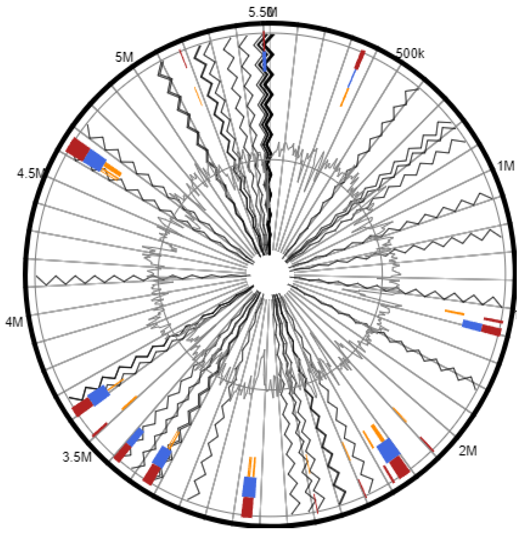
3.2.1. In Silico Analysis of Biosynthetic Gene Clusters

Bioactive compounds extracted from endophytic bacteria have gained attention due to their benefits to plants and humans in various sectors including agriculture, food, and pharmaceuticals [51]. Secondary metabolites in bacterial endophytes are mainly produced by biosynthetic gene clusters, which code for vital enzymes such as non-ribosomal peptide synthetases (NRPs), polyketide synthases (PKSs), transporters, and genes which code for tailoring enzymes [52,53]. The genome of the bacterial strain AS_2 was analysed using the antiSMASH 5.0 and PRISM online databases to explore the biosynthetic gene clusters of secondary metabolites. The antiSMASH database predicted a total of eight biosynthetic gene clusters in the genome, comprising NRPS-type koranimine, two terpenes, one siderophore cluster, one type III polyketide synthase (T3PKS), one linear azol(in)e-containing peptide (LAPS), and beta-lactone-type fengycin, whereas the PRISM database predicted three clusters which included two non-ribosomal peptides and NRPS-independent siderophore synthase gene clusters (Table 2). When comparing the predicted gene clusters, the koranimine-encoded gene cluster had a similarity percentage of 87% to Bacillus sp., whereas the Schizokinen and fengycin-encoded gene clusters had a similarity percentage of 60 (Nostoc sp.) and 46% (Bacillus velezensis), respectively. However, no similarity was detected in the other biosynthetic gene clusters (Table 2), terpene, TSPKS, NSPS, and LAPs, suggesting that some secondary metabolites may be uniquely produced by strain AS_2. Similar biosynthetic gene clusters were also identified in the P. frigoritolerans strain Q2H1, a bacterial endophyte in potato plants with a growth-promoting mechanism [54]. Koranimine, a biosynthetic gene cluster isolated from the bacterial endophyte P. frigoritolerans EB93, was discovered to have nematicidal action against Bursaphelenchus xylophilus, a common pine wilt nematode [55].

3.2.2. Antibiotic Resistance Genes

Antimicrobial resistance is still a major public concern, topping the World Health Organization’s (WHO) list of health concerns [59,60]. Recent studies have demonstrated that antimicrobial resistance genes (ARGs) are frequently acquired via horizontal gene transfer (HGT) using mobile genetic elements, resulting in the establishment and evolution of antimicrobial resistance bacteria [61,62]. Bacteria use HGT to transfer antibiotic resistance genes on plasmids using three primary mechanisms, which are conjugation, transduction, and natural transformation, and which occur in any environment including but not limited to the gut microbiome of humans and animals, soil, and water [63,64]. The transfer of plasmids between pathogenic microorganisms has resulted in a wide distribution of AGRs encoding resistance to a variety of drug classes including beta-lactams, tetracyclines, and quinolones [65]. A study by [66] revealed that the HGT of a plasmid encoded ARG was responsible for the outbreak of Shigella in the United Kingdom. The broad dissemination of ARGs in different host species and environments indicates that these genes can function well immediately after HGT [67]. Antibiotic resistance genes have been linked to the methylation of 16S rRNA [68], ribosomal proteins [69], lysyl-phosphatidylglycerols [70], and the encoding of the beta-subunit of RNA polymerase [71].
The genome of AS_2 was mapped onto the CARD database [72], which revealed gene clusters involved in small multidrug resistance antibiotic efflux pumps (qacJs), glycopeptide resistance (vanY, vanW, vanT, vanR), and fosfomycin thiol transferase (fosBx1). A class A Bacillus cereus Bc beta-lactamase gene cluster was also discovered, suggesting that the Peribacillus sp. strain AS_2 may be resistant to cephalosporin and penem (BcIII). Penem compounds are antimicrobials that are effective against a wide range of Gram-positive and Gram-negative bacteria [73].

3.2.3. Genes Involved in Endophytic Lifestyle

To better understand the factors that influence the Peribacillus sp. strain AS_2’s endophytic colonisation and lifestyle, we identified putative genes involved in the bacterial endophytic lifestyle (Table S3). Genes putatively involved in membrane transport proteins, detoxification, adhesion, substrate utilisation, secretion and delivery systems, transport, and transcriptional regulators were identified in AS_2. In addition, motility and chemotaxis are well known mechanisms which bacteria use to colonise their host plants [74]; consistent with this, two chemotactic signal-transducing proteins (CheC and CheD) were also identified.
Transcriptional regulators play an important role in the biological functions of bacterial endophytes, such as responding to environmental changes and colonising their host plants [75,76]. The TetR/AcrR family transcriptional regulators are known to influence a wide range of cell functions, including multidrug resistance, virulence, antibiotic production, osmotic stress, and bacterial pathogenicity [77]. In addition, the PadR transcriptional regulators are known to function as environmental sensors and assist in detoxifying harmful phenolic compounds [78]. Similarly, LysR family proteins were identified in the genome of strain AS_2, and are known to regulate the expression of genes mainly involved in the expression of motility, metabolism, and quorum sensing [79]. These have similarly been identified in other bacterial endophytes with plant-promoting, activities such as Burkholderia phytofirmans PsJN [47] and Bacillus paranthracis strain MHSD3 [80].
Evolutionary conserved antibiotic and virulence factors acquired by natural mutation and/or HGT may have the potential to serve as therapeutic new drug targets or vaccine antigens [81,82]. Lòpez-Fernàndez and colleagues hypothesised in 2015 that bacterial endophytes have the potential to express and share the same virulence-related genes as pathogens at the genomic level [83]. One example is the analysis of the Gram-negative colonising genome of B. phytofirmans PsJN, in which different mechanisms including siderophores, protein-secreting enzymes and polymer-degrading enzymes were identified, which are the same colonisation mechanisms that pathogens use to infect plants [84].
We discovered genes that encode the YihY/virulence factor BrkB family protein, a new virulence factor discovered in Bordetella pertussis that is relatively resistant to complement-dependent death via normal human serum [85]. The antibiotic biosynthesis monooxygenases were also identified, which are involved in the biosynthetic pathways for polyketide antibiotics, including tetracenomycin [86], alnumycin [87], and daunomycin [88]. The class D beta-lactamase antibiotic factors were also identified in strain AS_2, which are a group of antimicrobial agents that are mainly used to treat infections caused by bacterial pathogens [89]. Three genes encoded for a type III secretion system (EscU,YscU,HrcU) were also identified in the strain AS_2 genome, which is commonly found in Gram-negative pathogens including Pseudomonas, Escherichia coli, Shigella and Salmonella [90,91]. Although strain AS_2 was isolated as an endophyte, summing up these observations, strain AS_2 may have utilised these virulence genes to achieve colonisation in A. sessiliflora using the same mechanisms as pathogens.

3.2.4. Genes Involved in Biodegradation

Genomic studies have demonstrated that bacteria belonging to the genera Alcaligenes, Burkholderia, Mycobacteria, Rhodococcus and Pseudomonas [92] are capable of degrading organic contaminants such as aliphatic and aromatic compounds and converting them into either carbon dioxide, water, or biomass, which is essential for their growth [93]. In addition, synthetic chemicals used in pesticides, dyes, solvents, and refrigerators are often persistent in nature and hence are considered hazardous to the environment and humans, as they are toxic and insoluble and are often accumulated in high concentrations [94,95]. The biotransformation of these organic compounds can be further exploited for the treatment of contaminated water and soils through DNA recombinant techniques by constructing strains with an enhanced ability to degrade several toxic compounds.
Genes involved in the degradation of nitroalkanes (nitromethane, nitroethane, nitropropane)––which are a group of toxic compounds used in the chemical industries as intermediates to develop pharmaceutical compounds, insecticides, and dyes––were identified in the genome of strain AS_2 [96]. These nitroalkanes have been reported to be hazardous as they are toxic and carcinogenic [97]. Genes that code for the degradation of nitroalkanes, known as nitronate monooxygenases, were identified in strain AS_2, indicating its ability to catabolise toxic nitroalkane compounds. Nitronate monooxygenases have also been identified in other bacterial species, including Streptomyces ansochromogenes [98]. We further identified ssuD and tauD genes, which are mainly responsible for the degradation of organosulphonates such n-hexadecane, as the removal of this by-product is important for bacterial survival during oxidative stress in sulphur-limited conditions [99].
Pollution caused by organic contaminants coupled with heavy metals produced by industrial activities such as mining is a matter of great concern due to their toxic and carcinogenic effects on the environment and their position at top of the list of hazardous waste [100]. Research studies have demonstrated that genes involved in heavy metal resistance are often found in bacterial plasmids [101]. Several genes involved in heavy metal resistance were identified in the genome of strain AS_2, such as genes responsible for copper resistance, the CopC/CopD family protein, the heavy metal translocating P-type ATPase, and a magnesium transporter. In a similar study by [102], the bacterium Sphingobium yanoikuye strain SJTF8 was shown to be resistant to heavy metals such as copper, cadmium, and zinc and further showed degrading capabilities when co-cultured with these heavy metals with polycyclic aromatic hydrocarbons (PAHs), which are a group of hydrophobic compounds known for their toxic, carcinogenic, and mutagenic effects [103]. Other key enzymes required for initiating the degradation-like process, such as dioxygenases, have also been identified in the genome of AS_2, expanding the potential of this strain for bioremediation processes.

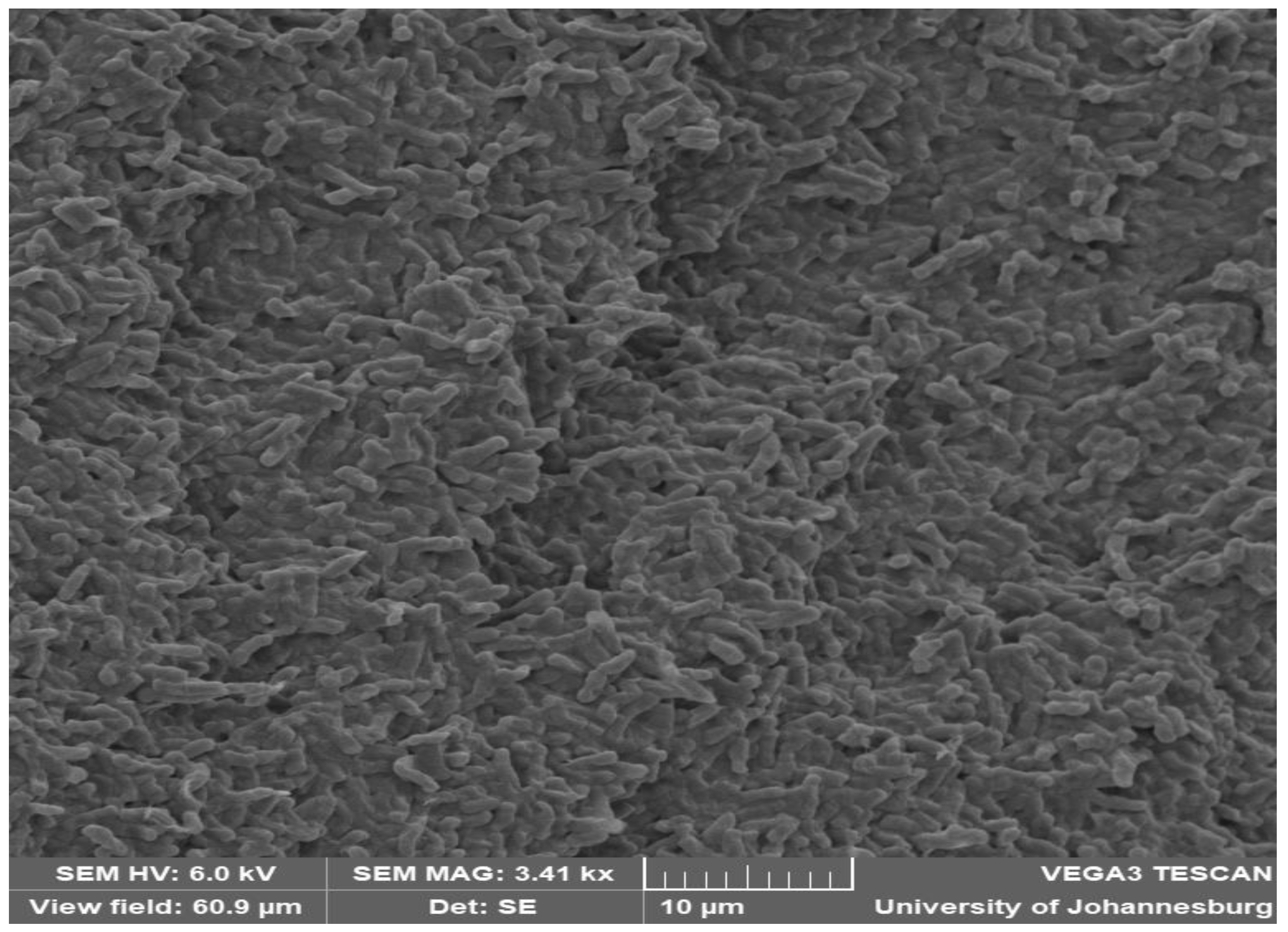
3.3. Phenotypic Characterisation

The strain AS_2 was a Gram-positive, spore-forming, and motile bacterium. Colonies of strain AS_2 on TSA plates were creamy, white, and circular with regular margins of 5–6 mm in diameter following incubation at 30 °C for 48 h. The morphology of strain AS_2 was observed using the SEM (Figure 5).
Peribacillus sp. strain AS_2 was catalase and oxidase positive, with growth temperatures ranging from 15 to 50 °C and a 30 °C optimum temperature. The cells of strain AS_2 were able to grow in NaCl concentrations ranging from 1 to 7% (w/v); the optimum was at 1% (w/v), demonstrating that it is a halotolerant bacterium [104]. The pH range in which strain AS_2 grew was 4–12, with an optimal pH of 7. Table 3 displays the phenotypic characteristics of strain AS_2 compared to closely related type strain species, P. frigoritolerans DSM 8801T, P. simplex DSM 1321T, P. muralis DSM 16288T, P. butanolivorans DSM 18926T, and P. loiseleuriae DSM 101776T. Strain AS_2 differs from some of the type strain species in the following characteristics: the ability to grow in anaerobic conditions and at 50 °C, the hydrolysis of casein, and being positive for oxidase and Voges–Proskauer. Strain AS_2 tested positive for eleven sugars in API 50CH, while some of the type strain species could not produce acids from the sugars tested. In contrast, AS_2 grew in a wider range of pH (4–12) and NaCl (1–7%) and it tested negative for hydrolysis of gelatine, arginine dihydrolase, indole production, and tryptophan deaminase.

4. Conclusions

The phenotypic and genomic analyses showed that strain AS_2, isolated from the medicinal plant Alectra sessliflora, belongs to the Peribacillus genus and is a subspecies of P. frigoritolerans DSM 8801T. Further studies are currently underway to describe it as a subspecies of P. frigoritolerans DSM 8801T. Genes involved in endophytic lifestyle were identified along with biosynthetic gene clusters. The genomic analysis has provided an initial step towards fully understanding the characteristics of AS_2 and its potential for broader application in the biotechnology sector. Furthermore, comparisons with other closely related Peribacillus-sequenced genomes will aid in delineating the distinct and shared characteristics of different Peribacillus species, shedding light on the evolutionary changes that may have occurred.

Supplementary Materials

The following supporting information can be downloaded at: https://www.mdpi.com/article/10.3390/microbiolres15010004/s1, Table S1: Clustered Regularly Interspaced Short Palindromic Repeats (CRISPR) sequences present within Peribacillus sp. strain AS_2 identified using CRISPRCasFinder; Table S2: Pairwise dDDH and ANI values between Peribacillus sp. AS_2 and the Peribacillus group strains; Table S3: Putative genes of Peribacillus sp. strain AS_2 involved in endophytic lifestyle; Table S4: Genes involved in biodegradation.

Author Contributions

M.P.M. contributed to the experimental work and data analysis. M.H.S.-D. conceptualised the study, acquired funding, project administration, and provided the materials used for the characterisation studies, and was the supervisor of the project. All authors have read and agreed to the published version of the manuscript.

Funding

This research was funded by the South African National Research Foundation Thuthuka grant (TTK210216586709). Ms MP Maela received the UJ Faculty of Science Bursary and the Gauteng City Region Academy Bursary (GCRA).

Institutional Review Board Statement

Not applicable.

Informed Consent Statement

Not applicable.

Data Availability Statement

This Whole Genome Shotgun project has been deposited at DDBJ/ENA/GenBank under the accession JAPTGA000000000. The version described in this paper is version JAPTGA000000000.

Acknowledgments

The authors would like to acknowledge the South African National Research foundation (NRF), Thuthuka, for the financial support for this study.

Conflicts of Interest

The authors declare no conflict of interest.

References

  1. Cui, L.; Yang, C.; Wang, Y.; Ma, T.; Cai, F.; Wei, L.; Jin, M.; Osei, R.; Zhang, J.; Tang, M. Potential of an endophytic bacteria Bacillus amyloliquefaciens 3–5 as biocontrol agent against potato scab. Microb. Pathog. 2022, 163, 105382. [Google Scholar] [CrossRef] [PubMed]
  2. Chu, L.L.; Bae, H. Bacterial endophytes from ginseng and their biotechnological application. J. Ginseng Res. 2022, 46, 1–10. [Google Scholar] [CrossRef] [PubMed]
  3. Poveda, J. Trichoderma parareesei favors the tolerance of rapeseed (Brassica napus L.) to salinity and drought due to a chorismate mutase. Agronomy 2020, 10, 118. [Google Scholar] [CrossRef]
  4. Sharma, S.; Dhar, M.K.; Kaul, S. Antagonistic, plant growth promoting and extracellular hydrolytic enzyme activity of fungal endophytes of Dioscorea bulbifera L. Biocatal. Agric. Biotechnol. 2023, 50, 102694. [Google Scholar] [CrossRef]
  5. Poveda, J.; Eugui, D.; Abril-Urías, P.; Velasco, P. Endophytic fungi as direct plant growth promoters for sustainable agricultural production. Symbiosis 2021, 85, 1–9. [Google Scholar] [CrossRef]
  6. Strobel, G.; Daisy, B.; Castillo, U.; Harper, J. Natural products from endophytic microorganisms. J. Nat. Prod. 2004, 67, 257–268. [Google Scholar] [CrossRef] [PubMed]
  7. Logan, N.A.; De Vos, P.; Genus, I. Bacillus. In Bergey’s Manual of Systematic Bacteriology; Springer: New York, NY, USA, 2009; Volume 3, pp. 21–128. [Google Scholar]
  8. Gupta, R.S.; Patel, S.; Saini, N.; Chen, S. Robust demarcation of 17 distinct Bacillus species clades, proposed as novel Bacillaceae genera, by phylogenomics and comparative genomic analyses: Description of Robertmurraya kyonggiensis sp. nov. and proposal for an emended genus Bacillus limiting it only to the members of the Subtilis and Cereus clades of species. Int. J. Syst. Evol. Microbiol. 2020, 70, 5753–5798. [Google Scholar]
  9. Patel, S.; Gupta, R.S. A phylogenomic and comparative genomic framework for resolving the polyphyly of the genus Bacillus: Proposal for six new genera of Bacillus species, Peribacillus gen. nov., Cytobacillus gen. nov., Mesobacillus gen. nov., Neobacillus gen. nov., Metabacillus gen. nov. and Alkalihalobacillus gen. nov. Int. J. Syst. Evol. Microbiol. 2020, 70, 406–438. [Google Scholar]
  10. Narsing Rao, M.P.; Dhulappa, A.; Banerjee, A.; Thamchaipenet, A. Transfer of Bacillus tepidiphilus Narsing Rao et al. 2021 to the genus Peribacillus as Peribacillus tepidiphilus comb. nov. Arch. Microbiol. 2022, 204, 545. [Google Scholar] [CrossRef]
  11. Ma, K.; Yin, Q.; Chen, L.; Lai, Q.; Xu, Y. Bacillus acanthi sp. nov., isolated from the rhizosphere soil of a mangrove plant Acanthus ilicifolius. Int. J. Syst. Evol. Microbiol. 2018, 68, 3047–3051. [Google Scholar] [CrossRef]
  12. Jiang, L.; Jung, W.Y.; Li, Z.; Lee, M.-K.; Park, S.-H.; Kang, S.W.; Lee, J.-S.; Jung, H.; Hur, T.-Y.; Kim, H.B.; et al. Peribacillus faecalis sp. nov., a moderately halophilic bacterium isolated from the faeces of a cow. Int. J. Syst. Evol. Microbiol. 2021, 71, 004721. [Google Scholar] [CrossRef] [PubMed]
  13. Zhang, Y.Z.; Chen, W.F.; Li, M.; Sui, X.H.; Liu, H.C.; Zhang, X.X.; Chen, W.X. Bacillus endoradicis sp. nov., an endophytic bacterium isolated from soybean root. Int. J. Syst. Evol. Microbiol. 2012, 62, 359–363. [Google Scholar] [CrossRef] [PubMed]
  14. Kämpfer, P.; Busse, H.J.; McInroy, J.A.; Glaeser, S.P. Bacillus gossypii sp. nov., isolated from the stem of Gossypium hirsutum. Int. J. Syst. Evol. Microbiol. 2015, 65, 4163–4168. [Google Scholar] [CrossRef] [PubMed]
  15. Rodríguez, M.; Reina, J.C.; Sampedro, I.; Llamas, I.; Martínez-Checa, F. Peribacillus castrilensis sp. nov.: A Plant-Growth-Promoting and Biocontrol Species Isolated From a River Otter in Castril, Granada, Southern Spain. Front. Plant Sci. 2022, 13, 896728. [Google Scholar] [CrossRef] [PubMed]
  16. Song, J.; Kim, S.; An, J.H.; Sang, M.K.; Weon, H.Y. Complete genome sequence of Peribacillus butanolivorans KJ40, a soil bacterium alleviating drought stress in plants. Korean J. Microbiol. 2020, 56, 407–409. [Google Scholar]
  17. Sato, S.; Ichiyanagi, N.; Sugiyama, K.; Aburai, N.; Fujii, K. Production of polyglutamic acid-like mucilage protein by Peribacillus simplex strain 8h. Folia. Microbiol. 2023, 68, 101–113. [Google Scholar] [CrossRef] [PubMed]
  18. Bhatt, D.; Srivastava, A.; Srivastava, P.C.; Sharma, A. Evaluation of three novel soil bacterial strains for efficient biodegradation of persistent boscalid fungicide: Kinetics and identification of microbial biodegradation intermediates. Environ. Pollut. 2023, 316, 120484. [Google Scholar] [CrossRef] [PubMed]
  19. Maela, M.P.; van der Walt, H.; Serepa-Dlamini, M.H. The antibacterial, antitumor activities, and bioactive constituents’ identification of ALECTRA sessiliflora bacterial endophytes. Front. Microbiol. 2022, 13, 870821. [Google Scholar] [CrossRef]
  20. Afgan, E.; Baker, D.; Batut, B.; van den Beek, M.; Bouvier, D.; Čech, M.; Chilton, J.; Clements, D.; Coraor, N.; Grüning, B.A.; et al. The Galaxy platform for accessible, reproducible and collaborative biomedical analyses: 2018 update. J. Nucleic Acids 2018, 46, W537–W544. [Google Scholar] [CrossRef]
  21. Andrews, S. FastQC: A Quality Control Tool for High Throughput Sequence Data. 2010. Available online: http://www.bioinformatics.babraham.ac.uk/projects/fastqc (accessed on 12 August 2023).
  22. Wick, R.R.; Judd, L.M.; Gorrie, C.L.; Holt, K.E. Unicycler: Resolving bacterial genome assemblies from short and long sequencing reads. PLoS Comput. Biol. 2017, 13, e1005595. [Google Scholar] [CrossRef]
  23. Gurevich, A.; Saveliev, V.; Vyahhi, N.; Tesler, G. QUAST: Quality assessment tool for genome assemblies. J. Bioinform. 2013, 15, 1072–1075. [Google Scholar] [CrossRef] [PubMed]
  24. Tatusova, T.; DiCuccio, M.; Badretdin, A.; Chetvernin, V.; Nawrocki, E.P.; Zaslavsky, L.; Lomsadze, A.; Pruitt, K.D.; Borodovsky, M.; Ostell, J. NCBI prokaryotic genome annotation pipeline. J. Nucleic Acids 2016, 44, 6614–6624. [Google Scholar] [CrossRef] [PubMed]
  25. Aziz, R.K.; Bartels, D.; Best, A.A.; DeJongh, M.; Disz, T.; Edwards, R.A.; Formsma, K.; Gerdes, S.; Glass, E.M.; Kubal, M.; et al. The RAST Server: Rapid annotations using subsystems technology. BMC Genom. 2008, 9, 75. [Google Scholar] [CrossRef] [PubMed]
  26. Simão, F.A.; Waterhouse, R.M.; Ioannidis, P.; Kriventseva, E.V.; Zdobnov, E.M. BUSCO: Assessing genome assembly and annotation completeness with single copy orthologs. Bioinformatics 2015, 31, 3210–3212. [Google Scholar] [CrossRef] [PubMed]
  27. Parks, D.H.; Imelfort, M.; Skennerton, C.T.; Hugenholtz, P.; Tyson, G.W. CheckM: Assessing the Quality of Microbial Genomes Recovered from Isolates, Single Cells, and Metagenomes. Genome Res. 2015, 25, 1043–1055. [Google Scholar] [CrossRef] [PubMed]
  28. Meier-Kolthoff, J.P.; Göker, M. TYGS is an automated high-throughput platform for state-of-the-art genome-based taxonomy. Nat. Commun. 2019, 10, 2182. [Google Scholar] [CrossRef]
  29. Lee, I.; Kim, Y.O.; Park, S.C.; Chun, J. OrthoANI: An improved algorithm and software for calculating average nucleotide identity. Int. J. Syst. Evol. Microbiol. 2016, 66, 1100–1103. [Google Scholar] [CrossRef] [PubMed]
  30. Kumar, S.; Stecher, G.; Li, M.; Knyaz, C.; Tamura, K. MEGA X: Molecular evolutionary genetics analysis across computing platforms. Mol. Biol. Evol. 2018, 35, 1547. [Google Scholar] [CrossRef]
  31. Grissa, I.; Vergnaud, G.; Pourcel, C. CRISPRFinder: A web tool to identify clustered regularly interspaced short palindromic repeats. Nucleic Acids Res. 2007, 35, W52–W57. [Google Scholar] [CrossRef]
  32. Abby, S.S.; Néron, B.; Ménager, H.; Touchon, M.; Rocha, E.P. MacSyFinder: A program to mine genomes for molecular systems with an application to CRISPR-Cas systems. PLoS ONE 2014, 9, e110726. [Google Scholar] [CrossRef]
  33. Couvin, D.; Bernheim, A.; Toffano-Nioche, C.; Touchon, M.; Michalik, J.; Néron, B.; Rocha, E.P.; Vergnaud, G.; Gautheret, D.; Pourcel, C. CRISPRCasFinder, an update of CRISRFinder, includes a portable version, enhanced performance and integrates search for Cas proteins. Nucleic Acids Res. 2018, 46, W246–W251. [Google Scholar] [CrossRef] [PubMed]
  34. Bertelli, C.; Laird, M.R.; Williams, K.P.; Simon, F.; Lau, B.Y.; Hoad, G.; Winsor, G.L.; Brinkman, F.S. IslandViewer 4: Expanded prediction of genomic islands for larger-scale datasets. Nucleic Acids Res. 2017, 45, W30–W35. [Google Scholar] [CrossRef] [PubMed]
  35. Blin, K.; Shaw, S.; Steinke, K.; Villebro, R.; Ziemert, N.; Lee, S.Y.; Medema, M.H.; Weber, T. antiSMASH 5.0: Updates to the secondary metabolite genome mining pipeline. Nucleic Acids Res. 2019, 47, W81–W87. [Google Scholar] [CrossRef] [PubMed]
  36. Skinnider, M.A.; Johnston, C.W.; Gunabalasingam, M.; Merwin, N.J.; Kieliszek, A.M.; MacLellan, R.J.; Li, H.; Ranieri, M.R.; Webster, A.L.; Cao, M.P.; et al. Comprehensive prediction of secondary metabolite structure and biological activity from microbial genome sequences. Nat. Commun. 2020, 11, 6058. [Google Scholar] [CrossRef] [PubMed]
  37. Logan, N.A.; Berge, O.; Bishop, A.H.; Busse, H.J.; De Vos, P.; Fritze, D.; Heyndrickx, M.; Kampfer, P.; Rabinovitch, L.; Salkinoja-Salonen, M.S.; et al. Proposed minimal standards for describing new taxa of aerobic, endospore-forming bacteria. Int. J. Syst. Evol. Microbiol. 2009, 59, 2114–2121. [Google Scholar] [CrossRef] [PubMed]
  38. Auch, A.F.; von Jan, M.; Klenk, H.P.; Göker, M. Digital DNA-DNA hybridization for microbial species delineation by means of genome-to-genome sequence comparison. Stand. Genom. Sci. 2010, 2, 117–134. [Google Scholar] [CrossRef] [PubMed]
  39. Meier-Kolthoff, J.P.; Auch, A.F.; Klenk, H.P.; Göker, M. Genome sequence-based species delimitation with confidence intervals and improved distance functions. BMC Bioinform. 2013, 14, 60. [Google Scholar] [CrossRef]
  40. Konstantinidis, K.T.; Tiedje, J.M. Genomic insights that advance the species definition for prokaryotes. Proc. Natl. Acad. Sci. USA 2005, 102, 2567–2572. [Google Scholar] [CrossRef]
  41. Richter, M.; Rosselló-Móra, R. Shifting the genomic gold standard for the prokaryotic species definition. Proc. Natl. Acad. Sci. USA 2009, 106, 19126–19131. [Google Scholar] [CrossRef]
  42. Montecillo, J.A.; Bae, H. Reclassification of Brevibacterium frigoritolerans as Peribacillus frigoritolerans comb. nov. based on phylogenomics and multiple molecular synapomorphies. Int. J. Syst. Evol. 2022, 72, 005389. [Google Scholar] [CrossRef]
  43. Liu, G.H.; Liu, B.; Wang, J.P.; Che, J.M.; Li, P.F. Reclassification of Brevibacterium frigoritolerans DSM 8801 T as Bacillus frigoritolerans comb. nov. Based on Genome Analysis. Curr. Microbiol. 2020, 77, 1916–1923. [Google Scholar] [CrossRef] [PubMed]
  44. Ulrich, K.; Kube, M.; Becker, R.; Schneck, V.; Ulrich, A. Genomic analysis of the endophytic Stenotrophomonas strain 169 reveals features related to plant-growth promotion and stress tolerance. Front. Microbiol. 2021, 12, 687463. [Google Scholar] [CrossRef] [PubMed]
  45. Fouts, D.E.; Tyler, H.L.; DeBoy, R.T.; Daugherty, S.; Ren, Q.; Badger, J.H.; Durkin, A.S.; Huot, H.; Shrivastava, S.; Kothari, S.; et al. Complete genome sequence of the N2-fixing broad host range endophyte Klebsiella pneumoniae 342 and virulence predictions verified in mice. PLoS Genet. 2008, 4, e1000141. [Google Scholar] [CrossRef]
  46. Guo, F.B.; Xiong, L.; Zhang, K.Y.; Dong, C.; Zhang, F.Z.; Woo, P.C. Identification and analysis of genomic islands in Burkholderia cenocepacia AU 1054 with emphasis on pathogenicity islands. BMC Microbiol. 2017, 17, 73. [Google Scholar] [CrossRef] [PubMed]
  47. Ali, S.; Duan, J.; Charles, T.C.; Glick, B.R. A bioinformatics approach to the determination of genes involved in endophytic behavior in Burkholderia spp. J. Theor. Biol. 2014, 343, 193–198. [Google Scholar] [CrossRef] [PubMed]
  48. Kitahara, K.; Miyazaki, K. Revisiting bacterial phylogeny: Natural and experimental evidence for horizontal gene transfer of 16S rRNA. Mob. Genet. Elem. 2013, 3, e24210. [Google Scholar] [CrossRef] [PubMed]
  49. Glaeser, S.P.; Kämpfer, P. Multilocus sequence analysis (MLSA) in prokaryotic taxonomy. Syst. Appl. Microbiol. 2015, 38, 237–245. [Google Scholar] [CrossRef] [PubMed]
  50. Zou, L.; Wang, Q.; Li, M.; Wang, S.; Ye, K.; Dai, W.; Huang, J. Culturable bacterial endophytes of Aconitum carmichaelii Debx. were diverse in phylogeny, plant growth promotion, and antifungal potential. Front. Microbiol. 2023, 14, 1192932. [Google Scholar] [CrossRef]
  51. Verma, V.C.; Gange, A.C. (Eds.) Advances in Endophytic Research; Springer Science & Business Media: New Delhi, India, 2013. [Google Scholar]
  52. Singh, M.; Kumar, A.; Singh, R.; Pandey, K.D. Endophytic bacteria: A new source of bioactive compounds. 3 Biotech 2017, 7, 315. [Google Scholar] [CrossRef]
  53. Akter, Y.; Barua, R.; Nasir Uddin, M.; Muhammad Sanaullah, A.F.; Marzan, L.W. Bioactive potentiality of secondary metabolites from endophytic bacteria against SARS-COV-2: An in-silico approach. PLoS ONE 2022, 17, e0269962. [Google Scholar] [CrossRef]
  54. Wang, Y.; Zhao, Q.; Sun, Z.; Li, Y.; He, H.; Zhang, Y.; Yang, X.; Wang, D.; Dong, B.; Zhou, H.; et al. Whole-genome analysis revealed the growth-promoting mechanism of endophytic bacterial strain Q2H1 in potato plants. Front. Microbiol. 2022, 13, 1035901. [Google Scholar] [CrossRef]
  55. Montecillo, J.A.; Bae, H. In silico analysis of koranimine, a cyclic imine compound from Peribacillus frigoritolerans reveals potential nematicidal activity. Sci. Rep. 2022, 12, 18883. [Google Scholar] [CrossRef]
  56. Evans, B.S.; Ntai, I.; Chen, Y.; Robinson, S.J.; Kelleher, N.L. Proteomics-based discovery of koranimine, a cyclic imine natural product. J. Am. Chem. Soc. 2011, 133, 7316–7319. [Google Scholar] [CrossRef]
  57. Koumoutsi, A.; Chen, X.H.; Henne, A.; Liesegang, H.; Hitzeroth, G.; Franke, P.; Vater, J.; Borriss, R. Structural and functional characterization of gene clusters directing nonribosomal synthesis of bioactive cyclic lipopeptides in Bacillus amyloliquefaciens strain FZB42. J. Bacteriol. 2004, 186, 1084–1096. [Google Scholar] [CrossRef]
  58. Kaneko, T.; Nakamura, Y.; Wolk, C.P.; Kuritz, T.; Sasamoto, S.; Watanabe, A.; Iriguchi, M.; Ishikawa, A.; Kawashima, K.; Kimura, T.; et al. Complete genomic sequence of the filamentous nitrogen-fixing cyanobacterium Anabaena sp. strain PCC 7120 (supplement). DNA Res. 2001, 8, 227–253. [Google Scholar] [CrossRef]
  59. Fresia, P.; Antelo, V.; Salazar, C.; Giménez, M.; D’Alessandro, B.; Afshinnekoo, E.; Mason, C.; Gonnet, G.H.; Iraola, G. Urban metagenomics uncover antibiotic resistance reservoirs in coastal beach and sewage waters. Microbiome 2019, 7, 35. [Google Scholar] [CrossRef]
  60. Zhao, C.X.; Su, X.X.; Xu, M.R.; An, X.L.; Su, J.Q. Uncovering the diversity and contents of gene cassettes in class 1 integrons from the endophytes of raw vegetables. Ecotoxicol. Environ. Saf. 2022, 247, 114282. [Google Scholar] [CrossRef]
  61. Liu, Y.Y.; Wang, Y.; Walsh, T.R.; Yi, L.X.; Zhang, R.; Spencer, J.; Doi, Y.; Tian, G.; Dong, B.; Huang, X.; et al. Emergence of plasmid-mediated colistin resistance mechanism MCR-1 in animals and human beings in China: A microbiological and molecular biological study. Lancet Infect. Dis. 2016, 16, 161–168. [Google Scholar] [CrossRef]
  62. Millan, A.S. Evolution of plasmid-mediated antibiotic resistance in the clinical context. Trends Microbiol. 2018, 26, 978–985. [Google Scholar] [CrossRef]
  63. Mathers, A.J.; Peirano, G.; Pitout, J.D. The role of epidemic resistance plasmids and international high-risk clones in the spread of multidrug-resistant Enterobacteriaceae. Clin. Microbiol. Rev. 2015, 28, 565–591. [Google Scholar] [CrossRef]
  64. Sun, D.; Jeannot, K.; Xiao, Y.; Knapp, C.W. Horizontal gene transfer mediated bacterial antibiotic resistance. Front. Microbiol. 2019, 10, 1933. [Google Scholar] [CrossRef] [PubMed]
  65. Huddleston, J.R. Horizontal gene transfer in the human gastrointestinal tract: Potential spread of antibiotic resistance genes. Infect. Drug Resist. 2014, 167, 76. [Google Scholar] [CrossRef] [PubMed]
  66. Baker, K.S.; Dallman, T.J.; Field, N.; Childs, T.; Mitchell, H.; Day, M.; Weill, F.X.; Lefèvre, S.; Tourdjman, M.; Hughes, G.; et al. Horizontal antimicrobial resistance transfer drives epidemics of multiple Shigella species. Nat. Commun. 2018, 9, 1462. [Google Scholar] [CrossRef] [PubMed]
  67. Lerminiaux, N.A.; Cameron, A.D. Horizontal transfer of antibiotic resistance genes in clinical environments. Can. J. Microbiol. 2019, 65, 34–44. [Google Scholar] [CrossRef] [PubMed]
  68. Nosrati, M.; Dey, D.; Mehrani, A.; Strassler, S.E.; Zelinskaya, N.; Hoffer, E.D.; Stagg, S.M.; Dunham, C.M.; Conn, G.L. Functionally critical residues in the aminoglycoside resistance-associated methyltransferase RmtC play distinct roles in 30S substrate recognition. J. Biol. Chem. 2019, 294, 17642–17653. [Google Scholar] [CrossRef]
  69. Finken, M.; Kirschner, P.; Meier, A.; Wrede, A.; Böttger, E.C. Molecular basis of streptomycin resistance in Mycobacterium tuberculosis: Alterations of the ribosomal protein S12 gene and point mutations within a functional 16S ribosomal RNA pseudoknot. Mol. Biol. 1993, 9, 1239–1246. [Google Scholar] [CrossRef]
  70. Samant, S.; Hsu, F.F.; Neyfakh, A.A.; Lee, H. The Bacillus anthracis protein MprF is required for synthesis of lysylphosphatidylglycerols and for resistance to cationic antimicrobial peptides. J. Bacteriol. 2009, 191, 1311–1319. [Google Scholar] [CrossRef]
  71. Jagielski, T.; Bakuła, Z.; Brzostek, A.; Minias, A.; Stachowiak, R.; Kalita, J.; Napiórkowska, A.; Augustynowicz-Kopeć, E.; Żaczek, A.; Vasiliauskiene, E.; et al. Characterization of mutations conferring resistance to rifampin in Mycobacterium tuberculosis clinical strains. Antimicrob. Agents Chemother. 2018, 62, e01093-18. [Google Scholar] [CrossRef]
  72. Alcock, B.P.; Raphenya, A.R.; Lau, T.T.; Tsang, K.K.; Bouchard, M.; Edalatmand, A.; Huynh, W.; Nguyen, A.L.V.; Cheng, A.A.; Liu, S.; et al. CARD 2020: Antibiotic resistome surveillance with the comprehensive antibiotic resistance database. Nucleic Acids Res. 2020, 48, D517–D525. [Google Scholar] [CrossRef]
  73. Lang, M.; Schneider, P.; Tosch, W.; Scartazzini, R.; Zak, O. The Penems, a New Class of β-Lactam Antibiotics 7. Synthesis and Antimicrobial Activity of 2-Heterocyclylmercaptoalkyl Derivatives. J. Antibiot. 1986, 39, 525–534. [Google Scholar] [CrossRef]
  74. Shastry, R.P.; Welch, M.; Rai, V.R.; Ghate, S.D.; Sandeep, K.; Rekha, P.D. The whole-genome sequence analysis of Enterobacter cloacae strain Ghats1: Insights into endophytic lifestyle-associated genomic adaptations. Arch. Microbiol. 2020, 202, 1571–1579. [Google Scholar] [CrossRef]
  75. Deng, W.; Li, C.; Xie, J. The underling mechanism of bacterial TetR/AcrR family transcriptional repressors. Cell. Signal. 2013, 25, 1608–1613. [Google Scholar] [CrossRef]
  76. Pinski, A.; Betekhtin, A.; Hupert-Kocurek, K.; Mur, L.A.; Hasterok, R. Defining the genetic basis of plant–endophytic bacteria interactions. Int. J. Mol. Sci. 2019, 20, 1947. [Google Scholar] [CrossRef]
  77. Ramos, J.L.; Martínez-Bueno, M.; Molina-Henares, A.J.; Terán, W.; Watanabe, K.; Zhang, X.; Gallegos, M.T.; Brennan, R.; Tobes, R. The TetR family of transcriptional repressors. Microbiol. Mol. Biol. Rev. 2005, 69, 326–356. [Google Scholar] [CrossRef]
  78. Park, S.C.; Kwak, Y.M.; Song, W.S.; Hong, M.; Yoon, S.I. Structural basis of effector and operator recognition by the phenolic acid-responsive transcriptional regulator PadR. Nucleic Acids Res. 2017, 45, 13080–13093. [Google Scholar] [CrossRef]
  79. Ko, M.; Park, C. H-NS-Dependent regulation of flagellar synthesis is mediated by a LysR family protein. J. Bacteriol. 2000, 182, 4670–4672. [Google Scholar] [CrossRef]
  80. Diale, M.O.; Kayitesi, E.; Serepa-Dlamini, M.H. Genome in silico and in vitro analysis of the probiotic properties of a bacterial endophyte, Bacillus paranthracis strain mhsd3. Front. Genet. 2021, 12, 672149. [Google Scholar] [CrossRef]
  81. O’Neill, J. Review on antimicrobial resistance: Tackling drug-resistant infections globally: Final report and recommendations. In Review on Antimicrobial Resistance: Tackling Drug-Resistant Infections Globally: Final Report and Recommendations; Wellcome Trust: London, UK, 2016. [Google Scholar]
  82. Yamaguchi, M.; Goto, K.; Hirose, Y.; Yamaguchi, Y.; Sumitomo, T.; Nakata, M.; Nakano, K.; Kawabata, S. Identification of evolutionarily conserved virulence factor by selective pressure analysis of Streptococcus pneumoniae. Commun. Biol. 2019, 2, 96. [Google Scholar] [CrossRef]
  83. Lòpez-Fernàndez, S.; Sonego, P.; Moretto, M.; Pancher, M.; Engelen, K.; Pertot, I.; Campisano, A. Whole-genome comparative analysis of virulence genes unveils similarities and differences between endophytes and other symbiotic bacteria. Front. Microbiol. 2015, 6, 419. [Google Scholar]
  84. Mitter, B.; Petric, A.; Shin, M.W.; Chain, P.S.; Hauberg-Lotte, L.; Reinhold-Hurek, B.; Nowak, J.; Sessitsch, A. Comparative genome analysis of Burkholderia phytofirmans PsJN reveals a wide spectrum of endophytic lifestyles based on interaction strategies with host plants. Front. Plant Sci. 2013, 4, 120. [Google Scholar] [CrossRef]
  85. Fernandez, R.C.; Weiss, A.A. Cloning and sequencing of a Bordetella pertussis serum resistance locus. Infect. Immun. 1994, 62, 4727–4738. [Google Scholar] [CrossRef]
  86. Kendrew, S.G.; Federici, L.; Savino, C.; Miele, A.; Marsh, E.N.; Vallone, B. Crystallization and preliminary X-ray diffraction studies of a monooxygenase from Streptomyces coelicolor A3 (2) involved in the biosynthesis of the polyketide actinorhodin. Acta Crystallogr. D Struct. Biol. 2000, 56, 481–483. [Google Scholar] [CrossRef]
  87. Ye, J.; Dickens, M.L.; Plater, R.; Li, Y.; Lawrence, J.; Strohl, W.R. Isolation and sequence analysis of polyketide synthase genes from the daunomycin-producing Streptomyces sp. strain C5. J. Bacteriol. 1994, 176, 6270–6280. [Google Scholar] [CrossRef]
  88. Grocholski, T.; Oja, T.; Humphrey, L.; Mäntsälä, P.; Niemi, J.; Metsä-Ketelä, M. Characterization of the two-component monooxygenase system AlnT/AlnH reveals early timing of quinone formation in alnumycin biosynthesis. J. Bacteriol. 2012, 194, 2829–2836. [Google Scholar] [CrossRef]
  89. Yoon, E.J.; Jeong, S.H. Class D β-lactamases. J. Antimicrob. Chemother. 2021, 76, 836–864. [Google Scholar] [CrossRef]
  90. Abubakar, I.I.; Tillmann, T.; Banerjee, A. Global, regional, and national age-sex specific all-cause and cause-specific mortality for 240 causes of death, 1990-2013: A systematic analysis for the Global Burden of Disease Study 2013. Lancet 2015, 385, 117–171. [Google Scholar]
  91. Troeger, C.; Forouzanfar, M.; Rao, P.C.; Khalil, I.; Brown, A.; Reiner, R.C.; Fullman, N.; Thompson, R.L.; Abajobir, A.; Ahmed, M.; et al. Estimates of global, regional, and national morbidity, mortality, and aetiologies of diarrhoeal diseases: A systematic analysis for the Global Burden of Disease Study 2015. Lancet Infect. Dis. 2017, 17, 909–948. [Google Scholar] [CrossRef]
  92. Hesham, A.E.L.; Mawad, A.M.; Mostafa, Y.M.; Shoreit, A. Biodegradation ability and catabolic genes of petroleum-degrading Sphingomonas koreensis strain ASU-06 isolated from Egyptian oily soil. BioMed Res. Int. 2014, 2014, 127674. [Google Scholar] [CrossRef]
  93. Karaś, M.A.; Wdowiak-Wróbel, S.; Sokołowski, W. Selection of endophytic strains for enhanced bacteria-assisted phytoremediation of organic pollutants posing a public health hazard. Int. J. Mol. Sci. 2021, 22, 9557. [Google Scholar] [CrossRef]
  94. Olaniran, A.O.; Balgobind, A.; Pillay, B. Bioavailability of heavy metals in soil: Impact on microbial biodegradation of organic compounds and possible improvement strategies. Int. J. Mol. Sci. 2013, 14, 10197–10228. [Google Scholar] [CrossRef]
  95. Santero, E.; Díaz, E. Special Issue: Genetics of biodegradation and bioremediation. Genes 2020, 11, 441. [Google Scholar] [CrossRef] [PubMed]
  96. Balogh-Hergovich, É.; Kaizer, J.; Speier, G. Chemical models relevant to nitroalkane dioxygenase. Comptes Rendus Chim. 2007, 10, 355–365. [Google Scholar] [CrossRef]
  97. Torres-Guzman, J.C.; Padilla-Guerrero, I.E.; Cervantes-Quintero, K.Y.; Martinez-Vazquez, A.; Ibarra-Guzman, M.; Gonzalez-Hernandez, G.A. Peculiarities of nitronate monooxygenases and perspectives for in vivo and in vitro applications. Appl. Microbiol. Biotechnol. 2021, 105, 8019–8032. [Google Scholar] [CrossRef] [PubMed]
  98. Li, Y.; Gao, Z.; Hou, H.; Li, L.; Zhang, J.; Yang, H.; Dong, Y.; Tan, H. Crystal structure and site-directed mutagenesis of a nitroalkane oxidase from Streptomyces ansochromogenes. Biochem. Biophys. Res. Commun. 2011, 405, 344–348. [Google Scholar] [CrossRef] [PubMed]
  99. Park, C.; Shin, B.; Park, W. Protective role of bacterial alkanesulfonate monooxygenase under oxidative stress. Appl. Microbiol. Biotechnol. 2020, 86, e00692-20. [Google Scholar] [CrossRef] [PubMed]
  100. Haarstad, K.; Bavor, H.J.; Mæhlum, T. Organic and metallic pollutants in water treatment and natural wetlands: A review. Water Sci. Technol. 2012, 65, 76–99. [Google Scholar] [CrossRef] [PubMed]
  101. Khatoon, K.; Malik, A. Screening of polycyclic aromatic hydrocarbon degrading bacterial isolates from oil refinery wastewater and detection of conjugative plasmids in polycyclic aromatic hydrocarbon tolerant and multi-metal resistant bacteria. Heliyon 2019, 5, e02742. [Google Scholar] [CrossRef] [PubMed]
  102. Yin, C.; Xiong, W.; Qiu, H.; Peng, W.; Deng, Z.; Lin, S.; Liang, R. Characterization of the phenanthrene-degrading Sphingobium yanoikuyae SJTF8 in heavy metal co-existing liquid medium and analysis of its metabolic pathway. Microorganisms 2020, 8, 946. [Google Scholar] [CrossRef]
  103. Haritash, A.K.; Kaushik, C.P. Biodegradation aspects of polycyclic aromatic hydrocarbons (PAHs): A review. J. Hazard. Mater. 2009, 169, 1–15. [Google Scholar] [CrossRef]
  104. Enquahone, S.; van Marle, G.; Simachew, A. Plant growth-promoting characteristics of halotolerant endophytic bacteria isolated from Sporobolus specatus (Vahr) Kunth and Cyperus laevigatus L. of Ethiopian rift valley lakes. Arch. Microbiol. 2022, 204, 403. [Google Scholar] [CrossRef]
  105. Heyrman, J.; Logan, N.A.; Rodríguez-Díaz, M.; Scheldeman, P.; Lebbe, L.; Swings, J.; Heyndrickx, M.; De Vos, P. Study of mural painting isolates, leading to the transfer of ‘Bacillus maroccanus’ and ‘Bacillus carotarum’to Bacillus simplex, emended description of Bacillus simplex, re-examination of the strains previously attributed to ‘Bacillus macroides’ and description of Bacillus muralis sp. nov. J. Syst. Evol. Microbiol. 2005, 55, 119–131. [Google Scholar]
  106. Kuisiene, N.; Raugalas, J.; Sproer, C.; Kroppenstedt, R.M.; Chitavichius, D. Bacillus butanolivorans sp. nov., a species with industrial application for the remediation of n-butanol. J. Syst. Evol. Microbiol. 2008, 58, 505–509. [Google Scholar] [CrossRef] [PubMed]
Figure 1. Heatmap generated with OAT software (version 0.93.1) showing the OrthoANI values of Peribacillus sp. strain AS_2 with other closely related Peribacillus species.
Microbiolres 15 00004 g001
Figure 2. Pie chart showing an overview of RAST subsystems assigned to genes in Peribacillus AS_2 genome. Each subsystem (left) is represented by a particular colour (right).
Microbiolres 15 00004 g002
Figure 3. Circular plot of genomic islands identified from the Peribacillus sp. strain AS_2 genome aligned against the reference genome Peribacillus butanolivorans strain KJ40 using IslandViewer 4. The black outer circle represents the scale line of the genome in Mbps, and the identified genomic islands are represented in three colours: red (integrated detection), blue (IslandPath-DIMOB), and orange (SIGI-HMM).
Microbiolres 15 00004 g003
Figure 4. Phylogenetic analysis of strain AS_2 and closely related species of the Peribacillus group based on MLSA of four housekeeping genes, gyrA-gyrB-rpoB-rpoC. Brevibacillus brevis NBRC 15304T was used as an outgroup.
Microbiolres 15 00004 g004
Figure 5. Scanning electron micrograph of Peribacillus sp. strain AS_2 cells grown for 24 h at 30 °C.
Microbiolres 15 00004 g005
Table 1. Genome characteristics of Peribacillus sp. strain AS_2.
Genome CharacteristicValue
Genome size (bp)5,482,853
G + C content (%)40.5
Total number of genes5290
Protein coding genes5072
Number of RNAs88
rRNA genes1, 1, 1 (5S, 16S, 23S)
tRNA genes79
ncRNAs6
Pseudogenes130
CRISPR repeats4
Cas cluster1
CRISPR: Clustered Regularly Interspaced Short Palindromic Repeats; rRNA: ribosomal RNA, tRNA: transfer RNA.
Table 2. Biosynthetic gene clusters involved in the production of secondary metabolites in Peribacillus sp. strain AS_2.
From (bp)To (bp)TypeProductionSimilarity (%)Reference
236,408296,726NRPsKoranimine87Bacillus sp. NK2003 [56]
140,936165,105Beta-lactoneFengycin46Bacillus velezensis FZB42 [57]
50,38065,892NI-siderophoreSchizokinen60Nostoc sp. PCC 7120 [58]
380,256402,151Terpene--
389,596430,684T3PKS--
87,936132,003NRPS--
888932,424LAP--
743128,249Terpene--
Table 3. Differential characteristics between the Peribacillus sp. strain AS_2 and closely related type strains.
Characteristic12+3+4+5+
Anaerobic growth+-vw-
Growth at 4 °C-+--+
Growth at 45 °C+---+
NaCl (w/v, %)0–70.5–7.5<5<70.5–5
Optimum NaCl (w/v, %)10001
pH range3–125–106–96–96–9
Optimum pH77877
Oxidase+--++
API 20E:
ONPG---+-
Arginine dihydrolase-----
Citrate utilisation-----
Voges– Proskauer+----
Urease-----
Tryptophan deaminase-----
Indole production-----
Gelatine hydrolysis-----
API 50:
L-arabinose++-+-
Ribose+--+-
Glucose++w+-
Fructose++w+-
Mannitol+--+-
N-acetylglucosamine+-w+w
Salicin+-w/v+w/v
Saccharose+----
Trehalose+-w+w
Inulin+-w-w
Raffinose++-+-
Hydrolysis of:
Starch--++-
Casein++vv-
DNA G + C content (mol %)40.4540.639.941.237.4
Strains: 1, Peribacillus sp. AS_2; 2, Peribacillus frigoritolerans DSM 8801T [40]; 3, P. simplex DSM 1321T [105]; 4, P. muralis DSM 16288T [105]; 5, P. butanolivorans DSM 18926T [106]. +, positive; -, negative; w, weak; v, variable; w/v, weak and variable.
Disclaimer/Publisher’s Note: The statements, opinions and data contained in all publications are solely those of the individual author(s) and contributor(s) and not of MDPI and/or the editor(s). MDPI and/or the editor(s) disclaim responsibility for any injury to people or property resulting from any ideas, methods, instructions or products referred to in the content.

