Review

The Role of Bacteriophages in the Gut Microbiota: Implications for Human Health

by
Stephen C. Emencheta
1,2,
Chinasa V. Olovo
3,
Osita C. Eze
1,
Chisom F. Kalu
1,
Dinebari P. Berebon
1,*,
Ebele B. Onuigbo
1,
Marta M. D. C. Vila
2,
Victor M. Balcão
2,4 and
Anthony A. Attama
5,6,*
1
Department of Pharmaceutical Microbiology and Biotechnology, University of Nigeria, Nsukka 410001, Nigeria
2
VBlab—Laboratory of Bacterial Viruses, University of Sorocaba, Sorocaba 18023-000, Brazil
3
Department of Microbiology, University of Nigeria, Nsukka 410001, Nigeria
4
Department of Biology and CESAM, University of Aveiro, Campus Universitário de Santiago, P-3810-193 Aveiro, Portugal
5
Department of Pharmaceutics, University of Nigeria, Nsukka 410001, Nigeria
6
Institute for Drug-Herbal Medicine-Excipient Research and Development, University of Nigeria, Nsukka 410001, Nigeria
*
Authors to whom correspondence should be addressed.
Pharmaceutics 2023, 15(10), 2416; https://doi.org/10.3390/pharmaceutics15102416
Submission received: 22 August 2023 / Revised: 18 September 2023 / Accepted: 30 September 2023 / Published: 3 October 2023

Abstract

:
Bacteriophages (phages) are nano-sized viruses characterized by their inherent ability to live off bacteria. They utilize diverse mechanisms to absorb and gain entry into the bacterial cell wall via the release of viral genetic material, which uses the replication mechanisms of the host bacteria to produce and release daughter progeny virions that attack the surrounding host cells. They possess specific characteristics, including specificity for particular or closely related bacterial species. They have many applications, including as potential alternatives to antibiotics against multi-resistant bacterial pathogens and as control agents in bacteria-contaminated environments. They are ubiquitously abundant in nature and have diverse biota, including in the gut. Gut microbiota describes the community and interactions of microorganisms within the intestine. As with bacteria, parasitic bacteriophages constantly interact with the host bacterial cells within the gut system and have obvious implications for human health. However, it is imperative to understand these interactions as they open up possible applicable techniques to control gut-implicated bacterial diseases. Thus, this review aims to explore the interactions of bacteriophages with bacterial communities in the gut and their current and potential impacts on human health.

1. Introduction

The ubiquitous nature of microorganisms extends to their presence in the gastrointestinal tract (GIT) [1,2], where they play a host of unique physiological characteristics and functions which contribute to the overall well-being and health [2], including, but not limited to, their help in digestive and metabolic processes [3], gut barrier protection [4], essential vitamin production [5], and the immune system [6].
The gut microbiota is influenced by a potential compounding combination of different factors determining the microbial makeup [7]. These include diet or nutrient type (e.g., plant and fiber-based diets promote the selection/presence of beneficial microorganisms, unlike chemically processed and sugary diets, and breast milk has significant influence on the infant microbiota in the first few years) [7], external chemicals (e.g., antibiotics) [8,9], genetic/hereditary profile (although largely unclear) [10], age [9], environmental pollutants/chemicals (e.g., agrochemicals), medical conditions (e.g., celiac and IBDs), and lifestyle [9].
Pathogenic/unhealthy gut microorganisms can hardly ever be eradicated and can even be potentially beneficial; however, maintenance of the equilibrium between the healthy and harmful gut microorganisms is essential and, through several mechanisms, is related to well-being and overall health and the prevention, development, and management of health/medical conditions [11]. Specifically, IBDs often result from the immune response to the dysbiosis of the gut microbiota, increasing the potentially pathogenic microorganisms (e.g., Pseudomonadota and Enterobacteriaceae) and decreasing the beneficial microorganisms (e.g., Lactobacillaceae and Bifidobacteria) [11]. Accumulative evidence has shown that disruption of the metabolites and the gut microbiota can influence insulin sensitivity [12], optimal brain and central nervous system (CNS) (which, of course, controls most of the GIT physiology) functions, and emotional behaviors [13,14]. High blood pressure, a top risk factor for heart disorders, can be initiated by disruption of the gut microbiota [15]. Also, specific microbial genes are associated with the generation and accumulation of Trimethylamine N-oxide (TMAO), a risk factor for cardiovascular diseases, especially at high concentrations [16,17]. The gut microbiota can be described as the “other organ”, contributing significantly to nutrient and energy acquisition and regulation. Thus, dysbiosis can negatively affect the above functions, triggering excessive tissue deposition and obesity [18].
One prominent and easily the most abundant life forms are the bacteriophages [8]. Bacteriophages have found applications in diverse fields and have peculiar characteristics in clinical practice against pathogens, including their respective specificity against limited bacterial genera and species [19,20]. They are also minimally allergenic with “little or no” side effects [21]. They form a major part of the gut microbiota and constantly interact with host bacterial strains [4]. Specifically, they naturally aid in the regulation/modulation of the gut bacterial population, acting as natural predators, selectively infecting and killing specific bacterial strains and, hence, aiding in maintaining the gut microbiota and its overall composition [22]. Also, specific interactions, which release cytokines and other immune molecules, contribute to the modulation of the immune system [4]. Furthermore, they significantly contribute to human metabolic processes and the general well-being in human health [4]. Currently, the diverse interactions of bacteriophages and their host bacteria and the implications for human health are poorly understood, and neither is the existing literature aggregated enough. While science struggles to keep up with the current and emerging interactions between gut viruses and the corresponding host bacteria strains [8], this review aims to discuss bacteriophages’ interactions with bacterial communities in the gut and their current and potential impacts on human health. In doing this, we shall discuss the basic concepts of bacteriophages, their characteristics, life cycle, and classifications, as well as the composition and functions of the gut microbiota. Next, we describe the impacts and roles of bacteriophages in maintaining the balance of the gut microbiota and their current and potential therapeutic applications. Finally, future research directions concerning the interactions between gut bacteriophages and the bacterial communities in the gut are discussed. The review method involved an overview of recently (2013–2023) published study reports using the relevant databases, including PubMed, Scopus, Google Scholar, and Thompson Reuters. The study search used keywords including “Bacteriophages”, “Gut Microbiota”, and “Human Health”, and the application of Boolean connectors were needed. Subscription-based articles were, however, not considered in the review.

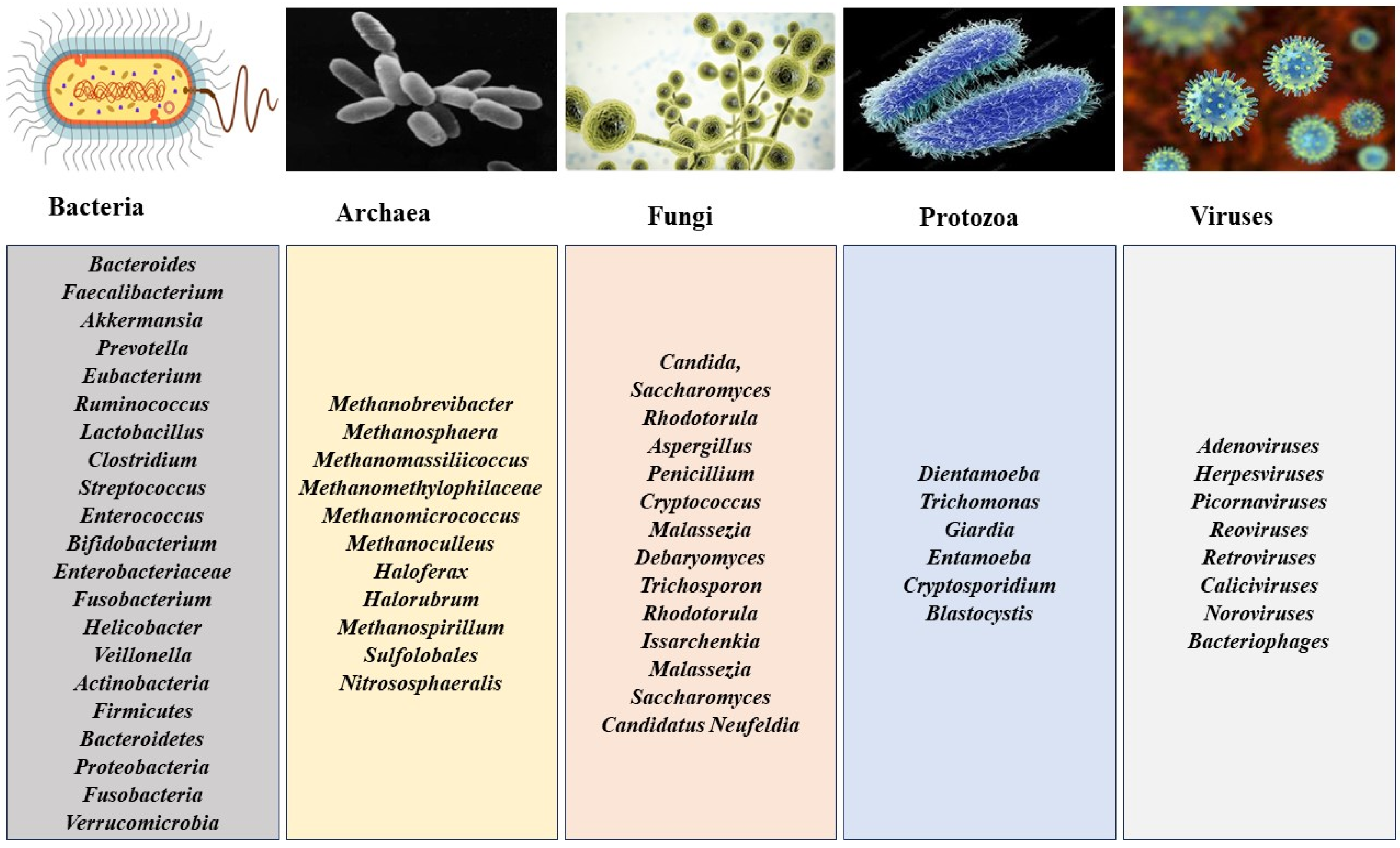
2. Gut Microbiota and Composition

The human gastrointestinal tract is a tube-like organ originating from the mouth, transversing the esophagus, stomach, and small and large intestines and terminating in the anus. These organs harbor a large number and numerous classes of microorganisms that make up the gut microbiota (Figure 1), including bacteria [23], archaea, fungi, protozoa, and viruses (virome), including bacteriophages [24,25,26,27,28,29,30,31,32]. Increasing knowledge of the gut microbiota is enhanced by advancements in biotechnological tools and shows that the viral population in the gut could easily match or exceed those of the bacteria, which is against earlier beliefs [32]. They consist of diverse viral types, including eukaryotic (10%) and prokaryotic viruses (90%) [32] and the DNA and RNA viruses [33]; however, the small amount of literature on gut RNA viruses could be attributed to their instability in cultures [34] and the individual specificity of the gut viral composition, taking into account location, lifestyle, diet, age, and other factors [32,35].

3. Functions of the Gut Microbiota

Studies have established the link between the gut microbiota and human health. Some essential functions of the microbiota include their effects on the host’s nutrient metabolism, immunomodulation, bioprotection against invading pathogens, effects on xenobiotics and drug metabolism, and maintenance of mucosal integrity.

3.1. Effects of Gut Microbiota on Host Metabolism

The microbial communities in the gut are implicated in the breakdown of complex macromolecules, such as carbohydrates and other polymers, into readily absorbable forms, yielding short-chain fatty acids (SCFAs) as the major products [36], which not only act as a source of energy to cells but perform other essential functions. In addition to the generation of energy to the hosts, they equally contribute to the prevention of the accumulation of toxic substances [37]; lipid metabolism, in the synthesis, biotransformation, and biodegradation of dietary lipids [38]; protein metabolism and the breakdown of undigested proteins in the distal part of the gut to generate important metabolites using the aid of their specialized enzymes [39,40,41,42]; the synthesis of vitamins, such as vitamin K [43]; the biotransformation of bile acids [44]; and the conversion of inactive phenolic compounds into biologically active compounds [45].

3.2. Immunomodulatory Potential of Gut Microbiota

The human immune system and the gut microbiota are in constant interaction, aided by having a more significant percentage of the human immune system and the microbiota residing in the gut environment. The diverse nature of the microbiota community in the gut is considered the most significant driver for the development and maturity of innate and adaptive immunity [46,47,48]. As part of innate immunity, the gut environment favors obligate anaerobes, especially members of the Bacillota and Bifidobacteriaceae, while discouraging/suppressing the growth of facultative anaerobic pathogens [49]. Also, the innate immune system uses pattern recognition receptors (PRRs) [50] to identify both microbial antigens [51], triggering an immediate immunological response [49]. In adaptive immunity, the gut microbiota stimulates the B-cells to produce copious amounts of antibodies [52] and promotes the differentiation of T-cells into the necessary arsenals of cellular immunity [53]. Further to these interactions, the flexibility and specificity of adaptive immunity are responsible for the recognition and subsequent destruction of potential pathogens or other foreign bodies, while selectively escaping the beneficial/commensal microbes. The relationship between gut microbes and the immune system is the foundation of immune homeostasis [54].

3.3. Bioprotection against Invading Pathogens

The gut microbiota co-evolves with the host and establishes a mutualistic relationship, with the host providing a stable habitat for microbial sustenance and the microbes providing the host with beneficial physiological functions essential for the overall well-being of the host [55]. The host’s protection from pathogenic infection is crucial through colonization resistance [56,57], which often deprives a potential pathogen of the leverage to colonize and establish infection. This bioprotective function can be achieved by several means: competition and depletion of nutrients starving off the pathogen(s), generation of inhibitory short-chain fatty acids (SCFAs) and secondary bile acids, interference with the expression of colonization factors (quorum-sensing signals), and the production of antibacterial peptides, e.g., bacteriocins or microcins [58].

3.4. Effect on Xenobiotics and Drug Metabolism

Xenobiotics, which are foreign exogenous or endogenous substances, when accumulated to toxic levels in the body, can expose the body to deleterious health challenges [59,60]. While exogenous xenobiotics include drugs, food additives such as artificial sweeteners, and inhaled pollutants such as pesticides, insecticides, and cosmetics, endogenous xenobiotics include bile acids, certain fatty acids, eicosanoids, and steroids [61]. The effects of the gut microbiota on xenobiotics/drug metabolism can be beneficial or harmful to the body. Although the liver is primarily charged with dissipating the xenobiotic residues in the body, the gut microbiota also influences xenobiotics/drug metabolism. The human genome project, which birthed the human microbiome project (HMP) [62], and the omics, especially pharmacomicrobiomics, produced significant findings on the importance of the gut microbiota on xenobiotics/drug metabolism [63,64]. Specifically, most orally administered hydrophilic drugs are converted to easily absorbable hydrophobic metabolites by the gut microbiota, enabling systemic circulation [65]. The influence on xenobiotics/drug metabolism can be by direct action on the drug or via the production of enzymes capable of drug biotransformation of the medications into more active, less active, inactive, or more toxic metabolites [66,67]. It can also indirectly influence drug biotransformation through modification of the amount of the gut/liver-produced metabolizing enzymes [68], the release of competitive metabolites competing with drugs for the same metabolizing enzymes [69], and the activation of inactive drug metabolites already secreted [70].

3.5. Protection of Mucosal Integrity

Maintaining the gut mucosal integrity is the core responsibility of the intestinal epithelial cells (IECs), which invariably play a vital role in maintaining the host’s health. The epithelial cells can segregate between the gut microbiota and the pathogen through sensing and microbial recognition, achieved through continuous crosstalk between the IECs and the gut microbiota [71]. The IECs, upon sensing, can secrete mediators leading to inflammation induction or immune tolerance via the stimulation or suppression of immunocompetent cells, respectively [71]. Disrupting the effective structural and functional equilibrium between the IECs and the gut microbiota can impair the mucosal integrity with attendant adverse effects [72,73,74].

4. Bacteriophages: Classification, Life Cycle, and General Mechanism of Action

Bacteriophages are a group of abundant nano-sized viruses that propagate by predating the bacterial population. They are structurally simple and often made up of the head or capsid, tail, and fiber regions. Bacteriophage classification (Table 1) can be performed based on three (3) significant characteristics: morphology, genomic properties, and life cycle. Regarding morphology, Caudovirales are the most common, prominent, and studied family and possess long, non-enveloped tails [75]. Their subdivisions are Myoviridae, with contractile, complex baseplate long flexible tails; Podoviridae, with short noncontractile tails; and Siphoviridae, with noncontractile and rigid/non-flexible tails [75]. Filamentous bacteriophages have long, flexible, and filament-like structures with the genetic material inside a protein coat that is part of the structure [76]. Tectiviridaes possess a “tectiviral”-appearing lipid envelope surrounding an icosahedral head [77]. Inoviridaes have long, flexible, helical, and icosahedral heads (encapsulating the genetic material) [78]. Leviviridaes (RNA phages) are small-sized and possess a single-stranded RNA genome in an icosahedral capsid [79]. Microviridaes are non-enveloped and small-sized and have a single-stranded DNA genome in an icosahedral capsid [80]. Pleolipoviridaes are pleomorphic; thus, they can be filamentous, spherical, or irregular and possesses a lipid-containing envelope [81]. Regarding genomic properties, DNA phages can be double-stranded DNA (dsDNA)-based genome phages or single-stranded DNA (ssDNA)-based genome phages [82]. Also, RNA phages can be double-stranded RNA (dsRNA)-based genome phages or single-stranded RNA (ssRNA)-based genome phages [83,84]. A specific group of RNA Phages, retroviruses, can convert their RNA genome into DNA via reverse-transcriptase enzymes, including DNA polymerase and ribonuclease H (RNase H) [85]. Circular replicating phages possess circular genomes and replicate independently of the host cell’s replication machinery using a circle replication mechanism, termed rolling circle replication (RCR) [86]. Temperate (pseudo-lysogenic) phages can be lytic, causing host cell death, or lysogenic, becoming dormant within the host after integration into the host DNA [8]. Finally, virulent phages are strict lytic phages that replicate within the host cell, eventually leading to cell lysis and the release of daughter phage particles [8]. Temperate, lysogenic, and lytic processes classify phages according to their life cycle [8].
Lysogenic phages replicate their genomes alongside the host bacterial chromosomes, either integrated into the host’s chromosome or in a free, plasmid-like state, forming a long-term stable coexistence with the host until induced by appropriate environmental conditions [87,88]. This state is also referred to as the prophage state, and they remain dormant until the induction of the lytic cycle. In the lytic cycle, the phage starts the production of new viral progeny immediately after infection and releases them by lysing the host [89]. A virulent or lytic phage subverts the cellular apparatus of its bacterial host for multiplication, typically culminating in cell lysis and the release of progeny virions [88]. The lysis–lysogeny transcriptional switch controls phage entry into the lytic or the lysogenic cycle and uses different mechanisms. For instance, Erez et al. [90] reported phages of Bacillus spp. using a peptide-based arbitrium communication system in deciding whether to enter the lytic or lysogenic life cycle (Figure 2).
The rate-limiting step in phage bacteria destruction, however, depends on the rate of adsorption onto the bacterial surface [91]. Also, the fate of the host bacteria is affected by the release of phage proteins, including holins and endolysins, which assist in compromising the bacterial cell wall externally or the internal breaking of the cell wall to release phage particles [92,93,94].

5. Impact and Roles in Maintaining the Balance of the Gut Microbiota

The human gut is densely populated with microorganisms at different concentrations that constantly interact [95]. Although the ratio of virotypes to species-level bacterial phylotypes in the ocean is determined to be greater by a factor of 10 (i.e., 10:1), in the gut, a closer 1:1 ratio is observed [96,97]. Compared with the bacterial makeup of the gut microbiome, little is still known about the composition and physiological function of the phage components (the phageome) in human gut populations [32]. Before now, owing to the limitations in techniques and toolkits available for the study of the phageome, much was not known in this field as only direct observation with the counting of virus-like particle (VLP) methods was prevalent for their investigation. These were performed with epi-fluorescence and transmission electron microscopy [32]. The isolation of bacteriophages infecting specific host strains in culture was also employed. With these microscope-based methods, the immense diversity of viral morphotypes per individual was used to estimate the total counts of bacteriophages in the colonic mucosa, cecal components, and fecal samples of humans. These were estimated to be between 109–1010 VLPs per gram, revealing the Caudovirales order, represented by Myoviridae, Podoviridae, and Siphoviridae as the most prevalent. However, these methods do not appropriately characterize the gut phageome, as many of the bacteria in the distal gut, such as Ruminococcaceae and Lachnospiraceae, are challenging to manage or culture in the laboratory [32,98,99]. Thus, the available collections of phage strains of human fecal samples do not reflect the true diversity of human gut bacteriophages. The complexity and abundance of human gut bacteriophage populations became more evident with emerging technologies such as high-throughput metagenomic sequencing technology [32,100]. The metagenomic analysis of fecal viromes carried out by Reyes et al. [97] showed that about 81–93% of bacterial viruses in the gut are novel and can be neither assigned a taxonomic position nor linked to a bacterial host [101]. Other studies on the gut phageomes of healthy humans also established that these viruses are specific to individuals and have minimal overlap [102]. Thus, substantial diversity of these phageomes exists between and among individuals; however, temporary stability is observed [97,103]. Minot and colleagues reported that most of the gut bacteriophages could persist for extended periods due to the lysogenic interactions with corresponding hosts, thus having a much slower evolution rate than the few lytic phages present [101,103]. Moreover, phages interact specifically with a single bacteria strain, and studies have also postulated that in the human gut, the phage–bacteria ratio is maintained at a ratio of 1:1 [101,104].
The imbalance in intestinal microecology could lead to several systemic diseases [105,106,107]. Phages are implicated in the balance or imbalance of the intestinal microecology and can affect human health either directly, by preying on the ecological landscape of the bacterial hosts, or indirectly, by influencing the immune system or metabolic pathway. Since the activities of phages alter the number and characteristics of the host bacteria, it is imperative to effectively and constantly regulate the relationship between these microbial species, as this not only keeps one in good health but can also reverse diseases [107].

6. Potential Therapeutic Applications

Many pathogenic and opportunistic bacteria are inherent in the gut system and have led to gut disease initiation, development, and advancement. Phage therapies are, however, developed and targeted at these bacteria. Potential applications of phages for preventive and curative purposes have produced single phages, cocktails, genetically modified phages, and even a combination with some other therapeutics, including antibiotics and probiotics [108]. The extensive literature review revealed that studies are developing therapies against the most prevalent bacteria associated with gut system infections, including Vibrio spp., Escherichia coli, Clostridioides difficile, Salmonella spp., Fusobacterium nucleatum, Shigella spp., Klebsiella pneumoniae, Listeria monocytogenes, Ruminococcus gnavus, and Campylobacter spp. Though most are still in the in vitro investigation, preclinical, and clinical trial stages, diverse potential applications have been found for many bacteriophage species, and they serve as potent therapeutic alternatives to managing multiple-drug-resistant strains [109]. These studies have enabled the identification and profiling of phages individually and in combinations using different dosage forms and delivery systems. The studies in the literature involving the therapeutic application of phages targeted against these implicated gut diseases are reported in the following section and summarized in Table 2.

6.1. Therapeutic Applications of Bacteriophages against Pathogenic Vibrio spp.

Considering the already established potential of phage therapy in gastrointestinal (GI) disorders, Jaiswal et al. [109] evaluated the in vitro and in vivo (using rabbits) therapeutic efficacy of a pure lytic vibriophage cocktail against the V. cholerae strain MAK 757 (ATCC 51352) that is implicated in cholera. The results of the lytic effects of individual vibriophages, B1, B2, B3, B4, and B5, applied singly and in combination as a cocktail showed synergistic effects, with the cocktail outperforming each of the individual phages. A comparison of the therapeutic management of orally induced V. cholerae infection in mice using a phage cocktail, conventional antibiotics (ciprofloxacin), and an oral rehydration system by Jaiswal et al. [110] showed that although the antibiotics had a significantly better anti-V. cholerae effect, the phage cocktail presented a significantly better safety and specificity profile and, thus, was more reliable in managing the infection. Jun et al. [111] showed a 74% (20 out of 27) lysis of multi-resistant Vibrio parahaemolyticus, a marine bacterium implicated in gastroenteritis and transmitted via raw oyster consumption, with optimal phage protein achieved following immediate phage therapy after infection. Yen et al. [112] utilized the potential prophylaxis of a cocktail of three virulent V. cholerae-specific phages, ICP1, ICP2, and ICP3, with specific effects against V. cholerae domiciled in the small intestine. From the in vivo results, the 24-h oral administration of the phages before cholera infection reduced intestinal tract colonization, thereby preventing cholera-like diarrhea. In addition to successfully demonstrating prophylaxis in V. cholerae-induced diarrhea, phage resistance was also not observed in the V. cholerae colonies.

6.2. Therapeutic Applications of Bacteriophages against Pathogenic Escherichia coli

Nasr-Eldin et al. [113] isolated and characterized highly stable Siphoviridae and Podoviridae phages for their lytic potential against Escherichia coli-causing gastrointestinal diseases using phage and E. coli incubation in a high saline environment. The characterizations, including the host range and synergistic profile, suggested that these bacteriophages were ideal candidates for therapeutic use. Similarly, Abdulamir et al. [114] targeted E. coli strains implicated in human gastroenteritis using a cocktail of 140 specific lytic phages administered to mice via their drinking water, oral injection, or vegetable capsules. The results revealed that the group receiving phage therapy via vegetable capsules obtained the least positive fecal cultures. While the peak reduction in E. coli was seen between 5–10 days post phage feeding, the second best-performing group following the phages’ administration was the group treated via the drinking water, with the study providing insight into the possible use of phage feed as a biocontrol for eliminating E. coli from animal intestines. Also, Abdelaziz et al. [115] isolated, characterized, and reported the broad-coverage lytic efficacy of phage phPE42 against E. coli clinical isolates implicated in gastrointestinal tract infections in an in vivo experiment. Bourdin et al. [116] acknowledged the downside of phage species and strain specificity and underlined the need for page therapies with broad host ranges. Also, the need to develop better-targeted phage therapies for disease conditions is needed. In treating childhood diarrhea-associated E. coli infection, they obtained and tested the lytic effects of 89 T4-like phages against four (4) batches of E. coli isolates. The result revealed that specific phage therapies for the tested pathogens were difficult and complex owing to the geographical, epidemiological, and time differences, thus recommending the need to identify flexible and specific therapeutic phages. With over 80% of traveler (TD) and childhood diarrhea cases caused by a variety of enteropathogens, and with multi-resistant E. coli usually responsible for about 30–40% [117], Aleshkin et al. [117] developed and assayed a phage cocktail for prophylaxis against TD caused by E. coli, Shigella flexneri, Shigella sonnei, Salmonella enterica, Listeria monocytogenes, and Staphylococcus aureus, and obtained specific prevention effects against TD caused by E. coli. In a similar experiment, Vahedi et al. [118] assayed the potential of combining a specific bacteriophage and antibiotics targeted against enteropathogenic E. coli (EPEC), using both in vitro and in vivo models. The in vitro study showed that 106 PFU/ml of the phage eliminated EPEC from infected HEp-2 cells. In vivo, administration of the phage:antibiotic combination presented a total reduction in EPEC after 24 h and is attributed to the potentiation effect of both the antibacterial agent and the phage. However, slight weight loss was observed in the mice, possibly due to the adverse impact of antibiotics on the microbiota. However, Sarker et al. [119], in their work against acute bacterial diarrhea in children, used safe, orally administered species-specific T4-like coliphages in human subjects and showed no improvement in diarrhea symptoms, which was attributed to insufficient phage coverage and low E. coli titers. However, they assume that higher oral phage doses might be necessary to obtain the desired outcome, which triggers the need for more knowledge using in vivo phage–bacterium interaction strategy to understand E. coli propagation in childhood diarrhea. Galtier et al. [120] utilized three virulent bacteriophages in therapy against the colonization of adherent invasive E. coli (AIEC) strain LF82 implicated in inflammatory bowel diseases, Crohn’s disease, and ulcerative colitis and showed sufficient phage replication in the ileum, cecum, and colon following murine gut analysis. A single day of treatment with the bacteriophages administered to LF82-colonized AIEC strain CEABAC10 transgenic mice, which express the human carcinoembryonic antigen-related cell adhesion molecule 6CEACAM6 glycoprotein receptor for AIEC, revealed a notable decrease in the AIEC count. Over two weeks of continuous treatment resulted in the absence of colitis symptoms in mice colonized with the bacterial strain. Cieplak et al. [121] demonstrated the safety of phages in relation to their ability to induce dysbiosis following a comparison with antibiotics in the in vitro decolonization of E. coli populations in the small intestine. From the study, unlike the synthetic drug, the phage preparation had a targeted lytic effect on the E. coli populations. It impacted no other commensal bacteria used in the study, thus supporting its application in personal medicine, as characterized by its targeting of the bacteria of interest and evasion of dysbiosis induction. The overall efficacy of an ascertained safe commercial phage cocktail, PreforPro®, on the gut microbiota and markers of intestinal and systemic inflammation in a healthy human population was studied by Gindin et al. [122]. Twenty-eight (28) days of phage consumption did not alter the normal gut microbiota of most individuals but significantly reduced the target E. coli population and the pro-inflammatory cytokine interleukin 4 (Il-4) responsible for inflammatory reactions in the gastrointestinal tract. Shiga-toxin-producing E. coli is implicated in severe, difficult-to-treat infections. The safety and tolerability of PreforPro® were reported by Grubb et al. [123]. They demonstrated the possible enhancement of the combined impacts of a probiotic microorganism and phage (Bifidobacterium lactis BL04 + PreforPro®) on gastrointestinal discomfort and stool consistency in a healthy adult population. They presented significant improvement in gastrointestinal symptoms over four weeks of therapy administration without disruption of the gut microbiota. There was also an associated increase in the relative abundance of some microorganisms, including Lactobacillaceae. Thus, the study suggests a strong connection between phage and the use of probiotics in improving the microbiota of the intestinal environment. The study by Alomari et al. [124] also further supported the simultaneous administration of bacteriophages and probiotics. They administered Lactobacillus spp. and phages of pathogenic E. coli combinations in suppositories in treating diarrheal calves. They reported a reduction in the diarrheal symptoms following therapy, complete elimination 24–48 h post-therapy, and a significant increase in the body weight of the treated calves compared with the control. Hsu et al. [95] proposed the use of genetics-based anti-virulence mechanisms in neutralizing the expression of the bacterial toxin and minimizing resistance as a better opinion to the conventional antibacterial approach. Unlike the conventional mechanism of bacterial lysis, temperate phages can be genetically engineered and integrated into the bacterial chromosome, and they are capable of neutralizing targeted gut bacterial toxins, impeding the virulence factors by modifying bacterial function at the genetic level; thus, they are good candidates for this therapy [125]. Hsu et al. [95] and Hsu et al. [125] utilized temperate phages capable of self-integration into the bacterial genome in in vivo and in situ studies, respectively, and both reported significant repression of the Shiga toxin secretion from E. coli in the mammalian gut. Cepko et al. [126], using a mouse model, isolated a strictly lytic phage that kills strains of enteroaggregative E. coli associated with both acute and chronic diarrhea [127]. A single dose of the phage one day post-infection-administration significantly reduced the bacterial count without altering microbiota diversity. Green et al. [128] explored a location-targeting mechanism aimed at enhancing the phage specificity and lytic cycle and treating gut infection, having observed that invasive pathobionts could reside deep within the mucosal epithelium of the gastrointestinal tract. With the ability to bind to heparan-sulfated glycans on the epithelial surface, the bacteriophage can position itself close to the target host. Here, phage HP3 showed lytic activities toward E. coli ST131 in vitro in a murine sepsis model. However, it proved ineffective in similar activities in the murine intestinal tract. The absence of lytic abilities in the latter was attributed to the intestinal mucins. Under a simulated intestinal environment, a podovirus phage isolated from wastewater, while not altering the intestinal microbiota compared with antibiotics, proved to be more effective due to its inherent ability to bind to heparan-sulfated proteoglycans on the surface of intestinal epithelial cells, thus strongly suggesting this feature to be responsible for the targeted lytic activity against the host bacterium.

6.3. Therapeutic Applications of Bacteriophages against Pathogenic Clostridioides Difficile

As an alternative to the use of antibiotics in treating GIT dysbiosis due to C. difficile infection (CDI), Nale et al. [129] assayed individual phages and a phage cocktail containing different phage combinations for their synergistic potential in the adequate clearance of C. difficile. Results obtained after 36 h post-infection supported the potential application of phage combinations for the targeted eradication of CDI and also concluded that specific phage combinations caused the complete lysis of C. difficile in vitro and prevented the appearance of resistant strains. Selle et al. [130] attempted to repurpose the endogenous type I-B CRISPR-Cas system in C. difficile as an antimicrobial agent through the use of bacteriophage capable of expressing a self-targeting CRISPR that redirected endogenous CRISPR-Cas3 activity against the bacterial chromosome and demonstrated that a recombinant bacteriophage expressing bacterial-genome-targeting CRISPR RNAs had significant lytic activities against C. difficile in both in vitro and mouse models. The study suggested that phage-delivered programmable CRISPR therapeutics have the potential to increase safety, specificity, and efficacy in complex gut microbial communities and offer a novel mechanism for the treatment of gut pathobionts.

6.4. Therapeutic Applications of Bacteriophages against Pathogenic Salmonella spp.

Using animal models, Dallal et al. [131] analyzed phage SE20 active against Salmonella enteritidis, a Gram-negative bacterium that occurs mainly in human gastroenteritis and is often implicated in salmonellosis, a disease commonly caused by the ingestion of animal-derived products, mainly poultry products (meat and eggs), that are significant carriers of Salmonella spp. [132]. The in vivo study revealed that a single dose (2 × 10⁹ PFU/mL) of the phage isolate provided a targeted and prophylactic effect against S. enteritidis. Additionally, while there was no bacterial resistance over twelve months of observation, compared with the animal groups receiving phage therapy, the test synthetic antibiotic used caused apparent weight loss in the administered experimental mice. Moye et al., 2019 [133] demonstrated that the direct ingestion of phages against Salmonella could enhance intrinsic gut resilience and provide protection against Salmonella-induced foodborne diseases. In their study, Simulator of the Human Intestinal Microbial Ecosystem (SHIME), a system aimed at exploring the potential of phage cocktails termed foodborne outbreak pills (FOPs) to eliminate foodborne pathogens and maintain the balance of the host microbiome, effectively depopulated the Salmonella without any distortion of stability of the gut microbiota. Thanki et al. [132] also reported that an increase in phage dosage resulted in the proportional and effective control of colonization by Salmonella spp. Using poultry and swine assays in vitro and in vivo, Nale et al. [134] determined the potential of twenty-one myoviruses and one siphovirus in eliminating Salmonella. Individual phages significantly reduced the growth of test isolates within six hours post-infection and the subsequent phage administration. However, bacterial regrowth within an hour following treatment suspension was reported, indicative of bacterial resistance to phage therapy. A novel three-constituent phage cocktail was employed in vitro for its lytic efficacy in an optimized Galleria mellonella larva model infected with Salmonella to remedy the resistance. Comparatively to the individual phages, the cocktail had broader bacterial range coverage, improved lytic efficiency, and prevented the emergence of resistant strains. The study is further supported by Pelyuntha et al. [135] in their comparison of the lytic profile of individual phages and a phage cocktail against Salmonella colonization, implicated in the broiler gastrointestinal tract, to enhance poultry consumption safety. Pronounced synergistic enhanced lytic activities and evasion of resistance were obtained with the cocktail compared with the individual agents. The study also affirmed that phages, generally considered safe by the FDA and specific in action, remain potential ideal biocontrol agents for bacteria colonization and biofilm formation in various edible products.

6.5. Therapeutic Applications of Bacteriophages against Pathogenic Fusobacterium nucleatum

Pro-tumoral F. nucleatum is significant in advancing colorectal cancer and potentially influences the therapeutic response [136]. By incorporating the principles of nanotechnology in a strategic attempt at gut microbiota manipulation, Zheng et al. [136], in demonstration of phage-guided nanotechnology and the potential to control F. nucleatum colonization in the gut, drastically improved the treatment of colorectal cancer. The oral or intravenous administration of irinotecan-loaded dextran nanoparticles covalently linked to azide-modified phages inhibited the growth of F. nucleatum and thus enhanced the effectiveness of first-line chemotherapy therapies for cancer. Similarly, Dong et al. [137] formulated F. nucleatum-binding M13-phage-loaded silver nanoparticles (AgNPs) to achieve targeted clearance of F. nucleatum and remodeling of the tumor-immune microenvironment. The in vitro and in vivo studies showed efficient eradication of the bacteria from the gut. Also, significant suppression of the myeloid-derived suppressor cells at the tumor site and the activation of antigen-presenting cells by the M13 phages were observed. These immunomodulatory activities boosted the capacity of the host immune system for colorectal cancer suppression.

6.6. Therapeutic Applications of Bacteriophages against Pathogenic Shigella spp.

Shahin et al. [138] determined the efficacy and specificity of individual Shigella-specific bacteriophages (vB_SflS-ISF001 and vB_SsoS-ISF002) and a cocktail of both. The phage preparations were investigated against multidrug-resistant Shigella sonnei and Shigella flexneri isolates. The individual bacteriophages showed high lytic activity in about 75% of the isolates. However, the phage cocktail inhibited 85% of the isolates, indicating higher effectiveness and specificity against a wide range of ESBL-positive and -negative isolates of S. sonnei and S. flexneri.

6.7. Therapeutic Applications of Bacteriophages against Pathogenic Klebsiella pneumoniae

Gut commensals like K. pneumoniae opportunistically worsen gut conditions such as inflammatory bowel diseases (IBDs). Federic et al. [139] identified multi-resistant K. pneumoniae strains strongly associated with the exacerbation of gastrointestinal disease following enhancement of intestinal inflammation in colitis-prone, germ-free mice challenged with IBD-associated K. pneumoniae strains. Stepwise production of lytic cocktails of five-phage targeting strains enabled the effective control of the bacteria in vivo and further supports the use of phage combination therapy in addressing resistance and generally managing gut disease-contributing pathobionts. The lytic effect of some commercially available bacteriophage preparations on strains of K. pneumoniae isolated from infants with functional gastrointestinal disorders (FGIDs) was assessed by Grigorova et al. [140] via the drip method and according to clinical recommendations. However, low-level lytic activities and sensitivity to K. pneumoniae correlated with age. Significant levels of lysis were observed in children of three to six months but still reflected the inefficiency of this therapy in eliminating K. pneumoniae from the intestinal microbiota of children with FGID and suggested that more ingenious and radical approaches to ensuring complete eradication of the associated K. pneumoniae are needed.

6.8. Therapeutic Applications of Bacteriophages against Pathogenic Listeria monocytogenes

L. monocytogenes is a facultative anaerobic Gram-positive bacterium prevalently implicated in foodborne and gastroenteritis diseases [141]. A phage cocktail designated as a foodborne outbreak pill (FOP) and targeted at the implicated L. monocytogenes was evaluated by Jakobsen et al. [142] in simulated small intestine, large intestine, and Caco-2 models. Extensive inhibition of L. monocytogenes with results comparable to that of a standard drug (ampicillin) was reported. Strikingly, unlike ampicillin, the FOP did not inhibit commensal bacteria in the small intestine, significantly and selectively lysing the L. monocytogenes population while being stable in the gastric environment. Furthermore, the FOP prevented the invasion and adhesion of L. monocytogenes through a Caco-2 monolayer. Generally, the study highlighted the essential health benefits of phage in this regard and their delivery as dietary supplements, enhancing natural defenses of the gastrointestinal tract against specific foodborne pathogens.

6.9. Therapeutic Applications of Bacteriophages against Pathogenic Ruminococcus gnavus

The bacteria Ruminococcus gnavus, a Gram-positive anaerobe, is also widely prevalent in the microbiota of humans with inflammatory bowel disease conditions due to Crohn’s disease [143]. Buttimer et al. [144] isolated, characterized, and analyzed six phages that infect the R. gnavus JCM 6515T strain. Although no significant decrease in the bacterial count was reported post-phage administration, the study provided insight into two significant mechanisms through which phages interact with R. gnavus in the human gut microbiome.

6.10. Therapeutic Applications of Bacteriophages against Pathogenic Campylobacter spp.

Campylobacter, a major component of the gut microbiota, especially in birds and livestock, is a major foodborne and diarrheal disease-causing bacterial species [145]. D’Angelantonio et al. [146], in their study against Campylobacter jejuni, demonstrated colony reduction in a broiler before slaughter, following a two-step phage administration process involving two double-stranded phages (Φ 16-izsam and Φ 7-izsam) belonging to the Caudovirale order. A 0.1 MOI of Φ 16-izsam was administered to a broiler group on the 38th day of rearing, while Φ 7-izsam at an MOI of 1 was administered on the 39th day to another group; these showed a significant one to two log reduction in C. jejuni counts on the cecal content compared with the control group after sacrificing the birds on the 40th day. The lowest colony count was, however, observed with an MOI of 0.1 of Φ 16-izsam. Also, Nowaczek et al. [147] isolated 48 strains from 140 broiler chickens (31 Campylobacter jejuni and 17 Campylobacter coli), which exhibited varying and high-level multi-resistance to the selected antibiotics ciprofloxacin, erythromycin, gentamicin, and tetracycline. They further identified and characterized bacteriophages, including bacteriophages φ4, φ44, φ22, φCj1, φ198, and φ287, placed in the Myoviridae and Siphoviridae of Caudovirales order, and demonstrated the susceptibility of a significant number of the Campylobacter spp. to the phage isolates, which had a lytic spectrum of 6, 4, 4, 3, 8, and 7, respectively.

6.11. General

Amidst the substantial efforts at identifying effective phages and their cocktails against bacteria-implicated gastrointestinal disorders, it remains desirable to develop effective delivery systems capable of protecting and stabilizing phage particles and products from degradation, destruction, and inactivation under the gastrointestinal tract’s acidic conditions. A smart biocontrol chitosan-encapsulated bacteriophage cocktail formulation was employed by Rbahimzade et al. [148]. The formulation was evaluated as a prophylactic and treatment option for gastrointestinal infections, specifically diarrhea. Experimental animals were challenged with S. enterica, Shigella flexneri, and E. coli, after which treatment commenced with the formulation. Findings reveal that phage encapsulation protected therapeutic life forms from enzymatic degradation, with the non-treated experimental animals experiencing weight loss. However, the highest lytic activity was obtained three days post-phage treatment compared with other studies, where lytic activities took seven to ten days to become evident.
Table 2. Therapeutic applications in the management of certain bacteria-implicated gut diseases.
Target PathogenPhage TherapyDiseaseStudy Type, ModelReportsReferences
Vibrio spp.Cocktail of five (5) lytic Vibrio phagesFighting V. cholerae infectionIn vivo, rabbitsOral administration of cocktails before infection resulted in prophylactic effects. The phage cocktail significantly reduced bacterial load 6 and 12 h after the challenge.Jaiswal et al. [109]
Oral phage cocktail therapyV. cholerae infectionIn vivo, micePhage cocktail (108 PFU/mL) given once daily significantly reduced bacterial load.Jaiswal et al. [110]
Bacteriophage pVp-1Multiple antibiotic-resistant V. parahaemolyticus implicated in gastroenteritisIn vivo, miceProtection from infection and death 1 h after inoculation with V. parahaemolyticus.Jun et al. [111]
Cocktail of three (3) lytic (virulent) phages—ICP1, ICP2, and ICP3Cholera pathogenesis/cholera-like diarrheaIn vivo, infant mice and rabbitsEffective at preventing mouse small intestinal colonization. Prophylaxis against the onset of cholera-like diarrhea was achieved after oral administration of the phages up to 24 h before V. cholera infestation.Yen et al. [112]
Escherichia coliCocktail of three (3)
bacteriophages from Siphoviridae and Podoviridae
E. coli implicated in
gastrointestinal diseases
In vitroPhage cocktail exhibited broad spectrum and strong lytic activity against E. coli isolates.Nasr-Eldin et al. [113]
Cocktail of lytic phages specific against E. coliGut pathogenic E. coliIn vivo, miceSuppression of E. coli was observed 5–10 d after phage therapy.Abdulamir et al. [114]
Lytic Myoviridae phage phPE42Extensively drug-resistant (XDR) E. coli implicated in foodborne infectionsIn vivo, ratsEffective eradication of XDR E. coli was observed in animal feces.Abdelaziz et al. [115]
T4-like phagesChildhood diarrhea-associated E. coli isolatesIn vitro, culturesT4-like phages combined in a cocktail resulted in increased bacterial lysis.Bourdin et al. [116]
Phage-based probiotic dietary supplement
consisting of 7 bacteriophage strains
Traveler’s diarrhea (TD) caused by E. coli, S. flexneri, S. sonnei, S. enterica, L. monocytogenes, S. aureusClinical study,
in vivo, humans and mice
Prophylactic effect against TD.Aleshkin et al. [117]
Specific bacteriophageEnteropathogenic E. coli (EPEC)In vivo, miceA single dose of the phage rendered a protective effect on the bacteria throughout the study.Vahedi et al. [118]
T4-like coliphagesAcute bacterial diarrheaClinical trial, humansFailure to improve diarrhea condition, possibly due to insufficient phage concentration.Sarker et al. [119]
Virulent bacteriophages targeting prototype of the Adherent Invasive E. coli (AIEC) strain LF82Crohn’s disease (CD)Ex vivo, in vivo, murine and human intestinal samplesThree virulent bacteriophage cocktails were active against the AIEC strain LF82. A single dose of the cocktail reduced colitis symptoms in mice colonized with AIEC.Galtier et al. [120]
Bacteriophage cocktail Ec17B153DK1 vs. the broad-spectrumantibiotic ciprofloxacinE. coli infecting the gut environmentIn vitro, simulated small intestine systemThe cocktail was effective in reducing E. coli in simulated gut conditions. No impact on commensal, non-targeted bacteria.Cieplak et al. [121]
Commercial cocktail of E. coli-targeting bacteriophages (PreforPro®) containing four phages (LH01-Myoviridae, LL5-Siphoviridae, T4D-Myoviridae, and LL12-Myoviridae)Effect on gut microbiota during GI distress and markers of intestinal and systemic inflammationClinical trial, humansThe potential of bacteriophages to selectively reduce target organisms without causing dysbiosisGindin et al. [122]
Supplemental bacteriophages (PreforPro®)Enhance the effects of a common probiotic, B. animalis subsp. Lactis (B. lactis) on GI healthClinical study, humansImprovements in GI inflammation and colon pain in individuals consuming B. lactis with PreforPro®.Grubb et al. [123]
Suppository containing probiotic strains of Lactobacillus spp. and bacteriophages specific for pathogenic E. coliDiarrheaIn vivo, calvesProbiotic-phage suppositories reduced the duration of diarrhea in calves. The complete stopping of diarrhea was observed 24–48 h after use.Alomari et al. [124]
Lytic phages (T4, F1, B40-8, and VD13
phages)
Effect on mice gut colonized with human commensal bacteriaIn vivo, gnotobiotic miceTargeted lysis of susceptible gut bacteria. Modulation of non-targeted bacteria through interbacterial interactions.Hsu et al. [95]
Genetically engineered temperate phagesShiga-toxin (Stx)-producing E. coli colonizing the mammalian gutIn vivo, miceSignificant repression of fecal Stx concentrations. Suppression of virulence factors in gut bacteria.Hsu et al. [125]
Phage PDX, a member of the Myoviridae familyDiarrheagenic enteroaggregative E. coli (EAEC)In vitro, in vivo, cultures and miceBacteriolytic activity of EAEC isolates (EN1E-0007) in vitro and in vivo. No dysbiosis was observed in the anaerobic culture.Cepko et al. [126]
Phage ES17, a Podoviridae phageExtraintestinal pathogenic E. coli (ExPEC) in the intestineIn vivo, miceSelective elimination of invasive pathobiont species from mucosal surfaces in the intestinal tract.Green et al. [128]
Clostridium difficileSix (6) myoviruses and one (1) siphovirusC. difficile infection (CDI)In vitro, in vivo, hamstersSpecific phage combinations resulted in total lysis of C. difficile in vitro. Prevention of resistance. In vivo, the evaluation revealed a reduction in C. difficile colonization 36 h post-infection.Nale et al. [129]
Recombinant bacteriophageC. difficile infectionIn vitro, in vivo, cultures and miceTargeting and killing of C. difficile.Selle et al. [130]
Salmonella spp.Phage SE20 (Podoviridae)S. enterica serotype EnteritidisIn vitro, in vivo, miceOral administration of a single dose of bacteriophage protected against salmonellosis and treatment of salmonellosis. Animals developed hepatomegaly and splenomegaly as side effects but had no gastrointestinal complications with the phage therapy.Dallal et al. [131]
Bacteriophage cocktail (foodborne outbreak pill (FOP) targeting E. coli O157:H7, L. monocytogenes, and
Salmonella)
Salmonella infectionIn vitroSimulator of the Human Intestinal Microbial Ecosystem (SHIME).Moye et al., 2019 [133]
Phage cocktailSalmonella colonization in experimentally challenged birdsIn vivo, birdsPhage treatment effectively reduced Salmonella colonization and enhanced growth performance weight gains in challenged birds.Thanki et al. [132]
Myoviruses and a siphovirusSalmonella infection gastrointestinal enteritisIn vitro, in vivo, swine, birds, culturesPhage cocktail (STW-77 and SEW-109) had the most lysing efficacy on the swine and bird models. Some phages from the cocktail could lyse resistant strains of the organism.Nale et al. [134]
Salmonella phages (vB_SenS_KP001,
vB_SenS_KP005, and vB_SenS_WP110)
Salmonella colonization in the gastrointestinal tract of broilersIn vivo, broilersThe phage cocktail reduced Salmonella colonization in broilers’ gastrointestinal tracts from over 70% to 0% 4 d post-treatment.Pelyuntha et al. [135]
Fusobacterium nucleatumIrinotecan-loaded dextran nanoparticles covalently linked to azide-modified phages.Colorectal cancer (CRC)In vivo, micePhage administration inhibited the growth of F. nucleatum. It significantly boosted the effectiveness of first-line chemotherapy treatments for CRC.Zheng et al. [136]
F. nucleatum (Fn)-binding M13-phage-loaded silver nanoparticles (AgNPs)Symbiotic F. nucleatum in the gut selectively increases immunosuppressive myeloid-derived suppressor cells
(MDSCs), thereby promoting colorectal cancer (CRC) progression.
In vitro, in vivo, miceTreatment with M13-phage-loaded AgNPs could mop up F. nucleatum in the gut, resulting in non-amplification in MDSCs at the tumor sites.Dong et al. [137]
Shigella spp.Shigella-specific bacteriophages: vB_SflS-ISF001, vB_SsoS-ISF002, and a cocktail of bothS. sonnei and S. flexneri causing human acute gastrointestinal infectionsIn vitro, culturesMore than 85% of the ESBL-positive and -negative isolates of S. sonnei and S. flexneri were inhibited by the phage cocktail (vB_SflS-ISF001 and vB_SsoS-ISF002.)Shahin et al. [138]
Klebsiella pneumoniaeLytic five-phage combinationInflammatory bowel disease (IBD)-associated K. pneumoniae (Kp) strainsIn vivo, miceSuppression of colitis in miceFederic et al. [139]
Commercial bacteriophage preparationsK. pneumoniae strains isolated from children with functional gastrointestinal disorders (FGIDs)In vitro, spot testPhages show negligible lytic activity, indicating the need for a more radical approach to eradicating K. pneumoniae in children with FGIDs.Grigorova et al. [140]
Listeria monocytogenesBacteriophage cocktail (Foodborne Outbreak Pill (FOP))L. monocytogenesIn vitro, simulated ilium and colon conditionsProtection against L. monocytogenes infecting the human gastrointestinal tract without causing dysbiosis.Jakobsen et al. [142]
Ruminococcus gnavusSix bacteriophagesMucin-degrading bacterium R. gnavus from the human gutIn vivo, miceResults show the coexistence of phages with R. gnavus in the human gut microbiome.Buttimer et al. [144]
Campylobacter spp.Double-stranded phages (Φ 16-izsam and Φ 7-izsam)C. jejuni associated with broilersIn vivo, broilersPhage administration showed a significant one to two log reduction in C. jejuni counts on the cecal content compared with the control group after sacrifice. The lowest colony count was, however, observed with an MOI of 0.1 of Φ 16-izsam.D’Angelantonio et al. [146]
Bacteriophages φ4, φ44, φ22, φCj1, φ198, and φ287C. jejuni associated with broilersIn vitro, in vivo, broilersDemonstrated the susceptibility of a significant number of the multi-resistant Campylobacter spp. to the phage isolates, which had a lytic spectrum of 6, 4, 4, 3, 8, and 7, respectively.Nowaczek et al. [147]
GeneralChitosan-encapsulated bacteriophage cocktailS. enterica, S. flexneri, and E. coli gastrointestinal infectionsIn vivo, ratsReduction in positive cultures from stools of the group receiving the chitosan-encapsulated bacteriophage cocktail was observed after two days.Rahimzade et al. [148]

7. Roles in Preventing and Treating Specific Gut Microbiota-Related Diseases

Many benefits are associated with the use of bacteriophages in the treatment of bacterial-related diseases in comparison with conventional antibiotics. Apart from being safe, bacteriophage actions are specific and, thus, retain the homeostatic nature of the gut microbiota following their use. Also, since they hijack the host’s replicative machinery within the host, there is usually no need for successive administrations, as they utilize the host for their propagation until their exhaustion, at which point the therapeutic aim is achieved [149,150]. They play different roles in preventing and treating specific gut microbiota-related diseases via the different mechanisms described above and summarized in Figure 3. In controlling gastrointestinal diseases, phages can infect, reduce bacterial loads, and eliminate implicated pathogens, including antibiotic-resistant ones. They also aid in restoring the gut microbiota, alleviating symptoms, and promoting healing. The gastrointestinal tract’s inflammatory burden, including ulcerative and Crohn’s disease, can be managed through microbiota modulation, biofilm disruption, and the control of proinflammatory bacterial flare-ups [120]. Dysbiosis interrupts the bacterial population with a consequential impact on metabolic processes, including glucose and fatty acid metabolism, adiposity leading to obesity, non-alcoholic fatty liver, and heart diseases [151]. Bacteriophage-aided gastrointestinal tract microbiota optimization is instrumental for efficient metabolic conditions. The bacteriophages’ ability to modulate both the innate and adaptive immune system is a veritable tool in the fight against cancer [152] and opens up a new frontier for their possible application in diagnosing and treating cancer [153]. Also, certain bacteria species have been significantly linked to the development of some forms of gastrointestinal cancers. Typically, colorectal and pancreatic cancers have significantly been linked to certain bacteria, including F. nucleatum and Porphyromonas spp., for which studies have reported bacteriophages with anti-colorectal and -pancreatic cancer potentials, with promising results [154,155,156]. Finally, bacteriophages contribute to the function of the gut–brain axis communication as it relates to gut microbiota regulation, immunomodulation, and metabolite production and have applicable uses against neurological disorders [157]. For example, Ghadge et al. [158] demonstrated a reduction in motor neuron loss, microgliosis, superoxide dismutase 1 (SOD1) burden and aggregation, and astrocytosis through the use of phage-specific single-chain variable fragment antibodies (scFvs) raised against SOD1G93A in mice.

8. Conclusions

Lytic bacteriophages are abundant inhabitants within the gut microbiota, and they are engaged in constant interactions with gut-associated bacteria, which act as their hosts, ensuring their propagation. These interactions have far-reaching implications for human health, as phages play essential roles in the gut system and overall well-being. Extensive research has implicated them in managing various gut-related diseases, though most of these findings are still in the early stages of development. Translating these research outputs into clinical applications poses a complex challenge, necessitating the synergistic engagement of related stakeholders in initiating and evaluating clinical trials, advancing research, and investigating potential clinical applications and commercialization. The significance of understanding the roles of bacteriophages in the gut and human health cannot be overstated.
One crucial aspect is their role in effectively regulating gut microbiota composition. Phages target specific bacterial species, either promoting the growth of beneficial bacteria or controlling the overgrowth of harmful ones. This delicate balance is vital for maintaining a healthy gut microbiota, which is essential for various physiological functions and overall health. Nutrient digestion and absorption are intricately linked to the gut microbiota’s activities, and bacteriophages play a significant role in this process. The gut microbiota breaks down complex carbohydrates, synthesizes specific vitamins, and aids nutrient absorption. Phages’ influence on the gut bacterial community affects these processes, which can have implications for nutrient availability and utilization by the host. Imbalances in the gut phage–bacterial community could lead to inefficient digestion and nutrient absorption, potentially contributing to malnutrition or other health issues. Another critical aspect of gut phage–bacterial interactions is their impact on the modulation of the immune system. The gut is a critical interface between the external environment and the body’s internal systems. The gut microbiota, including bacteriophages, is pivotal in educating and shaping the immune system and can influence the bacterial communities in the gut, leading to changes in immune responses.
A well-balanced gut phage–bacterial community can help prevent inappropriate immune reactions, reducing the risk of autoimmune disorders and allergies. They also hold the potential as a selective tool to combat gut bacterial pathogens, particularly multi-resistant ones. The rise of antibiotic-resistant bacteria has become a significant health concern worldwide, necessitating the exploration of alternative treatment options. Phage therapy, which involves using specific phages to target and kill pathogenic bacteria, offers a promising approach to addressing this challenge. Understanding the intricate interactions between bacteriophages and gut bacteria is crucial for developing personalized and targeted phage therapies that are both safe and effective.
Furthermore, dysbiosis, an imbalance in the gut phage–bacterial community, has been associated with various diseases, including inflammatory bowel diseases (IBDs), gut-related cancers, and metabolic disorders. Research on phages and their role in managing dysbiosis-related diseases holds immense potential for providing insights into disease development and potential therapeutic strategies. The intrinsic interactions between bacteriophages and host bacteria are a foundation for advancing personalized and targeted phage therapies. This emerging field of research aims to utilize phages as precision medicine tools, where specific phages are selected based on individual gut microbiota profiles and health conditions. Such personalized therapies have the potential to revolutionize disease treatment, particularly in cases where traditional treatments have shown limited efficacy.
In conclusion, understanding the roles of bacteriophages in the gut and human health is of utmost importance. These tiny viral agents play pivotal roles in regulating gut microbiota composition, nutrient digestion, immune system modulation, and protection against pathogens. Their potential in managing gut-related diseases, including dysbiosis, offers promising avenues for future therapeutic interventions. Collaboration among various stakeholders is essential to translating research findings into clinical applications. By harnessing the power of bacteriophages, we can unlock new and innovative approaches to improve human health and combat challenging diseases associated with the gut microbiota.

Author Contributions

Conceptualization, S.C.E., C.V.O., O.C.E., D.P.B. and E.B.O.; writing—original draft preparation, S.C.E., C.V.O., O.C.E., C.F.K. and D.P.B.; writing—review and editing, S.C.E., C.V.O., O.C.E., C.F.K., D.P.B., E.B.O., M.M.D.C.V., V.M.B. and A.A.A. All authors have read and agreed to the published version of the manuscript.

Funding

This research was funded by São Paulo Research Foundation (FAPESP), grant 2022/10775-9 (Project PsgPhageKill). Funding was also provided by FCT/MCTES to CESAM (UID/AMB/50017/2019). V.M.B. was visiting researcher fellow from FAPESP (2018/05522-9, Project PsaPhageKill) and received a research fellowship award from the National Council for Scientific and Technological Development (CNPq) (grant 301978/2022-0). We also acknowledge TETFund (Nigeria) for the AST&D benchwork (at VBlab—Laboratory of Bacterial Viruses, University of Sorocaba, 18023-000 Sorocaba/SP, Brazil) scholarship granted to S.C.E.

Institutional Review Board Statement

Not applicable.

Informed Consent Statement

Not applicable.

Data Availability Statement

Data sharing not applicable.

Acknowledgments

The authors fully acknowledge Ebele B. Onuigbo and Anthony A. Attama, who are both co-authors (E.B.O and A.A.A.) in this paper and progenitors of phage research at the Faculty of Pharmaceutical Sciences, University of Nigeria, Nsukka and who are jointly driving the burgeoning research in this area.

Conflicts of Interest

The authors declare no conflict of interest.

References

  1. Vemuri, R.; Shankar, E.M.; Chieppa, M.; Eri, R.; Kavanagh, K. Beyond Just Bacteria: Functional Biomes in the Gut Ecosystem Including Virome, Mycobiome, Archaeome and Helminths. Microorganisms 2020, 8, 483. [Google Scholar] [CrossRef] [PubMed]
  2. Hou, K.; Wu, Z.-X.; Chen, X.-Y.; Wang, J.-Q.; Zhang, D.; Xiao, C.; Zhu, D.; Koya, J.B.; Wei, L.; Li, J.; et al. Microbiota in health and diseases. Sig. Transduct. Target. Ther. 2022, 7, 135. [Google Scholar] [CrossRef] [PubMed]
  3. Duan, Y.; Young, R.; Schnabl, B. Bacteriophages and their potential for treatment of gastrointestinal diseases. Nat. Rev. Gastroenterol. Hepatol. 2022, 19, 135–144. [Google Scholar] [CrossRef]
  4. Sutton, T.D.S.; Hill, C. Gut Bacteriophage: Current Understanding and Challenges. Front. Endocrinol. 2019, 10, 784. [Google Scholar] [CrossRef]
  5. Rowland, I.; Gibson, G.; Heinken, A.; Scott, K.; Swann, J.; Thiele, I.; Tuohy, K. Gut microbiota functions: Metabolism of nutrients and other food components. Eur. J. Nutr. 2018, 57, 1–24. [Google Scholar] [CrossRef]
  6. Popescu, M.; Van Belleghem, J.D.; Khosravi, A.; Bollyky, P.L. Bacteriophages and the Immune System. Annu. Rev. Virol. 2021, 8, 415–435. [Google Scholar] [CrossRef] [PubMed]
  7. Hasan, N.; Yang, H. Factors affecting the composition of the gut microbiota, and its modulation. PeerJ 2019, 7, e7502. [Google Scholar] [CrossRef]
  8. Mukhopadhya, I.; Segal, J.P.; Carding, S.R.; Hart, A.L.; Hold, G.L. The gut virome: The ‘missing link’ between gut bacteria and host immunity? Ther. Adv. Gastroenterol. 2019, 12, 1756284819836620. [Google Scholar] [CrossRef]
  9. Moran-Ramos, S.; Lopez-Contreras, B.E.; Villarruel-Vazquez, R.; Ocampo-Medina, E.; Macias-Kauffer, L.; Martinez-Medina, J.N.; Villamil-Ramirez, H.; León-Mimila, P.; Del Rio-Navarro, B.E.; Ibarra-Gonzalez, I.; et al. Environmental and intrinsic factors shaping gut microbiota composition and diversity and its relation to metabolic health in children and early adolescents: A population-based study. Gut Microbes 2020, 11, 900–917. [Google Scholar] [CrossRef]
  10. Kurilshikov, A.; Medina-Gomez, C.; Bacigalupe, R.; Radjabzadeh, D.; Wang, J.; Demirkan, A.; Le Roy, C.I.; Raygoza Garay, J.A.; Finnicum, C.T.; Liu, X.; et al. Large-scale association analyses identify host factors influencing human gut microbiome composition. Nat. Genet. 2021, 53, 156–165. [Google Scholar] [CrossRef]
  11. Fan, Y.; Pedersen, O. Gut microbiota in human metabolic health and disease. Nat. Rev. Microbiol. 2021, 19, 55–71. [Google Scholar] [CrossRef]
  12. Cunningham, A.L.; Stephens, J.W.; Harris, D.A. Gut microbiota influence in type 2 diabetes mellitus (T2DM). Gut Pathog. 2021, 13, 50. [Google Scholar] [CrossRef]
  13. Morais, L.H.; Schreiber, H.L.; Mazmanian, S.K. The gut microbiota–brain axis in behaviour and brain disorders. Nat. Rev. Microbiol. 2021, 19, 241–255. [Google Scholar] [CrossRef]
  14. Park, J.; Kim, C.H. Regulation of common neurological disorders by gut microbial metabolites. Exp. Mol. Med. 2021, 53, 1821–1833. [Google Scholar] [CrossRef]
  15. Muralitharan, R.R.; Jama, H.A.; Xie, L.; Peh, A.; Snelson, M.; Marques, F.Z. Microbial Peer Pressure: The Role of the Gut Microbiota in Hypertension and Its Complications. Hypertension 2020, 76, 1674–1687. [Google Scholar] [CrossRef]
  16. Brown, J.M.; Hazen, S.L. Microbial modulation of cardiovascular disease. Nat. Rev. Microbiol. 2018, 16, 171–181. [Google Scholar] [CrossRef]
  17. Zuo, K.; Liu, X.; Wang, P.; Jiao, J.; Han, C.; Liu, Z.; Yin, X.; Li, J.; Yang, X. Metagenomic data-mining reveals enrichment of trimethylamine-N-oxide synthesis in gut microbiome in atrial fibrillation patients. BMC Genom. 2020, 21, 526. [Google Scholar] [CrossRef]
  18. Breton, J.; Galmiche, M.; Déchelotte, P. Dysbiotic Gut Bacteria in Obesity: An Overview of the Metabolic Mechanisms and Therapeutic Perspectives of Next-Generation Probiotics. Microorganisms 2022, 10, 452. [Google Scholar] [CrossRef] [PubMed]
  19. Koskella, B.; Meaden, S. Understanding Bacteriophage Specificity in Natural Microbial Communities. Viruses 2013, 5, 806–823. [Google Scholar] [CrossRef] [PubMed]
  20. Federici, S.; Nobs, S.P.; Elinav, E. Phages and their potential to modulate the microbiome and immunity. Cell Mol. Immunol. 2021, 18, 889–904. [Google Scholar] [CrossRef] [PubMed]
  21. AL-Ishaq, R.K.; Skariah, S.; Büsselberg, D. Bacteriophage Treatment: Critical Evaluation of Its Application on World Health Organization Priority Pathogens. Viruses 2020, 13, 51. [Google Scholar] [CrossRef] [PubMed]
  22. Sabino, J.; Hirten, R.P.; Colombel, J.-F. Review article: Bacteriophages in gastroenterology-from biology to clinical applications. Aliment. Pharmacol. Ther. 2020, 51, 53–63. [Google Scholar] [CrossRef] [PubMed]
  23. Arumugam, M.; Raes, J.; Pelletier, E.; Le Paslier, D.; Yamada, T.; Mende, D.R.; Fernandes, G.R.; Tap, J.; Bruls, T.; Batto, J.-M.; et al. Enterotypes of the human gut microbiome. Nature 2011, 473, 174–180. [Google Scholar] [CrossRef]
  24. Peroni, D.G.; Nuzzi, G.; Trambusti, I.; Di Cicco, M.E.; Comberiati, P. Microbiome Composition and Its Impact on the Development of Allergic Diseases. Front. Immunol. 2020, 11, 700. [Google Scholar] [CrossRef]
  25. Lozano, R.; Naghavi, M.; Foreman, K.; Lim, S.; Shibuya, K.; Aboyans, V.; Abraham, J.; Adair, T.; Aggarwal, R.; Ahn, S.; et al. Global and regional mortality from 235 causes of death for 20 age groups in 1990 and 2010: A systematic analysis for the Global Burden of Disease Study 2010. Lancet 2012, 380, 2095–2128. [Google Scholar] [CrossRef]
  26. Gaci, N.; Borrel, G.; Tottey, W.; O’Toole, P.; Brugère, J.-F. Archaea and the human gut: New beginning of an old story. World J. Gastroenterol. 2014, 20, 16062–16078. [Google Scholar] [CrossRef] [PubMed]
  27. Almeida, A.; Nayfach, S.; Boland, M.; Strozzi, F.; Beracochea, M.; Shi, Z.; Pollard, K.; Sakharova, E.; Parks, D.; Philip, H.; et al. A unified catalog of 204,938 reference genomes from the human gut microbiome. Nat. Biotechnol. 2021, 39, 105–114. [Google Scholar] [CrossRef]
  28. Pérez, J.C. Fungi of the human gut microbiota: Roles and significance. Int. J. Med. Microbiol. 2021, 311, 151490. [Google Scholar] [CrossRef]
  29. Fujimura, K.E.; Sitarik, A.R.; Havstad, S.; Lin, D.L.; Levan, S.; Fadrosh, D.; Panzer, A.R.; LaMere, B.; Rackaityte, E.; Lukacs, N.W.; et al. Neonatal gut microbiota associates with childhood multi–sensitized atopy and T–cell differentiation. Nat. Med. 2016, 22, 1187–1191. [Google Scholar] [CrossRef]
  30. Arrieta, M.-C.; Arévalo, A.; Stiemsma, L.; Dimitriu, P.; Chico, M.E.; Loor, S.; Vaca, M.; Boutin, R.C.T.; Morien, E.; Jin, M.; et al. Associations between infant fungal and bacterial dysbiosis and childhood atopic wheeze in a nonindustrialized setting. J. Allergy Clin. Immunol. 2018, 142, 424–434.e10. [Google Scholar] [CrossRef]
  31. Fiers, W.D.; Gao, I.H.; Iliev, I.D. Gut Mycobiota Under Scrutiny: Fungal Symbionts or Environmental Transients? Curr. Opin. Microbiol. 2019, 50, 79–86. [Google Scholar] [CrossRef] [PubMed]
  32. Shkoporov, A.N.; Hill, C. Bacteriophages of the Human Gut: The “Known Unknown” of the Microbiome. Cell Host Microbe 2019, 25, 195–209. [Google Scholar] [CrossRef] [PubMed]
  33. Cao, Z.; Sugimura, N.; Burgermeister, E.; Ebert, M.P.; Zuo, T.; Lan, P. The gut virome: A new microbiome component in health and disease. eBioMedicine 2022, 81, 104113. [Google Scholar] [CrossRef] [PubMed]
  34. Krishnamurthy, S.R.; Janowski, A.B.; Zhao, G.; Barouch, D.; Wang, D. Hyperexpansion of RNA Bacteriophage Diversity. PLoS Biol. 2016, 14, e1002409. [Google Scholar] [CrossRef]
  35. Zuo, T.; Sun, Y.; Wan, Y.; Yeoh, Y.K.; Zhang, F.; Cheung, C.P.; Chen, N.; Luo, J.; Wang, W.; Sung, J.J.Y.; et al. Human-Gut-DNA Virome Variations across Geography, Ethnicity, and Urbanization. Cell Host Microbe 2020, 28, 741–751.e4. [Google Scholar] [CrossRef]
  36. Rooks, M.G.; Garrett, W.S. Gut microbiota, metabolites and host immunity. Nat. Rev. Immunol. 2016, 16, 341–352. [Google Scholar] [CrossRef]
  37. Magwira, C.A.; Kullin, B.; Lewandowski, S.; Rodgers, A.; Reid, S.J.; Abratt, V.R. Diversity of faecal oxalate-degrading bacteria in black and white South African study groups: Insights into understanding the rarity of urolithiasis in the black group. J. Appl. Microbiol. 2012, 113, 418–428. [Google Scholar] [CrossRef]
  38. Brown, E.M.; Clardy, J.; Xavier, R.J. Gut microbiome lipid metabolism and its impact on host physiology. Cell Host Microbe 2023, 31, 173–186. [Google Scholar] [CrossRef]
  39. Shanahan, M.T.; Carroll, I.M.; Gulati, A.S. Critical design aspects involved in the study of Paneth cells and the intestinal microbiota. Gut Microbes 2014, 5, 208–214. [Google Scholar] [CrossRef]
  40. Fan, P.; Liu, P.; Song, P.; Chen, X.; Ma, X. Moderate dietary protein restriction alters the composition of gut microbiota and improves ileal barrier function in adult pig model. Sci. Rep. 2017, 7, 43412. [Google Scholar] [CrossRef]
  41. Laparra, J.M.; Sanz, Y. Interactions of gut microbiota with functional food components and nutraceuticals. Pharmacol. Res. 2010, 61, 219–225. [Google Scholar] [CrossRef] [PubMed]
  42. Blachier, F.; Mariotti, F.; Huneau, J.F.; Tomé, D. Effects of amino acid-derived luminal metabolites on the colonic epithelium and physiopathological consequences. Amino Acids 2007, 33, 547–562. [Google Scholar] [CrossRef] [PubMed]
  43. Shearer, M.J.; Newman, P. Recent trends in the metabolism and cell biology of vitamin K with special reference to vitamin K cycling and MK-4 biosynthesis. J. Lipid Res. 2014, 55, 345–362. [Google Scholar] [CrossRef] [PubMed]
  44. Ramírez-Pérez, O.; Cruz-Ramón, V.; Chinchilla-López, P.; Méndez-Sánchez, N. The Role of the Gut Microbiota in Bile Acid Metabolism. Ann. Hepatol. 2017, 16, S21–S26. [Google Scholar] [CrossRef]
  45. Marín, L.; Miguélez, E.M.; Villar, C.J.; Lombó, F. Bioavailability of Dietary Polyphenols and Gut Microbiota Metabolism: Antimicrobial Properties. BioMed Res. Int. 2015, 2015, 905215. [Google Scholar] [CrossRef]
  46. Belkaid, Y.; Harrison, O.J. Homeostatic immunity and the microbiota. Immunity 2017, 46, 562–576. [Google Scholar] [CrossRef]
  47. Jiao, Y.; Wu, L.; Huntington, N.D.; Zhang, X. Crosstalk Between Gut Microbiota and Innate Immunity and Its Implication in Autoimmune Diseases. Front. Immunol. 2020, 11, 282. [Google Scholar] [CrossRef]
  48. Zhao, Q.; Elson, C.O. Adaptive immune education by gut microbiota antigens. Immunology 2018, 154, 28–37. [Google Scholar] [CrossRef]
  49. Martin-Gallausiaux, C.; Béguet-Crespel, F.; Marinelli, L.; Jamet, A.; Ledue, F.; Blottière, H.M.; Lapaque, N. Butyrate produced by gut commensal bacteria activates TGF-beta1 expression through the transcription factor SP1 in human intestinal epithelial cells. Sci. Rep. 2018, 8, 9742. [Google Scholar] [CrossRef]
  50. den Besten, G.; Gerding, A.; van Dijk, T.H.; Ciapaite, J.; Bleeker, A.; van Eunen, K.; Havinga, R.; Groen, A.K.; Reijngoud, D.-J.; Bakker, B.M. Protection against the Metabolic Syndrome by Guar Gum-Derived Short-Chain Fatty Acids Depends on Peroxisome Proliferator-Activated Receptor γ and Glucagon-Like Peptide-1. PLoS ONE 2015, 10, e0136364. [Google Scholar] [CrossRef]
  51. Oviedo-Boyso, J.; Bravo-Patiño, A.; Baizabal-Aguirre, V.M. Collaborative Action of Toll-Like and Nod-Like Receptors as Modulators of the Inflammatory Response to Pathogenic Bacteria. Mediat. Inflamm. 2014, 2014, 432785. [Google Scholar] [CrossRef] [PubMed]
  52. Bunker, J.J.; Flynn, T.M.; Koval, J.C.; Shaw, D.G.; Meisel, M.; McDonald, B.D.; Ishizuka, I.E.; Dent, A.L.; Wilson, P.C.; Jabri, B.; et al. Innate and adaptive humoral responses coat distinct commensal bacteria with immunoglobulin A. Immunity 2015, 43, 541–553. [Google Scholar] [CrossRef] [PubMed]
  53. Shim, J.A.; Ryu, J.H.; Jo, Y.; Hong, C. The role of gut microbiota in T cell immunity and immune mediated disorders. Int. J. Biol. Sci. 2023, 19, 1178–1191. [Google Scholar] [CrossRef]
  54. Li, Y.; Ye, Z.; Zhu, J.; Fang, S.; Meng, L.; Zhou, C. Effects of Gut Microbiota on Host Adaptive Immunity Under Immune Homeostasis and Tumor Pathology State. Front. Immunol. 2022, 13, 844335. [Google Scholar] [CrossRef]
  55. Martinez-Guryn, K.; Hubert, N.; Frazier, K.; Urlass, S.; Musch, M.W.; Ojeda, P.; Pierre, J.F.; Miyoshi, J.; Sontag, T.; Cham, C.; et al. Small intestine microbiota regulate host digestive and absorptive adaptive responses to dietary lipids. Cell Host Microbe 2018, 23, 458–469.e5. [Google Scholar] [CrossRef] [PubMed]
  56. Scott, S.A.; Fu, J.; Chang, P.V. Microbial tryptophan metabolites regulate gut barrier function via the aryl hydrocarbon receptor. Proc. Natl. Acad. Sci. USA 2020, 117, 19376–19387. [Google Scholar] [CrossRef]
  57. Zheng, D.; Liwinski, T.; Elinav, E. Interaction between microbiota and immunity in health and disease. Cell Res. 2020, 30, 492–506. [Google Scholar] [CrossRef]
  58. Ubeda, C.; Djukovic, A.; Isaac, S. Roles of the intestinal microbiota in pathogen protection. Clin. Trans. Immunol. 2017, 6, e128. [Google Scholar] [CrossRef]
  59. Qadir, A.; Hashmi, M.Z.; Mahmood, A. Xenobiotics, Types, and Mode of Action. In Xenobiotics in the Soil Environment: Monitoring, Toxicity and Management; Springer: Cham, Switzerland, 2017; Volume 49, pp. 1–7. ISBN 978-3-319-47743-5. [Google Scholar]
  60. Collins, S.L.; Patterson, A.D. The gut microbiome: An orchestrator of xenobiotic metabolism. Acta Pharm. Sin. B 2020, 10, 19–32. [Google Scholar] [CrossRef]
  61. Pant, A.; Maiti, T.K.; Mahajan, D.; Das, B. Human Gut Microbiota and Drug Metabolism. Microb. Ecol. 2022, 86, 97–111. [Google Scholar] [CrossRef] [PubMed]
  62. Peterson, J.; Garges, S.; Giovanni, M.; McInnes, P.; Wang, L.; Schloss, J.A.; Bonazzi, V.; McEwen, J.E.; Wetterstrand, K.A.; Deal, C.; et al. The NIH Human Microbiome Project. Genome Res. 2009, 19, 2317–2323. [Google Scholar] [CrossRef] [PubMed]
  63. ElRakaiby, M.; Dutilh, B.E.; Rizkallah, M.R.; Boleij, A.; Cole, J.N.; Aziz, R.K. Pharmacomicrobiomics: The Impact of Human Microbiome Variations on Systems Pharmacology and Personalized Therapeutics. OMICS 2014, 18, 402–414. [Google Scholar] [CrossRef]
  64. Aziz, R.K. Rethinking Pharmacogenomics in an Ecosystem: Drug-microbiome Interactions, Pharmacomicrobiomics, and Personalized Medicine for the Human Supraorganism. Curr. Pharmacogenom. Pers. Med. 2012, 10, 258–261. [Google Scholar] [CrossRef]
  65. Yoo, D.-H.; Kim, I.S.; Le, T.K.V.; Jung, I.-H.; Yoo, H.H.; Kim, D.-H. Gut Microbiota-Mediated Drug Interactions between Lovastatin and Antibiotics. Drug Metab. Dispos. 2014, 42, 1508–1513. [Google Scholar] [CrossRef] [PubMed]
  66. Weersma, R.K.; Zhernakova, A.; Fu, J. Interaction between drugs and the gut microbiome. Gut 2020, 69, 1510–1519. [Google Scholar] [CrossRef]
  67. Dhurjad, P.; Dhavaliker, C.; Gupta, K.; Sonti, R. Exploring Drug Metabolism by the Gut Microbiota: Modes of Metabolism and Experimental Approaches. Drug Metab. Dispos. 2022, 50, 224–234. [Google Scholar] [CrossRef]
  68. Chaudhari, S.N.; Luo, J.N.; Harris, D.A.; Aliakbarian, H.; Yao, L.; Paik, D.; Subramaniam, R.; Adhikari, A.A.; Vernon, A.H.; Kiliç, A.; et al. A microbial metabolite remodels the gut-liver axis following bariatric surgery. Cell Host Microbe 2021, 29, 408–424.e7. [Google Scholar] [CrossRef]
  69. Clayton, T.A.; Baker, D.; Lindon, J.C.; Everett, J.R.; Nicholson, J.K. Pharmacometabonomic identification of a significant host-microbiome metabolic interaction affecting human drug metabolism. Proc. Natl. Acad. Sci. USA 2009, 106, 14728–14733. [Google Scholar] [CrossRef]
  70. Wallace, B.D.; Wang, H.; Lane, K.T.; Scott, J.E.; Orans, J.; Koo, J.S.; Venkatesh, M.; Jobin, C.; Yeh, L.-A.; Mani, S.; et al. Alleviating Cancer Drug Toxicity by Inhibiting a Bacterial Enzyme. Science 2010, 330, 831–835. [Google Scholar] [CrossRef]
  71. Gieryńska, M.; Szulc-Dąbrowska, L.; Struzik, J.; Mielcarska, M.B.; Gregorczyk-Zboroch, K.P. Integrity of the Intestinal Barrier: The Involvement of Epithelial Cells and Microbiota—A Mutual Relationship. Animals 2022, 12, 145. [Google Scholar] [CrossRef] [PubMed]
  72. Paone, P.; Cani, P.D. Mucus barrier, mucins and gut microbiota: The expected slimy partners? Gut 2020, 69, 2232–2243. [Google Scholar] [CrossRef] [PubMed]
  73. Zhu, L.; Han, J.; Li, L.; Wang, Y.; Li, Y.; Zhang, S. Claudin Family Participates in the Pathogenesis of Inflammatory Bowel Diseases and Colitis-Associated Colorectal Cancer. Front. Immunol. 2019, 10, 1441. [Google Scholar] [CrossRef] [PubMed]
  74. Okumura, R.; Takeda, K. Roles of intestinal epithelial cells in the maintenance of gut homeostasis. Exp. Mol. Med. 2017, 49, e338. [Google Scholar] [CrossRef]
  75. Ackermann, H.-W. Tailed Bacteriophages: The Order Caudovirales. Adv. Virus Res. 1998, 51, 135–201. [Google Scholar] [CrossRef]
  76. Marvin, D.A.; Symmons, M.F.; Straus, S.K. Structure and assembly of filamentous bacteriophages. Prog. Biophys. Mol. Biol. 2014, 114, 80–122. [Google Scholar] [CrossRef]
  77. King, A.M.Q.; Adams, M.J.; Carstens, E.B.; Lefkowitz, E.J. (Eds.) Family—Tectiviridae. In Virus Taxonomy; Elsevier: San Diego, CA, USA, 2012; pp. 317–321. ISBN 978-0-12-384684-6. [Google Scholar]
  78. Knezevic, P.; Adriaenssens, E.M. ICTV Virus Taxonomy Profile: Inoviridae. J. Gen. Virol. 2021, 102, 001614. [Google Scholar] [CrossRef]
  79. King, A.M.Q.; Adams, M.J.; Carstens, E.B.; Lefkowitz, E.J. (Eds.) Family—Leviviridae. In Virus Taxonomy; Elsevier: San Diego, CA, USA, 2012; pp. 1035–1043. ISBN 978-0-12-384684-6. [Google Scholar]
  80. King, A.M.Q.; Adams, M.J.; Carstens, E.B.; Lefkowitz, E.J. (Eds.) Family—Microviridae. In Virus Taxonomy; Elsevier: San Diego, CA, USA, 2012; pp. 385–393. ISBN 978-0-12-384684-6. [Google Scholar]
  81. Pietilä, M.K.; Roine, E.; Sencilo, A.; Bamford, D.H.; Oksanen, H.M. Pleolipoviridae, a newly proposed family comprising archaeal pleomorphic viruses with single-stranded or double-stranded DNA genomes. Arch. Virol. 2016, 161, 249–256. [Google Scholar] [CrossRef]
  82. Hatfull, G.F.; Hendrix, R.W. Bacteriophages and their Genomes. Curr. Opin. Virol. 2011, 1, 298–303. [Google Scholar] [CrossRef]
  83. Callanan, J.; Stockdale, S.R.; Shkoporov, A.; Draper, L.A.; Ross, R.P.; Hill, C. Expansion of known ssRNA phage genomes: From tens to over a thousand. Sci. Adv. 2020, 6, eaay5981. [Google Scholar] [CrossRef]
  84. Yang, Y.; Lu, S.; Shen, W.; Zhao, X.; Shen, M.; Tan, Y.; Li, G.; Li, M.; Wang, J.; Hu, F.; et al. Characterization of the first double-stranded RNA bacteriophage infecting Pseudomonas aeruginosa. Sci. Rep. 2016, 6, 38795. [Google Scholar] [CrossRef]
  85. Ilina, T.V.; Brosenitsch, T.; Sluis-Cremer, N.; Ishima, R. Retroviral RNase H: Structure, mechanism, and inhibition. Enzymes 2021, 50, 227–247. [Google Scholar] [CrossRef] [PubMed]
  86. Wawrzyniak, P.; Płucienniczak, G.; Bartosik, D. The Different Faces of Rolling-Circle Replication and Its Multifunctional Initiator Proteins. Front. Microbiol. 2017, 8, 2353. [Google Scholar] [CrossRef] [PubMed]
  87. Salmond, G.P.C.; Fineran, P.C. A century of the phage: Past, present and future. Nat. Rev. Microbiol. 2015, 13, 777–786. [Google Scholar] [CrossRef] [PubMed]
  88. Zhu, Y.; Shang, J.; Peng, C.; Sun, Y. Phage family classification under Caudoviricetes: A review of current tools using the latest ICTV classification framework. Front. Microbiol. 2022, 13, 1032186. [Google Scholar] [CrossRef]
  89. Zhang, M.; Zhang, T.; Yu, M.; Chen, Y.-L.; Jin, M. The Life Cycle Transitions of Temperate Phages: Regulating Factors and Potential Ecological Implications. Viruses 2022, 14, 1904. [Google Scholar] [CrossRef]
  90. Erez, Z.; Steinberger-Levy, I.; Shamir, M.; Doron, S.; Stokar-Avihail, A.; Peleg, Y.; Melamed, S.; Leavitt, A.; Savidor, A.; Albeck, S.; et al. Communication between viruses guides lysis-lysogeny decisions. Nature 2017, 541, 488–493. [Google Scholar] [CrossRef]
  91. Abedon, S.T. Bacteriophage Adsorption: Likelihood of Virion Encounter with Bacteria and Other Factors Affecting Rates. Antibiotics 2023, 12, 723. [Google Scholar] [CrossRef]
  92. Criel, B.; Taelman, S.; Van Criekinge, W.; Stock, M.; Briers, Y. PhaLP: A Database for the Study of Phage Lytic Proteins and Their Evolution. Viruses 2021, 13, 1240. [Google Scholar] [CrossRef]
  93. Oliveira, H.; São-José, C.; Azeredo, J. Phage-Derived Peptidoglycan Degrading Enzymes: Challenges and Future Prospects for in vivo Therapy. Viruses 2018, 10, 292. [Google Scholar] [CrossRef]
  94. Attama, A.; Agbo, I.; Eke, I.; Onuigbo, E.; Ogbonna, J. Bacteriophage: Clinical Applications. In Antimicrobial Research: Novel Bioknowledge and Educational Programs; Mendez-Vilas, A., Ed.; Formatex Research Center: Badajoz, Spain, 2017; ISBN 978-84-947512-0-2. [Google Scholar]
  95. Hsu, B.B.; Gibson, T.E.; Yeliseyev, V.; Liu, Q.; Lyon, L.; Bry, L.; Silver, P.A.; Gerber, G.K. Dynamic Modulation of the Gut Microbiota and Metabolome by Bacteriophages in a Mouse Model. Cell Host Microbe 2019, 25, 803–814.e5. [Google Scholar] [CrossRef] [PubMed]
  96. Carding, S.R.; Davis, N.; Hoyles, L. Review article: The human intestinal virome in health and disease. Aliment. Pharmacol. Ther. 2017, 46, 800–815. [Google Scholar] [CrossRef]
  97. Reyes, A.; Haynes, M.; Hanson, N.; Angly, F.E.; Heath, A.C.; Rohwer, F.; Gordon, J.I. Viruses in the faecal microbiota of monozygotic twins and their mothers. Nature 2010, 466, 334–338. [Google Scholar] [CrossRef] [PubMed]
  98. Hoyles, L.; McCartney, A.L.; Neve, H.; Gibson, G.R.; Sanderson, J.D.; Heller, K.J.; van Sinderen, D. Characterization of virus-like particles associated with the human faecal and caecal microbiota. Res. Microbiol. 2014, 165, 803–812. [Google Scholar] [CrossRef]
  99. Martinez-Castillo, A.; Quirós, P.; Navarro, F.; Miró, E.; Muniesa, M. Shiga Toxin 2-Encoding Bacteriophages in Human Fecal Samples from Healthy Individuals. Appl. Environ. Microbiol. 2013, 79, 4862–4868. [Google Scholar] [CrossRef] [PubMed]
  100. Breitbart, M.; Hewson, I.; Felts, B.; Mahaffy, J.M.; Nulton, J.; Salamon, P.; Rohwer, F. Metagenomic Analyses of an Uncultured Viral Community from Human Feces. J. Bacteriol. 2003, 185, 6220–6223. [Google Scholar] [CrossRef]
  101. Reyes, A.; Semenkovich, N.P.; Whiteson, K.; Rohwer, F.; Gordon, J.I. Going viral: Next-generation sequencing applied to phage populations in the human gut. Nat. Rev. Microbiol. 2012, 10, 607–617. [Google Scholar] [CrossRef]
  102. Manrique, P.; Bolduc, B.; Walk, S.T.; van der Oost, J.; de Vos, W.M.; Young, M.J. Healthy human gut phageome. Proc. Natl. Acad. Sci. USA 2016, 113, 10400–10405. [Google Scholar] [CrossRef]
  103. Minot, S.; Bryson, A.; Chehoud, C.; Wu, G.D.; Lewis, J.D.; Bushman, F.D. Rapid evolution of the human gut virome. Proc. Natl. Acad. Sci. USA 2013, 110, 12450–12455. [Google Scholar] [CrossRef]
  104. Liang, G.; Zhao, C.; Zhang, H.; Mattei, L.; Sherrill-Mix, S.; Bittinger, K.; Kessler, L.R.; Wu, G.D.; Baldassano, R.N.; DeRusso, P.; et al. The stepwise assembly of the neonatal virome is modulated by breastfeeding. Nature 2020, 581, 470–474. [Google Scholar] [CrossRef]
  105. Kåhrström, C.T.; Pariente, N.; Weiss, U. Intestinal microbiota in health and disease. Nature 2016, 535, 47. [Google Scholar] [CrossRef]
  106. Olovo, C.V.; Huang, X.; Zheng, X.; Xu, M. Faecal microbial biomarkers in early diagnosis of colorectal cancer. J. Cell. Mol. Med. 2021, 25, 10783–10797. [Google Scholar] [CrossRef]
  107. Shuwen, H.; Kefeng, D. Intestinal phages interact with bacteria and are involved in human diseases. Gut Microbes 2022, 14, 2113717. [Google Scholar] [CrossRef]
  108. Nikolich, M.P.; Filippov, A.A. Bacteriophage Therapy: Developments and Directions. Antibiotics 2020, 9, 135. [Google Scholar] [CrossRef]
  109. Jaiswal, A.; Koley, H.; Ghosh, A.; Palit, A.; Sarkar, B. Efficacy of cocktail phage therapy in treating Vibrio cholerae infection in rabbit model. Microbes Infect. 2013, 15, 152–156. [Google Scholar] [CrossRef] [PubMed]
  110. Jaiswal, A.; Koley, H.; Mitra, S.; Saha, D.R.; Sarkar, B. Comparative analysis of different oral approaches to treat Vibrio cholerae infection in adult mice. Int. J. Med. Microbiol. 2014, 304, 422–430. [Google Scholar] [CrossRef] [PubMed]
  111. Jun, J.W.; Shin, T.H.; Kim, J.H.; Shin, S.P.; Han, J.E.; Heo, G.J.; De Zoysa, M.; Shin, G.W.; Chai, J.Y.; Park, S.C. Bacteriophage therapy of a Vibrio parahaemolyticus infection caused by a multiple-antibiotic-resistant O3:K6 pandemic clinical strain. J. Infect. Dis. 2014, 210, 72–78. [Google Scholar] [CrossRef] [PubMed]
  112. Yen, M.; Cairns, L.S.; Camilli, A. A cocktail of three virulent bacteriophages prevents Vibrio cholerae infection in animal models. Nat. Commun. 2017, 8, 14187. [Google Scholar] [CrossRef] [PubMed]
  113. Nasr-Eldin, M.A.; EL-Maaty, S.A.A.; EL-Dougdoug, K.A.; Hazaa, M.M.; Abdel-mageed, A.H. Characterization and development of a phage cocktail for Escherichia coli causing gastrointestinal diseases. J. Basic Environ. Sci. 2018, 5, 115–122. [Google Scholar]
  114. Abdulamir, A.S.; Jassim, S.A.; Abu Bakar, F. Novel approach of using a cocktail of designed bacteriophages against gut pathogenic E. colifor bacterial load biocontrol. Ann. Clin. Microbiol. Antimicrob. 2014, 13, 39. [Google Scholar] [CrossRef] [PubMed]
  115. Abdelaziz, A.A.; Abo Kamer, A.M.; Nosair, A.M.; Al-Madboly, L.A. Exploring the potential efficacy of phage therapy for biocontrol of foodborne pathogenic extensively drug-resistant Escherichia coli in gastrointestinal tract of rat model. Life Sci. 2023, 315, 121362. [Google Scholar] [CrossRef]
  116. Bourdin, G.; Navarro, A.; Sarker, S.A.; Pittet, A.-C.; Qadri, F.; Sultana, S.; Cravioto, A.; Talukder, K.A.; Reuteler, G.; Brüssow, H. Coverage of diarrhoea-associated Escherichia coli isolates from different origins with two types of phage cocktails. Microb. Biotechnol. 2014, 7, 165–176. [Google Scholar] [CrossRef] [PubMed]
  117. Aleshkin, A.V.; Rubalskii, E.O.; Volozhantsev, N.V.; Verevkin, V.V.; Svetoch, E.A.; Kiseleva, I.A.; Bochkareva, S.S.; Borisova, O.Y.; Popova, A.V.; Bogun, A.G.; et al. A small-scale experiment of using phage-based probiotic dietary supplement for prevention of E. coli traveler’s diarrhea. Bacteriophage 2015, 5, e1074329. [Google Scholar] [CrossRef] [PubMed]
  118. Vahedi, A.; Soltan Dallal, M.M.; Douraghi, M.; Nikkhahi, F.; Rajabi, Z.; Yousefi, M.; Mousavi, M. Isolation and identification of specific bacteriophage against enteropathogenic Escherichia coli (EPEC) and in vitro and in vivo characterization of bacteriophage. FEMS Microbiol. Lett. 2018, 365, fny136. [Google Scholar] [CrossRef]
  119. Sarker, S.A.; Sultana, S.; Reuteler, G.; Moine, D.; Descombes, P.; Charton, F.; Bourdin, G.; McCallin, S.; Ngom-Bru, C.; Neville, T.; et al. Oral Phage Therapy of Acute Bacterial Diarrhea with Two Coliphage Preparations: A Randomized Trial in Children From Bangladesh. EBioMedicine 2016, 4, 124–137. [Google Scholar] [CrossRef] [PubMed]
  120. Galtier, M.; Sordi, L.D.; Sivignon, A.; de Vallée, A.; Maura, D.; Neut, C.; Rahmouni, O.; Wannerberger, K.; Darfeuille-Michaud, A.; Desreumaux, P.; et al. Bacteriophages Targeting Adherent Invasive Escherichia coli Strains as a Promising New Treatment for Crohn’s Disease. J. Crohn’s Colitis 2017, 11, 840–847. [Google Scholar] [CrossRef]
  121. Cieplak, T.; Soffer, N.; Sulakvelidze, A.; Nielsen, D.S. A bacteriophage cocktail targeting Escherichia coli reduces E. coli in simulated gut conditions, while preserving a non-targeted representative commensal normal microbiota. Gut Microbes 2018, 9, 391–399. [Google Scholar] [CrossRef]
  122. Gindin, M.; Febvre, H.P.; Rao, S.; Wallace, T.C.; Weir, T.L. Bacteriophage for Gastrointestinal Health (PHAGE) Study: Evaluating the Safety and Tolerability of Supplemental Bacteriophage Consumption. J. Am. Coll. Nutr. 2019, 38, 68–75. [Google Scholar] [CrossRef]
  123. Grubb, D.S.; Wrigley, S.D.; Freedman, K.E.; Wei, Y.; Vazquez, A.R.; Trotter, R.E.; Wallace, T.C.; Johnson, S.A.; Weir, T.L. PHAGE-2 Study: Supplemental Bacteriophages Extend Bifidobacterium animalis subsp. lactis BL04 Benefits on Gut Health and Microbiota in Healthy Adults. Nutrients 2020, 12, 2474. [Google Scholar] [CrossRef]
  124. Alomari, M.M.M.; Dec, M.; Nowaczek, A.; Puchalski, A.; Wernicki, A.; Kowalski, C.; Urban-Chmiel, R. Therapeutic and Prophylactic Effect of the Experimental Bacteriophage Treatment to Control Diarrhea Caused by E. coli in Newborn Calves. ACS Infect. Dis. 2021, 7, 2093–2101. [Google Scholar] [CrossRef]
  125. Hsu, B.B.; Way, J.C.; Silver, P.A. Stable Neutralization of a Virulence Factor in Bacteria Using Temperate Phage in the Mammalian Gut. mSystems 2020, 5, e00013-20. [Google Scholar] [CrossRef]
  126. Cepko, L.C.S.; Garling, E.E.; Dinsdale, M.J.; Scott, W.P.; Bandy, L.; Nice, T.; Faber-Hammond, J.; Mellies, J.L. Myoviridae phage PDX kills enteroaggregative Escherichia coli without human microbiome dysbiosis. J. Med. Microbiol. 2020, 69, 309–323. [Google Scholar] [CrossRef] [PubMed]
  127. Croxen, M.A.; Law, R.J.; Scholz, R.; Keeney, K.M.; Wlodarska, M.; Finlay, B.B. Recent Advances in Understanding Enteric Pathogenic Escherichia coli. Clin. Microbiol. Rev. 2013, 26, 822–880. [Google Scholar] [CrossRef] [PubMed]
  128. Green, S.I.; Gu Liu, C.; Yu, X.; Gibson, S.; Salmen, W.; Rajan, A.; Carter, H.E.; Clark, J.R.; Song, X.; Ramig, R.F.; et al. Targeting of Mammalian Glycans Enhances Phage Predation in the Gastrointestinal Tract. mBio 2021, 12, e03474-20. [Google Scholar] [CrossRef]
  129. Nale, J.Y.; Spencer, J.; Hargreaves, K.R.; Buckley, A.M.; Trzepiński, P.; Douce, G.R.; Clokie, M.R.J. Bacteriophage Combinations Significantly Reduce Clostridium difficile Growth in vitro and Proliferation in vivo. Antimicrob. Agents Chemother. 2016, 60, 968–981. [Google Scholar] [CrossRef] [PubMed]
  130. Selle, K.; Fletcher, J.R.; Tuson, H.; Schmitt, D.S.; McMillan, L.; Vridhambal, G.S.; Rivera, A.J.; Montgomery, S.A.; Fortier, L.-C.; Barrangou, R.; et al. In vivo Targeting of Clostridioides difficile Using Phage-Delivered CRISPR-Cas3 Antimicrobials. mBio 2020, 11, e00019-20. [Google Scholar] [CrossRef] [PubMed]
  131. Dallal, M.M.S.; Nikkhahi, F.; Alimohammadi, M.; Douraghi, M.; Rajabi, Z.; Foroushani, A.R.; Azimi, A.; Fardsanei, F. Phage Therapy as an Approach to Control Salmonella enterica serotype Enteritidis Infection in Mice. Rev. Soc. Bras. Med. Trop. 2019, 52, e20190290. [Google Scholar] [CrossRef]
  132. Thanki, A.M.; Hooton, S.; Whenham, N.; Salter, M.G.; Bedford, M.R.; O’Neill, H.V.M.; Clokie, M.R.J. A bacteriophage cocktail delivered in feed significantly reduced Salmonella colonization in challenged broiler chickens. Emerg. Microbes Infect. 2023, 12, 2217947. [Google Scholar] [CrossRef]
  133. Moye, Z.D.; Woolston, J.; Abbeele, P.V.D.; Duysburgh, C.; Verstrepen, L.; Das, C.R.; Marzorati, M.; Sulakvelidze, A. A Bacteriophage Cocktail Eliminates Salmonella Typhimurium from the Human Colonic Microbiome while Preserving Cytokine Signaling and Preventing Attachment to and Invasion of Human Cells by Salmonella in vitro. J. Food Prot. 2019, 82, 1336–1349. [Google Scholar] [CrossRef]
  134. Nale, J.Y.; Vinner, G.K.; Lopez, V.C.; Thanki, A.M.; Phothaworn, P.; Thiennimitr, P.; Garcia, A.; AbuOun, M.; Anjum, M.F.; Korbsrisate, S.; et al. An Optimized Bacteriophage Cocktail Can Effectively Control Salmonella in vitro and in Galleria mellonella. Front. Microbiol. 2021, 11, 609955. [Google Scholar] [CrossRef]
  135. Pelyuntha, W.; Yafa, A.; Ngasaman, R.; Yingkajorn, M.; Chukiatsiri, K.; Champoochana, N.; Vongkamjan, K. Oral Administration of a Phage Cocktail to Reduce Salmonella Colonization in Broiler Gastrointestinal Tract—A Pilot Study. Animals 2022, 12, 3087. [Google Scholar] [CrossRef] [PubMed]
  136. Zheng, D.-W.; Dong, X.; Pan, P.; Chen, K.-W.; Fan, J.-X.; Cheng, S.-X.; Zhang, X.-Z. Phage-guided modulation of the gut microbiota of mouse models of colorectal cancer augments their responses to chemotherapy. Nat. Biomed. Eng. 2019, 3, 717–728. [Google Scholar] [CrossRef]
  137. Dong, X.; Pan, P.; Zheng, D.-W.; Bao, P.; Zeng, X.; Zhang, X.-Z. Bioinorganic hybrid bacteriophage for modulation of intestinal microbiota to remodel tumor-immune microenvironment against colorectal cancer. Sci. Adv. 2020, 6, eaba1590. [Google Scholar] [CrossRef]
  138. Shahin, K.; Bouzari, M.; Komijani, M.; Wang, R. A New Phage Cocktail Against Multidrug, ESBL-Producer Isolates of Shigella sonnei and Shigella flexneri with Highly Efficient Bacteriolytic Activity. Microb. Drug Resist. 2020, 26, 831–841. [Google Scholar] [CrossRef] [PubMed]
  139. Federici, S.; Kredo-Russo, S.; Valdés-Mas, R.; Kviatcovsky, D.; Weinstock, E.; Matiuhin, Y.; Silberberg, Y.; Atarashi, K.; Furuichi, M.; Oka, A.; et al. Targeted suppression of human IBD-associated gut microbiota commensals by phage consortia for treatment of intestinal inflammation. Cell 2022, 185, 2879–2898.e24. [Google Scholar] [CrossRef]
  140. Grigorova, E.V.; Rychkova, L.V.; Belkova, N.L.; Nemchenko, U.M.; Savelkaeva, M.V.; Kungurtseva, E.A.; Voropaeva, N.M. Evaluation of the sensitivity of bacteriophage preparations to Klebsiella pneumoniae strains isolated from the colon microbiota in children with functional gastrointestinal disorders. Klin. Lab. Diagn. 2021, 66, 217–222. [Google Scholar] [CrossRef]
  141. De Noordhout, C.M.; Devleesschauwer, B.; Angulo, F.J.; Verbeke, G.; Haagsma, J.; Kirk, M.; Havelaar, A.; Speybroeck, N. The global burden of listeriosis: A systematic review and meta-analysis. Lancet Infect. Dis. 2014, 14, 1073–1082. [Google Scholar] [CrossRef] [PubMed]
  142. Jakobsen, R.R.; Trinh, J.T.; Bomholtz, L.; Brok-Lauridsen, S.K.; Sulakvelidze, A.; Nielsen, D.S. A Bacteriophage Cocktail Significantly Reduces Listeria monocytogenes without Deleterious Impact on the Commensal Gut Microbiota under Simulated Gastrointestinal Conditions. Viruses 2022, 14, 190. [Google Scholar] [CrossRef]
  143. Sorbara, M.T.; Littmann, E.R.; Fontana, E.; Moody, T.U.; Kohout, C.E.; Gjonbalaj, M.; Eaton, V.; Seok, R.; Leiner, I.M.; Pamer, E.G. Functional and Genomic Variation between Human-Derived Isolates of Lachnospiraceae Reveals Inter- and Intra-Species Diversity. Cell Host Microbe 2020, 28, 134–146.e4. [Google Scholar] [CrossRef] [PubMed]
  144. Buttimer, C.; Khokhlova, E.V.; Stein, L.; Hueston, C.M.; Govi, B.; Draper, L.A.; Ross, R.P.; Shkoporov, A.N.; Hill, C. Temperate bacteriophages infecting the mucin-degrading bacterium Ruminococcus gnavus from the human gut. Gut Microbes 2023, 15, 2194794. [Google Scholar] [CrossRef]
  145. Olson, E.G.; Micciche, A.C.; Rothrock, M.J.; Yang, Y.; Ricke, S.C. Application of Bacteriophages to Limit Campylobacter in Poultry Production. Front. Microbiol. 2022, 12, 458721. [Google Scholar] [CrossRef] [PubMed]
  146. D’Angelantonio, D.; Scattolini, S.; Boni, A.; Neri, D.; Di Serafino, G.; Connerton, P.; Connerton, I.; Pomilio, F.; Di Giannatale, E.; Migliorati, G.; et al. Bacteriophage Therapy to Reduce Colonization of Campylobacter jejuni in Broiler Chickens before Slaughter. Viruses 2021, 13, 1428. [Google Scholar] [CrossRef] [PubMed]
  147. Nowaczek, A.; Urban-Chmiel, R.; Dec, M.; Puchalski, A.; Stępień-Pyśniak, D.; Marek, A.; Pyzik, E. Campylobacter spp. and bacteriophages from broiler chickens: Characterization of antibiotic susceptibility profiles and lytic bacteriophages. Microbiologyopen 2019, 8, e00784. [Google Scholar] [CrossRef] [PubMed]
  148. Rahimzadeh, G.; Saeedi, M.; Moosazadeh, M.; Hashemi, S.M.H.; Babaei, A.; Rezai, M.S.; Kamel, K.; Asare-Addo, K.; Nokhodchi, A. Encapsulation of bacteriophage cocktail into chitosan for the treatment of bacterial diarrhea. Sci. Rep. 2021, 11, 15603. [Google Scholar] [CrossRef]
  149. Harada, L.K.; Silva, E.C.; Campos, W.F.; Del Fiol, F.S.; Vila, M.; Dąbrowska, K.; Krylov, V.N.; Balcão, V.M. Biotechnological applications of bacteriophages: State of the art. Microbiol. Res. 2018, 212–213, 38–58. [Google Scholar] [CrossRef]
  150. Shlezinger, M.; Khalifa, L.; Houri-Haddad, Y.; Coppenhagen-Glazer, S.; Resch, G.; Que, Y.-A.; Beyth, S.; Dorfman, E.; Hazan, R.; Beyth, N. Phage Therapy: A New Horizon in the Antibacterial Treatment of Oral Pathogens. Curr. Top. Med. Chem. 2017, 17, 1199–1211. [Google Scholar] [CrossRef]
  151. Baghi, H.B.; Naghili, B.; Shanehbandi, D.; Leylabadlo, H.E. Evaluation of a human gut-associated phage and gut dominant microbial phyla in the metabolic syndrome. Clin. Nutr. ESPEN 2022, 50, 133–137. [Google Scholar] [CrossRef] [PubMed]
  152. Górski, A.; Międzybrodzki, R.; Borysowski, J.; Dąbrowska, K.; Wierzbicki, P.; Ohams, M.; Korczak-Kowalska, G.; Olszowska-Zaremba, N.; Łusiak-Szelachowska, M.; Kłak, M.; et al. Phage as a modulator of immune responses: Practical implications for phage therapy. Adv. Virus Res. 2012, 83, 41–71. [Google Scholar] [CrossRef]
  153. Foglizzo, V.; Marchiò, S. Bacteriophages as Therapeutic and Diagnostic Vehicles in Cancer. Pharmaceuticals 2021, 14, 161. [Google Scholar] [CrossRef]
  154. Kabwe, M.; Meehan-Andrews, T.; Ku, H.; Petrovski, S.; Batinovic, S.; Chan, H.T.; Tucci, J. Lytic Bacteriophage EFA1 Modulates HCT116 Colon Cancer Cell Growth and Upregulates ROS Production in an Enterococcus faecalis Co-culture System. Front. Microbiol. 2021, 12, 650849. [Google Scholar] [CrossRef]
  155. Kabwe, M.; Dashper, S.; Tucci, J. The Microbiome in Pancreatic Cancer-Implications for Diagnosis and Precision Bacteriophage Therapy for This Low Survival Disease. Front. Cell Infect. Microbiol. 2022, 12, 871293. [Google Scholar] [CrossRef] [PubMed]
  156. Li, Y.; Yang, K.; Duan, H.; Du, Y.; Ye, J. Phage-based peptides for pancreatic cancer diagnosis and treatment: Alternative approach. Front. Microbiol. 2023, 14, 1231503. [Google Scholar] [CrossRef]
  157. Hong, D.; Zhang, C.; Wu, W.; Lu, X.; Zhang, L. Modulation of the gut–brain axis via the gut microbiota: A new era in treatment of amyotrophic lateral sclerosis. Front. Neurol. 2023, 14, 1133546. [Google Scholar] [CrossRef] [PubMed]
  158. Ghadge, G.D.; Kay, B.K.; Drigotas, C.; Roos, R.P. Single chain variable fragment antibodies directed against SOD1 ameliorate disease in mutant SOD1 transgenic mice. Neurobiol. Dis. 2019, 121, 131–137. [Google Scholar] [CrossRef] [PubMed]
Figure 1. The microbial genera/order composition of the gut microbiota.
Pharmaceutics 15 02416 g001
Figure 2. The lytic or the lysogenic pathways of a phage lifecycle.
Pharmaceutics 15 02416 g002
Figure 3. Roles of bacteriophage interactions in the gut in preventing and treating specific gut microbiota-related diseases.
Pharmaceutics 15 02416 g003
Table 1. Bacteriophage classifications.
MorphologyGenomic PropertiesLife Cycle
Caudovirales
Myoviridae
Siphoviridae
DNA Phages
Double-stranded DNA (dsDNA) Phages
Single-stranded DNA (ssDNA) Phages
Temperate
FilamentousRNA Phages
Double-stranded RNA (dsRNA) Phages
Single-stranded RNA (ssRNA) Phages
Lytic
TectiviridaeRetrovirusesLysogenic
InoviridaeCircular Replicating Phages
LeviviridaeTemperate Phages
MicroviridaeVirulent Phages
Pleolipoviridae
Disclaimer/Publisher’s Note: The statements, opinions and data contained in all publications are solely those of the individual author(s) and contributor(s) and not of MDPI and/or the editor(s). MDPI and/or the editor(s) disclaim responsibility for any injury to people or property resulting from any ideas, methods, instructions or products referred to in the content.

