Article

A Gene Encoding Scots Pine Antimicrobial Protein Sp-AMP2 (PR-19) Confers Increased Tolerance against Botrytis cinerea in Transgenic Tobacco

1
Department of Forest Sciences, University of Helsinki, P.O. Box 27, FI-00014 Helsinki, Finland
2
Department of Agricultural Sciences, University of Helsinki, P.O. Box 27, FI-00014 Helsinki, Finland
*
Author to whom correspondence should be addressed.
Forests 2018, 9(1), 10; https://doi.org/10.3390/f9010010
Submission received: 3 November 2017 / Revised: 29 November 2017 / Accepted: 21 December 2017 / Published: 23 December 2017
(This article belongs to the Section Forest Ecophysiology and Biology)

Abstract

:
Both the establishment of sustainable forestry practices and the improvement of commercially grown trees require better understanding of mechanisms used by forest trees to combat microbial pathogens. We investigated the contribution of a gene encoding Scots pine (Pinus sylvestris L.) antimicrobial protein Sp-AMP2 (PR-19) to the host defenses to evaluate the potential of Sp-AMP genes as molecular markers for resistance breeding. We developed transgenic tobacco plants expressing the Sp-AMP2 gene. Transgenic plants showed a reduction in the size of lesions caused by the necrotrophic pathogen Botrytis cinerea. In order to investigate Sp-AMP2 gene expression level, four transgenic lines were tested in comparison to control and non-transgenic plants. No Sp-AMP2 transcripts were observed in any of the control and non-transgenic plants tested. The transcript of Sp-AMP2 was abundantly present in all transgenic lines. Sp-AMP2 was induced highly in response to the B. cinerea infection at 3 d.p.i. This study provides an insight into the role of Sp-AMP2 and its functional and ecological significance in the regulation of plant–pathogen interactions.

1. Introduction

Plants engage an armory of defenses in response to microbial infection and pathogen attack. Invasion of plant tissues by microorganisms causes large-scale rearrangements in host metabolism, which are achieved via orchestrated changes in expression levels of hundreds of genes. Activation of defense responses is mediated by several types of signaling molecules via specific signaling pathways [1]. The best-characterized examples of such signaling molecules are jasmonic acid (JA) and methyl jasmonate (MeJA), ethylene (ET), and salicylic acid (SA). The activation of corresponding signaling pathways results in the induction of various plant defensive mechanisms, both physical and chemical. Physical defensive mechanisms include, among others, suberization and reinforcement of cell walls, and formation of wound periderm. Accumulation of induced antimicrobial secondary metabolites collectively known as phytoalexins (terpenoids, tannins, stilbenes, alkaloids, etc.), oxidative burst, and induction of antimicrobial and pathogenesis-related (PR) proteins provide examples of plant chemical defenses [2,3]. Pathogen-inducible defense-related proteins, known as PR proteins, have been reported in many plant species to enhance resistance of plants against microbial pathogens and insect pests [4]. Antimicrobial peptides (AMPs) play a major role in defense, and they are considered to be an important component of the innate immunity in living organisms [5]. Plant AMPs vary in their amino acid composition and structure [6] and are assigned to different families [7]. However, they share fundamental structural properties, such as small size and clustering of the cationic and hydrophobic amino acids within distinct domains [8]. The correlation between the increased production of antimicrobial peptides and improved pathogen resistance has been demonstrated in many commercial species [9,10]. AMPs are considered to be an important tool for the enhancement of the pest resistance in commercial crops, and there are numerous examples where heterologous expression of plant AMPs resulted in increased resistance to pathogens [11]. However, most of the AMPs studied so far originated from angiosperms, and the potential of AMPs from other plant groups (e.g., conifers, ferns, or mosses) remains poorly investigated.
In conifers, pathogenesis-related proteins associated with the defensive responses to Heterobasidion annosum (Fr.) Bref. infection include PR-2 (β-1,3-glucanases) [12], PR-3 (chitinases) [13,14], PR-5 (thaumatin-like proteins) [15], PR-9 (class III peroxidases) [16,17], PR-12 (defensin PsDef1) [18], SPI1 [19], and PR-19 (Scots pine antimicrobial proteins, Sp-AMPs) [20,21]. Other PR proteins induced upon pathogen infections and diverse abiotic stress in forest tree species are implicated in the systemic acquired resistance and tree resistance as well (as reviewed elsewhere [22]).
Five genes of Scots pine (Pinus sylvestris L.) antimicrobial peptides, Sp-AMPs, induced in response to inoculation with H. annosum, have been identified, of which four (Sp-AMP1–4) are known to share 93–100% nucleotide sequence identity [20]. Sp-AMPs represent a family of PR proteins, PR-19. PR-19 is proposed to be a family of non-catalytic β-glucan binding proteins that inhibit the hyphal growth of H. annosum [21]. Sp-AMP proteins are 105 amino acids-long cysteine-rich proteins, each containing an N-terminal region with a predicted secretion signal peptide. Based on the structure, conserved number and relative spacing of cysteines, Sp-AMP proteins belong to the MiAMP1 (Macadamia integrifolia Maiden & Betche AMP) family of proteins [23], which are believed to have a role in the defense against fungal pathogens. The proposed mode of action for these antimicrobial peptides involves interference with glucan assembly and alteration of the fungal cell wall structure, consequently compromising the fungal cell wall integrity [21]. Sp-AMP proteins’ applicability for the tree innate immune system is in providing rapid host protection with low energy expense [24].
We explored an effect of the heterologous expression of the Sp-AMP2-encoding gene in the angiosperm model plant tobacco. The main objective of expressing Sp-AMP2 was to evaluate the potential role of Sp-AMP in plant resistance and to elucidate its functional significance in the regulation of phytopathogen-host interaction. The response of the transgenic tobacco expressing Sp-AMP2 challenged with a fungal pathogen was investigated.

2. Materials and Methods

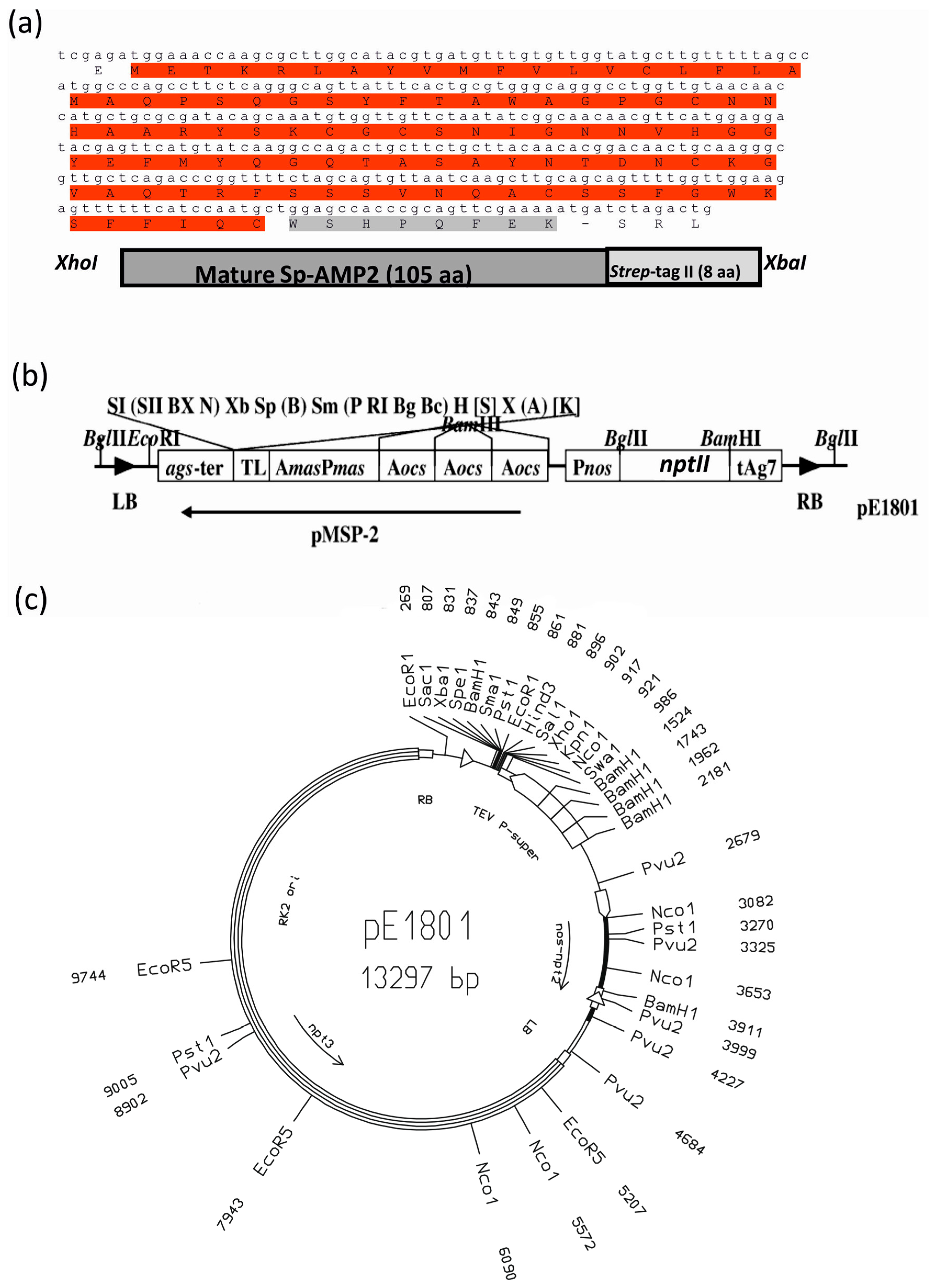
2.1. Agrobacterium Tumefaciens Vectors and Strains

Sp-AMP-encoding genes share a high level of sequence similarity. Sp-AMP2 has the highest score for identity of the Sp-AMP consensus sequence among the other members [20]. The Sp-AMP2 full-length complementary DNA (cDNA) was obtained by PCR amplification from a subtraction cDNA library (hasp001xd12f; GenBank: BI416519.1) of Scots pine root tissue infected with the root rot fungus H. annosum. It was cloned into pT-Adv vector (Clontech, Mountain View, CA, USA) using gene-specific primers containing XhoI and XbaI restriction sites and twenty-four nucleotides representing the Strep-tag II sequences in the reverse primer (IBA, Göttingen, Germany). The primers were designed based on the coding sequence of Sp-AMP2 gene (Genebank: AF410953.1) (forward primer: 5′ GCACTCGAGATGGAAACCAAGCGCTTGGCAT; reverse primer: 5′-CAGTCTAGATCATTTTTCGAACTGCGGGTGGCTCCAGCATTGGATGAAAAAACT).
The amplified products were subcloned into the XhoI/XbaI-digested plant binary vector pE1801 where the NcoI site was first deleted by mung bean nuclease trimming and re-ligation. pE1801 (Figure 1) contains a T-DNA segment that includes the kanamycin resistance gene nos-nptII, as well as strong synthetic promoter, the “super promoter” [25]. Cloning into the binary vector pE1801 was confirmed by sequencing. Recombinant construct was used to transform Agrobacterium tumefaciens Smith & Townsend strain EHA105 by freeze-thaw method [26]. A single colony of the A. tumefaciens EHA105 strain harboring the pE1801 and growing on LB agar plates supplemented with 100 mg/L kanamycin (Sigma-Aldrich, St Louis, MO, USA) and 100 mg/L rifampicin (Sigma-Aldrich, St Louis, MO, USA) was used to inoculate 5 mL of LB medium. After overnight incubation at 28 °C, 220 rpm, 5 mL of the bacterial culture was added to 30 mL MS (Murashige and Skoog Basal Salt Mixture) liquid medium (Sigma-Aldrich, M5524, St Louis, MO, USA) [27]. The resulting Agrobacterium suspension was used for tobacco transformation.

2.2. Tobacco Transformation

Tobacco (Nicotiana tabacum L. cv. Petit Havana SR1) was stably transformed using the leaf disc method [28]. Leaves were surface-sterilized for 1 min with 70% ethanol and for 10 min with 1.2% sodium hypochlorite solution, then rinsed five to seven times with sterile water.
The Agrobacterium suspension and tobacco leaf discs were incubated in a petri dish for 5 min, then infected leaf discs were placed upside down on agar-solidified MS medium (Sigma-Aldrich, M5524, St Louis, MO, USA) containing 1 mg/L BAP (6-benzylaminopurine, Sigma-Aldrich, M5524, St Louis, MO, USA) and supplemented with sucrose (20 g/L) (Sigma-Aldrich, M5524, St Louis, MO, USA). Plates were kept in the dark for 2 days; thereafter the infected explants were transferred to a fresh solid MS medium containing 1 mg/L BAP and sucrose (20 g/L), and supplemented with 100 mg/L kanamycin and 500 mg/L cefotaxime (Sigma-Aldrich, M5524, St Louis, MO, USA) at 23 °C in the light for 4–6 weeks. Each week, fresh selection plates were changed and, after the emergence of the shoots, a single shoot per leaf disc was transferred to MS Jars containing 100 mg/L kanamycin and 500 mg/L cefotaxime. Transgenic plants were regenerated under kanamycin selection and plantlets were moved into vermiculite in the growth room and then to the green house. Seeds (T1 generation) from the transgenic lines (T0) were collected after self-pollination. For pathogen resistance assay, the wild type plant (control) and four independently transformed T1 lines and their segregating siblings were used.

2.3. Selection of Transgenic Plants

The T1 seeds from transgenic plants were surface-sterilized by immersion in 70% (v/v) ethanol for 2 min, followed by immersion in 1.2% sodium hypochlorite solution for 10 min. Afterwards, seeds were washed four times with sterile distilled water and sown onto 1% agar-containing MS medium supplemented with kanamycin (100 mg/L) and sucrose (20 g/L). Plates were incubated at 18 °C with a photoperiod of 16 h. After 14 days, the well-developed rooted seedlings were transferred to soil prepared by mixing peat (Kekkilä Horticulture Peat, Kekkilä Oyj, Finland) and washed sand at a 5:1 ratio (v/v). T1 transgenic and non-transgenic plants were generated and sorted based on PCR analysis (XP cycler, BIOER, Hangzhou, China); the PCR product was absent in non-transgenic plants. All plants were grown in a growth room under controlled conditions, were watered when needed, and fertilized weekly with a 1.0% solution of N/P/K (8:4:6 or 16:9:22) fertilizer (Yara, Espoo, Finland).

2.4. cDNA Synthesis by Reverse Transcription

For the initial verification of Sp-AMP2 in the transgenic plants, RNA was extracted from one T0 transgenic plant, one T1 transgenic plant, one T1 non-transgenic, and one wild type control plant according to the methods of Chang et al. [29]. RNA concentration and quality were assessed spectrophotometrically using a NanodropTM ND-1000 (NanoDrop Technologies, Wilmington, DE, USA). A 1 µg sample of each total RNA isolate was used for cDNA synthesis, as described by Jaber et al. [30]. To verify the transcript of Sp-AMP2 in the tested tobacco plants, the resulting cDNA was diluted (1:5) and 5 µL was used to amplify the Sp-AMP2 fragment. The PCR products were separated on a 1.5% agarose gel.

2.5. Pathogen Bioassays of Transgenic Plants

Botrytis cinerea Pers. isolate B05.10 [31], a haploid aggressive strain isolated from Vitis vinifera L. (kindly provided by Mehmet Ali Keçeli, Department of Biosciences, Faculty of Biological and Environmental Sciences, University of Helsinki) was used for the pathogen assay.
B. cinerea was grown on potato-carrot agar (PCA). PCA was prepared as follows: 300 g of diced potatoes and 25 g of diced carrots were boiled in water for 30 min, then mashed and filtered through Miracloth (Millipore, Billerica, MA, USA). The final volume was adjusted to 1 L with H2O, and 15 g of agar was added. B. cinerea was grown for 10 days in the dark at approximately 22 °C. Spores were harvested from each dish by rinsing with 10 mL PDB (Potato Dextrose Broth, Difco, Detroit, MI, USA) while agitating the colonies gently with a glass rod. The spores in PDB medium were vortexed for 10 min at maximum speed and then filtered through Miracloth. The concentration of B. cinerea spores was adjusted to 1 × 10⁶ spores/mL. Four-week-old T1 generation tobacco plants and non-transformed control plants were inoculated with 10 μL of the spore suspension of B. cinerea onto the upper surface of a leaf of the growing seedling. Wild type plants (control) and four T1 lines were tested; five plants per line including four transgenic T1 plants and one non-transgenic T1 plant were used for each treatment and evaluated for disease development. To determine the susceptibility of plants, disease lesions were scored at 3 and 5 days post-inoculation (d.p.i.) and recorded using an optical Olympus CX31 microscope equipped with a digital Olympus SC30 camera (Olympus, Tokyo, Japan).

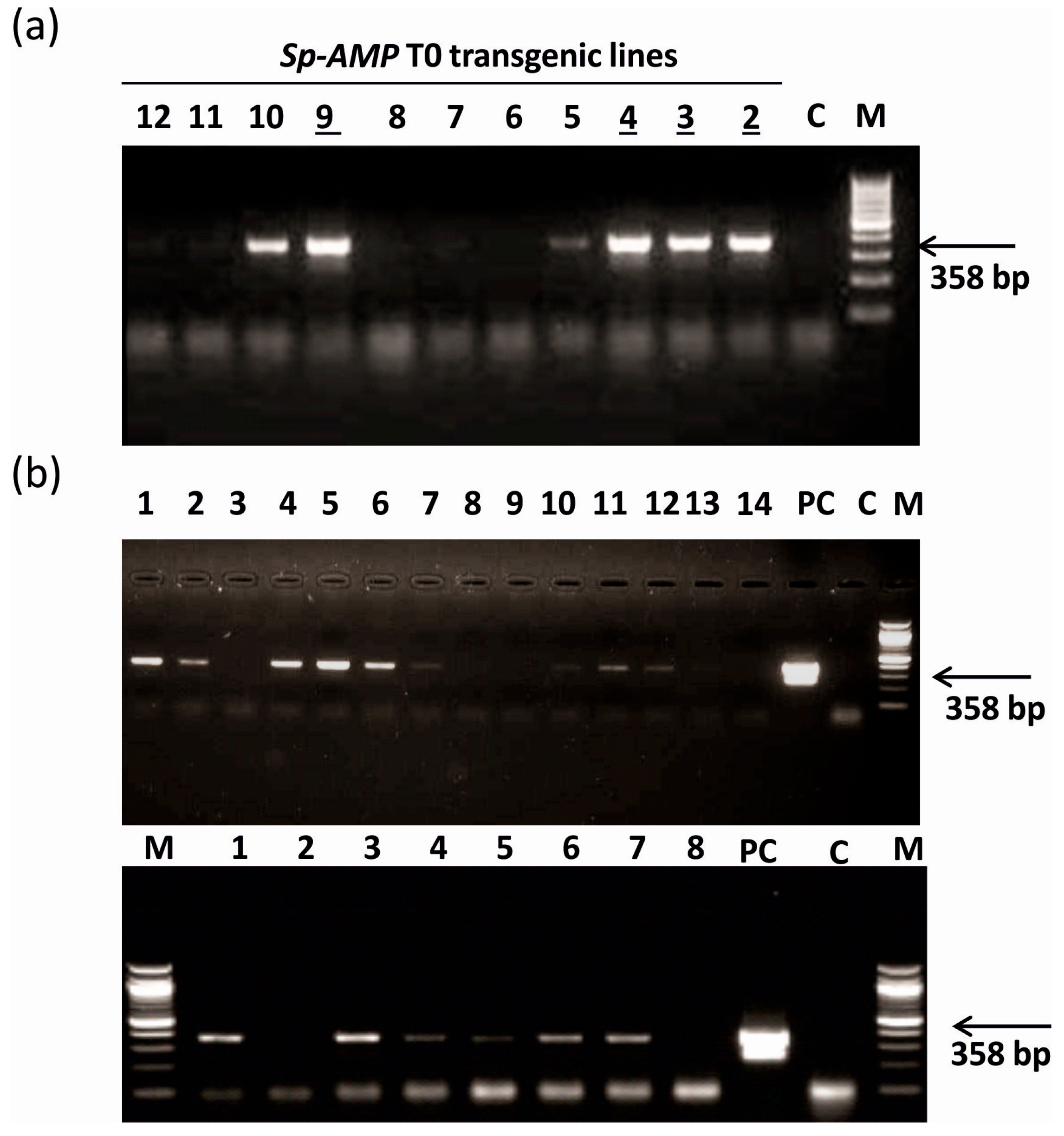
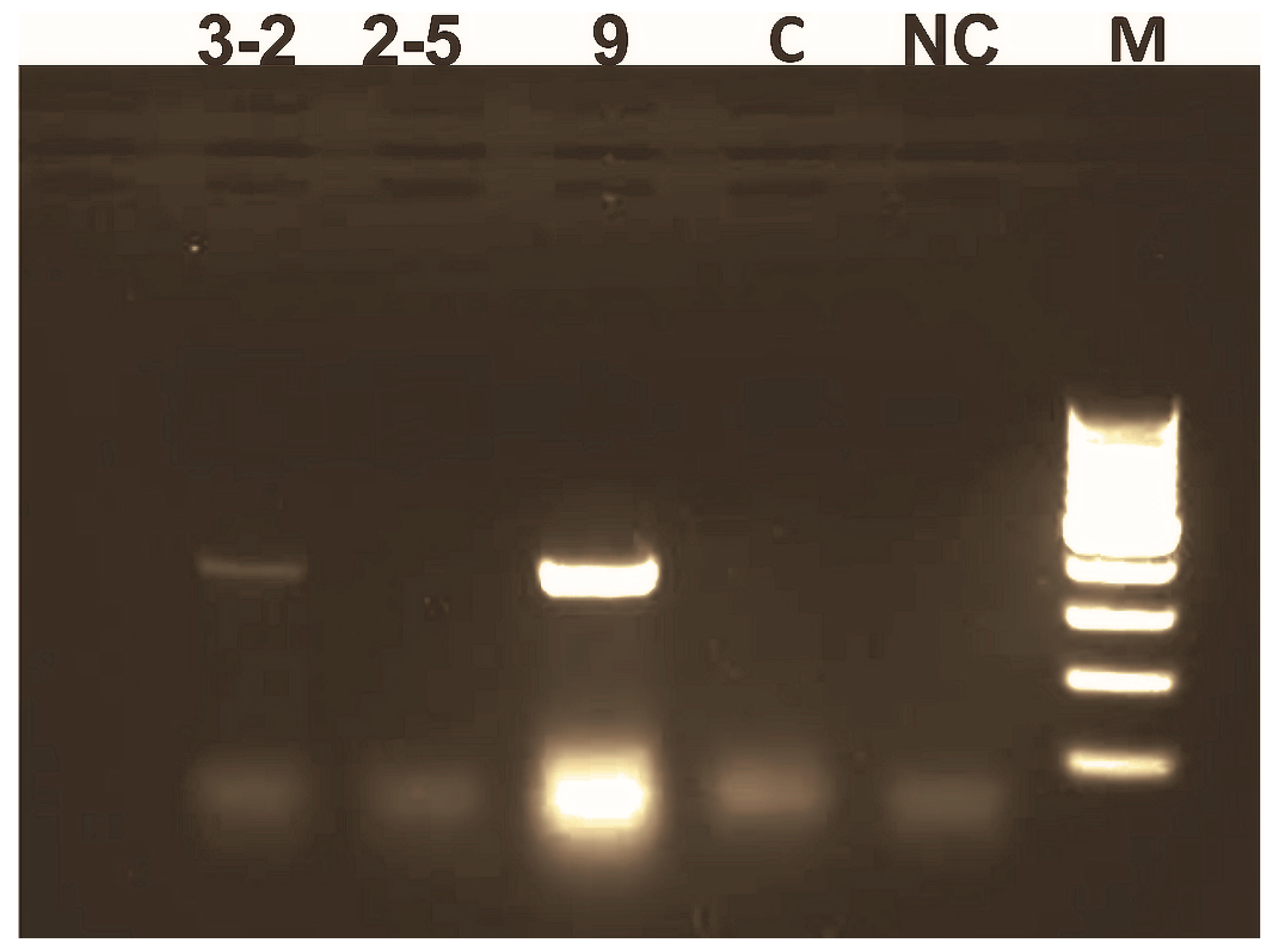
2.6. PCR Analysis of Transgenic Plants

Genomic DNA was isolated using a CTAB (cetyl trimethylammonium bromide) method [32]. The genomic DNA samples of the control plants and transformed T0 lines were analyzed by PCR (XP cycler, BIOER, Hangzhou, China) with the same primers that were used for cloning. The size of the amplified product matched the predicted length of 358 bp. Amplification was performed under the following conditions: 95 °C for 5 min, 39 cycles of 94 °C for 30 s, 62 °C for 30 s, and 72 °C for 40 s. Finally, the PCR products were separated on a 1.5% agarose gel and visualized by ethidium bromide staining. Segregating T1 lines were tested using PCR to verify segregating transgenic plants.

2.7. Real-Time Quantitative PCR Analysis

The analysis of the expression levels of the Sp-AMP2 and marker genes for the SA/JA/ET signaling pathways was performed by real-time quantitative reverse transcription PCR (qRT-PCR, LightCycler® 480 II system, Roche Diagnostics, Basel, Switzerland). For this purpose, RNA was extracted from transgenic, non-transgenic, and control plants. For each of the mentioned plant lines, 100 mg of non-treated young leaves (control), leaves inoculated with 10 μL of PDB (mock inoculation), and leaves inoculated with 10 μL of the spore suspension of B. cinerea (treatment) were taken for RNA isolation. cDNA synthesis by reverse transcription was preformed according to the protocol described above.
qRT-PCR was performed with the LightCycler® 480 II system (Roche Diagnostics, Basel, Switzerland) according to the manufacturer’s instructions. The list of gene names, accession numbers, qRT-PCR primers, and product sizes is provided in Supplementary file S1. PCR thermal conditions consisted of initial denaturation at 95 °C for 10 min followed by 40 cycles of 95 °C for 15 s and 1 min at the appropriate annealing temperature. The specificity of the primer pairs was verified both by gel and by melting curve analysis. The transcript abundance was estimated with LightCycler software version 3.5 (Roche) using SYBR® Green PCR Master Mix (Invitrogen) according to the manufacturer's recommendations, based on three biological and three technical replicates for each gene. The gene for elongation factor 1a (EF-1a), a very stably expressed gene [33], was used as a reference gene. In agreement with the previous reports, the expression level of this gene showed very little variation in our experiments (mean cycle threshold (Ct) value Ct ± SE = 26.91 ± 0.03, n = 324, where SE indicates standard error). The raw Ct values of the tested genes were normalized to the reference gene and used to compare the results from untreated control wild type against all samples using the 2−ΔΔCt method, as described previously [34].

2.8. Statistical Analyses

All experiments were repeated three times, and similar results were obtained. Means and standard errors were estimated from three independent experiments. Error bars indicate the standard error of the mean across three biological replicates. Statistical significance analyses of the necrosis were performed using paired t-test as well as by two-way ANOVA test. Bonferroni post hoc test was applied on the data to compare the value of each column factor “necrosis length” and each row factor “Genotype”. p > 0.05 was considered to be non-significant (Supplementary file S2). Statistical significance of the pathogen assays on transgenic plants and the gene expression were calculated by two-way ANOVA test. Thereafter, Bonferroni post hoc test correction was applied on the data for comparisons. The threshold for fold changes in the expression of the differentially expressed genes in comparison to the control samples was set at 2-fold. Asterisks indicate the significance of differences, * p values < 0.05, ** p values < 0.01, and *** p values < 0.001.

3. Results

3.1. Generation of Sp-AMP2 Transgenic Tobacco Plants

The Scots pine Sp-AMP2 cDNA under control of a strong promoter was introduced into tobacco (Nicotiana tabacum cv. Petit Havana SR1) by Agrobacterium tumefaciens-mediated transformation method (Figure 1). The promoter in the vector is a chimeric promoter derived from the octopine and mannopine synthase genes that is approximately 156-fold stronger than the cauliflower mosaic virus (CaMV) 35S promoter in tobacco leaf tissue, which makes it useful for high level constitutive expression of genes [25]. Presence of the Sp-AMP2 gene in the genomic DNA of tobacco plants was confirmed by PCR amplification. A 358 bp DNA fragment was detected in T0 transgenic tobacco lines, whereas no bands were detected in the untransformed control tobacco plant (Figure 2a). Four different tested tobacco lines T0 were selected for T1 seed collection after self-pollination, and used for further analysis. T1 seeds were collected and verified for transgene presence in plants regenerated on selective medium by PCR (Figure 2b). Transcription of Sp-AMP2 was confirmed by PCR amplification of SpAMP2-specific fragments from the cDNA of the non-transformed wild type, T0, non-transgenic and transgenic T1 plants (Figure 3). The T0 and T1 transgenic lines showed the transcript abundance of Sp-AMP2.

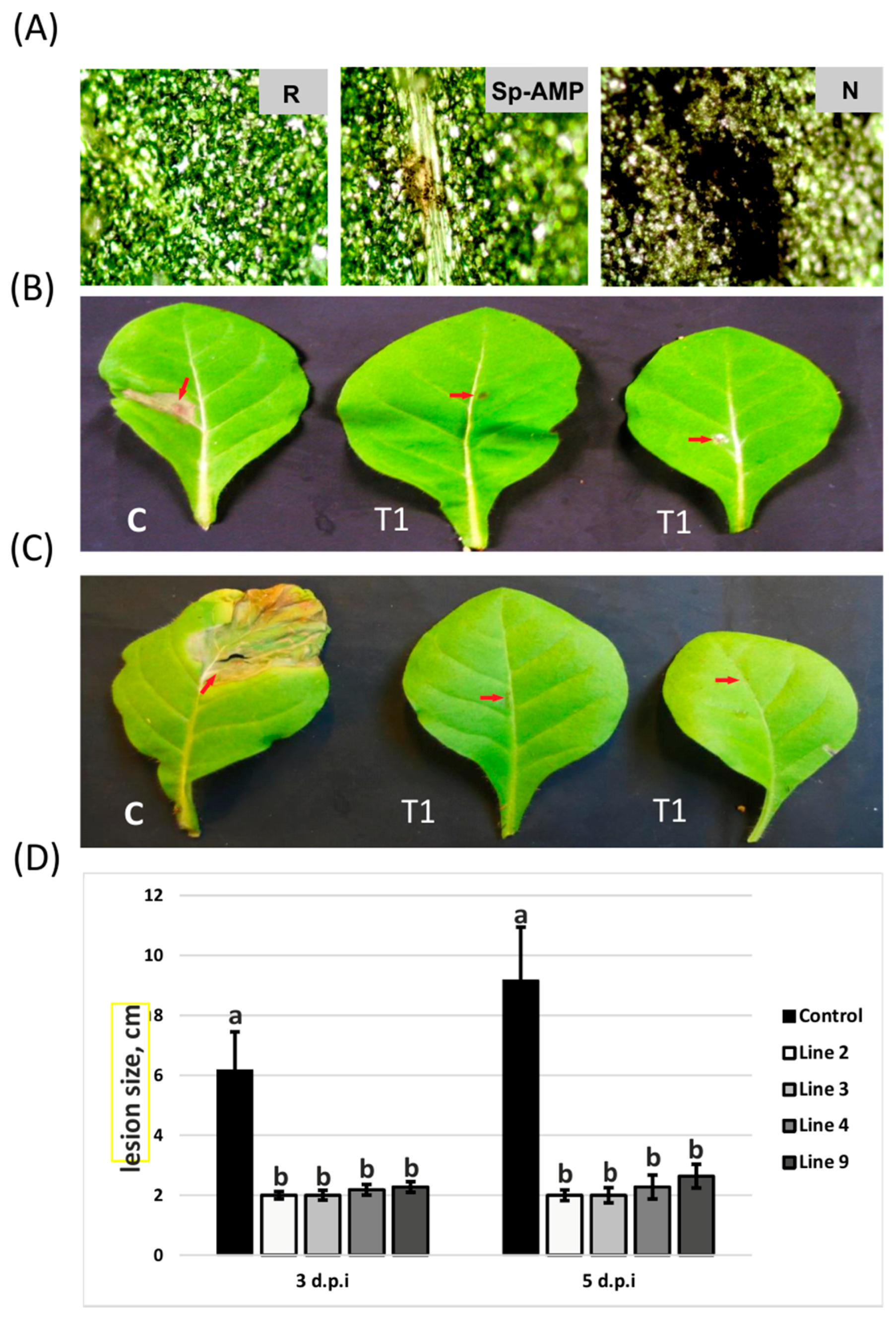
3.2. Sp-AMP2-Transformed Tobacco Plants Show Increased Tolerance to B. cinerea

The effects of infection caused by B. cinerea were investigated on the T1 transgenic plants and compared with both non-transformed plants and their non-transgenic segregating siblings. The T1 plants were verified by rooting in selective media and PCR. Each transformation event comprising of three transgenic T1 plants was compared to two non-transgenic T1 sibling plants and to one non-transformed control plant. The sizes of the lesions determined from 3 to 5 days post-inoculation (d.p.i.) under conditions described in the material section are indicated in Figure 4. Necrotic lesions caused by B. cinerea on the control tobacco and non-transgenic T1 leaves were more severe and larger than those formed on the leaves of T1 transgenic lines. The lesions on the transgenic lines usually increased slightly after 2 extra days of incubation, however, none reached the same size as those in the non-transformed control. Transgenic tobacco showed significant difference in lesion size in response to infection by B. cinerea as compared to the non-transgenic in the two time points (Supplementary file S3). Similar levels of lesion development were observed on non-transgenic T1 plants, compared to those of control plants. However, the disease spread was slower on transgenic plants (Figure 4). No statistically significant differences in the size of necrotic lesions were observed among different transgenic lines expressing the Sp-AMP2 gene.

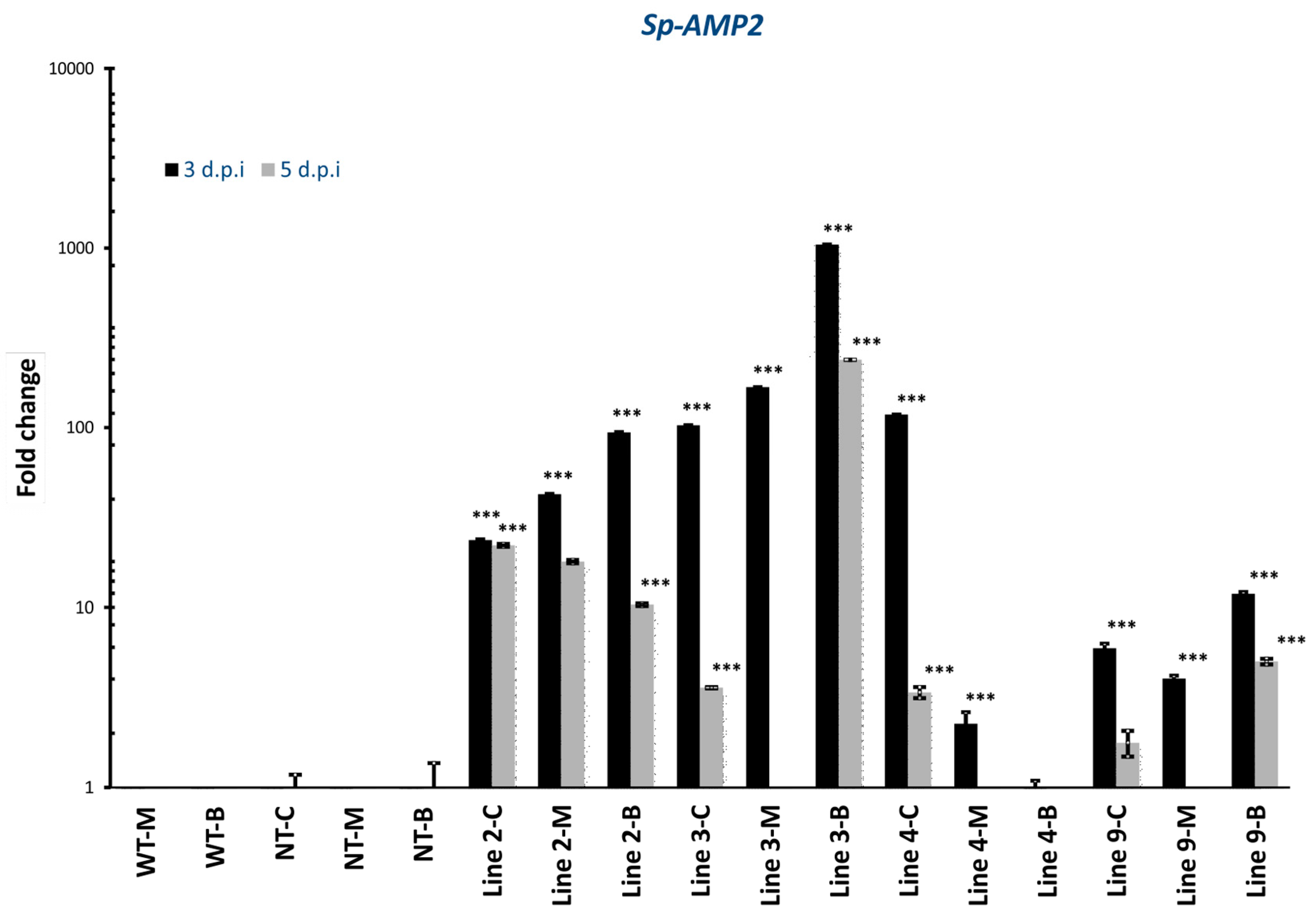
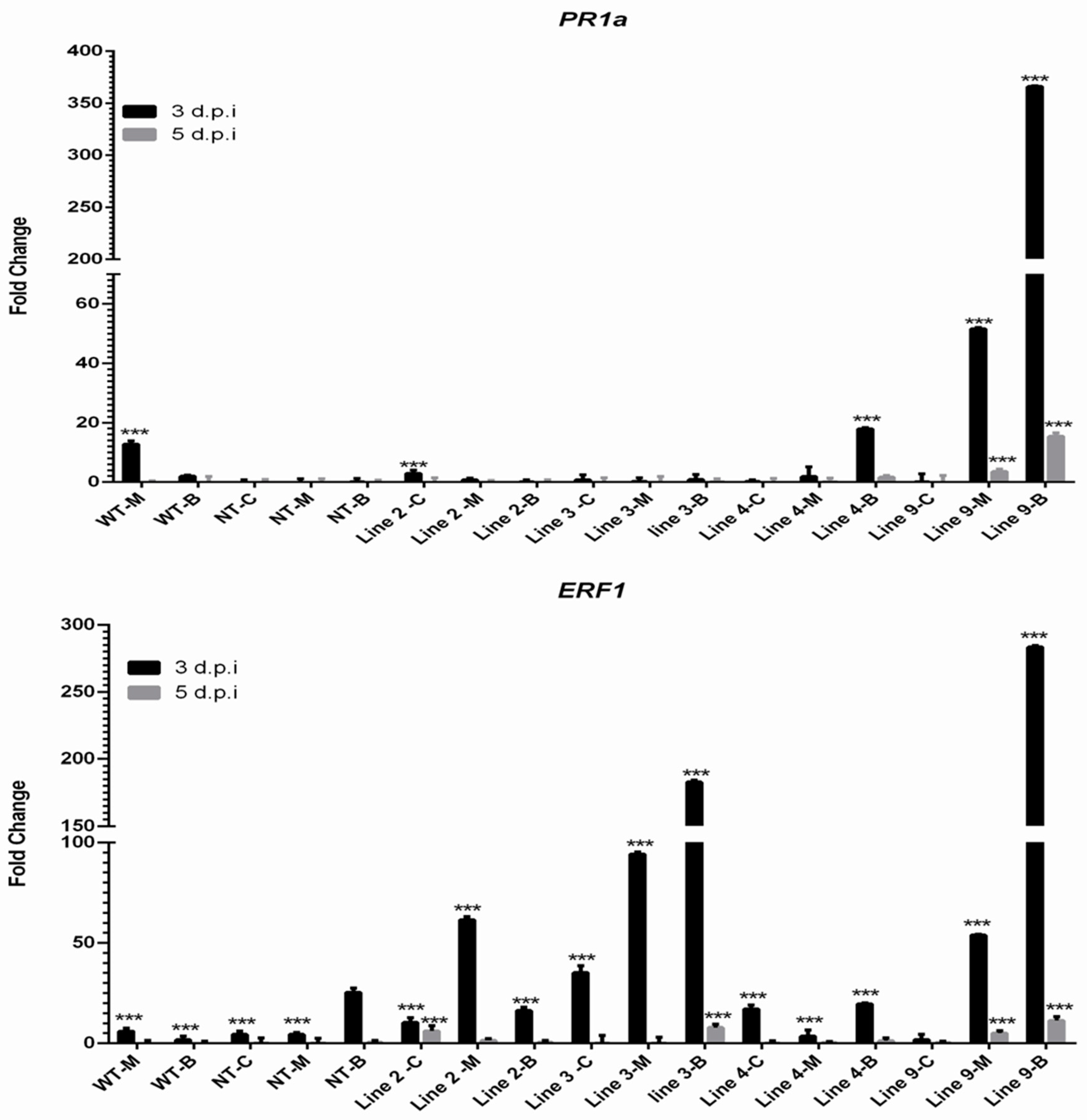
3.3. Expression Profiles of Sp-AMP2 Gene and SA/JA/ET Gene Markers in Transgenic Plants

In order to investigate Sp-AMP2 gene expression level, four transgenic lines were tested in comparison to control and non-transgenic plants. No Sp-AMP2 transcripts were observed in any of the control and non-transgenic plants tested. The transcript of Sp-AMP2 was abundantly present in all transgenic lines. Sp-AMP2 was induced highly in response to the B. cinerea infection at 3 d.p.i. in the three transgenic lines (except for line 4) and the expression was sustained over time by the pathogen (5 d.p.i) (Figure 5).
To study the interaction between the SA and JA/ET signaling pathways and the expression of Sp-AMP2 in the transgenic tobacco, we investigated the expressions levels of two important marker genes. PR1a is a SA-associated gene that serves as a common indicator for SA signaling pathway activity [35]. ERF1 (ethylene response factor 1) is a marker gene, which is induced by simultaneous action of the JA and ET signaling pathways [36]. The induction of ERF1 was observed in all tested tobacco plants at 3 d.p.i. However, increased levels of ERF1 transcripts were observed in the transgenic plants, notably in two lines challenged with B. cinerea (Lines 3 and 9) (Figure 6). PR1a was strongly induced in the mock-inoculated wild type and in two transgenic lines (Lines 4, 9). The highest expression was observed in the transgenic line 9 (Figure 6).

4. Discussion

In the present study, we investigated the role of a gene encoding Scots pine antimicrobial peptide Sp-AMP2 in the defense against pathogenic fungi. Our earlier results showed strong Sp-AMP induction in Scots pines challenged with the necrotrophic fungal pathogen H. annosum [20,21], justifying further detailed functional analyses of the corresponding gene. A successful transfer of a Sp-AMP2-encoding gene of the gymnosperm (Scots pine) to the angiosperm recipient (tobacco) was demonstrated. No homologs of Sp-AMP genes have been identified either in Nicotiana benthamiana Domin or in Nicotiana tabacum [37]. Our results suggest that expression of Sp-AMP2 in transgenic tobacco conferred enhanced resistance to Botrytis cinerea. This is a further functional demonstration of the potential role of Sp-AMP2 in plant defense. The plants expressing Sp-AMP2 showed a reduction in the spread and subsequent expansion of fungal infection over the 5 day evaluation period, with the most significant statistical differences being observed at 5 d.p.i., indicating that Sp-AMP2 is actively involved in the inhibition of B. cinerea and suggesting a broader spectrum of the Sp-AMP2 inhibitory activity. The observed delay in symptom development could potentially allow plants either to recover or adapt to changes in field conditions that could potentially reduce disease spread.
A considerable variation in the expression level of the heterologous Sp-AMP2 gene among different lines of transgenic tobacco plants was observed in our experiments. The reasons for these differences are not entirely clear, as the gene was put under control of a strong constitutive promoter. One of the possible explanations could be the integration of the transgenic construct in different genomic loci of the transgenic host plants, which in turn might have an effect on the expression level of the transgene. However, the transgenic lines with different expression levels of the Sp-AMP2 gene showed no statistically significant differences in the size of necrotic lesions caused by inoculation with B. cinerea. It is likely that even low-level expression of Sp-AMP2 is sufficient to protect transgenic tobacco plants from this fungal pathogen.
Tobacco plants expressing Sp-AMP2 showed increased level of ERF1 induction upon pathogen challenge, indicating a potential interplay between Sp-AMP expression and JA/ET signaling pathways. In Scots pine, increased expression of Sp-AMP in response to pathogen challenge, salicylic acid, ethylene, and other environmental stresses was shown in our earlier study [21]. SA-dependent signaling pathway, which is engaged with the cell death machinery against biotrophic pathogens, controls the synthesis of low molecular weight antimicrobial proteins such as PR-1, PR-2, and PR-5. At the same time, the JA/ET-dependent pathway is associated with defense against necrotrophic pathogens, and it activates genes that encode antimicrobial proteins such as thionin (THI2.1), defensin (PDF1.2), PR-3, and PR-4 [38]. Such differences may affect the specificity of plant-pathogen interactions.
PR-19 family members are potential alternatives to current H. annosum control and management strategies, since they exploit built-in defense systems of the host trees. A long-term objective is to find a gene that is tightly linked to a disease resistance trait that would serve as molecular DNA marker to be applied in breeding programs and facilitate the selection of naturally occurring genotypes with higher resistance against the root rot pathogen.
A similar cysteine-rich peptide, MiAMP1 protein, has been shown to be highly inhibitory to a wide range of phytopathogens, particularly fungi. In addition, transgenic expression of MiAMP1 in canola (Brassica napus L.) provided enhanced resistance against blackleg disease caused by the fungus Leptosphaeria maculans Ces. & De Not [39]. Also, another MiAMP1-related cysteine-rich antimicrobial peptide from western white pine (Pinus monticola Douglas ex D. Don) (PmAMP1) that belongs to the same family of MiAMP1, and is the closest homolog to Sp-AMPs, has been successfully transformed into canola and proved to confer resistance to multiple fungal pathogens (Alternaria brassicae (Berk.) Sacc., L. maculans, and Sclerotinia sclerotiorum (Lib.) de Bary) [40]. Further genetic association studies on western white pine PmAMP1 in a collection of western white pine trees revealed that high quantitative disease resistance against white pine blister rust (WPBR) caused by the obligate biotrophic fungus Cronartium ribicola A. Dietr. is genetically attributed (albeit slightly) to multiple PmAMP1 haplotypes, suggesting that functional regulation of many other defense-related genes is contributing to the quantitative variations of disease resistance. However, the study identified a haplotype of PmAMP1 (PmAMP1- hap2) which confers a higher resistance to C. ribicola than other haplotypes and which would therefore certainly benefit traditional tree breeding by marker-assisted selection [41].
The cysteine-rich antimicrobial peptides are evolutionarily dynamic within plant genomes, which are highly adaptive in the challenge of more evolutionary flexible pathogen populations [42]. Moreover, the structural resemblance between MiAMP1 protein family members and the novel Sp-AMP does not necessarily reflect functional similarities [43]. Therefore, the functional validation of the novel Sp-AMPs from the Scots pine through this transgenic experiment is imperative to manifest its potential in plant disease immunity.
Other authors have also demonstrated that somatic embryos of Norway spruce (Picea abies (L.) H. Karst.) plants at an age of 8 to 10 months over-expressing the SPI1 spruce defensin protein displayed reduced fungal growth in the sapwood after inoculation with H. annosum [44]. Also, SPI1 spruce defensin that was expressed in tobacco permitted less growth of the bacterial pathogen Erwinia carotovora (Jones) Bergey et al. [45], suggesting that SPI1 increases resistance in both homologous and heterologous systems. Furthermore, poplar trees transformed with a gene encoding synthetic peptide D4E1 were found to be resistant to bacterial pathogens Agrobacterium tumefaciens and Xanthomonas populi (Ridé) van den Mooter & Swings but not to the fungal pathogen Hypoxylon mammatum (Wahlenb.) P. Karst [46].
The combination of the broad spectrum action of MiAMP1 protein family members on numerous microorganisms [47,48] and the nature of the cysteine-rich AMPs, which reduce the growth of major microbes without any toxic effects toward the host [49], makes Sp-AMP proteins potential candidates for developing pathogen-resistant crops, a further demonstration of their role in tree resistance.

5. Conclusions

In this study, in order to address the role of Sp-AMPs in plant defense, Sp-AMP2 was inserted into the genome of the tobacco model plant. The transformed tobacco plants with Sp-AMP2 showed increased tolerance against Botrytis cinerea. Our future study will explore the possibility to transfer the Sp-AMP2 into a related conifer tree species using most promising technology approaches [50] and further assess its role in tree resistance.

Supplementary Materials

The following are available online at www.mdpi.com/1999-4907/9/1/10/s1, Supplementary file S1: List of primers used for the real-time quantitative PCR assays, Supplementary file S2: Statistical significance analyses of the genes expression, Supplementary file S3: Statistical significance analyses of the lesion size.

Acknowledgments

This work was supported by grants from the Academy of Finland (AKA), Helsinki University Research Fund, and The Doctoral Program in Plant Science (DPPS). We extend our sincere thanks to Chaowen Xiao for assistance with the Strep Tag constructs, Stan Gelvin for providing the super promoter vector, and to Hany Bashandy who tended to our plants.

Author Contributions

E.J. carried out the experimental work; T.R. participated in the data analysis; E.J. and A.K. drafted the manuscript; F.O.A. and T.T. conceived the study and contributed to the experiment design; F.O.A., T.T., T.R., and S.K. read, edited, and revised the manuscript.

Conflicts of Interest

The authors declare no conflict of interest. The founding sponsors had no role in the design of the study; in the collection, analyses, or interpretation of data; in the writing of the manuscript, and in the decision to publish the results.

References

  1. Koornneef, A.; Pieterse, C.M.J. Cross talk in defense signaling. Plant Physiol. 2008, 146, 839–844. [Google Scholar] [CrossRef] [PubMed]
  2. Broekaert, W.F.; Cammue, B.P.A.; DeBolle, M.F.C.; Thevissen, K.; DeSamblanx, G.W.; Osborn, R.W. Antimicrobial peptides from plants. Crit. Rev. Plant Sci. 1997, 16, 297–323. [Google Scholar] [CrossRef]
  3. Van Loon, L.C.; Van Strien, E.A. The families of pathogenesis-related proteins, their activities, and comparative analysis of pr-1 type proteins. Physiol. Mol. Plant Pathol. 1999, 55, 85–97. [Google Scholar] [CrossRef]
  4. Van Loon, L.C.; Rep, M.; Pieterse, C.M.J. Significance of inducible defense-related proteins in infected plants. Annu. Rev. Phytopathol. 2006, 44, 135–162. [Google Scholar] [CrossRef] [PubMed]
  5. Benko-Iseppon, A.M.; Galdino, S.L.; Calsa, T.; Kido, E.A.; Tossi, A.; Belarmino, L.C.; Crovella, S. Overview on plant antimicrobial peptides. Curr. Protein Pept. Sci. 2010, 11, 181–188. [Google Scholar] [CrossRef] [PubMed]
  6. Padovan, L.; Scocchi, M.; Tossi, A. Structural aspects of plant antimicrobial peptides. Curr. Protein Pept. Sci. 2010, 11, 210–219. [Google Scholar] [CrossRef] [PubMed]
  7. Hammami, R.; Ben Hamida, J.; Vergoten, G.; Fliss, I. Phytamp: A database dedicated to antimicrobial plant peptides. Nucleic Acids Res. 2009, 37, D963–D968. [Google Scholar] [CrossRef] [PubMed]
  8. Hancock, R.E.W.; Sahl, H.G. Antimicrobial and host-defense peptides as new anti-infective therapeutic strategies. Nat. Biotechnol. 2006, 24, 1551–1557. [Google Scholar] [CrossRef] [PubMed]
  9. Collinge, D.B.; Jørgensen, H.J.L.; Lund, O.S.; Lyngkjær, M.F. Engineering pathogen resistance in crop plants: Current trends and future prospects. Annu. Rev. Phytopathol. 2010, 48, 269–291. [Google Scholar] [CrossRef] [PubMed]
  10. Montesinos, E. Antimicrobial peptides and plant disease control. FEMS Microbiol. Lett. 2007, 270, 1–11. [Google Scholar] [CrossRef] [PubMed]
  11. Odintsova, T.; Egorov, T. Plant antimicrobial peptides. In Plant Signaling Peptides; Irving, H.R., Gehring, C., Eds.; Springer: Berlin/Heidelberg, Germany, 2012; pp. 107–133. [Google Scholar]
  12. Asiegbu, F.O.; Denekamp, M.; Daniel, G.; Johansson, M. Immune cytochemical-localization of pathogenesis-related proteins in roots of Norway spruce infected with Heterobasidion annosum. Eur. J. For. Pathol. 1995, 25, 169–178. [Google Scholar] [CrossRef]
  13. Davis, J.M.; Wu, H.G.; Cooke, J.E.K.; Reed, J.M.; Luce, K.S.; Michler, C.H. Pathogen challenge, salicylic acid, and jasmonic acid regulate expression of chitinase gene homologs in pine. Mol. Plant Microbe Interact. 2002, 15, 380–387. [Google Scholar] [CrossRef] [PubMed]
  14. Asiegbu, F.O.; Daniel, G.; Johansson, M. Defense-related reactions of seedling roots of Norway spruce to infection by Heterobasidion annosum (Fr.) bref. Physiol. Mol. Plant Pathol. 1994, 45, 1–19. [Google Scholar] [CrossRef]
  15. Asiegbu, F.O.; Nahalkova, J.; Li, G.S. Pathogen-inducible cdnas from the interaction of the root rot fungus Heterobasidion annosum with scots pine (Pinus sylvestris L.). Plant Sci. 2005, 168, 365–372. [Google Scholar] [CrossRef]
  16. Mensen, R.; Hager, A.; Salzer, P. Elicitor-induced changes of wall-bound and secreted peroxidase activities in suspension-cultured spruce (Picea abies) cells are attenuated by auxins. Physiol. Plant. 1998, 102, 539–546. [Google Scholar] [CrossRef]
  17. Adomas, A.; Heller, G.; Li, G.; Olson, A.; Chu, T.M.; Osborne, J.; Craig, D.; van Zyl, L.; Wolfinger, R.; Sederoff, R.; et al. Transcript profiling of a conifer pathosystem: Response of Pinus sylvestris root tissues to pathogen (Heterobasidion annosum) invasion. Tree Physiol. 2007, 27, 1441–1458. [Google Scholar] [CrossRef] [PubMed]
  18. Kovaleva, V.; Kiyamova, R.; Cramer, R.; Krynytskyy, H.; Gout, I.; Filonenko, V.; Gout, R. Purification and molecular cloning of antimicrobial peptides from scots pine seedlings. Peptides 2009, 30, 2136–2143. [Google Scholar] [CrossRef] [PubMed]
  19. Fossdal, C.G.; Nagy, N.E.; Sharma, P.; Lonneborg, A. The putative gymnosperm plant defensin polypeptide (SPI1) accumulates after seed germination, is not readily released, and the SPI1 levels are reduced in pythium dimorphum-infected spruce roots. Plant Mol. Biol. 2003, 52, 291–302. [Google Scholar] [CrossRef] [PubMed]
  20. Asiegbu, F.O.; Choi, W.; Li, G.; Nahalkova, J.; Dean, R.A. Isolation of a novel antimicrobial peptide gene (Sp-AMP) homologue from Pinus sylvestris (Scots pine) following infection with the root rot fungus Heterobasidion annosum. FEMS Microbiol. Lett. 2003, 228, 27–31. [Google Scholar] [CrossRef]
  21. Sooriyaarachchi, S.; Jaber, E.; Covarrubias, A.S.; Ubhayasekera, W.; Asiegbu, F.O.; Mowbray, S.L. Expression and beta-glucan binding properties of scots pine (Pinus sylvestris L.) antimicrobial protein (Sp-AMP). Plant Mol. Biol. 2011, 77, 33–45. [Google Scholar] [CrossRef] [PubMed]
  22. Veluthakkal, R.; Dasgupta, M.G. Pathogenesis-related genes and proteins in forest tree species. Trees 2010, 24, 993–1006. [Google Scholar] [CrossRef]
  23. Manners, J.M. Primitive defence: The MiAMP1 antimicrobial peptide family. Plant Mol. Biol. Rep. 2009, 27, 237–242. [Google Scholar] [CrossRef]
  24. Kovalchuk, A.; Kerio, S.; Oghenekaro, A.O.; Jaber, E.; Raffaello, T.; Asiegbu, F.O. Antimicrobial defenses and resistance in forest trees: Challenges and perspectives in a genomic era. Annu. Rev. Phytopathol. 2013, 51, 221–244. [Google Scholar] [CrossRef] [PubMed]
  25. Ni, M.; Cui, D.; Einstein, J.; Narasimhulu, S.; Vergara, C.E.; Gelvin, S.B. Strength and tissue-specificity of chimeric promoters derived from the octopine and mannopine synthase genes. Plant J. 1995, 7, 661–676. [Google Scholar] [CrossRef]
  26. An, G.; Ebert, P.; Mitra, A.; Ha, S. Binary vectors. In Plant Molecular Biology Manual; Gelvin, S., Schilperoort, R., Verma, D., Eds.; Springer: Dordrecht, The Netherlands, 1989; pp. 29–47. [Google Scholar]
  27. Murashige, T.; Skoog, F. A revised medium for rapid growth and bio assays with tobacco tissue cultures. Physiol. Plant. 1962, 15, 473–497. [Google Scholar] [CrossRef]
  28. Horsch, R.B.; Fry, J.E.; Hoffmann, N.L.; Eichholtz, D.; Rogers, S.G.; Fraley, R.T. A simple and general-method for transferring genes into plants. Science 1985, 227, 1229–1231. [Google Scholar]
  29. Chang, S.; Puryear, J.; Cairney, J. A simple and efficient method for isolating RNA from pine trees. Plant Mol. Biol. Rep. 1993, 11, 113–116. [Google Scholar] [CrossRef]
  30. Jaber, E.; Xiao, C.; Asiegbu, F.O. Comparative pathobiology of Heterobasidion annosum during challenge on Pinus sylvestris and arabidopsis roots: An analysis of defensin gene expression in two pathosystems. Planta 2014, 239, 717–733. [Google Scholar] [CrossRef] [PubMed]
  31. Quidde, T.; Buttner, P.; Tudzynski, P. Evidence for three different specific saponin-detoxifying activities in botrytis cinerea and cloning and functional analysis of a gene coding for a putative avenacinase. Eur. J. Plant Pathol. 1999, 105, 273–283. [Google Scholar] [CrossRef]
  32. Richards, E.; Reichardt, M.; Rogers, S. Preparation of genomic DNA from plant tissue. Curr. Protoc. Mol. Biol. 2001. [Google Scholar] [CrossRef]
  33. Schmidt, G.W.; Delaney, S.K. Stable internal reference genes for normalization of real-time RT-PCR in tobacco (Nicotiana tabacum) during development and abiotic stress. Mol. Genet. Genom. 2010, 283, 233–241. [Google Scholar] [CrossRef] [PubMed]
  34. Livak, K.J.; Schmittgen, T.D. Analysis of relative gene expression data using real-time quantitative PCR and the 2(-delta delta c(t)) method. Methods 2001, 25, 402–408. [Google Scholar] [CrossRef] [PubMed]
  35. Ward, E.R.; Uknes, S.J.; Williams, S.C.; Dincher, S.S.; Wiederhold, D.L.; Alexander, D.C.; Ahl-Goy, P.; Metraux, J.P.; Ryals, J.A. Coordinate gene activity in response to agents that induce systemic acquired resistance. Plant Cell 1991, 3, 1085–1094. [Google Scholar] [CrossRef] [PubMed]
  36. Lorenzo, O.; Piqueras, R.; Sanchez-Serrano, J.J.; Solano, R. Ethylene response factor1 integrates signals from ethylene and jasmonate pathways in plant defense. Plant Cell 2003, 15, 165–178. [Google Scholar] [CrossRef] [PubMed]
  37. Bombarely, A.; Menda, N.; Tecle, I.Y.; Buels, R.M.; Strickler, S.; Fischer-York, T.; Pujar, A.; Leto, J.; Gosselin, J.; Mueller, L.A. The sol genomics network (solgenomics.Net): Growing tomatoes using perl. Nucleic Acids Res. 2011, 39, D1149–D1155. [Google Scholar] [CrossRef] [PubMed]
  38. Thomma, B.P.H.J.; Broekaert, W.F. Tissue-specific expression of plant defensin genes pdf2.1 and pdf2.2 in arabidopsis thaliana. Plant Physiol. Biochem. 1998, 36, 533–537. [Google Scholar] [CrossRef]
  39. Kazan, K.; Rusu, A.; Marcus, J.P.; Goulter, K.C.; Manners, J.M. Enhanced quantitative resistance to leptosphaeria maculans conferred by expression of a novel antimicrobial peptide in canola (Brassica napus L.). Mol. Breed. 2002, 10, 63–70. [Google Scholar] [CrossRef]
  40. Verma, S.S.; Yajima, W.R.; Rahman, M.H.; Shah, S.; Liu, J.J.; Ekramoddoullah, A.K.M.; Kav, N.N.V. A cysteine-rich antimicrobial peptide from pinus monticola (PmAMP1) confers resistance to multiple fungal pathogens in canola (Brassica napus). Plant Mol. Biol. 2012, 79, 61–74. [Google Scholar] [CrossRef] [PubMed]
  41. Liu, J.J.; Zamany, A.; Sniezko, R.A. Anti-microbial peptide (AMP): Nucleotide variation, gene expression, and host resistance in the white pine blister rust (WPBR) pathosystem. Planta 2013, 237, 43–54. [Google Scholar] [CrossRef] [PubMed]
  42. Silverstein, K.A.T.; Moskal, W.A.; Wu, H.C.; Underwood, B.A.; Graham, M.A.; Town, C.D.; VandenBosch, K.A. Small cysteine-rich peptides resembling antimicrobial peptides have been under-predicted in plants. Plant J. 2007, 51, 262–280. [Google Scholar] [CrossRef] [PubMed]
  43. Stephens, C.; Kazan, K.; Goulter, K.C.; Maclean, D.J.; Manners, J.M. The mode of action of the plant antimicrobial peptide MiAMP1 differs from that of its structural homologue, the yeast killer toxin WmKT. FEMS Microbiol. Lett. 2005, 243, 205–210. [Google Scholar] [CrossRef] [PubMed]
  44. Elfstrand, M.; Fossdal, C.G.; Swedjemark, G.; Clapham, D.; Olsson, O.; Sitbon, F.; Sharma, P.; Lonneborg, A.; von Arnold, S. Identification of candidate genes for use in molecular breeding—A case study with the Norway spruce defensin-like gene, spi1. Silvae Genet. 2001, 50, 75–81. [Google Scholar]
  45. Walter, C.; Grace, L.J.; Donaldson, S.S.; Moody, J.; Gemmell, J.E.; van der Maas, S.; Kvaalen, H.; Lönneborg, A. An efficient biolistic® transformation protocol for picea abies embryogenic tissue and regeneration of transgenic plants. Can. J. For. Res. 1999, 29, 1539–1546. [Google Scholar] [CrossRef]
  46. Mentag, R.; Luckevich, M.; Morency, M.J.; Seguin, A. Bacterial disease resistance of transgenic hybrid poplar expressing the synthetic antimicrobial peptide d4e1. Tree Physiol. 2003, 23, 405–411. [Google Scholar] [CrossRef] [PubMed]
  47. Marcus, J.P.; Goulter, K.C.; Green, J.L.; Harrison, S.J.; Manners, J.M. Purification, characterisation and cDNA cloning of an antimicrobial peptide from macadamia integrifolia. Eur. J. Biochem. 1997, 244, 743–749. [Google Scholar] [CrossRef] [PubMed]
  48. Zamany, A.; Liu, J.J.; Ekramoddoullah, A.; Sniezko, R. Antifungal activity of a Pinus monticola antimicrobial peptide 1 (Pm-AMP1) and its accumulation in western white pine infected with Cronartium ribicola. Can. J. Microbiol. 2011, 57, 667–679. [Google Scholar] [CrossRef] [PubMed]
  49. Marshall, E.; Costa, L.M.; Gutierrez-Marcos, J. Cysteine-rich peptides (CRPs) mediate diverse aspects of cell-cell communication in plant reproduction and development. J. Exp. Bot. 2011, 62, 1677–1686. [Google Scholar] [CrossRef] [PubMed]
  50. Nehra, N.S.; Becwar, M.R.; Rottmann, W.H.; Pearson, L.; Chowdhury, K.; Chang, S.J.; Wilde, H.D.; Kodrzycki, R.J.; Zhang, C.S.; Gause, K.C.; et al. Forest biotechnology: Innovative methods, emerging opportunities. In Vitro Cell. Dev. Biol.-Plant 2005, 41, 701–717. [Google Scholar] [CrossRef]
Figure 1. Sequences of the amino acids and the nucleotides of the signal peptide of Sp-AMP2. (a) Red shadowed amino acids show the peptide sequence of Sp-AMP2 protein, grey shadowed amino acids show the Strep-tag II. (b) Map of the T-DNA binary vector pE1801. Cassette, pMSP-2, with the Translational (TL) enhancers element from Tobacco Etch Virus (TEV); Pnos, nos promoter; tAg7, poly (A) addition signal for T-DNA gene 7; nptII, gene conferring resistance to kanamycin; Aocs, ocs transcriptional activating element; AmasPmas, mas2′ activating and promoter elements; ags-ter, poly (A) addition signal from the agropine synthase gene, LB and RB, left and right border sequences delimiting T-DNA. Restriction endonuclease sites within parentheses are not unique to the plasmid. (c) Original pE1801 map before modification (NcoI site was deleted) which shows restriction sites relevant for cloning.
Forests 09 00010 g001
Figure 2. PCR analysis of DNA isolated from transgenic and control tobacco lines. (a) Fragment of 358 bp was generated from T0 plants using Sp-AMP2-specific primers. Four lines were selected to generate T1 lines (underlined). Lanes 2–12: T0 transgenic tobacco lines; lane C: non-transformed tobacco (control); lane M: 100 bp DNA ladder. (b) Fragment of 358 bp was generated from T1 plant using Sp-AMP2-specific primers. Upper gel: lane 1: segregating T1 line 4 tobacco plant; lanes 2–7: segregating T1 line 3 tobacco plants; lanes 8–13: segregating T1 line 2 tobacco plants; lane 14: no template DNA (water); lane PC: plasmid pE1801-Sp-AMP (positive control); lane C: non-transformed tobacco (negative control); lane M: 100 bp DNA ladder. Lower gel: lane M: 100 bp DNA ladder; lanes 1–7: segregating T1 line 9 tobacco plants; lane 8: no template DNA (water); lane PC: plasmid pE1801-Sp-AMP (positive control); lane C: non-transformed tobacco (control); lane M: 100 bp DNA ladder.
Forests 09 00010 g002
Figure 3. Analysis of the Sp-AMP2 gene expression in the transgenic tobacco plants by amplification of the gene-specific product from cDNA. Lane 3–2: transgenic T1 plants; lane 2–5: non-transgenic T1 plants; lane 9: T0 plants; lane C: control non-transgenic plants; lane NC: PCR without DNA template; lane M: 100 bp ladder.
Forests 09 00010 g003
Figure 4. Disease evaluation in Sp-AMP2-transgenic tobacco plants. (A) Left to right: leaf surface of non-transgenic tobacco with no infection (R); spores inoculation symptoms caused by B. cinerea on a leaf surface of transgenic tobacco 3 days post-inoculation (Sp-AMP); spores inoculation symptoms caused by B. cinerea on a leaf surface of non-transgenic tobacco 3 days post-inoculation (N). (B) Size of lesions (indicated with red arrows) formed in leaves 3 days after inoculation with B. cinerea on wild type (C) and transgenic plants (T1). (C) Size of lesions progressed in leaves 5 days after inoculation with B. cinerea on the same plants. (D) Analysis of lesion sizes on transgenic lines T1 leaves overexpressing Sp-AMP2 inoculated with B. cinerea compared to non-transgenic T1 and wild type plants (control). Error bars indicate the standard error of the mean across three biological replicates. Letters indicate statistically significantly different groups identified by ANOVA analysis (p < 0.001).
Forests 09 00010 g004
Figure 5. Quantitative real-time PCR analysis of expression patterns of Sp-AMP2 in different tissues and in response to various treatments at 3 and 5 days post-inoculation. Sp-AMP2 relative expression levels were determined as described earlier (Livak and Schmittgen 2001 [34]) and normalized with Elongation factor 1a (EF-1a). WT-M, untransformed wild-type tobacco mock treated with PDB (Potato Dextrose Broth); WT-B, untransformed wild-type tobacco infected with B. cinerea; NT-C, non-transgenic tobacco not treated control; NT-M, non-transgenic tobacco mock treated with PDB; NT-B, non-transgenic tobacco with independent transgenic tobacco infected with B. cinerea; Line 2; Line 3; Line 4; Line 9, independent transgenic line expressing Sp-AMP2 with either non-treated control (C), infected with B. cinerea (B), or mock treated with PDB (M). Data represent the fold change of Sp-AMP compared with expression in untreated wild type control plants at the corresponding time points. Error bars indicate the standard error of the mean across three independent biological replicates. Asterisks indicate the level of significance in comparison to non-transgenic plants infected with B. cinerea (NT-B), *** p values < 0.001.
Forests 09 00010 g005
Figure 6. Transcript abundance of tobacco PR1a (above) and ERF1 (below) genes in different tobacco plants at 3 and 5 days post-inoculation with either non-treated control (C), either infected with B. cinerea (B), or mock treated with PDB (M). PR1a and ERF1 relative expression levels were determined as described earlier (Livak and Schmittgen 2001 [34]) and normalized with Elongation factor 1a (EF-1a). WT-M, untransformed wild-type tobacco mock treated with PDB; WT-B, untransformed wild-type tobacco infected with B. cinerea; NT-C, non-transgenic tobacco non-treated control; NT-M, non-transgenic tobacco mock treated with PDB; NT-B, non-transgenic tobacco with independent transgenic tobacco infected with B. cinerea; Line 2; Line 3; Line 4; Line 9, independent transgenic line expressing Sp-AMP2 with either non-treated control (C), infected with B. cinerea (B). or mock-treated with PDB (M). Data represent the fold change of Sp-AMP compared with expression in untreated wild type control plants at the corresponding time points. Error bars indicate the standard error of the mean across three independent biological replicates. Asterisks indicate the level of significance in comparison to non-transgenic plants infected with B. cinerea (NT-B), *** p values < 0.001.
Forests 09 00010 g006

