
Triggered Release from Thermoresponsive Polymersomes with Superparamagnetic Membranes


Abstract
:
1. Introduction
2. Results

3. Discussion
4. Materials and Methods
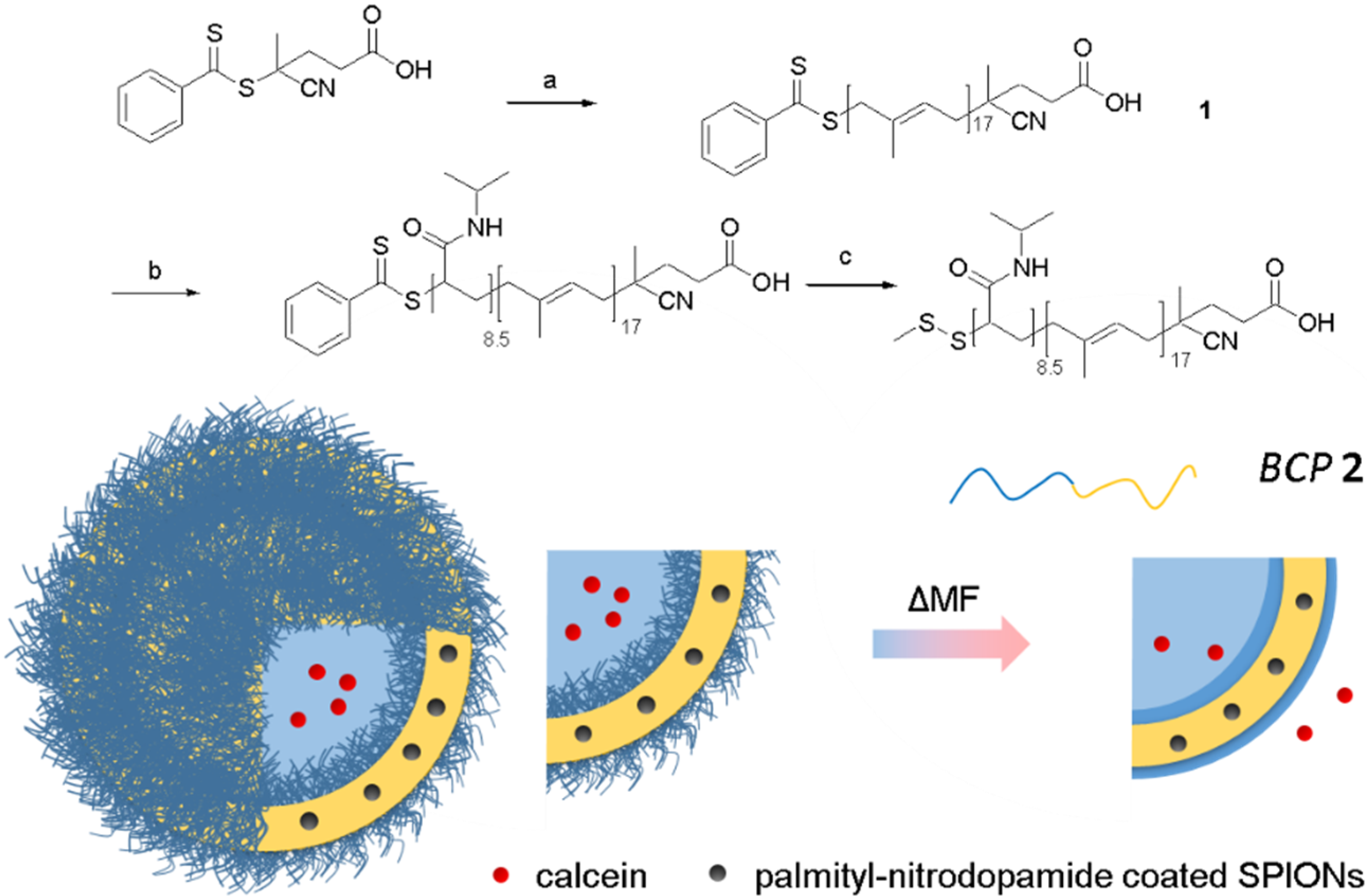
4.1. Synthesis
4.1.1. Polyisoprene MacroRAFT Agent (1): HOOC-PI(1300)-DTB
4.1.2. RAFT Terminated Diblock-Copolymer: HOOC-PI(1300)-b-PNIPAM(1000)-DTB
4.1.3. Disulfide Terminated Diblock-Copolymer (BCP 2): HOOC-PI(1300)-b-PNIPAM(1000)-SSMe (2)
4.2. Vesicle Formation and Release Study
4.2.1. Solvent Inversion
4.2.2. Release Assays
4.2.3. Magnetic Actuation
4.2.4. Fluorescence Measurements
4.3. Methods
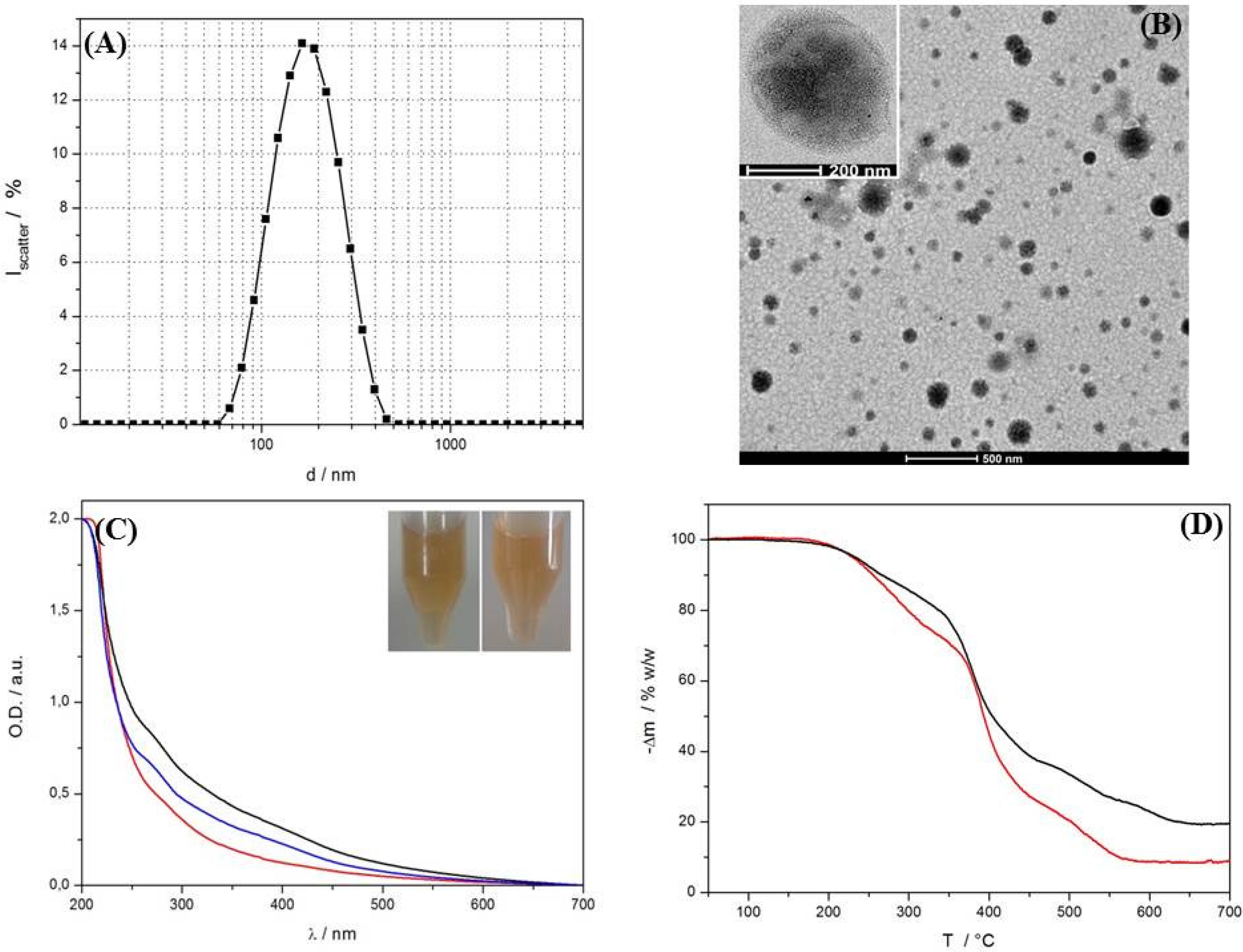
4.3.1. TEM
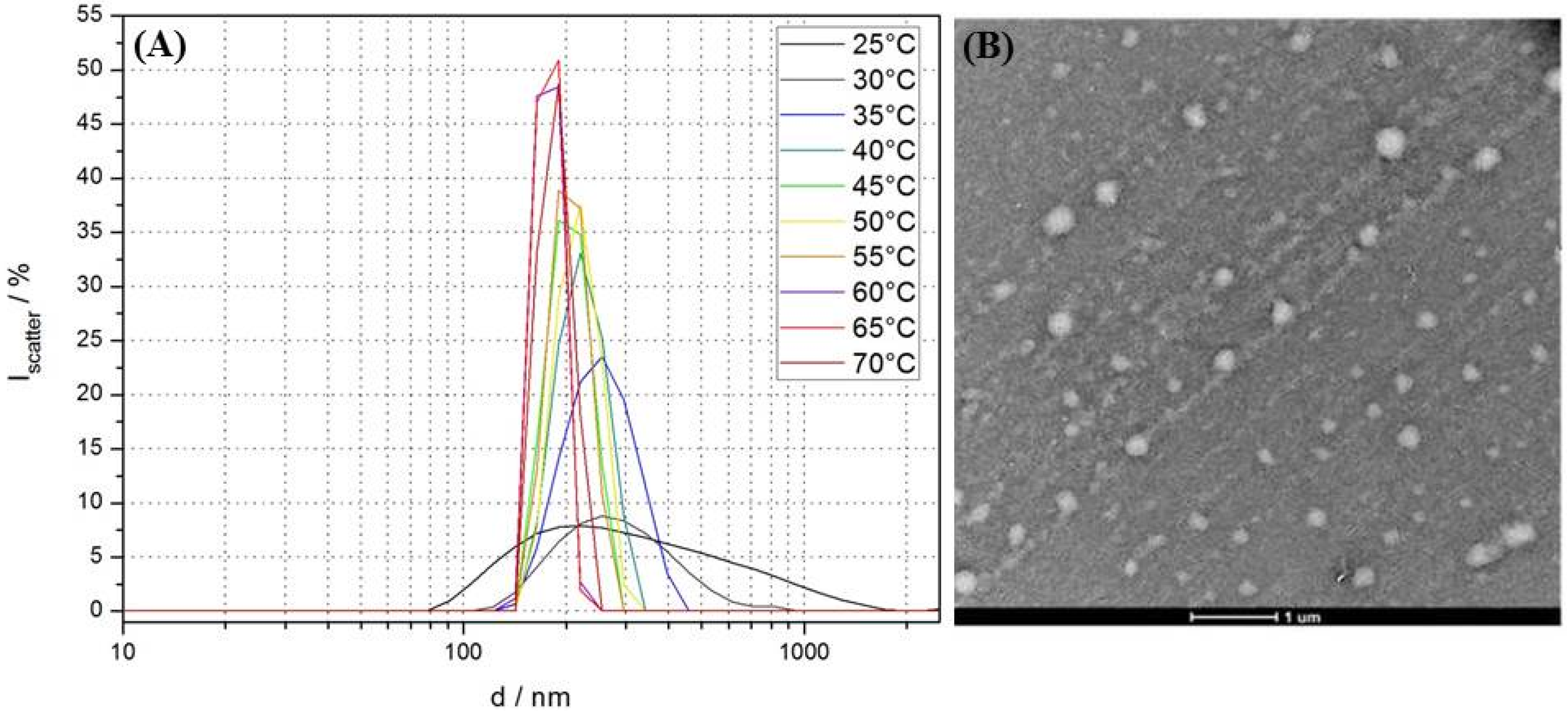
4.3.2. Dynamic Light Scattering
4.3.3. TGA/DSC Measurements
4.3.4. 1H- and 13C-NMR Measurements
4.3.5. MALDI-TOF-MS Measurements
4.3.6. ATR-FTIR Measurements
4.3.7. UV-Vis Measurements
5. Conclusions
Supplementary Materials
Acknowledgments
Author Contributions
Conflicts of Interest
References
- Lim Soo, P.; Eisenberg, A. Preparation of block copolymer vesicles in solution. J. Polym. Sci. B. Polym. Phys. 2004, 42, 923–938. [Google Scholar] [CrossRef]
- LoPresti, C.; Lomas, H.; Massignani, M.; Smart, T.; Battaglia, G. Polymersomes: Nature inspired nanometer sized compartments. J. Mater. Chem. 2009, 19, 3576–3590. [Google Scholar] [CrossRef]
- Mai, Y.; Eisenberg, A. Self-assembly of block copolymers. Chem. Soc. Rev. 2012, 41, 5969–5985. [Google Scholar] [CrossRef] [PubMed]
- Discher, D.E.; Ahmed, F. Polymersomes. Ann. Rev. Biomed. Eng. 2006, 8, 323–341. [Google Scholar] [CrossRef] [PubMed]
- Mura, S.; Nicolas, J.; Couvreur, P. Stimuli-responsive nanocarriers for drug delivery. Nat. Mater. 2013, 12, 991–1003. [Google Scholar] [CrossRef] [PubMed]
- Brinkhuis, R.P.; Rutjes, F.P.J.T.; van Hest, J.C.M. Polymeric vesicles in biomedical applications. Polym. Chem. 2011, 2, 1449–1462. [Google Scholar] [CrossRef]
- Onaca, O.; Enea, R.; Hughes, D.W.; Meier, W. Stimuli-responsive polymersomes as nanocarriers for drug and gene delivery. Macromol. Biosci. 2009, 9, 129–139. [Google Scholar] [CrossRef] [PubMed]
- Meng, F.; Zhong, Z.; Feijen, J. Stimuli-responsive polymersomes for programmed drug delivery. Biomacromolecules 2009, 10, 197–209. [Google Scholar] [CrossRef] [PubMed]
- Li, M.-H.; Keller, P. Stimuli-responsive polymer vesicles. Soft Matter 2009, 5, 927–937. [Google Scholar] [CrossRef]
- Ghoroghchian, P.P.; Lin, J.J.; Brannan, A.K.; Frail, P.R.; Bates, F.S.; Therien, M.J.; Hammer, D.A. Quantitative membrane loading of polymer vesicles. Soft Matter 2006, 2, 973–980. [Google Scholar] [CrossRef]
- Bermudez, H.; Brannan, A.K.; Hammer, D.A.; Bates, F.S.; Discher, D.E. Molecular weight dependence of polymersome membrane structure, elasticity, and stability. Macromolecules 2002, 35, 8203–8208. [Google Scholar] [CrossRef]
- Ahmed, F.; Discher, D.E. Self-porating polymersomes of PEG-PLA and PEG-PCL: Hydrolysis-triggered controlled release vesicles. J. Control. Release 2004, 96, 37–53. [Google Scholar] [CrossRef] [PubMed]
- Discher, B.M.; Hammer, D.A.; Bates, F.S.; Discher, D.E. Polymer vesicles in various media. Curr. Opin. Colloid Interface Sci. 2000, 5, 125–131. [Google Scholar] [CrossRef]
- Amstad, E.; Kim, S.-H.; Weitz, D.A. Photo- and thermoresponsive polymersomes for triggered release. Angew. Chem. Int. Ed. 2012, 51, 12499–12503. [Google Scholar] [CrossRef] [PubMed]
- Schulz, M.; Olubummo, A.; Binder, W.H. Beyond the lipid-bilayer: Interaction of polymers and nanoparticles with membranes. Soft Matter 2012, 8, 4849–4864. [Google Scholar] [CrossRef]
- Hickey, R.J.; Haynes, A.S.; Kikkawa, J.M.; Park, S.-J. Controlling the self-assembly structure of magnetic nanoparticles and amphiphilic block-copolymers: From micelles to vesicles. J. Am. Chem. Soc. 2011, 133, 1517–1525. [Google Scholar] [CrossRef] [PubMed]
- Hickey, R.J.; Koski, J.; Meng, X.; Riggleman, R.A.; Zhang, P.; Park, S.-J. Size-controlled self-assembly of superparamagnetic polymersomes. ACS Nano 2014, 8, 495–502. [Google Scholar] [CrossRef] [PubMed]
- Chen, D.; Shao, H.; Yao, W.; Huang, B. Fourier transform infrared spectral analysis of polyisoprene of a different microstructure. Int. J. Polym. Sci. 2013, 2013. [Google Scholar] [CrossRef]
- Qin, S.; Geng, Y.; Discher, D.E.; Yang, S. Temperature-controlled assembly and release from polymer vesicles of poly(ethylene oxide)-block-poly(N-isopropylacrylamide). Adv. Mater. 2006, 18, 2905–2909. [Google Scholar] [CrossRef]
- Kessel, S.; Truong, N.P.; Jia, Z.; Monteiro, M.J. Aqueous reversible addition-fragmentation chain transfer dispersion polymerization of thermoresponsive diblock copolymer assemblies: Temperature directed morphology transformations. J. Polym. Sci. Part A. Polym. Chem. 2012, 50, 4879–4887. [Google Scholar] [CrossRef]
- Morishima, Y. Thermally responsive polymer vesicles. Angew. Chem. Int. Ed. 2007, 46, 1370–1372. [Google Scholar] [CrossRef] [PubMed]
- Chen, X.; Ding, X.; Zheng, Z.; Peng, Y. Thermosensitive cross-linked polymer vesicles for controlled release system. New J. Chem. 2006, 30, 577–582. [Google Scholar] [CrossRef]
- Li, Y.; Lokitz, B.S.; McCormick, C.L. Thermally responsive vesicles and their structural “locking” through polyelectrolyte complex formation. Angew. Chem. Int. Ed. 2006, 45, 5792–5795. [Google Scholar] [CrossRef] [PubMed]
- Oliveira, H.; Perez-Andres, E.; Thevenot, J.; Sandre, O.; Berra, E.; Lecommandoux, S. Magnetic field triggered drug release from polymersomes for cancer therapeutics. J. Control. Release 2013, 169, 165–170. [Google Scholar] [CrossRef] [PubMed]
- Sanson, C.; Diou, O.; Thevenot, J.; Ibarboure, E.; Soum, A.; Brulet, A.; Miraux, S.; Thiaudiere, E.; Tan, S.; Brisson, A.; et al. Doxorubicin loaded magnetic polymersomes: theranostic nanocarriers for MR imaging and magneto-chemotherapy. ACS Nano 2011, 5, 1122–1140. [Google Scholar] [CrossRef] [PubMed]
- Ahmed, F.; Pakunlu, R.I.; Brannan, A.; Bates, F.; Minko, T.; Discher, D.E. Biodegradable polymersomes loaded with both paclitaxel and doxorubicin permeate and shrink tumors, inducing apoptosis in proportion to accumulated drug. J. Control. Release 2006, 116, 150–158. [Google Scholar] [CrossRef] [PubMed]
- Amstad, E.; Textor, M.; Reimhult, E. Stabilization and functionalization of iron oxide nanoparticles for biomedical applications. Nanoscale 2011, 3, 2819–2843. [Google Scholar] [CrossRef] [PubMed]
- Reimhult, E. Nanoparticle-triggered release from lipid membrane vesicles. New Biotechnol. 2015, 32, 665–672. [Google Scholar] [CrossRef] [PubMed]
- Amstad, E.; Kohlbrecher, J.; Muller, E.; Schweizer, T.; Textor, M.; Reimhult, E. Triggered release from liposomes through magnetic actuation of iron oxide nanoparticle containing membranes. Nano Lett. 2011, 11, 1664–1670. [Google Scholar] [CrossRef] [PubMed]
- Roy, D.; Brooks, W.L.A.; Sumerlin, B.S. New directions in thermoresponsive polymers. Chem. Soc. Rev. 2013, 42, 7214–7243. [Google Scholar] [CrossRef] [PubMed]
- Aseyev, V.; Tenhu, H.; Winnik, F.M. Non-ionic thermoresponsive polymers in water. In Self Organized Nanostructures of Amphiphilic Block Copolymers II; Muller, A., Borisov, O., Eds.; Springer-Verlag: Berlin/Heidelberg, Germany, 2011; Volume 242, pp. 29–89. [Google Scholar]
- Chiang, W.-H.; Huang, W.-C.; Chang, C.-W.; Shen, M.-Y.; Shih, Z.-F.; Huang, Y.-F.; Lin, S.-C.; Chiu, H.-C. Functionalized polymersomes with outlayered polyelectrolyte gels for potential tumor-targeted delivery of multimodal therapies and MRI imaging. J. Control. Release 2013, 168, 280–288. [Google Scholar] [CrossRef] [PubMed]
- Bixner, O.; Lassenberger, A.; Baurech, D.; Reimhult, E. Complete exchange of the hydrophobic dispersant shell on monodisperse superparamagnetic iron oxide nanoparticles. Langmuir 2015, 31, 9198–9204. [Google Scholar] [CrossRef] [PubMed]
- Kita-Tokarczyk, K.; Grumelard, J.; Haefele, T.; Meier, W. Block copolymer vesicles—Using concepts from polymer chemistry to mimic biomembranes. Polymer 2005, 46, 3540–3563. [Google Scholar] [CrossRef]
- Bixner, O.; Reimhult, E. Controlled magnetosomes: Embedding of magnetic nanoparticles into membranes of monodisperse lipid vesicles. J. Colloid Interface Sci. 2016, 466, 62–71. [Google Scholar] [CrossRef] [PubMed]
- Zhu, X.; Yan, C.; Winnik, F.M.; Leckband, D. End-grafted low-molecular-weight PNIPAM does not collapse above the LCST†. Langmuir 2007, 23, 162–169. [Google Scholar] [CrossRef] [PubMed]
- Deatsch, A.E.; Evans, B.A. Heating efficiency in magnetic nanoparticle hyperthermia. J. Magn. Magn. Mater. 2014, 354, 163–172. [Google Scholar] [CrossRef]
- Ling, D.; Hyeon, T. Chemical design of biocompatible iron oxide nanoparticles for medical applications. Small 2013, 9, 1450–1466. [Google Scholar] [CrossRef] [PubMed]
- Wang, J.; Pantopoulos, K. Regulation of cellular iron metabolism. Biochem. J. 2011, 434, 365–381. [Google Scholar] [CrossRef] [PubMed]
- Photos, P.J.; Bacakova, L.; Discher, B.; Bates, F.S.; Discher, D.E. Polymer vesicles in vivo: Correlations with PEG molecular weight. J. Control. Release 2003, 90, 323–334. [Google Scholar] [CrossRef]
- Cooperstein, M.A.; Canavan, H.E. Assessment of cytotoxicity of (N-isopropyl acrylamide) and Poly(N-isopropyl acrylamide)-coated surfaces. Biointerphases 2013, 8. [Google Scholar] [CrossRef] [PubMed]
- Li, J.; El harfi, J.; Howdle, S.M.; Carmichael, K.; Irvine, D.J. Controlled oligomerisation of isoprene-towards the synthesis of squalene analogues. Polym. Chem. 2012, 3, 1495–1501. [Google Scholar] [CrossRef]
- Rozentsvet, V.A.; Khachaturov, A.S.; Ivanova, V.P. Microstructure of polyisoprene produced by cationic polymerization: NMR spectroscopic study. Polym. Sci. Ser. A 2009, 51, 870–876. [Google Scholar] [CrossRef]
- Shan, J.; Zhao, Y.; Granqvist, N.; Tenhu, H. Thermoresponsive properties of N-isopropylacrylamide oligomer brushes grafted to gold nanoparticles: Effects of molar mass and gold core size. Macromolecules 2009, 42, 2696–2701. [Google Scholar] [CrossRef]
- Roth, P.J.; Kessler, D.; Zentel, R.; Theato, P. A method for obtaining defined end groups of polymethacrylates prepared by the raft process during aminolysis. Macromolecules 2008, 41, 8316–8319. [Google Scholar] [CrossRef]
© 2016 by the authors; licensee MDPI, Basel, Switzerland. This article is an open access article distributed under the terms and conditions of the Creative Commons by Attribution (CC-BY) license (http://creativecommons.org/licenses/by/4.0/).
Share and Cite
Bixner, O.; Kurzhals, S.; Virk, M.; Reimhult, E. Triggered Release from Thermoresponsive Polymersomes with Superparamagnetic Membranes. Materials 2016, 9, 29. https://doi.org/10.3390/ma9010029
Bixner O, Kurzhals S, Virk M, Reimhult E. Triggered Release from Thermoresponsive Polymersomes with Superparamagnetic Membranes. Materials. 2016; 9(1):29. https://doi.org/10.3390/ma9010029
Chicago/Turabian StyleBixner, Oliver, Steffen Kurzhals, Mudassar Virk, and Erik Reimhult. 2016. "Triggered Release from Thermoresponsive Polymersomes with Superparamagnetic Membranes" Materials 9, no. 1: 29. https://doi.org/10.3390/ma9010029