
The Observation of Cellular Precipitation in an Ni36Co18Cr20Fe19Al7 High-Entropy Alloy after Quenching and Annealing


Department of Mechanical Engineering, National Taiwan University of Science and Technology, 43 Keelung Road, Section 4, Taipei 106, Taiwan
*
Author to whom correspondence should be addressed.
Materials 2022, 15(19), 6613; https://doi.org/10.3390/ma15196613
Submission received: 24 August 2022
/
Revised: 18 September 2022
/
Accepted: 19 September 2022
/
Published: 23 September 2022
(This article belongs to the Special Issue Future Trends in High-Entropy Alloys)
Abstract
:High-entropy alloys (HEAs) comprise a minimum of five major elements. These alloys show some special characteristics, such as excellent mechanical and high temperature properties. The development of the HEAs requires a knowledge of phase transformations during alloy making procedures. The phase transformations of an Ni36Co18Cr20Fe19Al7 HEA were studied in this research. The alloy underwent hot forging, cold rolling, annealing at and quenching from 1323 K, and isothermal holding at 873 K. The alloy is a single face-centered cubic (FCC) phase in the as-quenched condition. After annealing at 873 K, not only fine coherent L12 particles precipitated homogeneously in the FCC matrix, but lamellae of FCC and L12 phases also developed from the grain boundaries. Both lamellar FCC and L12 grains have a cubic-on-cubic orientation relationship (OR). The composition of the lamellar L12 phase is Ni60Co8Cr6Fe6Al20, and that of the lamellar FCC phase is Ni31Co15Cr28Fe21Al4. Cellular precipitation occurs in the HEA, and the high-temperature FCC (γ) transforms to a lamella of low-temperature FCC (γ1), and an L12 phase, i.e., γ → γ1+L12.
1. Introduction
High-entropy alloys are composed of a minimum of five components, whose concentrations are approximately between 5 and 35 at.%. HEAs show some distinct features, for example, excellent mechanical strength, corrosion resistance, wear resistance, and high temperature oxidation properties. Since the high-entropy alloys may have various outstanding properties, they are of considerable importance in engineering applications [1,2,3]. In the study of the HEAs containing aluminum by transmission electron microscope (TEM) observations, phase transformations—such as precipitation transformation [4,5,6], spinodal decomposition [7,8,9,10,11], and ordering reaction [12,13]—were found, and their product phases were L12 [14,15,16], and B2 [17,18,19,20,21,22] superlattices.
The precipitation transformation begins with the appearance of a second phase (β), from the supersaturated matrix phase (α) of an alloy after cooling from high temperature. The high-temperature phase has, thus, been changed to the low-temperature phase (α1). The phase transformation is as follows: α → α1 + β. The concentration of the low-temperature α1 phase is different from the high-temperature α phase, but with the same crystal structure and orientation. The second phase (β) prefers to precipitate at the grain boundary and is called grain-boundary precipitate, and it has a different composition and a different crystal structure from the matrix phase. A necessary condition for the occurrence of the precipitation transformation is the long-distant diffusion of atoms through the matrix phase. Grain-boundary precipitation always results in the appearance of separate grains, allotriomorphs, Widmanstätten plates, and/or needles [23].
A special type of precipitation transformation is known as cellular precipitation. There are three distinct characteristics for the occurrence of cellular precipitation, which are as follows: The first characteristic is that the product phases are in the form of lamellar grains, which develop from the grain boundary. The second feature is that the lamellar α1 grains have the same orientation as that of one neighboring α grain, but a different orientation from the other. The third distinctive characteristic is that the grain boundary migrates, i.e., the grain boundary moves with the growing fronts of the cellular product phases; therefore, the newly formed lamellae nucleate at the grain boundary, where the α1 grain has the same orientation as one neighboring α grain and grows, simultaneously, into the other neighboring α grain with the movement of the original grain boundary [23,24,25]. Cellular precipitation occurs in some alloy steels. For example, in Fe-Mn-Al steel, after quenching and annealing, cellular precipitation occurs, and high-temperature FCC decomposes into lamellae of low-temperature FCC and κ-carbide, at the grain boundary [26]. The motivation for this paper was to report the occurrence of interesting cellular precipitation in an Al-containing HEA.
2. Experimental Procedures
By induction melting in a vacuum chamber, a 10-kg Ni36Co18Cr20Fe19Al7 ingot was formed by melting the following metals with high purity together: nickel, cobalt, chromium, iron, and aluminum. To ensure the uniform alloy composition, the ingot was homogenized at 1473 K, for 4 h, under an argon-protected atmosphere; hot forged and re-annealed at 1473 K, for at least 3 cycles; cut into plates; annealed at 1473 K for 4 h; and quenched. The plates were cold rolled into 2 mm plates at room temperature, and cut into samples, measuring 10 mm × 10 mm. In an argon atmosphere, samples were heated at 1323 K, for 1 h, and then water quenched. Under vacuum, specimens were sealed in quartz tubes and held isothermally at 873 K, for 24 h. After metallographic sample preparation, the samples were observed by using an optical microscope and a Jeol JEM 6500F high-resolution, field-emission scanning electron microscope (SEM, Jeol Ltd., Akishima, Tokyo, Japan). The crystal structure of the as-quenched HEA in powder form, sealed in a vacuum quartz tube, was analyzed by adopting X-ray diffraction (XRD) in the Taiwan photon synchrotron light source, of national synchrotron radiation center, Hsinchu, Taiwan, operated at 15 keV and a wavelength of 0.082656 nm. The sample, in the form of alloy plates annealed at 873 K, were also examined by XRD, in a Rigaku DMAX-B X-ray diffractometer, operated at a power of 12 kW. TEM samples were prepared by thinning 80 μm thin foils, punching them into discs with a diameter of 3 mm, and electro-polishing them in a 90% acetic acid and 10% perchloric acid solution. The TEM, with the brand of FEI (Thermo Fisher Scientific) Talos F200XG2 (Thermo Fisher Scientific, Waltham, MA, US), equipped with the energy dispersive spectroscopy (EDS) was utilized to analyze the TEM samples. The TEM operation voltage was 200 kV.
3. Results and Discussion
Figure 1 shows the results of the investigation on the Ni36Co18Cr20Fe19Al7 HEA, after the solution treatment at 1323 K. An optical micrograph (OM) and a secondary electron image (SEI) from the SEM in Figure 1a,b, respectively, illustrate that the HEA has similar grains with annealing twins. Only FCC peaks were detected by the synchrotron-based XRD, as shown in Figure 1c. Therefore, the HEA is a single phase of FCC after the solution treatment at 1323 K, and the lattice parameter of the FCC phase is approximately 0.3589 nm 5 nm.
After understanding the constituent phase of the as-quenched HEA, we examined the crystal structures of the HEA that was held isothermally, at 873 K. The results from the OM, SEI, and XRD studies are shown in Figure 2a–c, respectively. From the OM observation in Figure 2a, the FCC grains with twins remain as the matrix phase. However, besides the matrix FCC phase, the other phases precipitate at the grain boundaries. Separate grains and lamellar colonies appear in the alloy simultaneously, as shown in Figure 2b. The analysis of the XRD, as shown in Figure 2c, reveals that the HEA has a major phase of FCC, and that the crystal structures of the second phases, precipitating at the grain boundaries, cannot be clearly identified. Therefore, we applied the TEM to study the grain boundary precipitates in detail.
The TEM analysis of the HEA annealed at 873 K is illustrated in Figure 3. We focused the TEM study on the grain-boundary colony, as well as the FCC matrix. A selected area diffraction pattern (SADP), taken on the FCC matrix, is shown in Figure 3a. We applied the smallest SADP aperture, with a diameter of approximately 100 nm, to take the SADP. The zone axis of the FCC phase on the SADP in Figure 3a is along the [011] direction. The SADP in Figure 3a reveals the appearance of extra superlattice (100) reflections. These reflections are from the L12 superlattice phase. The zone axis of the L12 phase on the SADP is also along the [011] direction. Therefore, there is a cubic-on-cubic orientation relationship (OR) between the L12 precipitate and the FCC matrix, i.e., [011]L12//[011]γ and (200)L12//(200)γ. As shown in Figure 3a, the Miller indexes of the L12 reflections are underlined to differentiate them from those of the FCC reflections. In Figure 3b, a dark-field (DF) image, taken from the superlattice reflection of the L12 (100), manifests fine coherent particles that are distributed uniformly in the FCC matrix. Thus, the L12 superlattice phase precipitates homogeneously in the FCC matrix, as fine coherent particles during annealing. The corresponding bright-field (BF) image in Figure 3c shows that the fine L12 particles associated with the strain energy field can also be clearly seen. We estimated the particle shape as a sphere, and the spherical particles have a diameter of approximately 2 nm. The L12 phase has a Cu3Au crystal structure, which is derived from its parent FCC phase, and has the space group, Pmm. In the Al-content HEAs, fine coherent L12 particles precipitating homogeneously in the FCC phase were reported previously [14,15,16].
In addition to the homogeneous precipitation of the fine L12 particles in the FCC grains, grain-boundary precipitates not only appear in the form of separate grains, but also as colonies from the SEI observation, as shown in Figure 2b. The TEM investigation of the colony is shown in Figure 3d–f. An SADP (taken on the lamellae) similar to that in Figure 3a is shown in Figure 3d. The zone axes of both the FCC and L12 superlattice on the SADP are all along the [011] directions. A DF image in Figure 3e, taken from the 100 superlattice reflection of the L12 phase, illustrates the locations of the lamellar L12 grains (with a white contrast) in the lamellae. It is worth noting that, in the colony, the dark lamellar grains between the white ones are FCC. The corresponding BF image in Figure 3f presents the following three sections: an FCC grain at the left-hand side, with a bright contrast; a colony in the middle, with a dark contrast; and an FCC grain at the right-hand side, with the same contrast as the colony. The colony is located between two FCC grains. During the TEM operation at the same tilting condition, the same contrast for both the lamellae and FCC grain demonstrated that both orientations are identical, as was also confirmed by both SADPs. This means that the lamellar FCC grains nucleate at the grain boundary, with the same orientation as the FCC grain at the right-hand side and have a cubic-on-cubic OR, along with the lamellar L12 grains. Thus, the fact that colonies of lamellar FCC and L12 grains develop from the grain boundaries has been confirmed. The precipitation of the lamellar FCC and L12 phases is achieved via cellular precipitation [23,24,25]. Therefore, cellular precipitation occurs in the HEA, and the phase transformation is as follows: γ → γ1 + L12. The lamellar L12 and γ1 FCC grains are the product phases of the cellular precipitation.
The lamellae of both the FCC and L12 phases nucleate and grow simultaneously at the grain boundary. The lamellar FCC grains have the same orientation as that of the right FCC matrix and have a cubic-on-cubic OR with the lamellar L12 grains. The lamellar phases grow side-by-side, forward, and to the left of the FCC grain. Figure 3f illustrates more evidence for the occurrence of cellular precipitation. The BF image in Figure 3f shows the original grain boundary between two FCC grains, located near the centered vertical line, or, in other words, located at the right-hand side boundary of the colony. The lamellae nucleate at the grain boundary and grow into the FCC grain at the left-hand side, with a curved boundary. As shown in Figure 3e, it is not only fine white L12 particles—which are the same as those in Figure 3b—that appear uniformly in the right FCC grain, but white L12 lamellar grains also appear in the lamellae. This reveals more evidence that the lamellar FCC grains in the newly formed lamellae have the same orientation as that of the right-neighboring FCC grain. However, the lamellae grow into the left-neighboring FCC grain in a different orientation. Therefore, all the above satisfy the distinct characteristics of cellular precipitation, i.e., the lamellar FCC grains, along with the L12 grains, nucleate at the grain boundary with the same orientation as one neighboring FCC grain and grow into the other neighboring FCC grain (in a different orientation), with the movement of the grain boundary. These are the distinctive features of cellular precipitation [23]. Therefore, the occurrence of cellular precipitation for the transformation of the high-temperature FCC phase to the low-temperature FCC and L12 phases has been addressed for the first time in the Al-contained NiCoCrFe HEAs.
The concentrations of the lamellar phases in the colonies were studied by EDS, in the STEM mode of the Tolas TEM. For example, the composition line scans of the lamellar grains are shown in Figure 4. Figure 4a illustrates a BF image covering a section of the lamellae, in which the thinner lamellar grains with a dark contrast are the L12 phase and the thicker lamellar grains with a grey contrast are the FCC phase. The concentrations of the constituent elements were measured by the line scans on the white solid line, as drawn on the BF image in Figure 4a. The concentration curves for the following elements: Ni, Al, Co, Cr, and Fe, are shown in Figure 4b–f, respectively. The average composition (at.%) of the lamellar L12 phase in the following sequential elements of the Al, Co, Cr, Fe, and Ni elements is approximately 19.9, 8.1, 5.8, 6.4, and 59.8 at.%, respectively, and the FCC is approximately 4.3, 15.3, 28.4, 21.2, and 30.8 at.%, respectively. The margin of error in the elemental composition is approximately 10%. In the other form, the lamellar L12 phase has the following composition: Ni60Co8Cr6Fe6Al20, and the FCC grain is as follows: Ni31Co15Cr28Fe21Al4. This result shows that the lamellar L12 grains are rich in Ni and Al, and lean in Co, Cr, and Fe, and that the lamellar FCC grains are vice versa. The ratio of the concentration of Ni to that of Al in the lamellar L12 phase is approximately three, and it means that the L12 phase is approaching the Ni3Al phase.
4. Conclusions
The phase transformations of the Ni36Co18Cr20Fe19Al7 HEA have been studied in this research paper. The alloy underwent hot forging, cold rolling, annealing at and quenching from 1323 K, and isothermal holding, at 873 K. The as-quenched HEA is a single FCC. After the 873-K isothermal holding, the coherent fine L12 particles precipitated homogeneously in the FCC matrix. In addition, cellular precipitation occurred, resulting in the transformation of the high-temperature FCC phase into lamellae of the low-temperature FCC phase and L12 phase. The colonies nucleated and grew from the grain boundaries. The grain boundary moved with the growing tips of the newly formed lamellar FCC and L12 phases, and the lamellar FCC grains had the same orientation as that of one neighboring FCC grain and grew into the other neighboring FCC grain, with a different orientation. The lamellar L12 grains had the following composition: Ni60Co8Cr6Fe6Al20, and the FCC grains had the following composition: Ni31Co15Cr28Fe21Al4. The lamellar L12 phase was rich in Ni and Al, and lean in Co, Cr, and Fe, and the lamellar FCC phase was contrary to this.
Author Contributions
Conceptualization, W.-C.C.; methodology, G.R.K. and K.R.C.; formal analysis, G.R.K.; investigation, G.R.K. and K.R.C.; writing—original draft preparation, W.-C.C.; writing—review and editing, W.-C.C.; visualization, W.-C.C.; supervision, W.-C.C.; project administration, W.-C.C.; funding acquisition, W.-C.C. All authors have read and agreed to the published version of the manuscript.
Funding
This research was supported by the Ministry of Science and Technology (Taiwan), under Grant no. MOST-108-2218-E-011-008.
Institutional Review Board Statement
Not applicable.
Informed Consent Statement
Not applicable.
Data Availability Statement
Not applicable.
Conflicts of Interest
The authors declare no conflict of interest.
References
- Tsai, M.H.; Yeh, J.W. High-Entropy Alloys: A Critical Review. Mater. Res. Lett. 2014, 2, 107–123. [Google Scholar] [CrossRef]
- Miracle, D.B.; Senkov, O.N. A Critical Review of High Entropy Alloys and Related Concepts. Acta Mater. 2017, 122, 448–511. [Google Scholar] [CrossRef]
- Qiao, J.W.; Ma, S.G.; Huang, E.W.; Chuang, C.P.; Liaw, P.K.; Zhang, Y. Microstructural Characteristics and Mechanical Behaviors of AlCoCrFeNi High-Entropy Alloys at Ambient and Cryogenic Temperatures. Mater. Sci. Forum 2011, 688, 419–425. [Google Scholar] [CrossRef]
- Zhu, J.M.; Fu, H.M.; Zhang, H.F.; Wang, A.M.; Li, H.; Hu, Z.Q. Microstructure and Compressive Properties of Multiprincipal Component AlCoCrFeNiCX Alloys. J. Alloys Compd. 2011, 509, 3476–3480. [Google Scholar] [CrossRef]
- Shun, T.T.; Du, Y.C. Microstructure and Tensile Behaviors of FCC Al0.3CoCrFeNi High Entropy Alloy. J. Alloys Compd. 2009, 479, 157–160. [Google Scholar] [CrossRef]
- Gwalani, B.; Soni, V.; Choudhuri, D.; Lee, M.; Hwang, J.Y.; Nam, S.J.; Ryu, H.; Hong, S.H.; Banerjee, R. Stability of Ordered L12 and B2 Precipitates in Face Centered Cubic Based High Entropy Alloys-Al0.3CoFeCrNi and Al0.3CuFeCrNi2. Scr. Mater. 2016, 123, 130–134. [Google Scholar] [CrossRef]
- Butler, T.M.; Weaver, M.L. Investigation of the Phase Stabilities in AlNiCoCrFe High Entropy Alloys. J. Alloys Compd. 2017, 691, 119–129. [Google Scholar] [CrossRef]
- Manzoni, A.; Daoud, H.; Völkl, R.; Glatzel, U.; Wanderka, N. Phase Separation in Equiatomic AlCoCrFeNi High-Entropy Alloy. Ultramicroscopy 2013, 132, 212–215. [Google Scholar] [CrossRef]
- Lin, C.M.; Tsai, H.L. Evolution of Microstructure, Hardness, and Corrosion Properties of High-Entropy Al0.5CoCrFeNi Alloy. Intermetallics 2011, 19, 288–294. [Google Scholar] [CrossRef]
- Kao, Y.F.; Chen, T.J.; Chen, S.K.; Yeh, J.-W. Microstructure and Mechanical Property of As-Cast, -Homogenized, and -Deformed AlXCoCrFeNi (0 ≤ x ≤ 2) High-Entropy Alloys. J. Alloys Compd. 2009, 488, 57–64. [Google Scholar] [CrossRef]
- Dong, Y.; Zhou, K.; Lu, Y.; Gao, X.; Wang, T.; Li, T. Effect of Vanadium Addition on the Microstructure and Properties of AlCoCrFeNi High Entropy Alloy. Mater. Des. 2014, 57, 67–72. [Google Scholar] [CrossRef]
- Chen, J.; Niu, P.; Liu, Y.; Lu, Y.; Wang, X.; Peng, Y.; Liu, J. Effect of Zr Content on Microstructure and Mechanical Properties of AlCoCrFeNi High Entropy Alloy. Mater. Des. 2016, 94, 39–44. [Google Scholar] [CrossRef]
- Butler, T.M.; Weaver, M.L. Oxidation Behavior of Arc Melted AlCoCrFeNi Multi-Component High-Entropy Alloys. J. Alloys Compd. 2016, 674, 229–244. [Google Scholar] [CrossRef]
- Mohanty, S.; Maity, T.N.; Mukhopadhyay, S.; Sarkar, S.; Gurao, N.P.; Bhowmick, S.; Biswas, K. Powder Metallurgical Processing of Equiatomic AlCoCrFeNi High Entropy Alloy: Microstructure and Mechanical Properties. Mater. Sci. Eng. A 2017, 679, 299–313. [Google Scholar] [CrossRef]
- Xu, X.D.; Liu, P.; Guo, S.; Hirata, A.; Fujita, T.; Nieh, T.G.; Liu, C.T.; Chen, M.W. Nanoscale Phase Separation in a Fcc-Based CoCrCuFeNiAl0.5 High-Entropy Alloy. Acta Mater. 2015, 84, 145–152. [Google Scholar] [CrossRef]
- Wani, I.S.; Bhattacharjee, T.; Sheikh, S.; Bhattacharjee, P.P.; Guo, S.; Tsuji, N. Tailoring Nanostructures and Mechanical Properties of AlCoCrFeNi2.1 Eutectic High Entropy Alloy Using Thermo-Mechanical Processing. Mater. Sci. Eng. A 2016, 675, 99–109. [Google Scholar] [CrossRef]
- Wang, W.R.; Wang, W.L.; Wang, S.C.; Tsai, Y.C.; Lai, C.H.; Yeh, J.W. Effects of Al Addition on the Microstructure and Mechanical Property of AlXCoCrFeNi High-Entropy Alloys. Intermetallics 2012, 26, 44–51. [Google Scholar] [CrossRef]
- Wang, W.R.; Wang, W.L.; Yeh, J.W. Phases, Microstructure and Mechanical Properties of AlXCoCrFeNi High-Entropy Alloys at Elevated Temperatures. J. Alloys Compd. 2014, 589, 143–152. [Google Scholar] [CrossRef]
- Lv, Y.; Hu, R.; Yao, Z.; Chen, J.; Xu, D.; Liu, Y.; Fan, X. Cooling Rate Effect on Microstructure and Mechanical Properties of AlXCoCrFeNi High Entropy Alloys. Mater. Des. 2017, 132, 392–399. [Google Scholar] [CrossRef]
- Munitz, A.; Salhov, S.; Hayun, S.; Frage, N. Heat Treatment Impacts the Micro-Structure and Mechanical Properties of AlCoCrFeNi High Entropy Alloy. J. Alloys Compd. 2016, 683, 221–230. [Google Scholar] [CrossRef]
- Lee, K.S.; Bae, B.; Kang, J.H.; Lim, K.R.; Na, Y.S. Multi-Phase Refining of an AlCoCrFeNi High Entropy Alloy by Hot Compression. Mater. Lett. 2017, 198, 81–84. [Google Scholar] [CrossRef]
- Tang, Z.; Senkov, O.N.; Parish, C.M.; Zhang, C.; Zhang, F.; Santodonato, L.J.; Wang, G.; Zhao, G.; Yang, F.; Liaw, P.K. Tensile Ductility of an AlCoCrFeNi Multi-Phase High-Entropy Alloy through Hot Isostatic Pressing (HIP) and Homogenization. Mater. Sci. Eng. A 2015, 647, 229–240. [Google Scholar] [CrossRef]
- Ecob, R.C.; Bee, J.V.; Ralph, B. The Cellular Reaction in Dilute Copper-Titanium Alloys. Metall. Trans. A 1980, 11, 1407–1414. [Google Scholar] [CrossRef]
- Miki, M.; Laughlin, D.E. Cellular Decomposition in a Cu-25Ni-15Co Side-Band Alloy. Metall. Trans. A 1985, 16, 1751–1757. [Google Scholar] [CrossRef]
- Aaronson, H.I.; Pande, C.S. A Synthesis of Mechanisms for Initiation of the Cellular (or Discontinuous Precipitation) Reaction. Acta Mater. 1998, 47, 175–181. [Google Scholar] [CrossRef]
- Cheng, W.C.; Cheng, C.Y.; Hsu, C.W.; Laughlin, D.E. Phase Transformation of the L12 Phase to Kappa-Carbide after Spinodal Decomposition and Ordering in an Fe-C-Mn-Al Austenitic Steel. Mater. Sci. Eng. A 2015, 642, 128–135. [Google Scholar] [CrossRef]
Figure 1.
The investigation of the HEA after solution treatment at 1323 K, for 1 h: (a) OM, (b) SEI, and (c) XRD. Note that the unmarked broad peak at approximately 11 degrees of the diffraction angle is the signal from the quartz tube.
Figure 1.
The investigation of the HEA after solution treatment at 1323 K, for 1 h: (a) OM, (b) SEI, and (c) XRD. Note that the unmarked broad peak at approximately 11 degrees of the diffraction angle is the signal from the quartz tube.
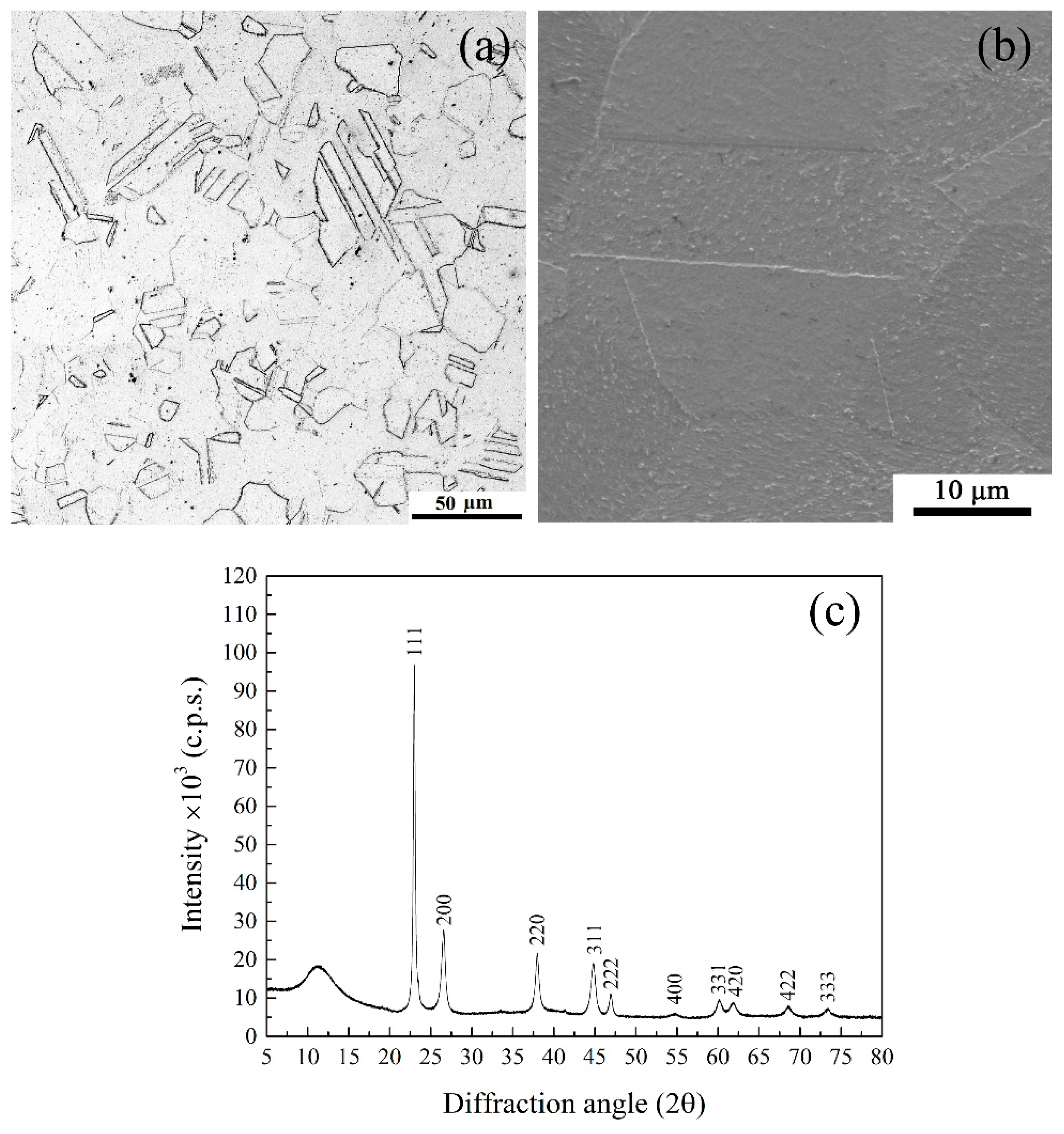
Figure 2.
(a) OM, (b) SEI, and (c) XRD of the HEA after quenching, from 1323 K, and isothermal holding at 873 K, for 24 h.
Figure 2.
(a) OM, (b) SEI, and (c) XRD of the HEA after quenching, from 1323 K, and isothermal holding at 873 K, for 24 h.
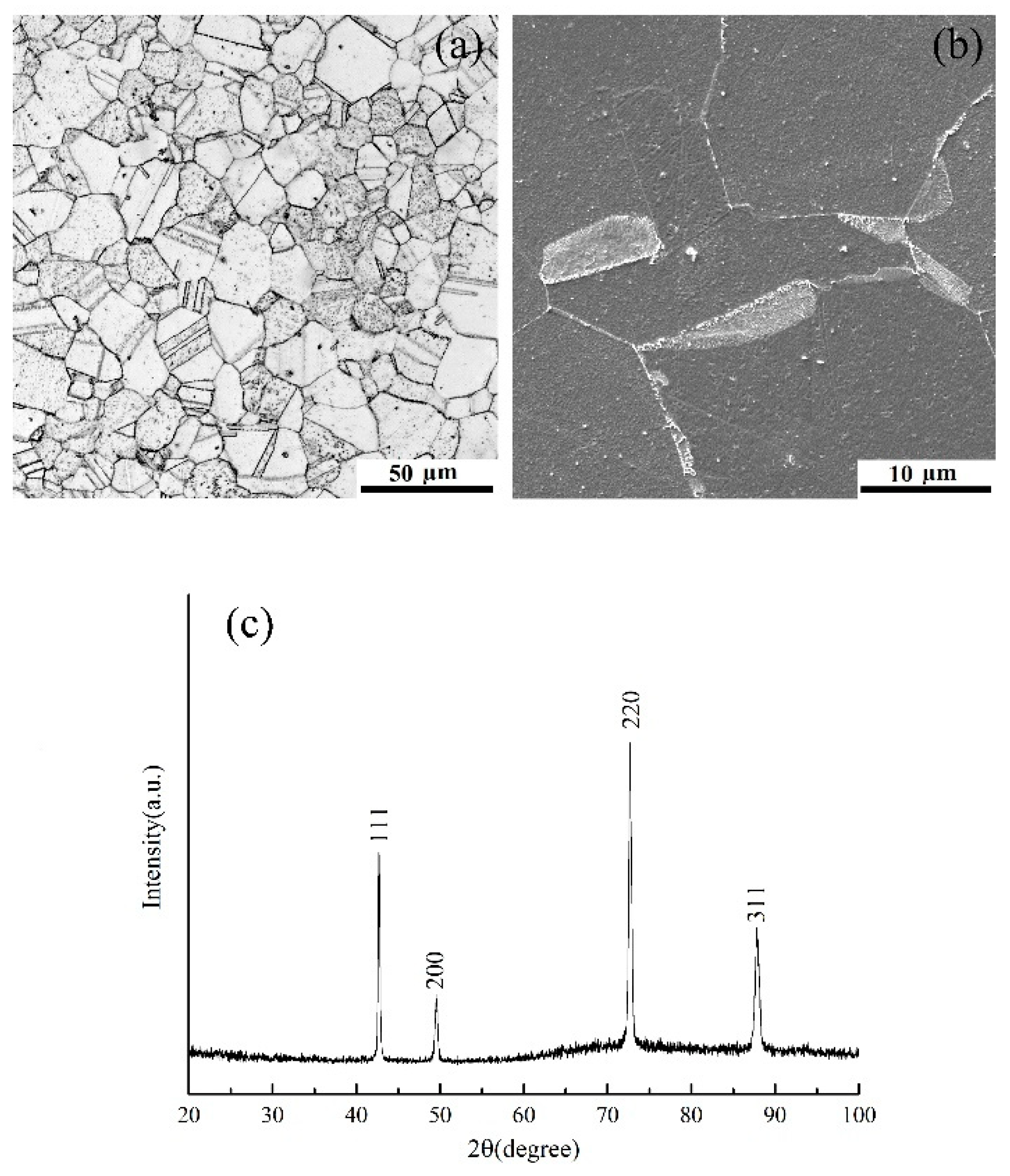
Figure 3.
The TEM analysis of the HEA with the same heat treatment as Figure 2: (a) [011] SADP taken from the FCC matrix; (b) DF image taken from the L12 (100) reflection in (a); (c) the BF image corresponding to the DF image in (b); (d) [011] SADP taken from the lamellae at the grain boundary; (e) the DF image taken from the L12 (100) reflection; and (f) the BF image corresponding to (e).
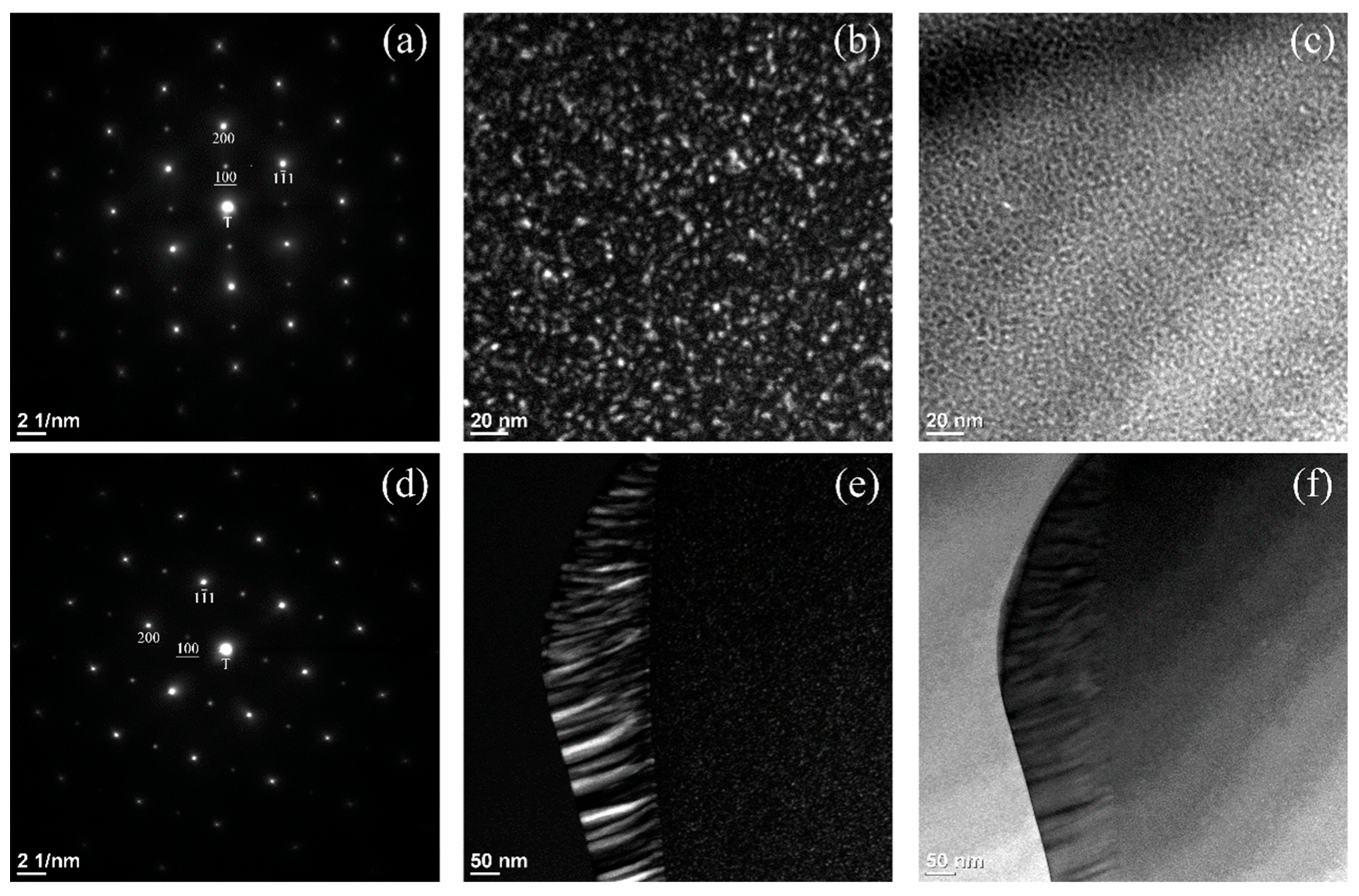
Figure 4.
The TEM study of the HEA in STEM mode to illustrate the composition differences between the lamellar grains: (a) BF image taken on the lamellae. The concentration line scans by EDS, along the white solid line marked in (a), as follows: (b) Ni, (c) Al, (d) Co, (e) Cr, and (f) Fe. The specimen had the same heat treatment as that in Figure 2.

Publisher’s Note: MDPI stays neutral with regard to jurisdictional claims in published maps and institutional affiliations. |
© 2022 by the authors. Licensee MDPI, Basel, Switzerland. This article is an open access article distributed under the terms and conditions of the Creative Commons Attribution (CC BY) license (https://creativecommons.org/licenses/by/4.0/).
Share and Cite
MDPI and ACS Style
Kenedy, G.R.; Chemeli, K.R.; Cheng, W.-C. The Observation of Cellular Precipitation in an Ni36Co18Cr20Fe19Al7 High-Entropy Alloy after Quenching and Annealing. Materials 2022, 15, 6613. https://doi.org/10.3390/ma15196613
AMA Style
Kenedy GR, Chemeli KR, Cheng W-C. The Observation of Cellular Precipitation in an Ni36Co18Cr20Fe19Al7 High-Entropy Alloy after Quenching and Annealing. Materials. 2022; 15(19):6613. https://doi.org/10.3390/ma15196613
Chicago/Turabian StyleKenedy, Gurumayum Robert, Korir Rosemary Chemeli, and Wei-Chun Cheng. 2022. "The Observation of Cellular Precipitation in an Ni36Co18Cr20Fe19Al7 High-Entropy Alloy after Quenching and Annealing" Materials 15, no. 19: 6613. https://doi.org/10.3390/ma15196613
Note that from the first issue of 2016, this journal uses article numbers instead of page numbers. See further details here.