Synthesis, Structural Property, Photophysical Property, Photocatalytic Property of Novel ZnBiErO4 under Visible Light Irradiation


Abstract
:1. Introduction
2. Results and Discussion
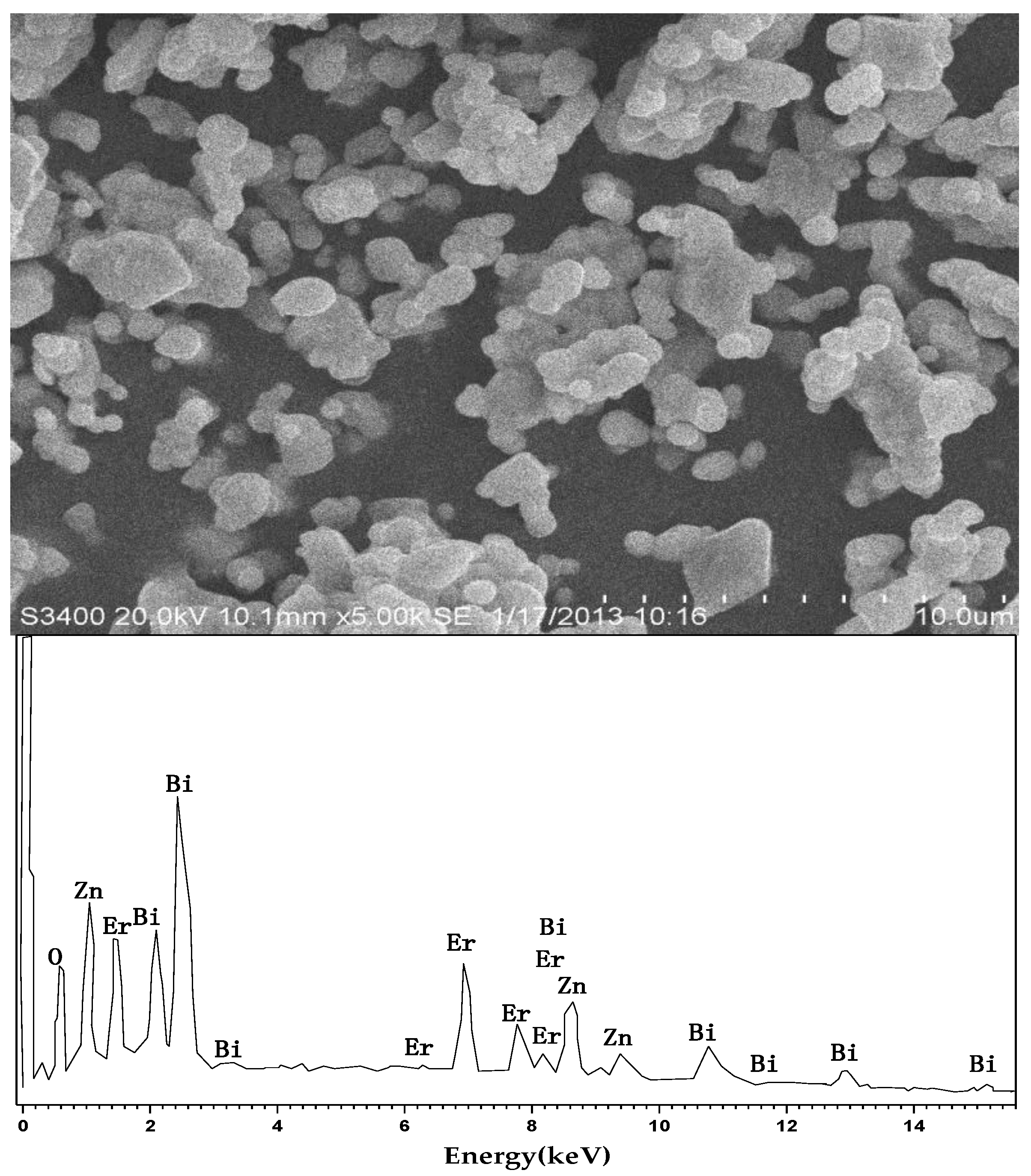
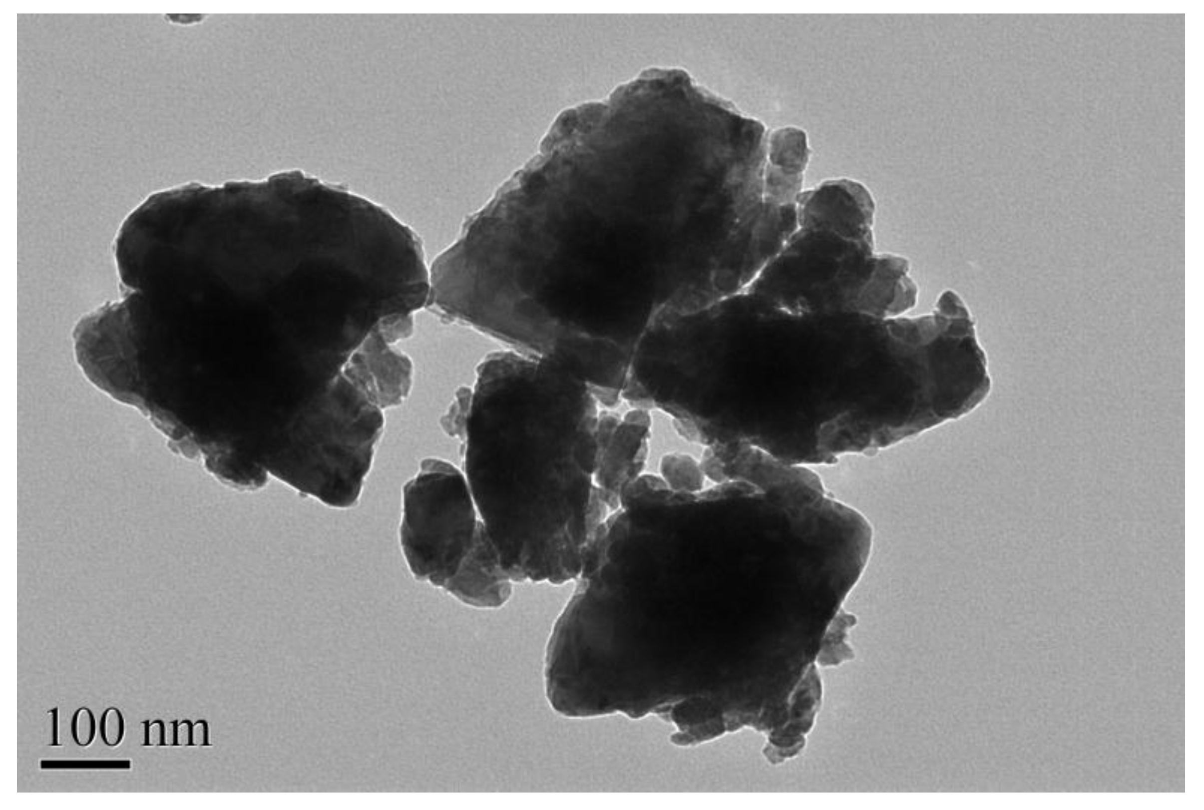
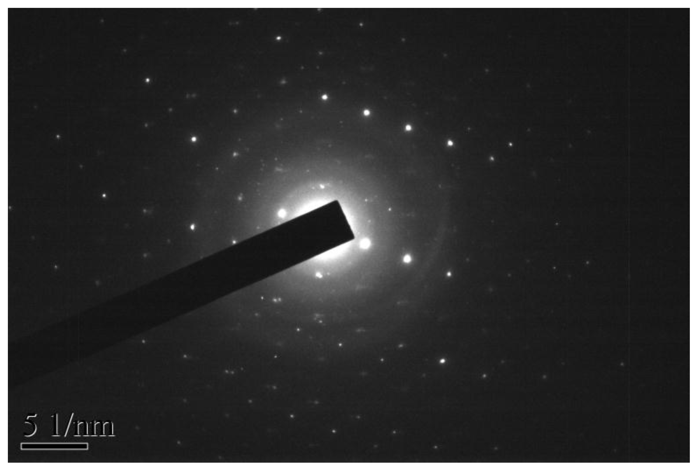
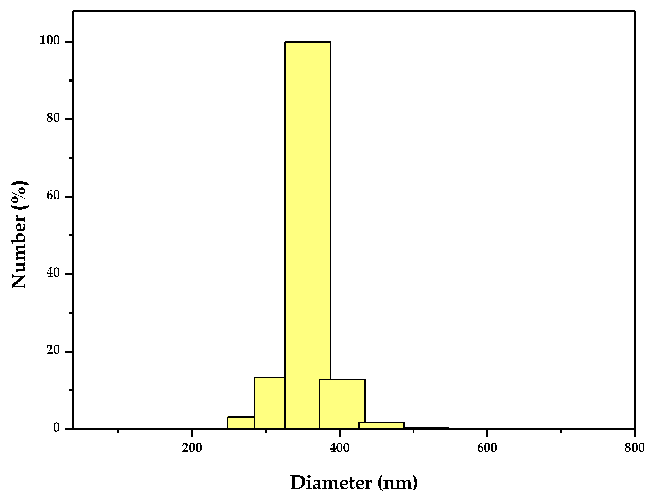
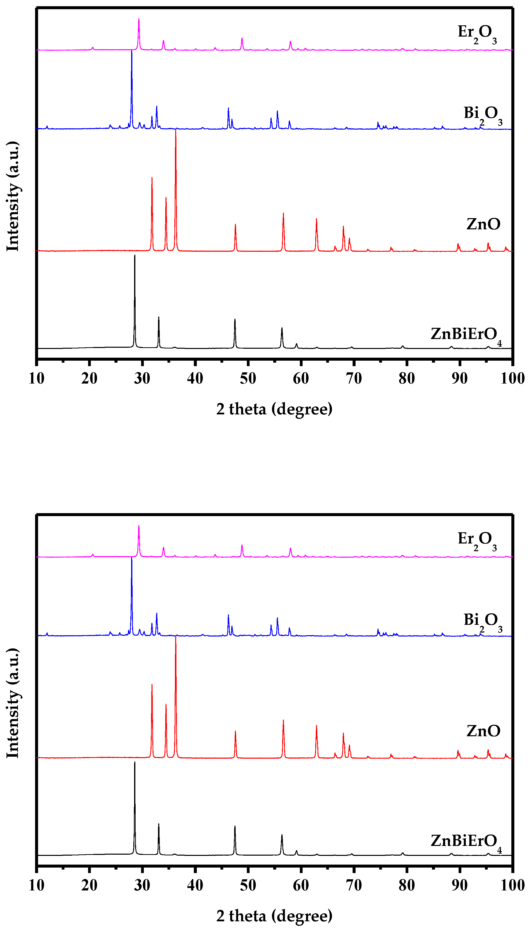
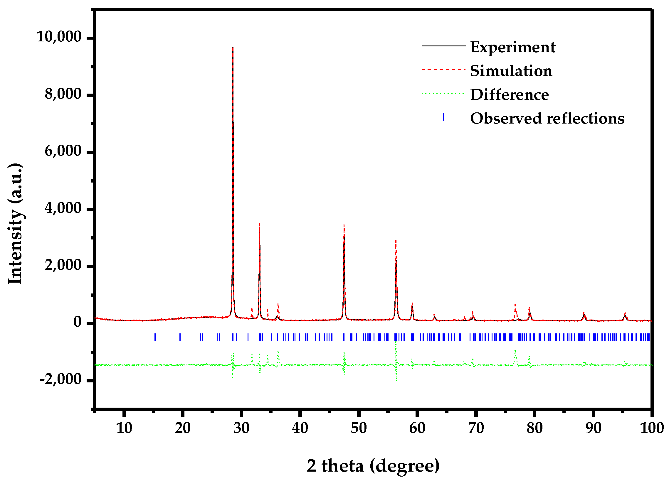
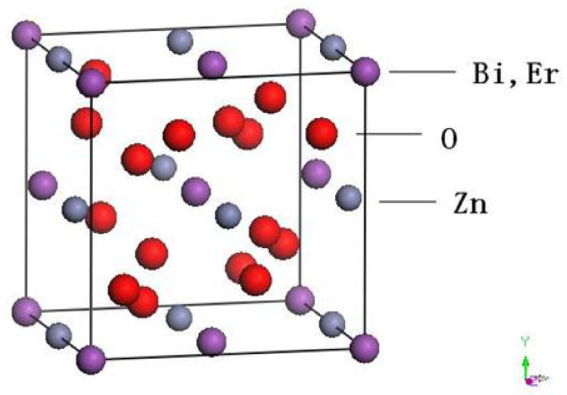
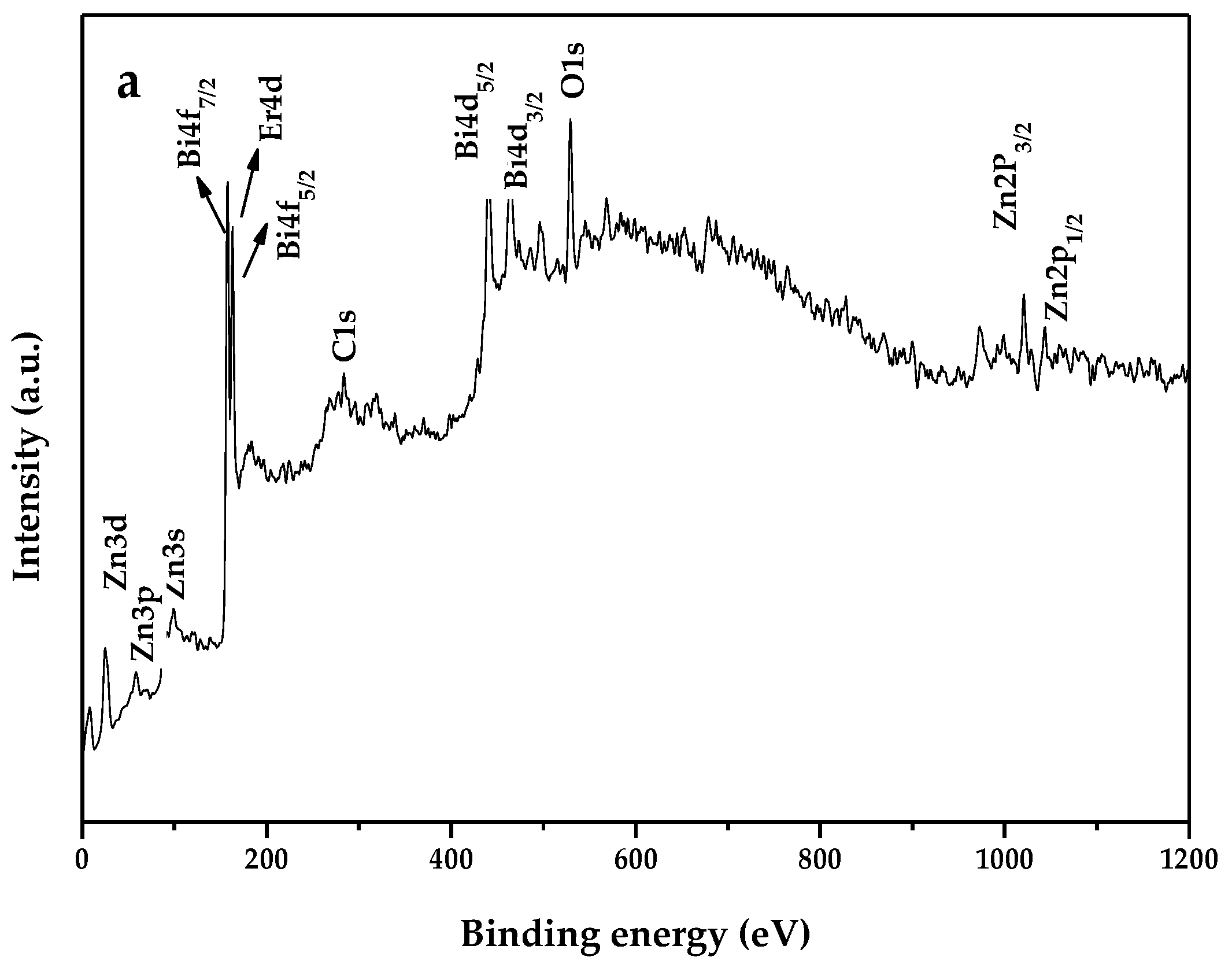
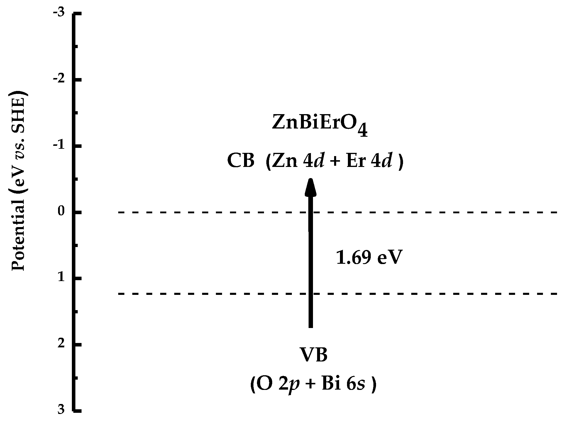
2.1. Characterization
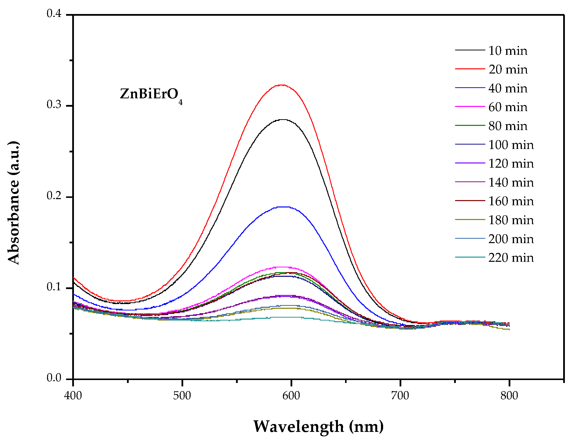
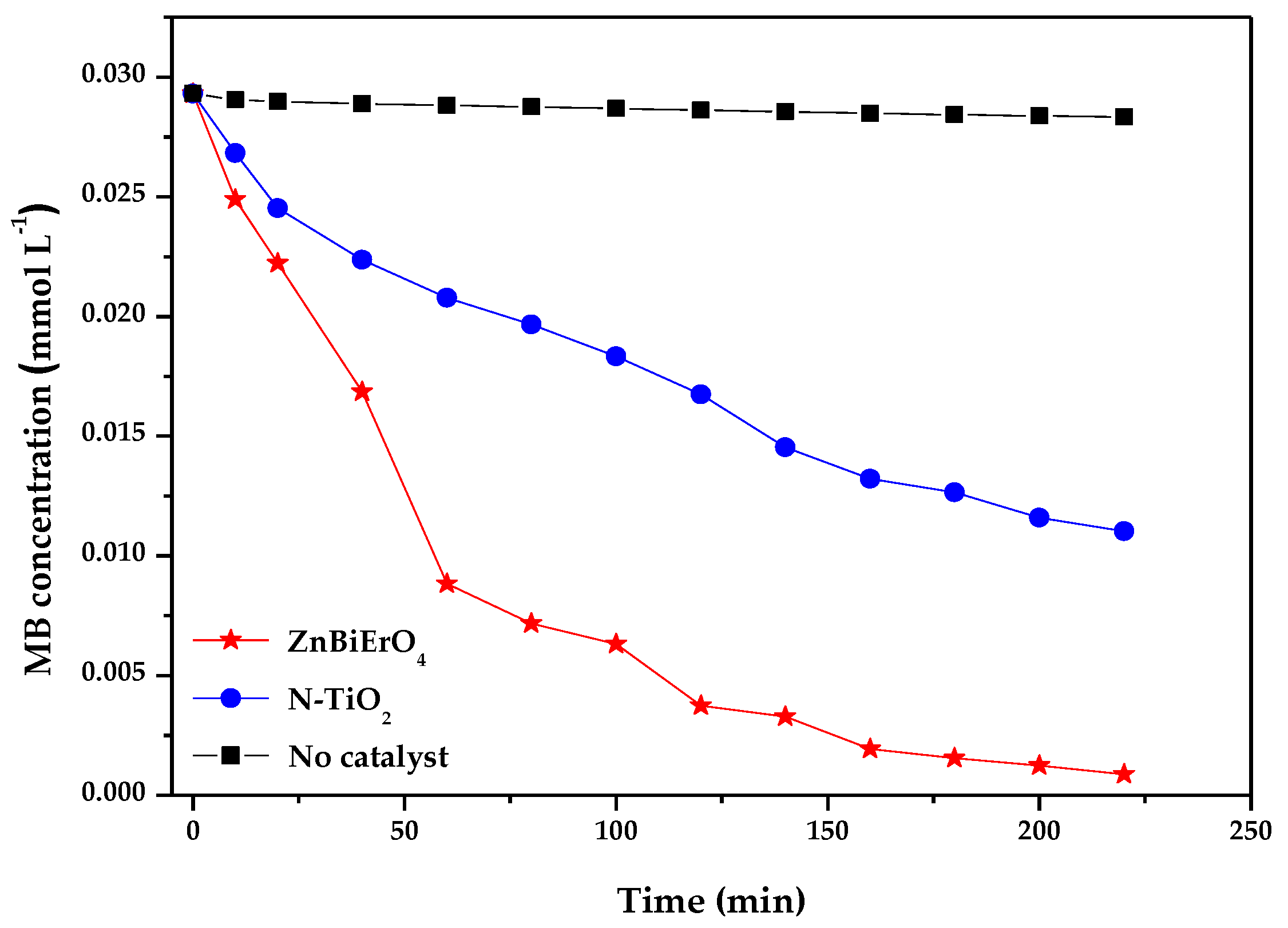
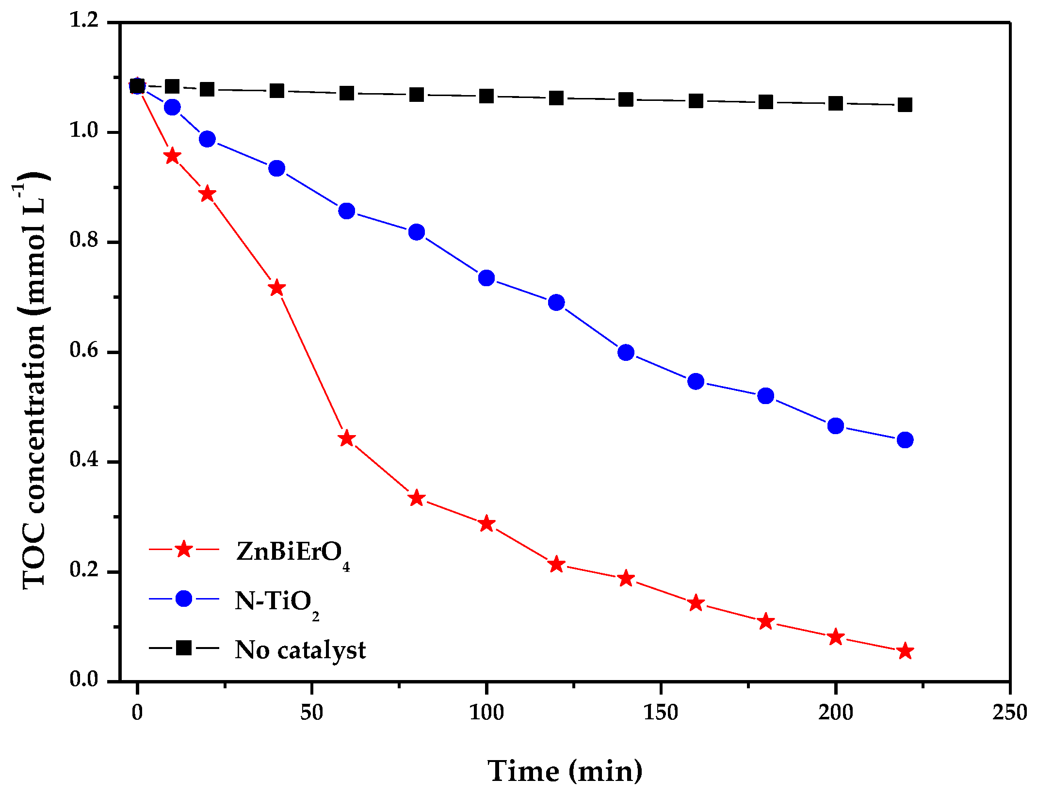
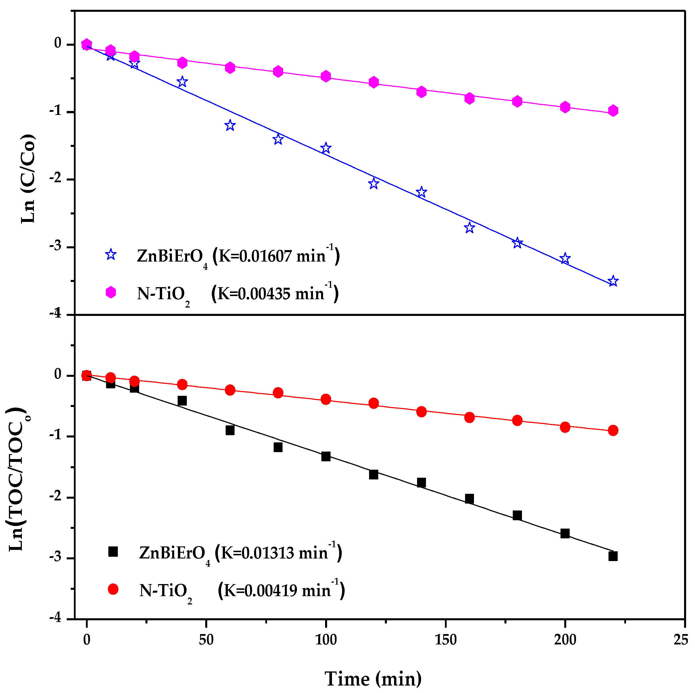
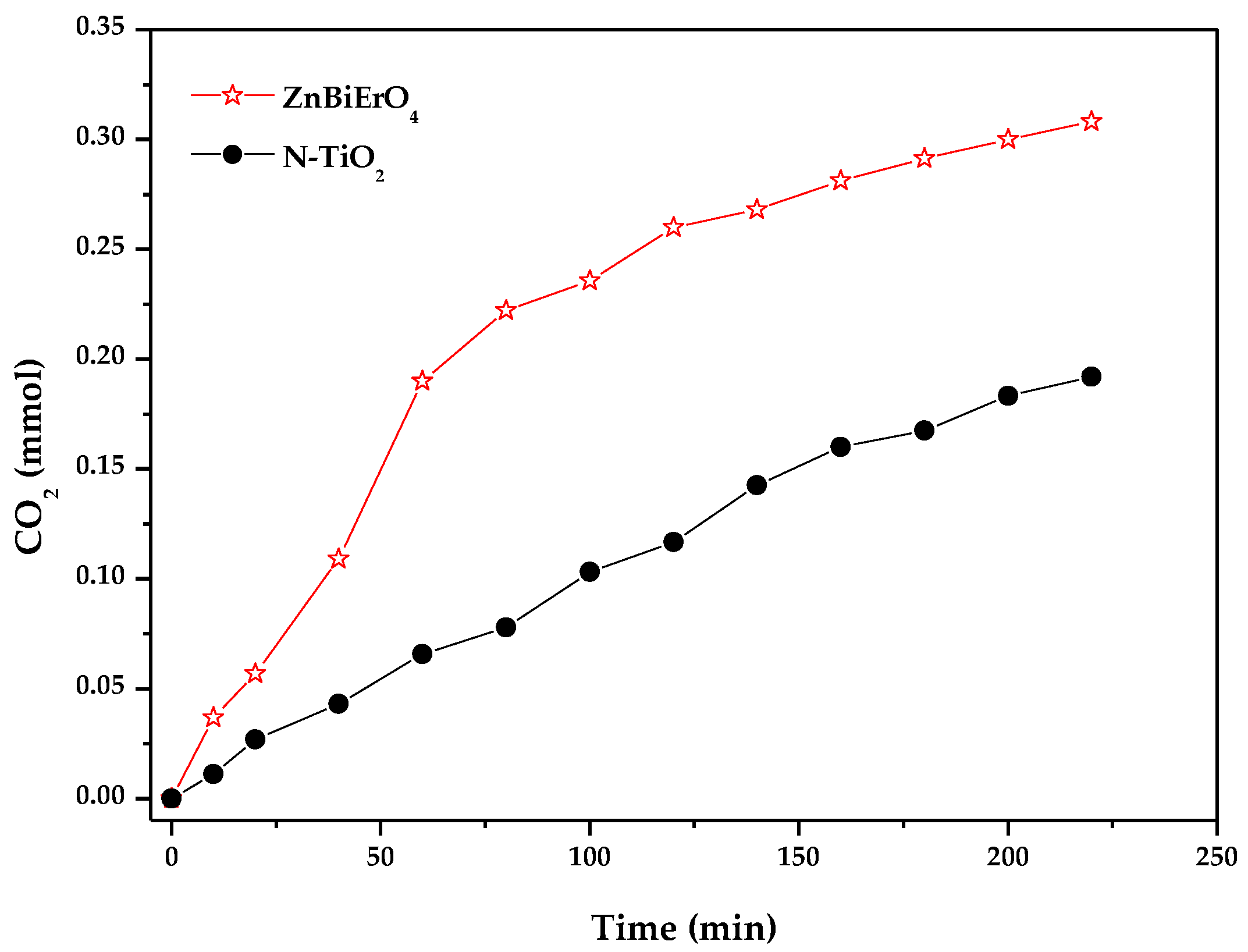
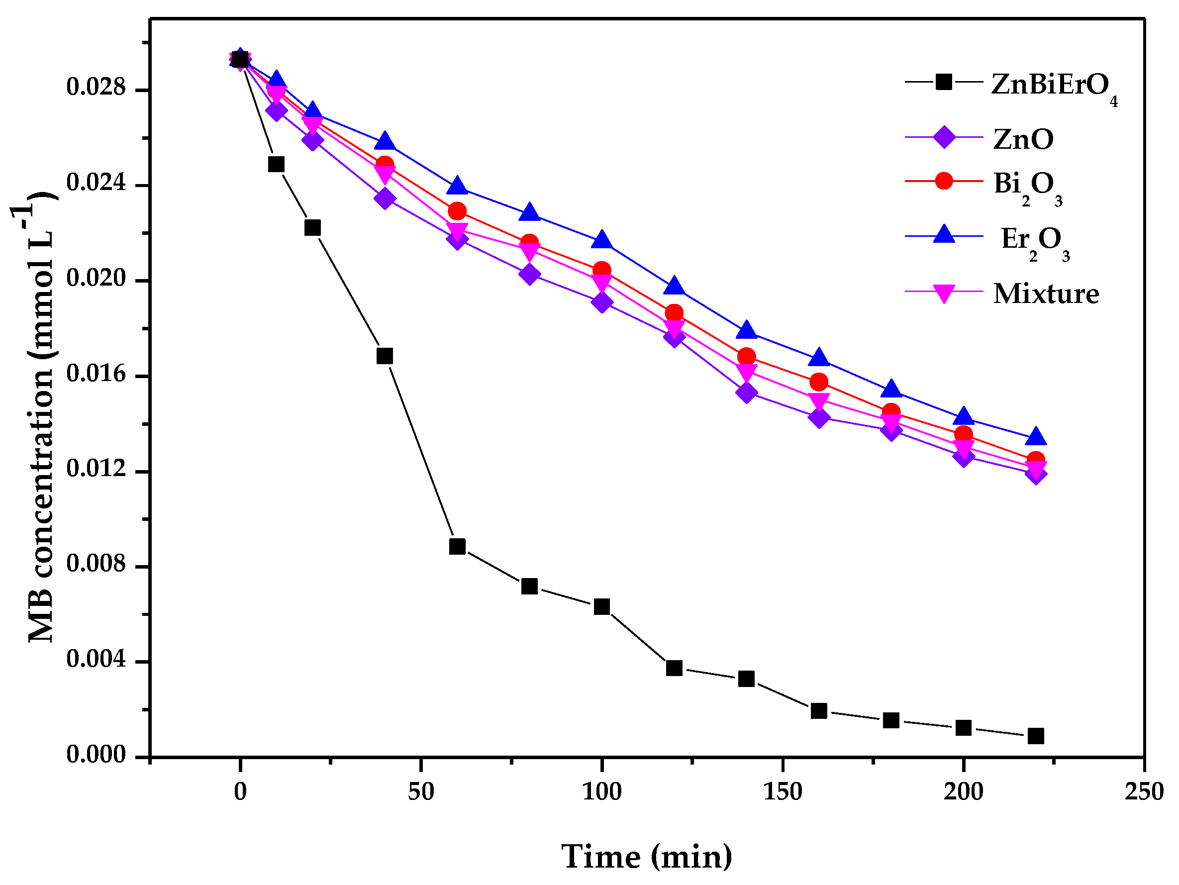
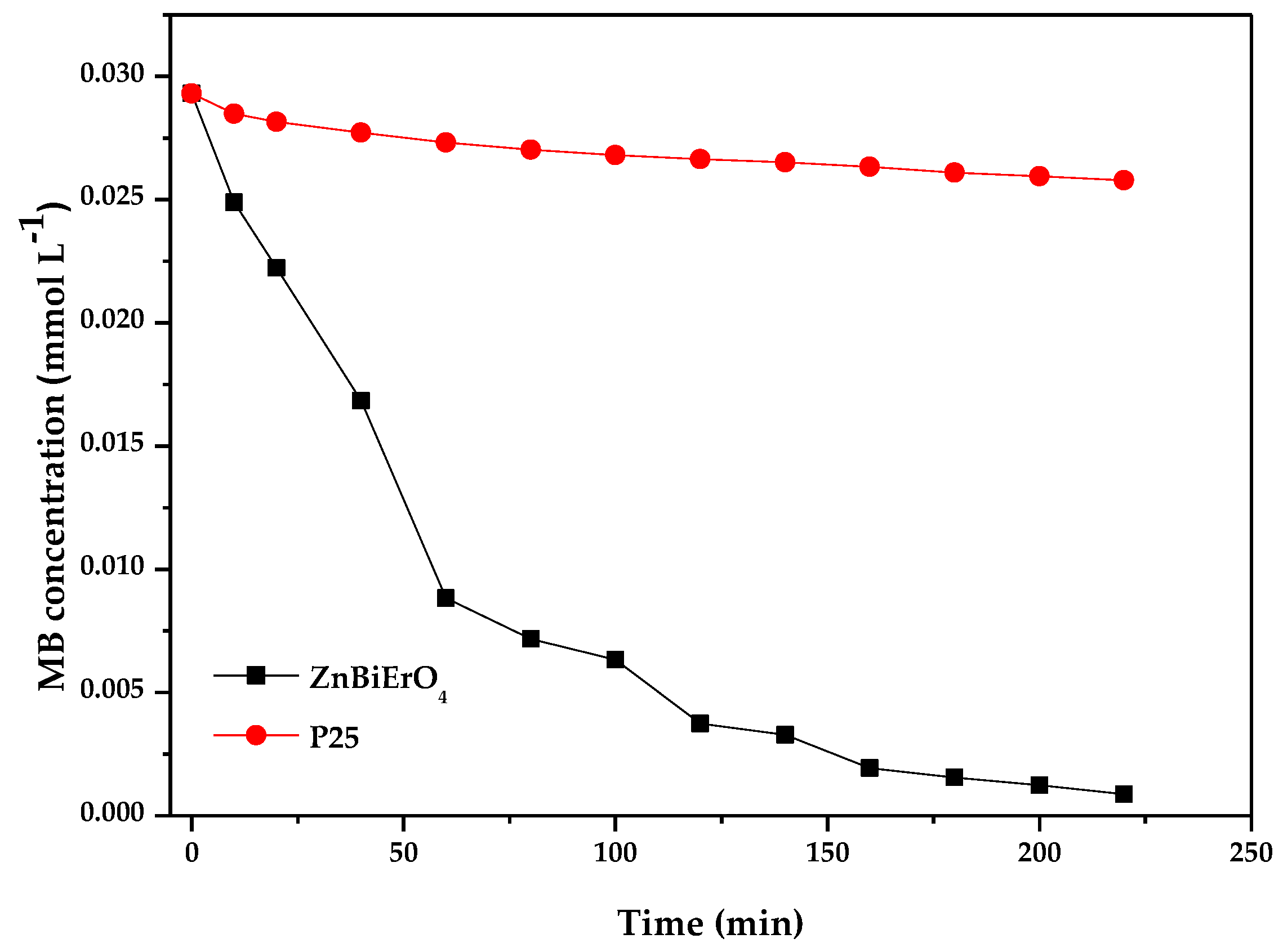
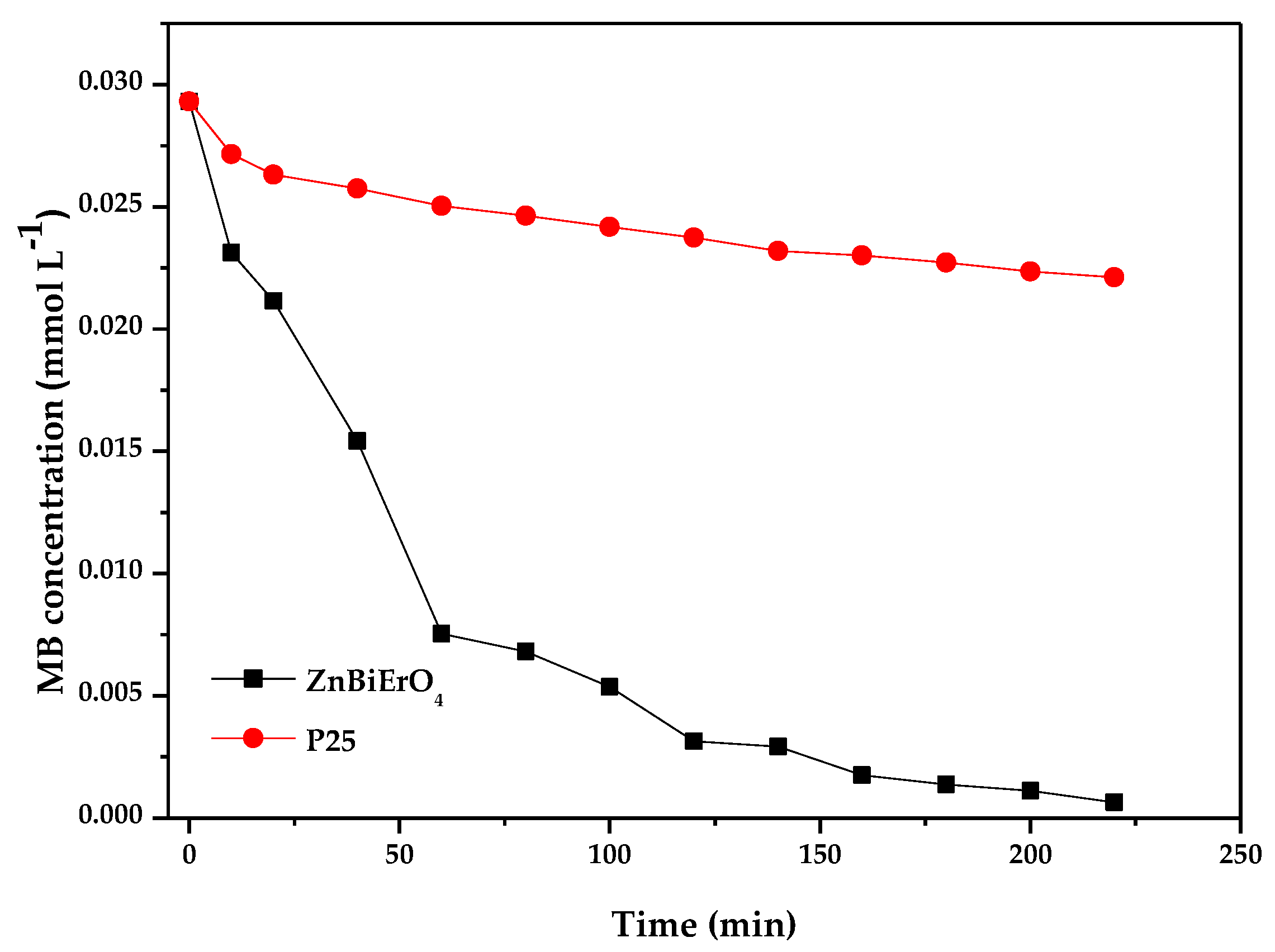
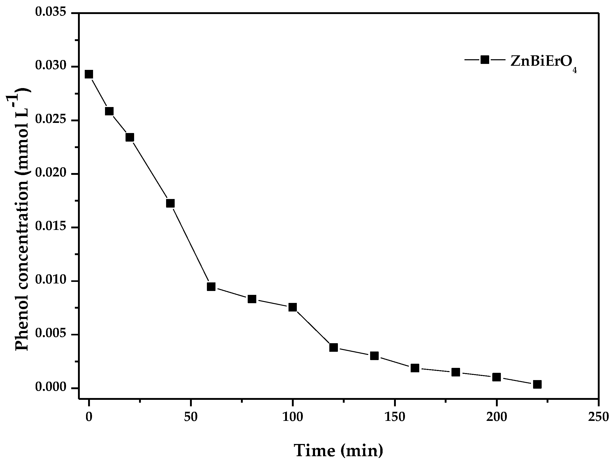
2.2. Photocatalytic Activity
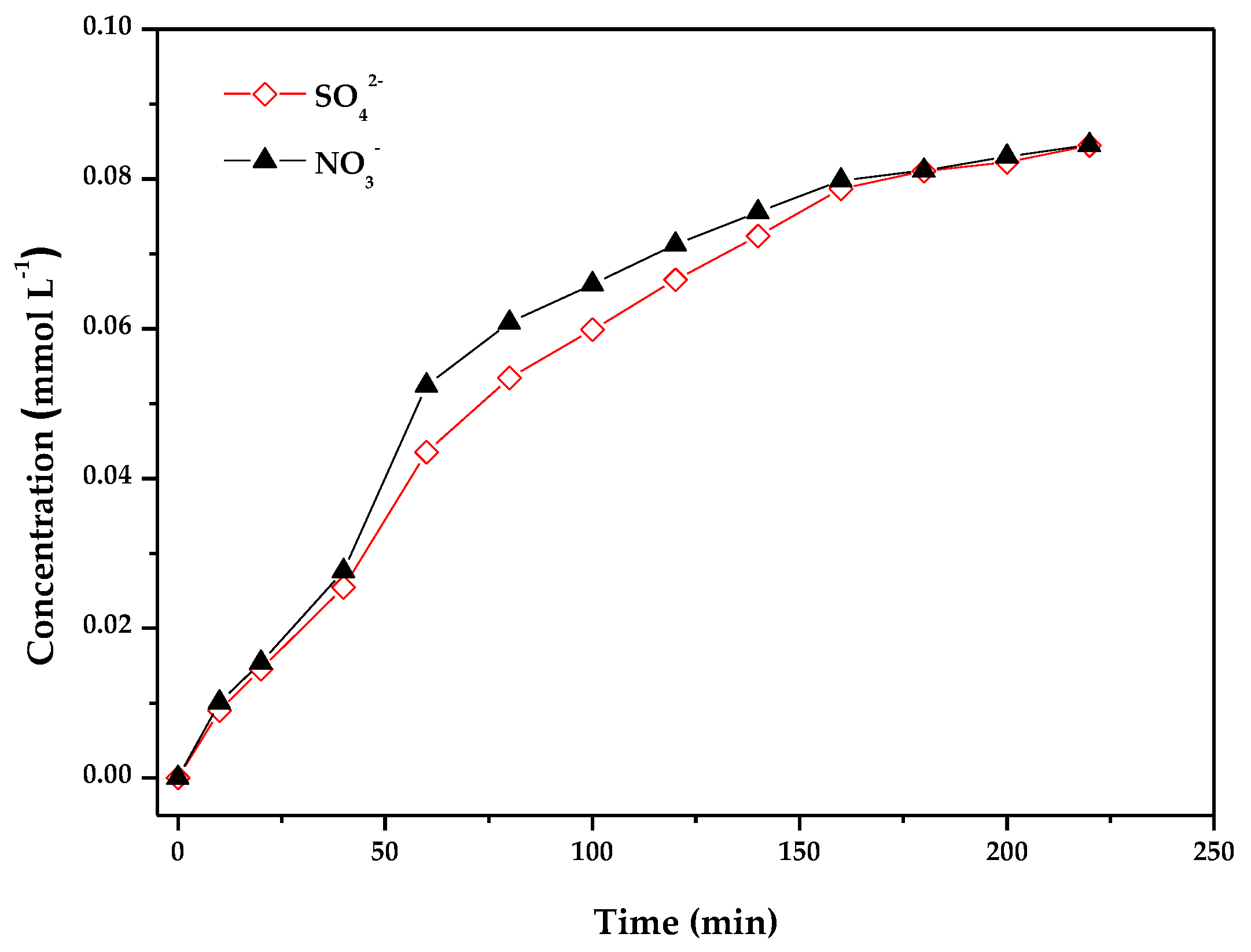
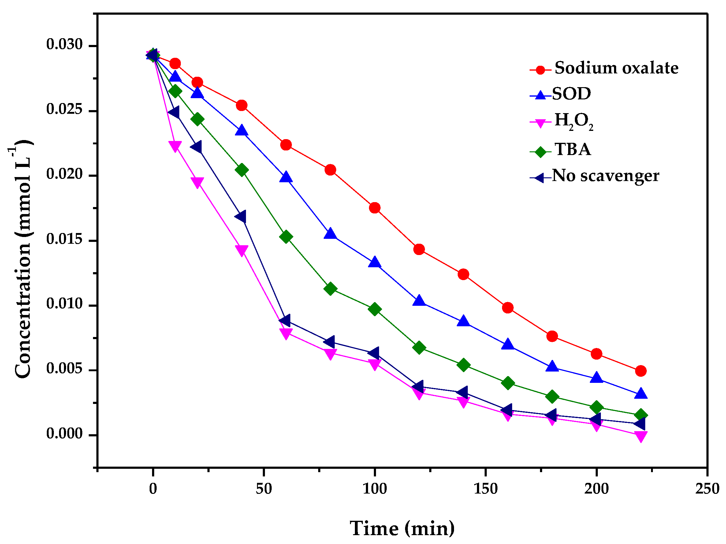
2.3. Photocatalytic Degradation Mechanism
3. Experimental
3.1. Synthesis of ZnBiErO4 and N-Doped TiO2
3.2. Characterization of ZnBiErO4
3.3. Photocatalytic Activity Tests
4. Conclusions
Acknowledgments
Author Contributions
Conflicts of Interest
References
- Han, F.; Kambala, V.S.R.; Srinivasan, M.; Rajarathnam, D.; Naidu, R. Tailored titanium dioxide photocatalysts for the degradation of organic dyes in wastewater treatment: A review. Appl. Catal. A-Gen. 2009, 359, 25–40. [Google Scholar] [CrossRef]
- Gupta, V.K.; Suhas. Application of low-cost adsorbents for dye removal—A review. J. Environ. Manag. 2009, 90, 2313–2342. [Google Scholar] [CrossRef] [PubMed]
- Martinez-Huitle, C.A.; Brillas, E. Decontamination of wastewaters containing synthetic organic dyes by electrochemical methods: A general review. Appl. Catal. B 2009, 87, 105–145. [Google Scholar] [CrossRef]
- Nasuha, N.; Hameed, B.H. Adsorption of methylene blue from aqueous solution onto NaOH-modified rejected tea. Chem. Eng. J. 2011, 166, 783–786. [Google Scholar] [CrossRef]
- Safarik, I.; Horska, K.; Safarikova, M. Magnetically modified spent grain for dye removal. J. Cereal Sci. 2011, 53, 78–80. [Google Scholar] [CrossRef]
- Khan, R.; Bhawana, P.; Fulekar, M.H. Microbial decolorization and degradation of synthetic dyes: A review. Rev. Environ. Sci. Bio/Technol. 2013, 12, 75–97. [Google Scholar] [CrossRef]
- Popli, S.; Patel, U.D. Destruction of azo dyes by anaerobic-aerobic sequential biological treatment: A review. Int. J. Environ. Sci. Technol. 2015, 12, 405–420. [Google Scholar] [CrossRef]
- Khanna, A.; Shetty, V.K. Solar photocatalysis for treatment of Acid Yellow-17 (AY-17) dye contaminated water using Ag@TiO2 core-shell structured nanoparticles. Environ. Sci. Pollut. Res. 2013, 20, 5692–5707. [Google Scholar] [CrossRef] [PubMed]
- Qin, X.; Cheng, H.; Wang, W.; Huang, B.; Zhang, X.; Dai, Y. Three dimensional BiOX (X = Cl, Br and I) hierarchical architectures: Facile ionic liquid-assisted solvothermal synthesis and photocatalysis towards organic dye degradation. Mater. Lett. 2013, 100, 285–288. [Google Scholar] [CrossRef]
- Chang, Y.; Yu, K.; Zhang, C.X.; Li, R.; Zhao, P.Y.; Lou, L.L.; Liu, S.X. Three-dimensionally ordered macroporous WO3 supported Ag3PO4 with enhanced photocatalytic activity and durability. Appl. Catal. B 2015, 176, 363–373. [Google Scholar] [CrossRef]
- Zhu, Y.P.; Li, M.; Liu, Y.L.; Ren, T.Z.; Yuan, Z.Y. Carbon-doped ZnO hybridized homogeneously with graphitic carbon nitride nanocomposites for photocatalysis. J. Phys. Chem. C 2014, 118, 10963–10971. [Google Scholar] [CrossRef]
- Abdullah, H.; Kuo, D.H.; Kuo, Y.R.; Yu, F.A.; Cheng, K.B. Facile synthesis and recyclability of thin nylon film-supported n-type ZnO/p-type Ag2O nano composite for visible light photocatalytic degradation of organic dye. J. Phys. Chem. C 2016, 120, 7144–7154. [Google Scholar] [CrossRef]
- Teng, F.; Liu, Z.L.; Zhang, A.; Li, M. Photocatalytic performances of Ag3PO4 polypods for degradation of dye pollutant under natural indoor weak light irradiation. Environ. Sci. Technol. 2015, 49, 9489–9494. [Google Scholar] [CrossRef] [PubMed]
- Shao, D.L.; Gao, J.; Xin, G.Q.; Wang, Y.P.; Li, L.; Shi, J.; Lian, J.; Koratkar, N.; Sawyer, S. Cl-doped ZnO nanowire arrays on 3D graphene foam with highly efficient field emission and photocatalytic properties. Small 2015, 11, 4785–4792. [Google Scholar] [CrossRef] [PubMed]
- Ao, Y.H.; Wang, K.D.; Wang, P.F.; Wang, C.; Hou, J. Synthesis of novel 2D-2D p-n heterojunction BiOBr/La2Ti2O7 composite photocatalyst with enhanced photocatalytic performance under both UV and visible light irradiation. Appl. Catal. B 2016, 194, 157–168. [Google Scholar] [CrossRef]
- Sathyaseelan, B.; Manikandan, E.; Lakshmanan, V.; Baskaran, I.; Sivakumar, K.; Ladchumananandasivam, R.; Kennedy, J.; Maaza, M. Structural, optical and morphological properties of post-growth calcined TiO2 nanopowder for opto-electronic device application: Ex-situ studies. J. Alloys Compd. 2016, 671, 486–492. [Google Scholar] [CrossRef]
- Yoshinaga, M.; Yamamoto, K.; Sato, N.; Aoki, K.; Morikawa, T.; Muramatsu, A. Remarkably enhanced photocatalytic activity by nickel nanoparticle deposition on sulfur-doped titanium dioxide thin film. Appl. Catal. B 2009, 87, 239–244. [Google Scholar] [CrossRef]
- Erjavec, B.; Hudoklin, P.; Perc, K.; Tisler, T.; Dolenc, M.S.; Pintar, A. Glass fiber-supported TiO2 photocatalyst: Efficient mineralization and removal of toxicity/estrogenicity of bisphenol A and its analogs. Appl. Catal. B 2016, 183, 149–158. [Google Scholar] [CrossRef]
- Yao, B.H.; Peng, C.; Zhang, W.; Zhang, Q.K.; Niu, J.F.; Zhao, J.E. A novel Fe(III) porphyrin-conjugated TiO2 visible-light photocatalyst. Appl. Catal. B 2015, 174, 77–84. [Google Scholar] [CrossRef]
- Yaghoubi, H.; Li, Z.; Chen, Y.; Ngo, H.T.; Bhethanabotla, V.R.; Joseph, B.; Ma, S.Q.; Schlaf, R.; Takshi, A. Toward a visible light-driven photocatalyst: The effect of midgap-states-induced energy gap of undoped TiO2 nanoparticles. ACS Catal. 2015, 5, 327–335. [Google Scholar] [CrossRef]
- Lu, F.; Meng, F. Research evolution of doping modification on TiO2 photocatalyst. B. Chin. Ceram. Soc. 2011, 30, 116–119. [Google Scholar]
- Park, H.; Park, Y.; Kim, W.; Choi, W. Surface modification of TiO2 photocatalyst for environmental applications. J. Photochem. Photobiol. C 2013, 15, 1–20. [Google Scholar] [CrossRef]
- Sheng, G.; Li, J.; Wang, S.; Wang, X. Modification to promote visible-light catalytic activity of TiO2. Prog. Chem. 2009, 21, 2492–2504. [Google Scholar]
- Ding, D.W.; Liu, K.; He, S.N.; Gao, C.B.; Yin, Y.D. Ligand-Exchange Assisted Formation of Au/TiO2 Schottky Contact for Visible-Light Photocatalysis. Nano Lett. 2014, 14, 6731–6736. [Google Scholar] [CrossRef] [PubMed]
- Pastrana-Martinez, L.M.; Morales-Torres, S.; Figueiredo, J.L.; Faria, J.L.; Silva, A.M.T. Graphene oxide based ultrafiltration membranes for photocatalytic degradation of organic pollutants in salty water. Water Res. 2015, 77, 179–190. [Google Scholar] [CrossRef] [PubMed]
- Rengifo-Herrera, J.A.; Blanco, M.; Wist, J.; Florian, P.; Pizzio, L.R. TiO2 modified with polyoxotungstates should induce visible-light absorption and high photocatalytic activity through the formation of surface complexes. Appl. Catal. B 2016, 189, 99–109. [Google Scholar] [CrossRef]
- Asahi, R.; Morikawa, T.; Ohwaki, T.; Aoki, K.; Taga, Y. Visible-light photocatalysis in nitrogen-doped titanium oxides. Science 2001, 293, 269–271. [Google Scholar] [CrossRef] [PubMed]
- Mohapatra, P.; Parida, K.M. Photocatalytic activity of sulfate modified titania 3: Decolorization of methylene blue in aqueous solution. J. Mol. Catal. A-Chem. 2006, 258, 118–123. [Google Scholar] [CrossRef]
- Jang, Y.J.; Simer, C.; Ohm, T. Comparison of zinc oxide nanoparticles and its nano-crystalline particles on the photocatalytic degradation of methylene blue. Mater. Res. Bull. 2006, 41, 67–77. [Google Scholar] [CrossRef]
- Li, H.P.; Liu, J.Y.; Hou, W.G.; Du, N.; Zhang, R.J.; Tao, X.T. Synthesis and characterization of g-C3N4/Bi2MoO6 heterojunctions with enhanced visible light photocatalytic activity. Appl. Catal. B 2014, 160, 89–97. [Google Scholar] [CrossRef]
- Saison, T.; Chemin, N.; Chaneac, C.; Durupthy, O.; Mariey, L.; Mauge, F.; Brezova, V.; Jolivet, J.P. New Insights into BiVO4 Properties as Visible Light Photocatalyst. J. Phys. Chem. C 2015, 119, 12967–12977. [Google Scholar] [CrossRef]
- Ding, X.; Zhao, K.; Zhang, L.Z. Enhanced photocatalytic removal of sodium pentachlorophenate with self-doped Bi2WO6 under visible light by generating more superoxide ions. Environ. Sci. Technol. 2014, 48, 5823–5831. [Google Scholar] [CrossRef] [PubMed]
- Yang, S.B.; Xu, D.B.; Chen, B.Y.; Luo, B.F.; Yan, X.; Xiao, L.S.; Shi, W.D. Synthesis and visible-light-driven photocatalytic activity of p-n heterojunction Ag2O/NaTaO3 nanocubes. Appl. Surf. Sci. 2016, 383, 214–221. [Google Scholar] [CrossRef]
- Wu, L.N.; Fang, S.; Ge, L.; Han, C.C.; Qiu, P.; Xin, Y.J. Facile synthesis of Ag@CeO2 core-shell plasmonic photocatalysts with enhanced visible-light photocatalytic performance. J. Hazard. Mater. 2015, 300, 93–103. [Google Scholar] [CrossRef] [PubMed]
- Christoforidis, K.C.; Montini, T.; Bontempi, E.; Zafeiratos, S.; Jaen, J.J.D.; Fornasiero, P. Synthesis and photocatalytic application of visible-light active beta-Fe2O3/g-C3N4 hybrid nanocomposites. Appl. Catal. B 2016, 187, 171–180. [Google Scholar] [CrossRef]
- Cui, B.; Lin, H.; Li, Y.Z.; Li, J.B.; Sun, P.; Zhao, X.C. Photophysical and photocatalytic properties of core-ring struc-tured NiCo2O4 nanoplatelets. J. Phys. Chem. C 2009, 32, 14083–14087. [Google Scholar] [CrossRef]
- Chen, C.H.; Liang, Y.H.; Zhang, W.D. ZnFe2O4/MWCNTs composite with enhanced photocatalytic activity under visible-light irradiation. J. Alloys Compd. 2010, 1, 168–172. [Google Scholar] [CrossRef]
- Carta, G.; Casarin, M.; El Habra, N.; Natali, M.; Rossetto, G.; Sada, C.; Tondello, E.; Zanella, P. MOCVD deposition of CoAl2O4 films. Electrochim. Acta 2005, 50, 4592–4599. [Google Scholar] [CrossRef]
- Tang, J.W.; Zou, Z.G.; Ye, J.H. Efficient photocatalytic decomposition of organic contaminants over CaBi2O4 under visible-light irradiation. Angew. Chem.-Int. Ed. 2004, 43, 4463–4466. [Google Scholar] [CrossRef] [PubMed]
- Cui, B.; Lin, H.; Zhao, X.C.; Li, J.B.; Li, W.D. Visible light induced photocatalytic activity of ZnCo2O4 nanoparticles. Acta Phys.-Chim. Sin. 2011, 27, 2411–2415. [Google Scholar]
- Kale, B.B.; Baeg, J.O.; Kong, K.J.; Moon, S.J.; Lee, S.M.; So, W.W. Synthesis and structural analysis of visible light photocatalyst, ZnBiGaO4 for photocatalytic solar hydrogen production. Int. J. Energy Res. 2010, 34, 404–411. [Google Scholar] [CrossRef]
- Arshadnia, I.; Movahedi, M.; Rasouli, N. MgFe2O4 and MgFe2O4/ZnFe2O4 coated with polyaniline as a magnetically separable photocatalyst for removal of a two dye mixture in aqueous solution. Res. Chem. Intermed. 2017, 43, 4459–4474. [Google Scholar] [CrossRef]
- Tang, J.W.; Zou, Z.G.; Yin, J.; Ye, J. Photocatalytic degradation of methylene blue on Caln(2)O(4) under visible light irradiation. Chem. Phys. Lett. 2003, 382, 175–179. [Google Scholar] [CrossRef]
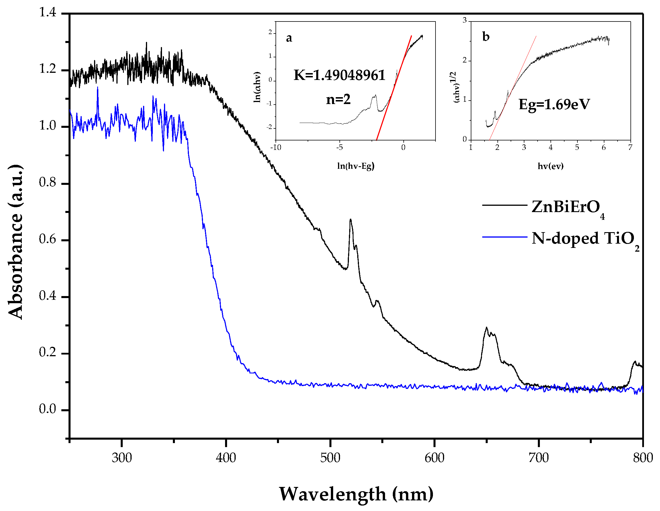
- Tauc, J.; Grigorov, R.; Vancu, A. Optical properties and electronic structure of amorphous germanium. Phys. Status Solidi 1966, 15, 627–637. [Google Scholar] [CrossRef]
- Xujiang Electromechanical Plant, Nanjing, China. Available online: http://www.njxu.com (accessed on 12 February 2018).
- Cai, J.B.; Wu, X.Q.; Li, Y.H.; Lin, Y.; Yang, H.; Li, S.X. Noble metal sandwich-like TiO2@Pt@C3N4 hollow spheres enhance photocatalytic performance. J. Colloid Interface Sci. 2018, 514, 791–800. [Google Scholar] [CrossRef] [PubMed]
- Vaiano, V.; Sacco, O.; Sannino, D.; Ciambelli, P. Nanostructured N-doped TiO2 coated on glass spheres for the photocatalytic removal of organic dyes under UV or visible light irradiation. Appl. Catal. B 2015, 170, 153–161. [Google Scholar] [CrossRef]
- Klein, M.; Nadolna, J.; Golabiewska, A.; Mazierski, P.; Klimczuk, T.; Remita, H.; Zaleska-Medynska, A. The effect of metal cluster deposition route on structure and photocatalytic activity of mono- and bimetallic nanoparticles supported on TiO2 by radiolytic method. Appl. Surf. Sci. 2016, 378, 37–48. [Google Scholar] [CrossRef]
- Jantawasu, P.; Sreethawong, T.; Chavadej, S. Photocatalytic activity of nanocrystalline mesoporous-assembled TiO2 photocatalyst for degradation of methyl orange monoazo dye in aqueous wastewater. Chem. Eng. J. 2009, 155, 223–233. [Google Scholar] [CrossRef]
- Guesh, K.; Marquez-Alvarez, C.; Chebude, Y.; Diaz, I. Enhanced photocatalytic activity of supported TiO2 by selective surface modification of zeolite Y. Appl. Surf. Sci. 2016, 378, 473–478. [Google Scholar] [CrossRef]
- Abdelhaleem, A.; Chu, W. Photodegradation of 4-chlorophenoxyacetic acid under visible LED activated N-doped TiO2 and the mechanism of stepwise rate increment of the reused catalyst. J. Hazard. Mater. 2017, 338, 491–501. [Google Scholar] [CrossRef] [PubMed]
- Lachheb, H.; Puzenat, E.; Houas, A.; Ksibi, M.; Elaloui, E.; Guillard, C.; Herrmann, J.M. Photocatalytic degradation of various types of dyes (Alizarin S, Crocein Orange G, Methyl Red, Congo Red, Methylene Blue) in water by UV-irradiated titania. Appl. Catal. B 2002, 39, 75–90. [Google Scholar] [CrossRef]
- Calza, P.; Rigo, L.; Sangermano, M. Investigations of photocatalytic activities of photosensitive semiconductors dispersed into epoxy matrix. Appl. Catal. B Environ. 2011, 106, 657–663. [Google Scholar] [CrossRef]
- Nasr, C.; Vinodgopal, K.; Fisher, L.; Hotchandani, S.; Chattopadhyay, A.K.; Kamat, P.V. Environmental photochemistry on semiconductor surfaces. Visible light induced degradation of a textile diazo dye, naphthol blue black, on TiO2 nanoparticles. J. Phys. Chem. 1996, 100, 8436–8442. [Google Scholar] [CrossRef]
- Marugan, J.; Hufschmidt, D.; Sagawe, G.; Selzer, V.; Bahnemann, D. Optical density and photonic efficiency of silica-supported TiO2 photocatalysts. Water Res. 2006, 40, 833–839. [Google Scholar] [CrossRef] [PubMed]
- Sakthivel, S.; Shankar, M.V.; Palanichamy, M.; Arabindoo, B.; Bahnemann, D.W.; Murugesan, V. Enhancement of photocatalytic activity by metal deposition: Characterisation and photonic efficiency of Pt, Au and Pd deposited on TiO2 catalyst. Water Res. 2004, 38, 3001–3008. [Google Scholar] [CrossRef] [PubMed]


| Atom | x | y | z | Occupation Factor |
|---|---|---|---|---|
| Zn | 0 | 0 | 0.5 | 1 |
| Bi | 0 | 0 | 0 | 1 |
| Er | 0 | 0 | 0 | 1 |
| O | 0.76731 | 0.14013 | 0.08188 | 1 |
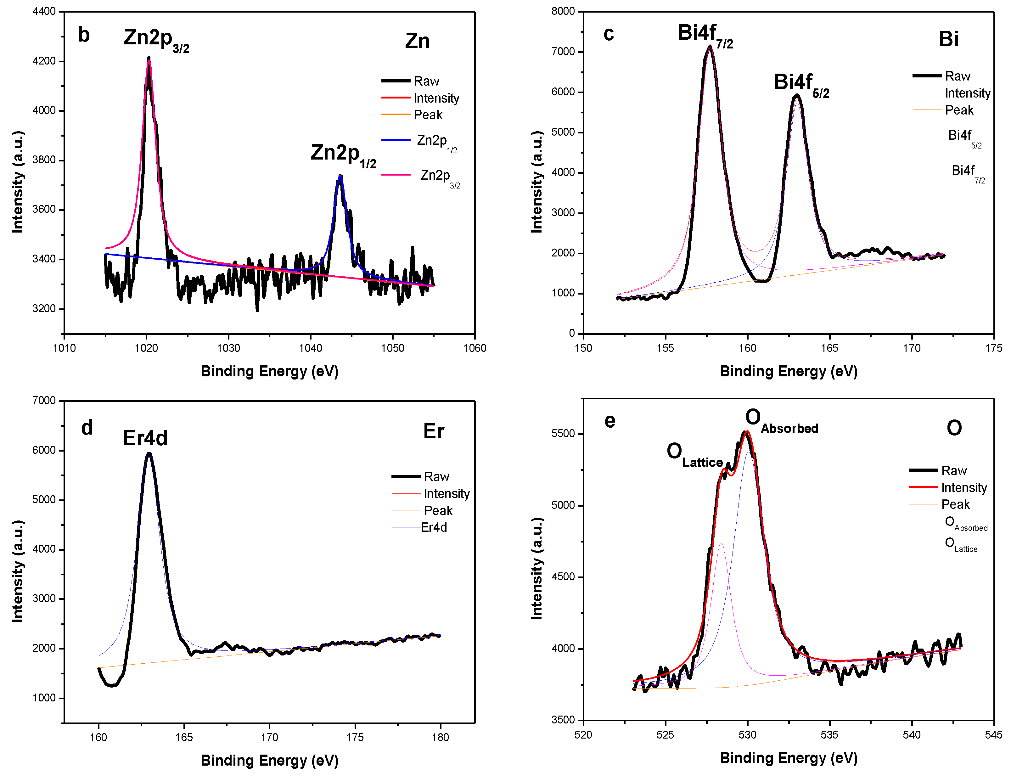
| Compound | Zn2p1/2 BE (eV) | Zn2p3/2 BE (eV) | Bi4f7/2 BE (eV) | Bi4f5/2 BE (eV) | Er4d BE (eV) | O1s BE (eV) |
|---|---|---|---|---|---|---|
| ZnBiErO4 | 1043.6 | 1020.3 | 157.7 | 163.0 | 162.9 | 529.8 |
© 2018 by the authors. Licensee MDPI, Basel, Switzerland. This article is an open access article distributed under the terms and conditions of the Creative Commons Attribution (CC BY) license (http://creativecommons.org/licenses/by/4.0/).
Share and Cite
Luan, J.; Zhuang, Y. Synthesis, Structural Property, Photophysical Property, Photocatalytic Property of Novel ZnBiErO4 under Visible Light Irradiation. Materials 2018, 11, 303. https://doi.org/10.3390/ma11020303
Luan J, Zhuang Y. Synthesis, Structural Property, Photophysical Property, Photocatalytic Property of Novel ZnBiErO4 under Visible Light Irradiation. Materials. 2018; 11(2):303. https://doi.org/10.3390/ma11020303
Chicago/Turabian StyleLuan, Jingfei, and Yan Zhuang. 2018. "Synthesis, Structural Property, Photophysical Property, Photocatalytic Property of Novel ZnBiErO4 under Visible Light Irradiation" Materials 11, no. 2: 303. https://doi.org/10.3390/ma11020303
APA StyleLuan, J., & Zhuang, Y. (2018). Synthesis, Structural Property, Photophysical Property, Photocatalytic Property of Novel ZnBiErO4 under Visible Light Irradiation. Materials, 11(2), 303. https://doi.org/10.3390/ma11020303