
Designing Vitamin D3 Formulations: An In Vitro Investigation Using a Novel Micellar Delivery System

 , , , ,
Abstract
: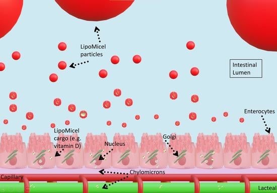
1. Introduction
2. Materials and Methods
2.1. Vitamin D3 (Cholecalciferol) Formulations
2.2. Solubility Analysis
2.3. Permeability Analysis
2.4. Ultra High Performance Liquid Chromatography (UHPLC)
2.5. Particle-Size Distribution by Laser Diffraction
2.6. Cryo-SEM (Cryogenic Scanning Electron Microscopy)
2.7. Zeta Potential
2.8. Data Analysis
3. Results
3.1. Solubility Measurements
3.2. Permeability Measurements
3.3. Laser Diffraction, Micellar Morphology, Size and Stability

| Formula LM1 | Formula LM2 | Formula LM3 | Formula BC | |
|---|---|---|---|---|
| D10% (μm) | 0.864 | 17.7 | 6.65 | 12.7 |
| D50% (μm) | 1.89 | 77.1 | 23.4 | 46.4 |
| D90% (μm) | 2.62 | 107 | 54.2 | 66 |
| PDI | 0.617 | 1.59 | 0.315 | 1.13 |
4. Discussion
5. Patents
Author Contributions
Funding
Institutional Review Board Statement
Informed Consent Statement
Data Availability Statement
Acknowledgments
Conflicts of Interest
References
- Manson, J.E.; Bassuk, S.S. Commentary. Eliminating Vitamin D Deficiency during the COVID-19 Pandemic: A Call to Action. Metabolism 2020, 112, 154322. [Google Scholar] [CrossRef] [PubMed]
- Role of Vitamin D in Preventing of COVID-19 Infection, Progression and Severity|Elsevier Enhanced Reader. Available online: https://reader.elsevier.com/reader/sd/pii/S1876034120305311?token=5DCE253A2FD14CEF551141818FE8E448EAD74AA08DD01227B004D090A85FE5413047698880DB1C6A0E41C838BB915941&originRegion=us-east-1&originCreation=20220811220227 (accessed on 11 August 2022).
- Zemb, P.; Bergman, P.; Camargo, C.A.; Cavalier, E.; Cormier, C.; Courbebaisse, M.; Hollis, B.; Joulia, F.; Minisola, S.; Pilz, S.; et al. Vitamin D Deficiency and the COVID-19 Pandemic. J. Glob. Antimicrob. Resist. 2020, 22, 133–134. [Google Scholar] [CrossRef] [PubMed]
- Ebadi, M.; Montano-Loza, A.J. Perspective: Improving Vitamin D Status in the Management of COVID-19. Eur. J. Clin. Nutr. 2020, 74, 856–859. [Google Scholar] [CrossRef]
- Vimaleswaran, K.S.; Forouhi, N.G.; Khunti, K. Vitamin D and COVID-19. BMJ 2021, 372, n544. [Google Scholar] [CrossRef]
- Steroid Hormone Vitamin D. Available online: https://www.ahajournals.org/doi/epdf/10.1161/CIRCRESAHA.118.311585 (accessed on 24 March 2022).
- Del Pinto, R.; Ferri, C.; Cominelli, F. Vitamin D Axis in Inflammatory Bowel Diseases: Role, Current Uses and Future Perspectives. Int. J. Mol. Sci. 2017, 18, 2360. [Google Scholar] [CrossRef] [PubMed] [Green Version]
- Hossein-nezhad, A.; Holick, M.F. Vitamin D for Health: A Global Perspective. Mayo Clin. Proc. 2013, 88, 720–755. [Google Scholar] [CrossRef] [PubMed] [Green Version]
- Naeem, Z. Vitamin D Deficiency- An Ignored Epidemic. Int. J. Health Sci. 2010, 4, V–VI. [Google Scholar]
- Prentice, A. Vitamin D Deficiency: A Global Perspective. Nutr. Rev. 2008, 66, S153–S164. [Google Scholar] [CrossRef]
- Vitamin D and Health—The Missing Vitamin in Humans|Elsevier Enhanced Reader. Available online: https://reader.elsevier.com/reader/sd/pii/S187595721830651X?token=DA9100E09E971FA1DFD0B89CE3639AA109DE80A384EAC895E73A4FF922C10CFDD12C94ED8E20534B2B05FCAE2ED5A7C3&originRegion=us-east-1&originCreation=20220811215304 (accessed on 11 August 2022).
- Reboul, E.; Goncalves, A.; Coméra, C.; Bott, R.; Nowicki, M.; Landrier, J.-F.; Jourdheuil-Rahmani, D.; Dufour, C.; Collet, X.; Borel, P. Vitamin D Intestinal Absorption Is Not a Simple Passive Diffusion: Evidences for Involvement of Cholesterol Transporters. Mol. Nutr. Food Res. 2011, 55, 691–702. [Google Scholar] [CrossRef]
- Shah, D.; Gupta, P. Vitamin D Deficiency: Is The Pandemic for Real? Indian J. Community Med. 2015, 40, 215–217. [Google Scholar] [CrossRef]
- Kamiński, M.; Kręgielska-Narożna, M.; Bogdański, P. Determination of the Popularity of Dietary Supplements Using Google Search Rankings. Nutrients 2020, 12, 908. [Google Scholar] [CrossRef] [Green Version]
- Kantor, E.D.; Rehm, C.D.; Du, M.; White, E.; Giovannucci, E.L. Trends in Dietary Supplement Use among US Adults From 1999–2012. JAMA 2016, 316, 1464–1474. [Google Scholar] [CrossRef] [PubMed]
- North America Vitamin D Market Size Report, 2020–2027. Available online: https://www.grandviewresearch.com/industry-analysis/north-america-vitamin-d-market (accessed on 4 April 2022).
- Vieth, R. Vitamin D Supplementation: Cholecalciferol, Calcifediol, and Calcitriol. Eur. J. Clin. Nutr. 2020, 74, 1493–1497. [Google Scholar] [CrossRef] [PubMed]
- Silva, M.C.; Furlanetto, T.W. Intestinal Absorption of Vitamin D: A Systematic Review. Nutr. Rev. 2018, 76, 60–76. [Google Scholar] [CrossRef] [PubMed]
- Dałek, P.; Drabik, D.; Wołczańska, H.; Foryś, A.; Jagas, M.; Jędruchniewicz, N.; Przybyło, M.; Witkiewicz, W.; Langner, M. Bioavailability by Design—Vitamin D3 Liposomal Delivery Vehicles. Nanomedicine 2022, 43, 102552. [Google Scholar] [CrossRef]
- Chang, H.-I.; Yeh, M.-K. Clinical Development of Liposome-Based Drugs: Formulation, Characterization, and Therapeutic Efficacy. Int. J. Nanomed. 2012, 7, 49–60. [Google Scholar] [CrossRef] [Green Version]
- Samad, A.; Sultana, Y.; Aqil, M. Liposomal Drug Delivery Systems: An Update Review. Curr. Drug Deliv. 2007, 4, 297–305. [Google Scholar] [CrossRef]
- Lian, T.; Ho, R.J.Y. Trends and Developments in Liposome Drug Delivery Systems. J. Pharm. Sci. 2001, 90, 667–680. [Google Scholar] [CrossRef]
- Kumari, A.; Yadav, S.K.; Yadav, S.C. Biodegradable Polymeric Nanoparticles Based Drug Delivery Systems. Colloids Surf. B Biointerfaces 2010, 75, 1–18. [Google Scholar] [CrossRef]
- Liu, D.; Yang, F.; Xiong, F.; Gu, N. The Smart Drug Delivery System and Its Clinical Potential. Theranostics 2016, 6, 1306–1323. [Google Scholar] [CrossRef]
- Bi, Y.; Xia, H.; Li, L.; Lee, R.J.; Xie, J.; Liu, Z.; Qiu, Z.; Teng, L. Liposomal Vitamin D3 as an Anti-Aging Agent for the Skin. Pharmaceutics 2019, 11, 311. [Google Scholar] [CrossRef] [Green Version]
- Loewen, A.; Chan, B.; Li-Chan, E.C.Y. Optimization of Vitamins A and D3 Loading in Re-Assembled Casein Micelles and Effect of Loading on Stability of Vitamin D3 during Storage. Food Chem. 2018, 240, 472–481. [Google Scholar] [CrossRef]
- Crintea, A.; Dutu, A.G.; Sovrea, A.; Constantin, A.-M.; Samasca, G.; Masalar, A.L.; Ifju, B.; Linga, E.; Neamti, L.; Tranca, R.A.; et al. Nanocarriers for Drug Delivery: An Overview with Emphasis on Vitamin D and K Transportation. Nanomaterials 2022, 12, 1376. [Google Scholar] [CrossRef]
- Gupta, R.; Behera, C.; Paudwal, G.; Rawat, N.; Baldi, A.; Gupta, P. Recent Advances in Formulation Strategies for Efficient Delivery of Vitamin D. AAPS PharmSciTech 2019, 20, 11. [Google Scholar] [CrossRef]
- Ozturk, B.; Argin, S.; Ozilgen, M.; McClements, D.J. Nanoemulsion Delivery Systems for Oil-Soluble Vitamins: Influence of Carrier Oil Type on Lipid Digestion and Vitamin D3 Bioaccessibility. Food Chem. 2015, 187, 499–506. [Google Scholar] [CrossRef] [PubMed]
- Kotake-Nara, E.; Komba, S.; Hase, M. Uptake of Vitamins D2, D3, D4, D5, D6, and D7 Solubilized in Mixed Micelles by Human Intestinal Cells, Caco-2, an Enhancing Effect of Lysophosphatidylcholine on the Cellular Uptake, and Estimation of Vitamins D’ Biological Activities. Nutrients 2021, 13, 1126. [Google Scholar] [CrossRef] [PubMed]
- Fratter, A.; Pellizzato, M. Novel Micellar System for Vitamin D3 Oral Delivery: Assessment of Enteric Absorption through a Digestion-like in Vitro Model. J. Drug Deliv. Sci. Technol. 2020, 59, 101840. [Google Scholar] [CrossRef]
- Margier, M.; Collet, X.; Le May, C.; Desmarchelier, C.; André, F.; Lebrun, C.; Defoort, C.; Bluteau, A.; Borel, P.; Lespine, A.; et al. ABCB1 (P-glycoprotein) Regulates Vitamin D Absorption and Contributes to Its Transintestinal Efflux. FASEB J. 2019, 33, 2084–2094. [Google Scholar] [CrossRef] [PubMed] [Green Version]
- Glowka, E.; Stasiak, J.; Lulek, J. Drug Delivery Systems for Vitamin D Supplementation and Therapy. Pharmaceutics 2019, 11, 347. [Google Scholar] [CrossRef] [Green Version]
- Badran, M. Formulation and in vitro evaluation of flufenamic acid loaded deformable liposomes for improved skin delivery. Dig. J. Nanomater. Biostructures (DJNB) 2014, 9, 83–91. [Google Scholar]
- Maurya, V.K.; Shakya, A.; Bashir, K.; Jan, K.; McClements, D.J. Fortification by Design: A Rational Approach to Designing Vitamin D Delivery Systems for Foods and Beverages. Comp. Rev. Food Sci. Food Safe 2023, 22, 135–186. [Google Scholar] [CrossRef]
- Lea, T. Caco-2 Cell Line. In The Impact of Food Bioactives on Health: In Vitro and Ex Vivo Models; Springer: Cham, Switzerland, 2015; pp. 103–111. [Google Scholar]
- Raval, N.; Maheshwari, R.; Kalyane, D.; Youngren-Ortiz, S.R.; Chougule, M.B.; Tekade, R.K. Chapter 10—Importance of Physicochemical Characterization of Nanoparticles in Pharmaceutical Product Development. In Basic Fundamentals of Drug Delivery; Tekade, R.K., Ed.; Advances in Pharmaceutical Product Development and Research; Academic Press: Cambridge, MA, USA, 2019; pp. 369–400. ISBN 978-0-12-817909-3. [Google Scholar]
- Danaei, M.; Dehghankhold, M.; Ataei, S.; Hasanzadeh Davarani, F.; Javanmard, R.; Dokhani, A.; Khorasani, S.; Mozafari, M. Impact of Particle Size and Polydispersity Index on the Clinical Applications of Lipidic Nanocarrier Systems. Pharmaceutics 2018, 10, 57. [Google Scholar] [CrossRef] [PubMed] [Green Version]
- Putri, D.C.A.; Dwiastuti, R.; Marchaban, M.; Nugroho, A.K. Optimization of mixing temperature and sonication duration in liposome preparation. J. Pharm. Sci. Community 2017, 14, 79–85. [Google Scholar] [CrossRef] [Green Version]
- Schneider, C.A.; Rasband, W.S.; Eliceiri, K.W. NIH Image to ImageJ: 25 Years of Image Analysis. Nat. Methods 2012, 9, 671–675. [Google Scholar] [CrossRef] [PubMed]
- Lam, J.; Katti, P.; Biete, M.; Mungai, M.; AshShareef, S.; Neikirk, K.; Garza Lopez, E.; Vue, Z.; Christensen, T.A.; Beasley, H.K.; et al. A Universal Approach to Analyzing Transmission Electron Microscopy with ImageJ. Cells 2021, 10, 2177. [Google Scholar] [CrossRef]
- Department, M.O.R. Caco-2 Cells Permeability Assay Protocol—ReadyCell. ReadyCell in Vitro Tools. 2022. Available online: https://readycell.com/caco-2-permeability-protocol/ (accessed on 8 June 2023).
- Wahlang, B.; Pawar, Y.B.; Bansal, A.K. Identification of Permeability-Related Hurdles in Oral Delivery of Curcumin Using the Caco-2 Cell Model. Eur. J. Pharm. Biopharm. 2011, 77, 275–282. [Google Scholar] [CrossRef]
- Fossati, L.; Dechaume, R.; Hardillier, E.; Chevillon, D.; Prevost, C.; Bolze, S.; Maubon, N. Use of Simulated Intestinal Fluid for Caco-2 Permeability Assay of Lipophilic Drugs. Int. J. Pharm. 2008, 360, 148–155. [Google Scholar] [CrossRef]
- Xia, F.; Jin, H.; Zhao, Y.; Guo, X. Supercritical Antisolvent-Based Technology for Preparation of Vitamin D3 Proliposome and Its Characteristics. Chin. J. Chem. Eng. 2011, 19, 1039–1046. [Google Scholar] [CrossRef]
- Mohammadi, M.; Pezeshki, A.; Mesgari Abbasi, M.; Ghanbarzadeh, B.; Hamishehkar, H. Vitamin D3-Loaded Nanostructured Lipid Carriers as a Potential Approach for Fortifying Food Beverages; in Vitro and in Vivo Evaluation. Adv. Pharm. Bull. 2017, 7, 61–71. [Google Scholar] [CrossRef] [Green Version]
- Cohen, Y.; Levi, M.; Lesmes, U.; Margier, M.; Reboul, E.; Livney, Y.D. Re-Assembled Casein Micelles Improve in Vitro Bioavailability of Vitamin D in a Caco-2 Cell Model. Food Funct. 2017, 8, 2133–2141. [Google Scholar] [CrossRef]
- Walia, N.; Chen, L. Pea Protein Based Vitamin D Nanoemulsions: Fabrication, Stability and in Vitro Study Using Caco-2 Cells. Food Chem. 2020, 305, 125475. [Google Scholar] [CrossRef]
- Li, W.; Peng, H.; Ning, F.; Yao, L.; Luo, M.; Zhao, Q.; Zhu, X.; Xiong, H. Amphiphilic Chitosan Derivative-Based Core–Shell Micelles: Synthesis, Characterisation and Properties for Sustained Release of Vitamin D3. Food Chem. 2014, 152, 307–315. [Google Scholar] [CrossRef] [PubMed]
- Mulrooney, S.L.; O’Neill, G.J.; Brougham, D.F.; Lyng, J.G.; O’Riordan, D. Improving Vitamin D3 Stability to Environmental and Processing Stresses Using Mixed Micelles. Food Chem. 2021, 362, 130114. [Google Scholar] [CrossRef] [PubMed]
- Goncalves, A.; Gleize, B.; Roi, S.; Nowicki, M.; Dhaussy, A.; Huertas, A.; Amiot, M.-J.; Reboul, E. Fatty Acids Affect Micellar Properties and Modulate Vitamin D Uptake and Basolateral Efflux in Caco-2 Cells. J. Nutr. Biochem. 2013, 24, 1751–1757. [Google Scholar] [CrossRef]
- Allen, T.M.; Cullis, P.R. Drug Delivery Systems: Entering the Mainstream. Science 2004, 303, 1818–1822. [Google Scholar] [CrossRef] [PubMed] [Green Version]
- Andar, A.U.; Hood, R.R.; Vreeland, W.N.; DeVoe, D.L.; Swaan, P.W. Microfluidic Preparation of Liposomes to Determine Particle Size Influence on Cellular Uptake Mechanisms. Pharm. Res. 2014, 31, 401–413. [Google Scholar] [CrossRef]
- Atanase, L.I. Micellar Drug Delivery Systems Based on Natural Biopolymers. Polymers 2021, 13, 477. [Google Scholar] [CrossRef]
- Reineke, J.J.; Cho, D.Y.; Dingle, Y.-T.; Morello, A.P.; Jacob, J.; Thanos, C.G.; Mathiowitz, E. Unique Insights into the Intestinal Absorption, Transit, and Subsequent Biodistribution of Polymer-Derived Microspheres. Proc. Natl. Acad. Sci. USA 2013, 110, 13803–13808. [Google Scholar] [CrossRef] [Green Version]
- Vieira, E.F.; Souza, S. Formulation Strategies for Improving the Stability and Bioavailability of Vitamin D-Fortified Beverages: A Review. Foods 2022, 11, 847. [Google Scholar] [CrossRef]
- Estupiñan, O.R.; Garcia-Manrique, P.; Blanco-Lopez, M.D.C.; Matos, M.; Gutiérrez, G. Vitamin D3 Loaded Niosomes and Transfersomes Produced by Ethanol Injection Method: Identification of the Critical Preparation Step for Size Control. Foods 2020, 9, 1367. [Google Scholar] [CrossRef]
- Malvern Instruments Limited. Zeta Potential: An Introduction in 30 Minutes; Zetasizer Nano Serles Technical Note MRK654-01; Malvern Instruments Limited: Worcestershire, UK, 2011; Volume 2, pp. 1–6. Available online: https://scholar.google.com/scholar_lookup?title=Zeta+Potential:+An+Introduction+in+30+Minutes&author=Malvern+Instruments+Limited&publication_year=2011#d=gs_cit&t=1686937273975&u=%2Fscholar%3Fq%3Dinfo%3AxTipZNW5B30J%3Ascholar.google.com%2F%26output%3Dcite%26scirp%3D0%26hl%3Den (accessed on 8 June 2023).
- Prakash, S.; Mishra, R.; Malviya, R.; Sharma, K. Journal of Chronotherapy and Drug Delivery Measurement Techniques and Pharmaceutical Applications of Zeta Potential: A Review. J. Chronother. Drug Deliv. 2014, 5, 33–40. [Google Scholar]
- Ghiasi, F.; Eskandari, M.H.; Golmakani, M.-T.; Rubio, R.G.; Ortega, F. Build-Up of a 3D Organogel Network within the Bilayer Shell of Nanoliposomes. A Novel Delivery System for Vitamin D3: Preparation, Characterization, and Physicochemical Stability. J. Agric. Food Chem. 2021, 69, 2585–2594. [Google Scholar] [CrossRef] [PubMed]
- Didar, Z. Enrichment of Dark Chocolate with Vitamin D3 (Free or Liposome) and Assessment Quality Parameters. J. Food Sci. Technol. 2021, 58, 3065–3072. [Google Scholar] [CrossRef] [PubMed]
- Chen, J.; Dehabadi, L.; Ma, Y.-C.; Wilson, L.D. Development of Novel Lipid-Based Formulations for Water-Soluble Vitamin C versus Fat-Soluble Vitamin D3. Bioengineering 2022, 9, 819. [Google Scholar] [CrossRef] [PubMed]
- Bhattacharjee, S. DLS and Zeta Potential—What They Are and What They Are Not? J. Control. Release 2016, 235, 337–351. [Google Scholar] [CrossRef]
- Krupa, L.; Bajka, B.; Staroń, R.; Dupont, D.; Singh, H.; Gutkowski, K.; Macierzanka, A. Comparing the Permeability of Human and Porcine Small Intestinal Mucus for Particle Transport Studies. Sci. Rep. 2020, 10, 20290. [Google Scholar] [CrossRef] [PubMed]
- Martinez, M.N.; Amidon, G.L. A Mechanistic Approach to Understanding the Factors Affecting Drug Absorption: A Review of Fundamentals. J. Clin. Pharmacol. 2002, 42, 620–643. [Google Scholar] [CrossRef] [Green Version]
- Maurya, V.K.; Aggarwal, M. Factors Influencing the Absorption of Vitamin D in GIT: An Overview. J. Food Sci. Technol. 2017, 54, 3753–3765. [Google Scholar] [CrossRef] [PubMed]
- Tsume, Y.; Mudie, D.M.; Langguth, P.; Amidon, G.E.; Amidon, G.L. The Biopharmaceutics Classification System: Subclasses for in Vivo Predictive Dissolution (IPD) Methodology and IVIVC. Eur. J. Pharm. Sci. 2014, 57, 152–163. [Google Scholar] [CrossRef] [Green Version]
- Yu, L.X.; Amidon, G.L.; Polli, J.E.; Zhao, H.; Mehta, M.U.; Conner, D.P.; Shah, V.P.; Lesko, L.J.; Chen, M.-L.; Lee, V.H.; et al. Biopharmaceutics Classification System: The Scientific Basis for Biowaiver Extensions. Pharm. Res. 2002, 19, 921–925. [Google Scholar] [CrossRef] [Green Version]
- Buckley, S.T.; Fischer, S.M.; Fricker, G.; Brandl, M. In Vitro Models to Evaluate the Permeability of Poorly Soluble Drug Entities: Challenges and Perspectives. Eur. J. Pharm. Sci. 2012, 45, 235–250. [Google Scholar] [CrossRef] [PubMed]


| Formula LM1 | Formula LM2 | Formula LM3 | Formula BC |
|---|---|---|---|
| Vitamin D3 | Vitamin D3 | Vitamin D3 | Vitamin D3 |
| Medium chain triglycerides | Medium chain triglycerides | Medium chain triglycerides | Flaxseed oil |
| Xylitol | Xylitol | Xylitol | |
| Methylsulfonylmethane | Methylsulfonylmethane | Methylsulfonylmethane | |
| Glycerin | Stevia | Cocoa | |
| Saponin Ethanol | Lecithin |
| Formula LM1 | Formula LM2 | Formula LM3 | Formula BC | |
|---|---|---|---|---|
| Water (pH 6.3) | 7.55 × 10⁻⁴ ± 2.81 × 10⁻⁴ | 2.89 × 10⁻⁴ ± 1.27 × 10⁻⁵ | 1.97 × 10⁻⁴ ± 4.20 × 10⁻⁵ | 1.64 × 10⁻⁴ ± 2.57 × 10⁻⁵ |
| Gastric juice (pH 1.2) | 6.93 × 10⁻⁴ ± 1.31 × 10⁻⁴ | 4.22 × 10⁻⁴ ± 4.34 × 10⁻⁵ | 3.37 × 10⁻⁴ ± 1.76 × 10⁻⁴ | 2.40 × 10⁻⁴ ± 4.86 × 10⁻⁵ |
| Intestinal juice (pH 6.8) | 1.09 × 10⁻³ ± 5.09 × 10⁻⁴ a | 7.19 × 10⁻⁴ ± 1.90 × 10⁻⁴ ab | 2.89 × 10⁻⁴ ± 7.92 × 10⁻⁴ b | 1.91 × 10⁻⁴ ± 4.24 × 10⁻⁵ b |
| Formula LM1 | Formula LM2 | Formula LM3 | Formula BC |
|---|---|---|---|
| 1.9 ± 0.3 × 10⁻⁷ ab | 3.6 ± 0.8 × 10⁻⁶ ab | 1.6 ± 0.3 × 10⁻⁵ cm/s b | 1.3 ± 1.1 × 10⁻⁹ a |
Disclaimer/Publisher’s Note: The statements, opinions and data contained in all publications are solely those of the individual author(s) and contributor(s) and not of MDPI and/or the editor(s). MDPI and/or the editor(s) disclaim responsibility for any injury to people or property resulting from any ideas, methods, instructions or products referred to in the content. |
© 2023 by the authors. Licensee MDPI, Basel, Switzerland. This article is an open access article distributed under the terms and conditions of the Creative Commons Attribution (CC BY) license (https://creativecommons.org/licenses/by/4.0/).
Share and Cite
Du, M.; Chang, C.; Zhang, X.; Zhang, Y.; Radford, M.J.; Gahler, R.J.; Kuo, Y.C.; Wood, S.; Solnier, J. Designing Vitamin D3 Formulations: An In Vitro Investigation Using a Novel Micellar Delivery System. Nutraceuticals 2023, 3, 290-305. https://doi.org/10.3390/nutraceuticals3020023
Du M, Chang C, Zhang X, Zhang Y, Radford MJ, Gahler RJ, Kuo YC, Wood S, Solnier J. Designing Vitamin D3 Formulations: An In Vitro Investigation Using a Novel Micellar Delivery System. Nutraceuticals. 2023; 3(2):290-305. https://doi.org/10.3390/nutraceuticals3020023
Chicago/Turabian StyleDu, Min, Chuck Chang, Xin Zhang, Yiming Zhang, Melissa J. Radford, Roland J. Gahler, Yun Chai Kuo, Simon Wood, and Julia Solnier. 2023. "Designing Vitamin D3 Formulations: An In Vitro Investigation Using a Novel Micellar Delivery System" Nutraceuticals 3, no. 2: 290-305. https://doi.org/10.3390/nutraceuticals3020023