Review

A Review on the Respiratory System Toxicity of Carbon Nanoparticles

1
Biology Department, College of Science, Engineering, and Technology, Jackson State University, Jackson, MS 39217, USA
2
Research Center in Minority Institutions-Center for Environmental Health, College of Science, Engineering, and Technology, Jackson State University, Jackson, MS 39217, USA
3
Biology Department, The University of Texas Rio Grande Valley, Edinburg, TX 78539, USA
*
Author to whom correspondence should be addressed.
Int. J. Environ. Res. Public Health 2016, 13(3), 325; https://doi.org/10.3390/ijerph13030325
Submission received: 15 August 2015 / Revised: 13 February 2016 / Accepted: 18 February 2016 / Published: 15 March 2016
(This article belongs to the Special Issue Environmental Fate and Effect of Nanoparticles and Nanomaterials)

Abstract

:
The respiratory system represents the main gateway for nanoparticles’ entry into the human body. Although there is a myriad of engineered nanoparticles, carbon nanoparticles/nanotubes (CNPs/CNTs) have received much attention mainly due to their light weight, very high surface area, durability, and their diverse applications. Since their discovery and manufacture over two decades ago, much has been learned about nanoparticles’ interactions with diverse biological system models. In particular, the respiratory system has been of great interest because various natural and man-made fibrous particles are known to be responsible for chronic and debilitating lung diseases. In this review, we present up-to-date the literature regarding the effects of CNTs or carbon nanofibers (CNFs) on the human respiratory system with respect to respiratory toxicity pathways and associated pathologies. This article is intended to emphasize the potentially dangerous effects to the human respiratory system if inadequate measures are used in the manufacture, handling, and preparation and applications of CNP or CNP-based products.

1. Introduction

Carbon nanomaterials, mainly represented by carbon nanoparticles or carbon nanotubes (CNPs/CNTs), and carbon nanofibers (CNFs), have captured the interest of the scientific community for more than two decades since the discovery of the first nanometer-sized carbon nanotubes [1]. Carbon nanomaterials have unprecedented potential for a large variety of industrial and biomedical applications. In industry, carbon-based nanomaterials have potential uses in many applications ranging from aerospace, construction, automobiles, electronics, and others. In the medical field, carbon nanomaterials are being considered as contrast agents, chemo-carriers, biological platforms, and for many other applications [2]. Currently, financial investment in nanotechnology is substantial, suggesting that an abundance of nano-based products will reach the market in the next few years [3]. While CNTs have been the focus of intense research, another type of carbon-based nanomaterial, CNF, is becoming increasingly prevalent as a nanomaterial of choice for nanoproducts/nanoscaffolds. CNFs seem to be a very suitable nanomaterial for biomaterial production. However, their effects on biological processes are mostly unknown to date. Thus, this review aims to present the current information on CNPs/CNTs and CNFs effects on the respiratory system.

2. Are CNT and CNF Structurally Different?

Carbon allotropes are present in a variety of carbon nanostructures including graphene, amorphous carbon, CNTs, and CNFs. Like CNTs, CNFs are increasingly used in many industrial applications, particularly, as polymer nanocomposites to provide strength, stiffness, heat resistance, and durability [4]. CNTs and CNFs belong to the same family, yet both these carbon nanostructures have distinct features. The physical and chemical properties of CNTs have been comprehensively presented in numerous reviews [5,6] and will not be discussed in the present review. Single-walled (SW)-CNTs are more distinct from CNFs, as opposed to multi-walled (MW)-CNTs. SWCNTs are a single sheet of graphene and, therefore, possess less rigidity and aggregate in bundles, properties that set them apart from MWCNTs and CNFs. MWCNTs are multiple sheets of graphene arranged concentrically with diameters between 10 and 150 nm [7]. Structurally, CNFs are layers of graphene arranged in an axial arrangement that form these carbon nanostructure fibers. CNFs have diameters ranging from 70 to 200 nm, and lengths of 10 to 100 μm [8]. CNFs may pose different health hazard implications compared to CNTs due to the differences in structural aspects between CNTs and CNFs; moreover, these nanostructures are less investigated than CNTs. It is envisioned that CNT and CNF end-products will dominate many aspects of industry due to their attractive intrinsic properties; thus, increased human exposure either at the synthesis or end-product is highly possible. Therefore, understanding the toxicological and pathological effects related to CNTs and CNFs is imperative to avoid potential respiratory effects such as those observed, for example, with asbestos exposure [5,9,10].

3. CNTs and CNFs as Bio-Scaffold and Bio-Sensor Materials

CNTs and different CNFs nanostructures are being investigated as biomaterial hybrid scaffolds. The sustainability of CNFs as scaffold materials stems from their fibrous nature, which mimics the fibrillar structure of extracellular matrix proteins. This nano-fibrillar structure provides a structural mesh for cell adherence and interactions making CNFs potential nanomaterials for orthopedic applications and more recently for peripheral nerve regeneration [11,12]. For example, a type of CNF with a hat-stacked nanostructure (H-CNF) were implanted subcutaneously, and after four weeks these H-CNF clusters were surrounded by a thin layer of connective tissue suggesting no tissue toxicity but indeed biocompatibility with surrounding tissue [12]. However, slight inflammation and the presence of phagocytizing macrophages were noticed at the H-CNF site. These processes were however considered as part of the wound healing process rather than a toxicological response. Carbon nanofiber-based thin films and mats have been tested for directed growth of Schwann cells [11]. Schwann cells proliferated when grown on CNF- and carbon-films without apoptosis or reactive oxygen species (ROS) formation. These results suggest that the fibrous nature of CNF provides suitable physical guidance for peripheral nerve cell growth, and may open-up new ways for neural tissue engineering, particularly peripheral nerve regeneration [11].
Cell adherence to CNFs patterned on polymer substrates was evaluated using osteoblast cells. Khang et al. [13] maintained cultures of osteoblasts for 21 days on matrices patterned with CNFs and found osteoblast cells to be fully functional with mineral deposition along the CNF patterns. Price et al. [14] used two types of carbon fiber compacts, conventional carbon and nanometer carbon fibers, to determine the adhesion of osteoblast cells. Osteoblasts cultured on carbon nanofiber scaffolds showed selective adherence to nanometer-dimension rough surfaces. These results suggest that CN-based scaffolds, perhaps due to their nanosize, are the preferred nanomaterials for use in directed bone growth applications, or for successful orthopedic and dental implants.
MWCNTs have been tested for muscle tissue engineering. Ostrovidov et al. [15] investigated a hybrid of gelatin and multi-walled fibers for myotube formation and functionality. MWCNTs enhanced the formation of fully functional myotubes with mechano-transductive properties, suggesting that MWCNTs may represent a novel scaffold material for skeletal tissue formation.
SWCNTs and MWCNTs have been tested as biosensors for the detection of proteins, nucleic acids, enzymes, and other biomolecules [16,17]. Carbon nanofibers, just recently, have been used for biosensors. For example, vertically aligned carbon nanofibers (VACNFs) seem to be a suitable material for the fabrication of biosensors for the detection of cardiac troponin [18] or C-reactive protein (CRP) [19]. Gupta et al. [19] tested a CNF-based biosensor platform for the detection of CRP and demonstrated that a CNF-sensor could potentially be developed to detect clinically relevant CRP level with high specificity. Moreover, the applicability of CNF-based biosensors has been extended to evaluate drug metabolism and inhibition. Xue et al. [20] reported the development of a CNF-modified film electrode in which the drug metabolizing enzyme, CYP3A4, was physically adsorbed producing a CYP3A4/CNF-modified film electrode. In comparison with CNT or carbon black (CB), the CNF-modified film electrode was the most efficient substrate for the physical adsorption of CYP3A4 onto film. The application of CNF-based film electrodes has the potential to revolutionize the time and type of drug metabolism screening as well as screening for CYP3A4 inhibitors. CNF applications have also been extended to phyto-pharmaceutical testing with potential applications in anticancer phytochemical discovery. Shao et al. [21] developed MWCNT-based nanofibers to which green tea polyphenols were adsorbed to test green tea antioxidant cancer therapy. The viability of an array of cell types (e.g., osteoblast, A549, and HepG2) on GTP-bound nanofibers were tested. Of all tested cells, HepG2 cells were more sensitive to GTP-bound nanofibers compared to osteoblast or A549 cells, suggesting the appropriateness of carbon-nanofiber platforms for antioxidant-linked cancer therapy. The applicability of CNF-based biosensors has been tested for the detection of catechol, a phenolic compound found in pesticides, and other pharmaceuticals [22]. The detection of catechol in solution was possible with the use of the laccase enzyme immobilized on electrospun CNF (ECNF) films. The detection limit of catechol in the solution was 0.63 μM within a 2 s response time, an indicator that such platforms could revolutionize testing for environmental/food chemistry safety.
Another recent application of carbon nanoparticles was the use of CNTs for microRNA (miRNAs) detection. Li et al. [23] designed MWCNT-based biosensors to which synthetic DNA probes complementary to miRNAs were immobilized. The hybridization of specific microRNAs was measured using guanine oxidation signals when a specific microRNA bound to its complementary DNA. Guanine oxidation was quantified using differential pulse voltammetry. Detection of microRNA expression using MWCNT-biosensors appears to be simple and cost-effective, given that microRNA are emerging important regulators of gene expression in plants, animals, and humans. Therefore, determining microRNA expression levels will provide information that will help to understand how microRNAs are associated with biological processes and human disease. To validate this method, Li et al. [24] tested microRNA-24 hybridization to the MWCNT-biosensor, which showed a good sensitivity, selectivity, and reproducibility. However, hybridization time seemed to play an important role in the detection of microRNA-24, suggesting that microRNA are susceptible to degradation. Despite this, the authors were able to obtain an acceptable recovery concentration of microRNA-24.
MWCNTs applications to biological systems, such as skeletal muscle fiber formation on gelatin nanofibers-MWCNT hybrid scaffolds, have been also investigated. Ostrovidov et al. [15] examined two concentrations of MWCNT, 0.5 and 5 mg/mL, combined with gelatin (20%) as a scaffold support for myofiber formation. Addition of MWCNT significantly increased the mechanical strength of gelatin nanofibers according to Young’s modulus measurements, and provided the necessary frame for C2C12 myoblast cell alignment, an indication for supporting myotube formation. C2C12 cell viability was not affected when the cells were cultured for two days, and myotube length increased with increasing MWCNT concentrations on gelatin nanofibers. Moreover, the gelatin-MWCNT nanofiber-guided myotubes exhibited contractibility under electrical stimulation that increased with increasing MWCNTs concentrations. These results suggest that gelatin-MWCNT hybrid scaffolds may be useful for skeletal muscle formation which could be applicable for muscle regeneration in skeletal muscle diseases.
CNTs/CNFs have the potential to function as biosensors for respiratory system pathological diagnosis, such as their use for the detection of lung cancer. Choudary et al. [25] showed that lung cancer antigens, MAGE A2 and MAGE A11, covalently attached to SWCNT-chitosan composites detected anti-MAGE A2 and anti-MAGE A11, respectively. A more sophisticated application of CNT-based biosensors for diagnosis and detection was developed by Tian et al. [26]. Using CNTs and hairpin sequences against a specific microRNA, Tian and coworkers showed femtomolar detection of the lung cancer biomarker, let-7 microRNA.
These studies indicate the possible applicability of using CNTs/CNFs as bioscaffolds in regenerative medicine and biosensors in clinical assays. Although CNTs induce toxicological responses in the respiratory system in cellular and animal models, CNTs in combination with biopolymers seem to have different interactions, particularly, with osteogenic or neuronal cells. Moreover, since most of these studies were short term, it is prudent to suggest the need for long-term studies for the applicability of CNTs/CNFs as bioscaffolds.

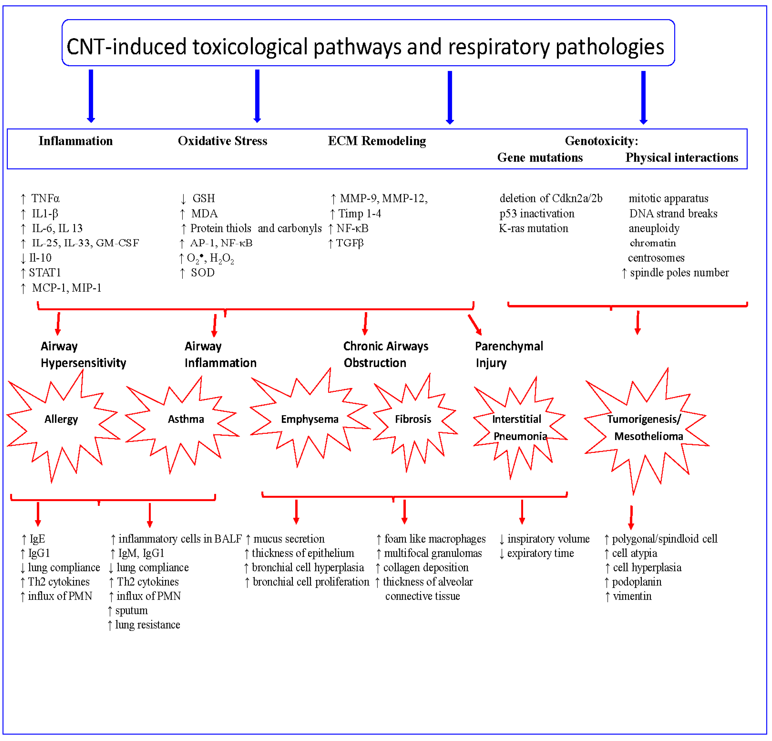
4. CNT Toxicological Pathways

4.1. Oxidative Stress

Oxidative stress is the most accepted mechanism for CNTs, asbestos, and other nanoparticles’ toxicity [5,27,28,29,30]. It is well established that ROS can react with cellular macromolecules and disrupt intracellular homeostasis. For example, numerous studies have shown that the presence of transition metals (e.g., Fe, Ni, Cu) in CNTs induces the formation of molecular oxygen-dependent superoxide anion radicals (O2), hydrogen peroxide (H2O2) and hydroxyl radicals (∙HO), all of which have high redox potentials and reactivities [29,30] (Figure 1). However, studies have also shown that even purified MWCNTs induce cyto- and genotoxic responses in BEAS-2B bronchio-epithelial cells [31,32]. Specific molecular signaling factors associated with oxidative stress, such as activator protein-1 (AP-1), nuclear factor κB (NF-κB), and MAP kinases ERK1/2 and p38, are activated by CNTs/CNFs and lead to antioxidant defense mechanism depletion or downregulation [32]. In mesothelioma cells, MWCNTs induced the formation of superoxide and DNA damage [33]. In vivo studies by Khaliullin et al. [34] and Shvedova et al. [35] showed that MWCNTs given by aspiration or SWCNTs either administered by pharyngeal aspiration or inhalation, decreased the level of glutathione, increased the level of protein thiols, and increased the level of malondialdehyde (MDA). Although CNFs are similar to MWCNTs, CNFs’ specific physicochemical characteristics, particularly having a low level of catalytic trace metals, may or may not induce ROS. This fact still requires further toxicological experimental investigation.

4.2. Extracellular Matrix Remodeling/Tissue Remodeling

Tissue remodeling of the respiratory system is a key pathophysiological characteristic of lung diseases including asthma, emphysema, and fibrosis. Mechanistically, airway remodeling is associated with biological processes such as cellular structural changes, cell proliferation, cell hyperplasia, cell hypertrophy, subepithelial fibrosis, inflammation and vascular changes [36]. The role of CNTs in lung remodeling has been shown by numerous studies which have demonstrated how they induce respiratory system remodeling via an array of cellular and molecular factors that favor the tissue remodeling processes. For example, CNTs alone or in allergen-sensitized animal models foster airway and lung remodeling via cytokines TNFα, IL1-β, MCP-1, Il-13, and recruitment of blood inflammatory cells [35,37,38,39,40,41]. Extracellular remodeling induced by CNTs is mediated primarily via TGFβ, TNFα, and osteopontin (OPN) signaling pathways [34,42]. Although TGFβ is a key cytokine that regulates extracellular matrix remodeling via STAT1, Thompson et al. [40] showed a role of STAT1 in MWCNT-induced airways remodeling. However in STAT1-depleted and OVA-sensitized mice or house-dust mites-sensitized mice, MWCNTs increased collagen, TGFβ, Il-13, eotaxin, and mucus production. These results suggest that other cytokines compensate for the TGFβ signaling pathway [40,43]. TGFβ1 is a key molecule in the development and progression of pulmonary fibrosis. It promotes the transcription of collagen type I and fibronectin in fibroblasts through an intricate signaling cascade. Furthermore, at the cellular level, CNTs were shown to induce plasma membrane remodeling via rearrangement of the actin cytoskeleton and clathrin [38,41]. In addition to cytokines, matrix metalloproteinases (MMPs) play an important role in tissue remodeling; however, MMP regulation by CNTs is less studied yet. A recent study by Meng et al. [44] showed that naїve macrophages secreted MMP-9 and VEGF in the presence of CNTs. MMP-9 is a known key factor in TGFβ activation and extracellular matrix remodeling in normal physiology but also in the pathophysiology of lung disease [44]. In addition to MMP-9 production, it has been shown that long-term, i.e., 4 months, exposure of mesothelial cells to both SWCNTs and MWCNTs increased MMP-2 expression and activity, which was involved in cell migration and invasion [45]. Poulsen et al. [46] showed that MWCNTs of different sizes (short: 67 ± 26.2 nm and long: 4.05 ± 2.4 μm) at either 3 days or 28 days post-exposure, induced lung remodeling regardless of the dose (54 or 163 μg/mouse). In addition to fibrosis genes of the MMP family (Mmp-12, Mmp-13, Mmp-14), MWCNT also upregulated the expression of tissue inhibitors of metalloproteinase (Timp-1, Timp-2, Timp-3, Timp-4), and TFGβ receptors signaling (Tgfbr-1, Tgfbr-3). Long MWCNTs were more potent in inducing gene expression than short MWCNTs. MWCNTs have also been shown to induce airway tissue remodeling via cyclooxygenase-2 (COX-2). Sayers et al. [47] showed that MWCNT potentiated allergen-induced increase in Th2, Th1, Th17 cytokines, prostaglandin D2, thromboxane B2, inflammation, and mucus-cell metaplasia in the lungs of COX-2(-/-) mice compared to wild type mice. However, no differences in airway fibrosis were noted between COX-2(-/-) and wild type mice. Similarly, Mizutani et al. [48] reported that MWCNTs sensitized airways in OVA-challenged mice via airway resistance, inflammation, and goblet cell hyperplasia, and induced a biphasic increase in airway responses and antigen-specific antibodies. Biphasic acute airways’ responses to CNTs manifested in the form of inflammation and inflammatory cells accumulation and chronic responses like lung fibrosis were also reported by Mercer et al. [37,49]. Furthermore, Wang et al. [50] showed a role of IL-33 in MWNCT-induced airway constriction, inflammation, and fibrosis. Il-33 signaling mediates allergen-induced Th2 cytokines which induce asthma. Asthma, a chronic airway inflammation and hyper-responsiveness is a major public health concern. Approximately 10% of the U.S. population and approximately 300 million people worldwide are affected by asthma [51].
These studies indicate that CNTs induce airways resistance in animal models and this should be taken into consideration as CNTs are being rapidly incorporated into numerous nanoproducts. Thus, it is prudent to strongly suggest the adoption of strict prevention strategies and workplace exposure controls in workplaces dedicated to CNTs/CNFs manufacture.

4.3. Genotoxicity

The potential of CNTs and CNFs to induce genetic alterations has been investigated in different system models; most often the results were partially dependent on the type and physical characteristics of the nanomaterial tested [52,53,54]. Although SWCNTs have been shown to induce genetic alterations, no tumors have been reported in any study [55,56]. However, some MWCNTs have been shown to induce genetic damage, but only MWCNT Mitsui 7 has been shown to induce carcinogenicity via induction of mesothelioma (reviewed by Toyokuni, [54]). A study by Sargent and coworkers [57] showed that MWCNTs induced genotoxicity; however in a subsequent study in [58], the same MWCNTs did not induce tumors unless there was an initiator. The latter study is crucial to extrapolate to human exposure, since many humans are exposed to mutagens including cigarette smoke, chemicals such as benz[a]anthracene, radon, naphthalene, diesel exhaust benzo[a]pyrene, and others. If these materials can serve as initiators, humans exposed to MWCNTs could develop tumors [58,59,60].
The potential of CNFs to cause DNA damage was initially evaluated in Chinese hamster lung fibroblast cells, V79, using the Comet assay and micronuclei formation (MN) procedures by Kisin et al. [61]. In this study, CNFs induced DNA damage but no differences were found between CNF or asbestos at similar doses (3 or 48 μg/cm2) and exposure times (3 or 24 h). CNF and asbestos dose-dependently induced MN formation in V79 cells. In the same study, CNF aneugenic or clastogenic effects were evaluated in human small airway epithelial cells (SAECs) in comparison with asbestos. CNF increased both aneugenic and clastogenic events and could be found inside the nucleus of SAECs. The mechanism by which CNFs induce genotoxicity is not known yet, however, direct physical interactions of nanofibers with mitotic machinery or with chromosomes during cell division leading to aneuploidy are possible. Aneuploidy is the result of imbalanced chromosome separation during cell division leading to the formation of daughter cells with an abnormal chromosome number. DNA damage and adduct formation are related to the generation of ROS via the Fenton reaction. The presence of transition metals such as Fe in CNFs, has been found to generate ROS; therefore, the genotoxic effects of CNF may be partially accounted for by ROS generation [61]. A major health concern of CNFs is further supported by findings in the study by Kisin et al. [61] where CNF genotoxicity was comparable to asbestos and CNFs were more potent than SWCNTs. In addition to ROS-mediated DNA damage, CNTs physically interact with DNA, particularly MWCNTs which have been shown to exhibit preference toward G-C rich DNA regions [24,62,63,64,65,66,67]. CNTs have been shown to induce DNA strand breaks, which can either be caused by the direct effects of ROS or apoptosis, and by the activity of endonucleases that are activated by the formation of DNA adducts [52,66]. Both SWCNTs and MWCNTs induce DNA strand breaks, PPAR activation, and formation of γH2AX foci in lung epithelial cells [30,31,52,57,58,66,67,68,69,70]. Sargent and co-authors [57,58] observed the induction of aneuploidy, formation of three spindle poles, microtubules and centrosome fragmentation, and accumulation of cells in the G2/M phase in SAEC exposed to a dose that would be a worker-relevant exposure dose of SWCNTs (0.024 μg/cm2). Similarly, Siegrest and co-authors [67] obtained similar results when MWCNTs were tested on the same cell type, SAECs, with the exception of the accumulation of cells in G1/S phase rather than in the G2/M phase as was the case of SWCNTs. Mechanistically, these studies indicate physical association of SWCNTs and MWCNTs with DNA, mitotic spindles, and centrosomes thus impeding proper chromosome segregation.
Long-term post-exposure of mice (C57BL/6) to SWCNTs via inhalation or aspiration, and CNFs via aspiration demonstrated that SWCNTs induced K-ras mutations regardless of the mode of administration, whereas CNFs induced such mutations only at very high doses [56]. However, despite oncogenic K-ras mutations, no tumors were found after one year in mice exposed to SWCNTs, CNFs, or asbestos, regardless of the mode of administration. Furthermore, in vivo studies with MWCNT-induced mesothelioma showed effects on gene stability (Cdkna2a/2b) and protein expression (vimentin and podoplanin) [58,71,72].
These findings suggest that in addition to particle chemistry, other characteristics such as physical properties, and the physiological interaction of CNTs/CNFs via administration (inhalation or aspiration) also modulate pathological responses [56]. However, these studies clearly indicate that although CNTs/CNFs induce genotoxic insults, no tumors were initiated in the animals unless there was an initiator, thus these findings should be taken with prudence.

4.4. MicroRNA Regulation

The role of microRNA in mediating molecular carbon-based nanoparticles toxicity is relatively unknown. microRNAs are short noncoding RNA strands that regulate post-transcriptional gene expression in plants, animals, and humans [73]. The relationship between MWCNTs and microRNA was first studied by Zhao et al. [74]. In this study, the authors exposed Caenorhabditis elegans (CE) to different concentrations of MWCNTs (0.1, 1, and 10 mg/L) and identified microRNA targets for MWCNTs using next-generation sequencing analysis. MWCNTs differentially dysregulated 55 microRNAs, of which 21 were up-regulated and 34 down-regulated. Using a prediction approach, the MWCNT-dysregulated microRNAs were mainly targeting genes involved in biological processes associated with development, cell adhesion, cell cycle, and immune response. The functional relationship between dysregulated microRNAs and MWCNT toxicity was also studied using a gain-of-function approach. Gain-of-function of microRNA-51 resulted in resistance to MWCNT toxicity whereas loss-of-function of microRNA-259 resulted in susceptibility of MWCNT toxicity. Although no specific mRNA targets for these dysregulated microRNAs were identified in the study, the data suggests that human exposure to MWCNTs could have complex effects on gene expression. Similarly, Nymark et al. [75] showed that exposure of BEAS-2B cells to MWCNTs lead to mitochondrial membrane potential (MtMP) dysfunction, regulation of over 300 genes of which 26 genes were linked to mitochondrial function, and a small microRNA signature. Of the four microRNAs signatures, miR-1275 negatively correlated with MtMP-dysfunction genes. These studies show that microRNA are involved in regulating CNT toxicity and in vivo translocation, and may be useful in circumventing CNTs toxicity for potential biological uses.

5. CNT-Induced Toxicological and Pathological Responses

5.1. Airway Hypersensitivity

Ultrafine particles including CNTs have been shown to induce allergic responses in several experimental model systems [76,77]. Intratracheal instillation of MWCNTs caused an allergic response in mice with increased populations of B-cells in the spleen and blood, and increased levels of Th2-cytokines (IL-4, Il-5, IL-10) and IgE [78]. MWCNT also aggravated allergic airway inflammation via infiltration of inflammatory cells and antigen presenting cells in the bronchial epithelium; increased Th cytokines/chemokines; and increased IgG1 and IgE levels [79]. Another study by Rydman et al. [80] found that in short term inhalation of two types of MWCNTs (rod-like rigid and flexible tangled) only the rigid type induced allergic-like-airway responses marked by increased eosinophil infiltration, mucus hypersecretion, and Th2 cytokines expression. Intratracheal administration of SWCNTs with an allergen to mice exacerbated Th2 cytokine levels in the lungs and increased IgG1 and IgE in the serum [81,82]. In a mouse model of asthma, MWCNTs induced an influx of inflammatory cells, increased mucus and IgG1 production, and increased the activation of inflammatory genes IL-13, Il-25, IL-33 and GM-CSF [43].
Investigations comparing the effects of CNFs and CNTs on airway hypersensitivity showed that CNFs increased adjuvant-mediated IgE production, whereas CNTs (SWCNTs and MWCNTs) increased IgE production and eosinophil recruitment to the lungs [83]. In this study, the authors showed that some of the CNFs had high Ni levels, but induced a medium allergic response as opposed to MWCNTs which were low in Ni, but powerful in inducing allergic responses. A more recent study by Thompson et al. [40] showed a differential activation of white blood cells (WBCs) in a murine-model of allergen-induced airways remodeling. Ovalbumin (OVA) alone increased the number of eosinophils while the addition of MWCNTs increased only neutrophil numbers in bronchoalveolar lavage. Mechanistically, these authors showed a role of STAT1 in MWCNT-induced airway remodeling via IL-13. MWCNTs increased goblet cells hyperplasia and airway fibrosis in mice bearing a deletion of STAT1 and OVA-sensitized mice. Molecularly, MWCNTs increased IL-13, TGFβ, and TNFα; and decreased IL-10 [40]. These studies clearly show the potency of CNTs over CNFs in inducing allergic responses, mostly due to their structure rather than due to metallic contaminants [83].

5.2. Inflammatory Responses

Acute and chronic inflammatory responses to CNTs have been documented in numerous in vitro and in vivo studies [35,40,84,85,86,87,88]. In vivo inflammatory responses were primarily characterized by the recruitment of lymphoid cells into the bronchial epithelium, lung parenchyma, and lung perivascular tissue [35,56,89,90]. Regardless of the mode of administration (e.g., inhalation, instillation, intratracheal instillation), MWCNTs induced the recruitment of eosinophils, neutrophils, macrophages, and antigen-activated dendritic or goblet cells [10,39]. Poulsen et al. [46] showed that MWCNTs induced the activation of inflammatory genes TNFα, CCL and CXCL chemokines, and increased the levels of serine protease inhibitors (SERPINs), serum amyloid A3, TNFα, and Il-1β regardless of their physical size (short or long).

5.3. Fibrogenic Responses

Numerous studies have demonstrated the fibrogenic potential of CNTs either in in vitro or in vivo models (Figure 1) [37,46,84,91,92]. Although particle settlement in the lungs is partly determined by physical properties, i.e., diameter and length, studies have shown that SWCNTs possess a greater fibrotic potential than MWCNTs [37,92]. However, Mercer et al. [37] showed that MWCNTs with a mean size of 3.9 μm × 49 nm deposited at approximately 8% in the alveolar septa at 56 days post-exposure and progressively increased collagen deposition in the lungs over a period of 11 months [37,49]. Long term post-exposure studies of CNTs showed the presence of MWCNTs in the lung interstitium; however, most of the MWCNTs were of small size and seemed to be less potent in inducing lung fibrosis than SWCNTs [37,39,56].

5.4. Tumorigenic Responses

Whether CNTs stimulate the growth of lung tumors still remains unknown; however, several studies suggest that CNTs may act as tumor promoter. Shvedova et al. [10,35] showed that one single acute exposure to SWCNTs resulted in lung tumor burden but was conditional on SWCNTs interactions with myeloid-derived suppressor cells (MDSCs) and TGFβ production. Studies investigating the tumorigenic effects of MWCNTs showed no lung cancer but mesothelioma, which at times was dependent on the type of MWCNTs involved (Figure 1). Muller et al. [88] reported that short MWCNTs (<1 μM) administered intraperitoneally did not induce mesothelioma. Similarly, Nagai et al. [88,93] reported no mesothelioma following thick or tangled MWCNTs (diameter: 150 and 15 nm) administration, but mesothelioma with thin MWCNTs (diameter 50 nm) intraperitoneally administered to rats. Takagi et al. [94] showed the induction of mesothelioma in p53-deficient mice. Sargent et al. [58] showed that inhalation of MWCNTs (Mitsui-7) induced lung neoplasms in mice only if mice were pretreated with the neoplasm initiator methylcholanthrene (MCA). However, mice exposed to MCA followed by MWCNTs inhalation developed malignant sarcomatoid mesotheliomas morphologically appearing as nodular masses composed of polygonal to spindloid cells in the diaphragm [58].
To further investigate the tumorigenic mechanism of CNTs, Luanpitpong et al. [95,96] showed that chronic exposure of human lung cells to SWCNTs for 6 months induced the appearance of cancer stem cells-like cells, which when implanted in mice promoted tumor development. The mechanism by which SWCNTs promoted the formation of cancer stem cells-like cells was partly via p53 inactivation and caveolin-1 over-expression [97].

6. Human Exposure to CNTs/CNFs

Few studies have reported human exposure to CNTs/CNFs. In 2012, Dahm et al. [97] measured exposure levels to CNTs/CNFs at six primary and secondary manufacturing sites. Using personal breathing area and area sampling the authors found most of the collected samples were below the REL (7 μg/m3) and only two tested sites had higher exposure limits than REL. The 7 μg/m3 REL was the initial exposure level set by NIOSH in 2010 [98].
The actual effect of MWCNTs on human health was first presented by Lee et al. [99] for a U.S. MWCNTs manufacturing site. The authors surveyed workers’ health in a workplace where MWCNTs are manufactured. A higher level of elemental carbon was found in the personal worker area than in the sampling area (6.2–9.3 μg/m3 and 5.5–7.3 μg/m3, respectively). The exhaled breath condensate of manufacturing workers contained higher levels of oxidative stress markers (MDA, 4-HHE, and n-hexanal) and higher blood molybdenum than office workers. The levels of CNTs/CNFs in the personal breathing area of workers were found to be above the proposed recommended exposure limits in secondary manufacturing sites that use the nanoparticles for commercial applications [99]. Kuijpers et al. [100] assessed occupational exposure of workers to MWCNTs during production and handling at a commercial facility in The Netherlands. The authors reported that in the production and handling areas, the exposure levels to MWCNTs were similar at 41 and 43 μg/m3 respectively, whereas in the research and development area (R&D) and the office area, exposure levels of MWCNTs were at 5 and 7 μg/m3, respectively. However, in 2013 The U.S. agency NIOSH has recommended an occupational exposure limit (REL) of 1 μg/m3 CNT which initially was set at 7 μg/m3 [101] for an 8 h time-weighted average of elemental carbon EC, the respirable fraction of CNT. Although, in the above studies, the exposure level of MWCNTs was much higher than the REL, neither study indicates whether the workers were wearing personal protective equipment. Han et al. [102] showed that use of engineering controls effectively decreases airbone MWCNTs during synthesis and processing in a laboratory setting. Methner [103] showed that local exhaust ventilation decreased MWNCTs during reactor cleaning by 88%, and Regasamy et al. [104] showed that face respirators with 95 or 100 series filters effectively captured 95% of nanoparticles 4–30 nm in size.

7. Conclusions and Perspectives on CNTs

In summary, the experimental data presented in this review clearly shows that CNTs/CNFs induce respiratory pathologies in animal models, although some studies (a few) report negative results (Figure 1 and Table 1), and that CNTs/CNFs are released during production and handling. Moreover numerous studies conducted over the past two decades since the discovery of CNTs in 1991 have provided a wealth of scientific knowledge on CNT/CNF interactions with biological systems and have contributed to development of methods of handling, limits of exposure, and toxicity outcomes, and importantly classification of one type of MWCNTs as carcinogen. Among the different types of MWCNTs presented in this article, based on the experimental data, MWCNT Mitsui 7 is classified as a carcinogen [105,106].
With regards to the carcinogenicity of CNTs/CNFs, there are no studies which show that chronic exposure to CNTs/CNFs leads to the development of lung cancer. Two studies, Shvedova et al. [56] and Sargent et al. [58] show the absence of lung tumors induced only by CNTs/CNFs (Table 1). However, the study by Sargent et al. [58] clearly shows that if a tumor is initiated, MWCNTs promote lung adenocarcinoma. This study is most likely to reflect humans, since people smoke or have genetic predisposition, etc., thus in such situations, CNTs may accelerate tumor progression. This is also supported by numerous in vitro studies which point out that CNTs activate molecular mechanisms associated with tumorigenesis and since typical fiber-induced lung cancer in humans is 30–40 years, it is prudent to advise the adoption of strategies and implementation of exposure controls in environments where CNTs/CNFs are present. Given that the production and applications of CNT-based nanoproducts will likely increase and thus the presence of CNTs in the environment either from product wear, disposal, or manufacturing will likely increase. Therefore, CNTs might become more bioavailable andthis could enhance the potential for adverse respiratory effects. Since many chronic lung diseases, i.e., fibrosis and chronic obstructive pulmonary disease (COPD), are time-dependent, novel and innovative approaches to circumvent the advent of CNT accumulation in the environment need to be considered. Just as the studies by Martino et al. [110] and Favero-Longo et al. [111] showed that soil fungi and lichens were detoxifiers of asbestos via chelation or removal of reactive metal iron, the scientific community must envision the development of novel protocols leading to nanobioremediation. For example, Yang et al. [112] developed a chemical approach based on the use of Ca2+ and paper filtration to extract CNTs from an aqueous environment, while Zhang et al. [113] showed that pristine or oxidized (O) MWCNTs at low doses enhanced bacterial growth and biodegradation of organic molecules in an aqueous environment; however, high doses inhibited bacterial growth. In addition to nanobioremediation, the use of CNTs to increase microalgae biomass for sustainable energy production has also been proposed [114]. Based on the history of asbestos and its human health effects, the scientific community may explore the use of procedures developed for other environmental contaminants, such as asbestos, as a guide to more safely produce and process nanomaterials [115]. Although asbestos has been banned in the USA and other countries, it still remains an environmental hazard. Consequently, research is still being conducted to develop methods to reduce asbestos contamination. We can potentially use this knowledge to design and post-process CNTs as a preemptive approach to minimizing nanodiseases and to avoid future asbestos-like phenomena.

Acknowledgments

This research was financially supported by a Career Development Grant (MP) through National Institute of Health Grant No. G12MD007581 to RCMI/Center for Environmental Health (PT), and National Science Foundation Grant No. 1008708 to Jackson State University, Jackson, MS. Any opinions, findings, and conclusions or recommendations expressed in this material are those of the authors and do not necessarily reflect the views of the National Institutes of Health or National Science Foundation. We also thank reviewers whose valuable suggestions improved the quality of the manuscript.

Author Contributions

Maricica Pacurari conceived and wrote the manuscript, which was revised by Kristine Lowe, Paul B. Tchounwou, and Ramzi Kafoury. All authors read and approved the final manuscript.

Conflicts of Interest

The authors declare that there is no conflict of interest regarding the publication of this article.

References

  1. Ijima, S.; Ichihashi, T. Single-shell carbon nanotubes of 1-nm diameter. Nature 1993, 363, 603–605. [Google Scholar] [CrossRef]
  2. Baughman, R.H.; Zakhidov, A.A.; de Heer, W.A. Carbon nanotubes-the route toward applications. Science 2002, 297, 787–792. [Google Scholar] [CrossRef] [PubMed]
  3. Huizar, I.; Malur, A.; Patel, J.; McPeek, M.; Dobbs, L.; Wingard, C.; Barna, B.; Thomassen, M.J. The role of PPARγ in carbon nanotube-elicited granulomatous lung inflammation. Resp. Res. 2013, 14, 14–17. [Google Scholar] [CrossRef] [PubMed]
  4. Jensen, E.K.; Larsen, S.Y.; Nygaard, U.C.; Marioara, C.D.; Syversen, T. Early combination of material characteristics and toxicology is useful in the design of low toxicity carbon nanofiber. Materials 2012, 5, 1560–1580. [Google Scholar] [CrossRef]
  5. Donaldson, K.; Poland, C.A.; Murphy, F.A.; MacFarlane, M.; Chernova, T.; Schinwald, A. Pulmonary toxicity of carbon nanotubes and asbestos-similarities and differences. Adv. Drug Deliv. Rev. 2013, 65, 2078–2086. [Google Scholar] [CrossRef] [PubMed]
  6. Johnston, H.J.; Hutchison, G.R.; Christensen, F.M.; Peters, S.; Hankin, S.; Aschberger, K.; Stone, V. A critical review of the biological mechanisms underlying the in vivo and in vitro toxicity of carbon nanotubes: The contribution of physico-chemical characteristics. Nanotoxicology 2010, 4, 207–246. [Google Scholar] [CrossRef] [PubMed]
  7. International Organization for Standardization (ISO). Nanotechnologies—Terminology and Definitions for Nano-object—Nanoparticle, Nanofibre and Nanoplate; ISO/TS 27687:2008; International Organization for Standardization (ISO): Geneva, Switzerland, 2008. [Google Scholar]
  8. De Jong, K.; Geu, J. Carbon nanofibers: Catalytic synthesis and applications. Catal. Rev. Sci. Eng. 2000, 42, 481–510. [Google Scholar] [CrossRef]
  9. Kane, A.B. Epidemiology and pathology of asbestos-related diseases. Rev. Mineral. 1993, 18, 347–359. [Google Scholar]
  10. Shvedova, A.A.; Kisin, E.R.; Yanamala, N.; Tkach, A.V.; Gutkin, D.W.; Star, A.; Shuring, G.V.; Kagan, V.E.; Shurin, M.R. MDSC and TGFbeta are required for facilitation of tumor growth in the lungs of mice exposed to carbon nanotubes. Cancer Res. 2015, 75, 1615–1623. [Google Scholar] [CrossRef] [PubMed]
  11. Jain, S.; Webster, T.J.; Sharma, A.; Basu, B. Intracellular reactive oxidative stress, cell proliferation and apoptosis of Schwann cells on carbon nanofibrous substrates. Biomaterials 2013, 34, 4891–4901. [Google Scholar] [CrossRef] [PubMed]
  12. Yokoama, A.; Sato, Y.; Nodasaka, Y.; Yamamoto, S.; Kawasaki, T.; Shindoh, M.; Kohgo, T.; Akasaka, T.; Uo, M.; Watari, F.; et al. Biological behavior of hat-stacked carbon nanofibers in the subcutaneous tissue in rats. Nano Lett. 2005, 5, 157–161. [Google Scholar] [CrossRef] [PubMed]
  13. Khang, D.; Sato, M.; Price, R.L.; Ribbe, A.; Webster, T.J. Selective adhesion and mineral deposition by osteoblasts on carbon nanofiber patterns. Intern. J. Nanomed. 2006, 1, 65–72. [Google Scholar] [CrossRef]
  14. Price, R.L.; Ellison, K.; Haberstroh, K.W.; Webster, T.J. Nanometer surface roughness increases select ostoeblast adhesion on carbon nanofiber compacts. J. Biomed. Mater. Res. 2004, 70, 129–138. [Google Scholar] [CrossRef] [PubMed]
  15. Ostrovidov, S.; Shi, X.; Zhang, L.; Liang, X.; Kim, S.B.; Fujie, T.; Ramalingam, M.; Chen, M.; Nakajima, K.; Al-Hazmi, F.; et al. Myotube formation on gelatin nanofibers-Multi-walled carbon nanotubes hybrid scaffolds. Biomaterials 2014, 35, 1–10. [Google Scholar] [CrossRef] [PubMed]
  16. Jacobs, C.B.; Peairs, J.; Venton, B.J. Review: Carbon nanotube based electrochemical sensors for biomolecules. Anal. Chim. Acta 2010, 662, 105–127. [Google Scholar] [CrossRef] [PubMed]
  17. Kumar, S.; Ahlawat, W.; Kumar, R.; Dilbaghi, N. Graphene, carbon nanotubes, zinc oxide and gold as elite nanomaterials for fabrication of biosensors for healthcare. Biosens. Bioelectron. 2015, 70, 498–503. [Google Scholar] [CrossRef] [PubMed]
  18. Periyakaruppan, A.; Gandhiraman, R.P.; Meyyappan, M.; Koehne, J.E. Label-free detection of cardiac troponin-I using carbon nanofiber based nanoelectrode arrays. Anal. Chem. 2013, 85, 3858–3863. [Google Scholar] [CrossRef] [PubMed]
  19. Gupta, R.K.; Periyakaruppan, A.; Meyyappan, M.; Koehne, J.E. Label-free detection of C-reactive protein using a carbon nanofiber based biosensor. Biosens. Bioelectron. 2014, 59, 112–119. [Google Scholar] [CrossRef] [PubMed]
  20. Xue, Q.; Kato, D.; Kamata, T.; Guo, Q.; You, T.; Niwa, O. Human cytochrome P450 3A4 and a carbon nanofiber modified film electrode as a platform for the simple evaluation of drug metabolism and inhibition reactions. Analyst 2013, 138, 6463–6468. [Google Scholar] [CrossRef] [PubMed]
  21. Shao, S.; Li, L.; Yang, G.; Li, Y.; Luo, C.; Gong, T.; Zhou, S. Controlled green tea polyphenols release from electrospun PCL/MWCNTs composite nanofibers. Pharm. Nanotechnol. 2011, 421, 310–320. [Google Scholar] [CrossRef] [PubMed]
  22. Li, D.; Pang, Z.; Chen, X.; Luo, L.; Cai, Y.; Wei, Q. A catechol biosensor based on electrospun carbon nanofibers. Beilstein J. Nanotechnol. 2014, 5, 346–354. [Google Scholar] [CrossRef] [PubMed]
  23. Li, F.; Peng, J.; Wang, J.; Tang, H.; Tan, L.; Xie, Q. Carbon nanotube-based label-free electrochemical biosensor for sensitive detection of miRNA-24. Biosens. Bioelectron. 2014, 54, 158–164. [Google Scholar] [CrossRef] [PubMed]
  24. Li, X.; Peng, Y.; Ren, J.; Qu, X. Carboxyl-modified single-walled carbon nanotubes selectively induce human telomeric i-motif formation. Proc. Natl. Acad. Sci. USA 2006, 103, 19658–19663. [Google Scholar] [CrossRef] [PubMed]
  25. Choudhary, M.; Singh, A.; Kaur, S.; Arora, K. Enhancing lung cancer diagnosis: Electrochemical simultaneous bianalyte immunosensing using carbon nanotubes-chitosan nanocomposite. Appl. Biochem. Biotechnol. 2014, 174, 1188–1200. [Google Scholar] [CrossRef] [PubMed]
  26. Tian, Q.; Wang, Y.; Deng, R.; Lin, L.; Liu, Y.; Li, J. Carbon nanotube enhanced laber-free detection of microRNAs based on hairpin probe triggered solid-phase rolling-circle amplification. Nanoscale 2015, 7, 987–993. [Google Scholar] [CrossRef] [PubMed]
  27. Madani, S.Y.; Mandel, A.; Seifalian, A.M. A concise review of carbon nanotube’s toxicology. Nano Rev. 2013, 4. [Google Scholar] [CrossRef]
  28. Møller, P.; Christophersen, D.V.; Jensen, D.M.; Kermanizadeh, A.; Roursgaard, M.; Jacobsen, N.R.; Hemmingsen, J.G.; Danielsen, P.H.; Cao, Y.; Jantzen, K.; et al. Role of oxidative stress in carbon nanotube-generated health effect. Arch. Toxicol. 2014, 88, 1939–1964. [Google Scholar] [CrossRef] [PubMed]
  29. Schins, R.P. Mechanisms of genotoxicity of particles and fibers. Inhal. Toxicol. 2002, 14, 57–78. [Google Scholar] [CrossRef] [PubMed]
  30. Pacurari, M.; Schwegler-Berry, D.; Friend, D.; Vallyathan, V.; Castronova, V. Raw single-walled carbon nanotube-induced cytotoxic effects in human bronchial epithelial cells: Comparison to asbestos. Toxicol. Environ. Chem. 2011, 93, 1045–1072. [Google Scholar] [CrossRef]
  31. Pacurari, M.; Yin, X.; Ding, M.; Schwegler-Berry, D.; Morinobu, E.; Castranova, V.; Vallyathan, V. Oxidative and molecular interactions of multi-wall carbon nanoutbes (MWCNT) in normal and malignant human mesothelial cells. Nanotoxicology 2008, 3, 155–170. [Google Scholar] [CrossRef]
  32. Tsukahara, T.; Haniu, H. Cellular cytotoxic response induced by highly purified multi-wall carbon nanotube in human lung cells. Mol. Cell. Biochem. 2011, 352, 57–63. [Google Scholar] [CrossRef] [PubMed]
  33. Pacurari, M.; Yin, X.; Zhao, J.; Ding, M.; Leonard, S.; Schwegler-Berry, D.; Ducatman, B.; Sbara, D.; Hoover, M.; Castranova, V.; et al. Raw single-wall carbon nanotubes induce oxidative stress and activate MAPKs, AP-1, NF-kB, and Akt in normal and malignant human mesothelial cells. Environ. Health Perspect. 2008, 116, 1211–1217. [Google Scholar] [CrossRef] [PubMed]
  34. Khaliullin, T.O.; Shvedova, A.A.; Kisin, E.R.; Zalyalov, R.R.; Fatkhutdinova, L.M. Evaluation of fibrogenic potenatial of industrial multi-walled carbon nanotubes in acute aspiration experiment. Bull. Exp. Biol. Med. 2015, 158, 684–687. [Google Scholar] [CrossRef] [PubMed]
  35. Shvedova, A.A.; Kisin, E.; Murray, A.R.; Johnson, V.J.; Gorelik, O.; Arepalli, S.; Hubbs, A.F.; Mercer, R.R.; Keohavong, P.; Sussman, N.; et al. Inhalation vs. aspiration of single-walled carbon nanotubes in C57BL/6 mice: Inflammation, fibrosis, oxidative stress, and mutagenesis. Am. J. Physiol. Lung Cell. Mol. Physiol. 2008, 295, L552–L556. [Google Scholar] [CrossRef] [PubMed]
  36. Kulkarni, T.; O’Reilly, P.; Antony, V.B.; Gaggar, A.; Thannickal, V.J. Matrix remodeling in pulmonary fibrosis and emphysema. Am. J. Respir. Cell Mol. Biol. 2016. [Google Scholar] [CrossRef] [PubMed]
  37. Mercer, R.R.; Hubbs, A.F.; Scabilloni, J.F.; Wang, L.; Battelli, L.A.; Friend, S.; Castranova, V.; Porter, D.W. Pulmonary fibrotic response to aspiration of multi-walled carbon nanotubes. Part. Fibre Toxicol. 2011, 8, 21–31. [Google Scholar] [CrossRef] [PubMed]
  38. Pacurari, M.; Qian, Y.; Schegler-Berry, D.; Castranova, V.; Guo, N.L. Cell permeability, migration, and reactive oxygen species induced by multi-walled carbon nanotubes in human microvascular endothelial cells. J. Toxicol. Environ. Health A 2012, 75, 112–128. [Google Scholar] [CrossRef] [PubMed]
  39. Shvedova, A.A.; Tkach, A.V.; Kisin, E.R.; Khaliullin, T.; Stanley, S.; Gutkin, D.W.; Star, A.; Chen, Y.; Shurin, G.V.; Kagan, V.E.; Shuring, M.R. Carbon nanotubes enhance metastatic growth of lung carcinoma via up-regulation of myeloid-derived suppressor cells. Small 2013, 9, 1691–1965. [Google Scholar] [CrossRef] [PubMed]
  40. Thompson, E.A.; Sayers, B.C.; Glista-Baker, E.E.; Shipkowski, K.A.; Ihrie, M.D.; Duke, K.S.; Taylor, A.J.; Bonner, J.C. Role of signal transducer and activator of transcription 1 in murine allergen-induced airway remodeling and exarcerbation by carbon nanotubes. Am. J. Respir. Cell Mol. Biol. 2015, 53, 625–636. [Google Scholar] [CrossRef] [PubMed]
  41. Umemoto, E.Y.; Speck, M.; Shimoda, L.M.; Kahue, K.; Sung, C.; Stokes, A.J.; Turner, H. Single-walled carbon nanotube exposure induces membrane re-arrangement and suppression of receptor-mediated signaling pathways in model mast cells. Toxicol. Lett. 2014, 229, 198–209. [Google Scholar] [CrossRef] [PubMed]
  42. Manke, A.; Luanpitpong, S.; Dong, C.; Wang, L.; He, X.; Battelli, L.; Derk, R.; Stueckle, T.A.; Porter, D.W.; Sager, T.; et al. Effect of fiber length on carbon nanotube-induced fibrogenesis. Int. J. Mol. Sci. 2014, 15, 7444–7461. [Google Scholar] [CrossRef] [PubMed]
  43. Ronzani, C.; Casset, A.; Pons, F. Exposure to multi-walled carbon nanotubes results in aggravation of airways inflammation and remodeling and in increased production of epithelium-derived innate cytokines in a mouse model of asthma. Arch. Toxicol. 2014, 88, 489–499. [Google Scholar] [CrossRef] [PubMed]
  44. Meng, J.; Li, X.; Wang, C.; Guo, H.; Liu, J.; Xu, H. Carbon nanotubes activate macrophages into a M1/M2 mixed status: Recruiting naїve macrophages and supporting angiogenesis. ACS Appl. Mater. Interfaces 2015, 7, 3180–3188. [Google Scholar] [CrossRef] [PubMed]
  45. Lohcharoenkal, W.; Wang, L.; Stueckle, T.A.; Dinu, C.Z.; Castranova, V.; Liu, Y.; Rojanasakul, Y. Chronic exposure to carbon nanotubes induces invasion of human mesothelial cells through matrix metalloproteinase-2. ACS Nano 2013, 7, 7711–7723. [Google Scholar] [CrossRef] [PubMed]
  46. Poulsen, S.S.; Saber, A.T.; Williams, A.; Andersen, O.; Kobler, C.; Alturi, R.; Pozzebon, M.E.; Mucelli, S.P.; Simion, M.; Rickerby, D.; et al. MWCNTs of different physicochemical properties cause similar inflammatory responses, but differences in transcriptional and histological markers of fibrosis in mouse lungs. Toxicol. Appl. Pharmacol. 2015, 284, 16–32. [Google Scholar] [CrossRef] [PubMed] [Green Version]
  47. Sayers, B.C.; Taylor, A.J.; Glista-Baker, E.E.; Shipley-Phillips, J.K.; Dackor, R.T.; Edin, M.L.; Lih, F.B.; Tomer, K.B.; Zeldin, D.C.; Langenbach, R.; et al. Role of cyclooxygenase-2 in exarcerbation of allergen-induced airway remodeling by multiwalled carbon nanotubes. Am. J. Respir. Cell Mol. Biol. 2013, 49, 525–535. [Google Scholar] [CrossRef] [PubMed]
  48. Mizutani, N.; Nabe, T.; Yoshino, S. Exposure to multiwalled carbon nanotubes and allergen promotes early- and late-phase increases in airway resistance in mice. Biol. Pharm. Bull. 2012, 35, 2133–2140. [Google Scholar] [CrossRef] [PubMed]
  49. Mercer, R.R.; Scabolloni, J.F.; Hubbs, A.F.; Battelli, L.A.; McKinney, W.; Friend, S.; Wolfarth, M.G.; Andrew, M.; Castranova, V.; Porter, D.W. Distribution and fibrotic response following inhablation exposure to multi-walled carbon nanotubes. Part. Fibre Toxicol. 2013, 10, 33–39. [Google Scholar] [CrossRef] [PubMed]
  50. Wang, X.; Shannahan, J.H.; Bronw, J.M. IL-33 modulates chronic airway resistance changes induced by multiwalled carbon nanotubes. Inhal. Toxicol. 2014, 26, 240–249. [Google Scholar] [CrossRef] [PubMed]
  51. Beamer, C.A.; Girtsman, T.A.; Seaver, B.P.; Finsaas, K.J.; Migliaccio, C.T.; Perry, K.V.; Rottman, B.J.; Smith, E.D.; Holian, A. IL-33 mediates multi-walled carbon nanoutubes (MWCNT)-induced airway hyper-reactivity via the mobilization of innate helper cells in the lung. Nanotoxicology 2013, 7, 1070–1081. [Google Scholar] [CrossRef] [PubMed]
  52. Lindberg, H.K.; Falck, G.C.; Suhonen, S.; Vippola, M.; Vanhala, E.; Catalán, J. Genotoxicity of nanomaterials: DNA damage and micronuclei induced by carbon nanotubes and graphite nanofibres in human bronchial epithelial cells in vitro. Toxicol. Lett. 2009, 186, 166–173. [Google Scholar] [CrossRef] [PubMed]
  53. Oesch, F.; Landsiedel, R. Genotoxicity investigations on nanomaterials. Arch. Toxicol. 2012, 86, 985–994. [Google Scholar] [CrossRef] [PubMed]
  54. Toyokuni, S. Genotoxicity and carcinogenicity risk of carbon nanotubes. Adv. Drug Deliv. Rev. 2013, 65, 2098–2110. [Google Scholar] [CrossRef] [PubMed]
  55. Naya, M.; Kobayashi, N.; Endoh, S.; Mary, J.; Honda, K.; Ema, M.; Tanaka, J.; Fukumuro, M.; Hasegawa, K.; Nakajima, M.; et al. In vivo genotoxicity study of single-wall carbon nanotubes using comet assay following intratracheal instillation in rats. Regul. Toxicol. Pharmacol. 2012, 64, 124–129. [Google Scholar] [CrossRef] [PubMed]
  56. Shvedova, A.A.; Yanamala, N.; Kisin, E.R.; Tkach, A.V.; Murray, A.R.; Hubbs, A.; Chirila, M.M.; Castranova, V. Long term effects of carbon containing engineered nanomaterials and asbestos in the lung: One year postexposure comparisons. Am. J. Physiol. Lung Cell. Mol. Physiol. 2014, 306, L170–L182. [Google Scholar] [CrossRef] [PubMed]
  57. Sargent, L.M.; Hubbs, A.F.; Young, S.H.; Kashon, M.L.; Dinu, C.Z.; Salisbury, J.L.; Benkovic, S.A.; Lowry, D.T.; Murray, A.R.; Kisin, E.R.; et al. Single-walled carbon nanotube-induced mitotic disruption. Mutat. Res. 2012, 745, 28–37. [Google Scholar] [CrossRef] [PubMed]
  58. Sargent, L.M.; Porter, D.E.; Staska, L.M.; Hubbs, A.F.; Lowry, D.T.; Battelli, L.; Siegrist, K.J.; Kashon, M.L.; Mercer, R.R.; Castronava, V.; et al. Promotion of lung adenocarcinoma following inhalation exposure to multi-walled carbon nanotubes. Part. Fibre Toxicol. 2014, 11, 3–13. [Google Scholar] [CrossRef] [PubMed]
  59. Hecht, S.S. Carcinogenicity studies of inhaled cigarette smoke in laboratory animals: Old and new. Carcinogenesis 2005, 26, 1488–1492. [Google Scholar] [CrossRef] [PubMed]
  60. Ulfvarson, U.; Alexandersson, R.; Aringer, L.; Svensson, E.; Hedenstierna, G.; Hogstedt, C.; Holmberg, B.; Rosen, G.; Sorsa, M. Effects of exposure to vehicle exhaust on health. Scand. J. Work Environ. Health 1987, 13, 505–512. [Google Scholar] [CrossRef] [PubMed]
  61. Kisin, E.R.; Murray, A.R.; Sargent, L.; Lowry, D.; Chirila, M.; Siegrist, K.J.; Schwegler-Berry, D.; Leonard, S.; Castranova, V.; Fadeel, B.; et al. Genotoxicity of carbon nanofibers: Are they potentially more or less dangerous than carbon nanotubes or asbestos? Toxicol. Appl. Pharmacol. 2011, 252, 1–10. [Google Scholar] [CrossRef] [PubMed]
  62. Di Giorgio, M.L.; Di Bucchianico, S.; Ragnelli, A.M.; Aimola, P.; Santucci, S.; Poma, A. Single and and multi walled carbon nanotubes on macrophages: Cyto and genotoxicity and electron microscopy. Mutat. Res. 2011, 722, 20–31. [Google Scholar] [CrossRef] [PubMed]
  63. Kato, T.; Totsuka, Y.; Ishino, K.; Matsumoto, Y.; Tada, Y.; Nakae, D.; Goto, S.; Masuda, S.; Ogo, S.; Kawanishi, M. Genotoxicity of multi-walled carbon nanotubes in both in vitro and in vivo assay systems. Nanotoxicology 2013, 7, 452–461. [Google Scholar] [CrossRef] [PubMed]
  64. Monteiro-Riviere, N.A.; Nemanich, R.J.; Inman, A.O.; Wang, Y.Y.; Riviere, J.E. Multi-walled carbon nanotube interactions with human epidermal keratinocytes. Toxicol. Lett. 2005, 155, 377–384. [Google Scholar] [CrossRef] [PubMed]
  65. Muller, J.; Decordier, I.; Hoet, P.H.; Lombaert, N.; Thomassen, L.; Huaux, F.; Lison, D.; Kirsch-Volders, M. Clastogenic and aneugenic effects of multi-wall carbon nanotubes in epithelial cells. Carcinogenesis 2008, 29, 427–433. [Google Scholar] [CrossRef] [PubMed]
  66. Van Berlo, D.; Clift, M.J.; Albrecht, C.; Schins, R.P. Carbon nanotubes: An insight into the mechanism of their potential genotoxicity. Swiss Med. Wkly. 2012, 142, 13698–13704. [Google Scholar]
  67. Siegrist, K.J.; Reynolds, S.H.; Kashon, M.L.; Lowry, D.T.; Dong, C.; Hubbs, A.F.; Young, S.H.; Salisbury, J.L.; Porter, D.W.; Benkovic, S.A.; et al. Genotoxicity of multi-walled carbon nanotubes at occupationally relevant doses. Part. Fibre Toxicol. 2014, 11. [Google Scholar] [CrossRef] [PubMed]
  68. Sargent, L.M.; Shevedova, A.A.; Hubbs, A.F.; Salisbury, J.L.; Benkovic, S.A.; Kashon, M.L.; Lowry, D.T.; Murray, A.R.; Kisin, E.R.; Friend, S.; et al. Induction of aneuploidy by single-walled carbon nanotubes. Environ. Mol. Mutagen. 2009, 50, 708–717. [Google Scholar] [CrossRef] [PubMed]
  69. Pacurari, M.; Qian, Y.; Porter, D.W.; Wolfarth, M.; Wan, Y.; Luo, D.; Ding, M.; Castranova, V.; Guo, N.L. Multi-walled carbon nanoutube-induced gene expression in the mouse lung: association with lung pathology. Toxicol. Appl. Pharmacol. 2011, 255, 18–31. [Google Scholar] [CrossRef] [PubMed]
  70. Cavallo, D.; Fanizza, C.; Ursini, C.L.; Casciardi, S.; Paba, E.; Ciervo, A. Multi-walled carbon nanotubes induce cytotoxicity and genotoxicity in human lung epithelial cells. J. Appl. Toxicol. 2012, 32, 454–464. [Google Scholar] [CrossRef] [PubMed]
  71. Rittinghausen, S.; Hackbarth, A.; Creutzenberg, O.; Ernst, H.; Heinrich, U.; Leonhardt, A.; Schaudien, D. The carcinogenic effect of various multi-walled carbon nanotubes (MWCNT) after intraperitoneal injection in rats. Part. Fibre Toxicol. 2014, 11, 59. [Google Scholar] [CrossRef] [PubMed]
  72. Nagai, H.; Okazaki, Y.; Chew, S.H.; Misawa, N.; Yamashita, Y.; Akatsuka, S.; Ishihara, T.; Yamashita, K.; Yoshikawa, Y.; Yasui, H.; et al. Diameter and rigidity of multiwalled carbon nanotubes are critical factors in mesothelial injury and carcinogenesis. Proc. Natl. Acad. Sci. USA 2011, 108, E1330–E1338. [Google Scholar] [CrossRef] [PubMed]
  73. Bartel, C.P. MicroRNAs. Genomics, biogenesis, mechanism, and function. Cell 2004, 116, 281–297. [Google Scholar] [CrossRef]
  74. Zhao, Y.; Wu, Q.; Abdelli, Y.L.; Nouara, A.; Jia, R.; Wang, D. In vivo translocation and toxicity of multi-walled carbon nanotubes are regulated by microRNAs. Nanoscale 2014, 6, 4275–4284. [Google Scholar] [CrossRef] [PubMed]
  75. Nymark, P.; Wijshoff, P.; Cavill, R.; van Herwijnen, M.; Coonen, M.L.; Claessen, S.; Catalan, J.; Norppa, H.; Kleinjans, J.C.; Birede, J.J. Extensive temporal transcriptome and microRNA analyses identify molecular mechanisms underlying mitochondrial dysfunction induced by multi-walled carbon nanotubes in human lung cells. Nanotoxicology 2015, 1, 1–12. [Google Scholar] [CrossRef] [PubMed]
  76. Alessandrini, F.; Schulz, H.; Takenaka, S.; Lentner, B.; Karg, E.; Behrendt, H.; Jakob, T. Effects of ultrafine carbon particle inhalation on allergic inflammation of the lung. Allergy Clin. Immunol. 2006, 17, 824–830. [Google Scholar] [CrossRef] [PubMed]
  77. Alessandrini, F.; Semmler-Behnke, M.; Jakob, T.; Schulz, H.; Behrendt, H.; Kreyling, W. Total and regional deposition of ultrafine particles in a mouse model of allergic inflammation of the lung. Inhal. Toxicol. 2008, 20, 585–593. [Google Scholar] [CrossRef] [PubMed]
  78. Park, E.J.; Cho, W.S.; Jeong, J.Y.J.; Choi, K.; Park, K. Pro-inflammatory and potential allergic responses resulting from B cell activation in mice treated with multi-walled carbon nanotubes by intratracheal instillation. Toxicology 2009, 259, 113–121. [Google Scholar] [CrossRef] [PubMed]
  79. Inoue, K.; Koike, E.; Yanagisawa, R.; Hirano, S.; Nishikawa, M.; Takano, H. Effects of multi-walled carbon nanotubes on a murine allergic airway inflammation model. Toxicol. Appl. Pharmacol. 2009, 237, 306–316. [Google Scholar] [CrossRef] [PubMed]
  80. Rydman, E.M.; Ilves, M.; Koivisto, A.J.; Kinaret, P.A.; Fortino, V.; Savinko, T.S.; Lehto, M.T.; Pulkkinen, V.; Vippola, M.; Hameri, K.J.; et al. Inhalation of rod-like carbon nanotubes causes unconventional allergic airway inflammation. Part. Fibre Toxicol. 2014, 11, 48–58. [Google Scholar] [CrossRef] [PubMed]
  81. Hsieh, W.Y.; Chou, C.C.; Ho, C.C.; Yu, S.L.; Chen, H.Y.; Chou, H.Y.; Chen, J.W.J.; Yang, P.C. Single-walled carbon nanotubes induce airway hypereactivity and parenchymal injury in mice. Am. J. Respir. Cell Mol. Biol. 2011, 46, 257–267. [Google Scholar] [CrossRef] [PubMed]
  82. Inoue, K.; Yanagisawa, R.; Koike, E.; Nishikawa, M.; Takano, H. Repeated pulmonary exposure to single-walled carbon nanotubes exacerbates allergic inflammation of the airway: Possible role of oxidative stress. Free Radic. Biol. Med. 2010, 48, 924–934. [Google Scholar] [CrossRef] [PubMed]
  83. Nygaard, U.C.; Samuelsen, M.; Marioara, C.D.; Løvik, M. Carbon nanofibers have IgE adjuvant capacity but are less potent than nanotubes in promoting allergic airway responses. Biomed. Res. Int. 2013, 2013. [Google Scholar] [CrossRef] [PubMed]
  84. Hamilton, R.F.; Wu, Z.; Mitra, S.; Shaw, P.K.; Holian, A. Effect of MWCNT size, carboxylation, and purification on in vitro and in vivo toxicity, inflammation and lung pathology. Part. Fibre Toxicol. 2013, 13, 13–57. [Google Scholar] [CrossRef] [PubMed]
  85. Lee, B.W.; Kadoya, C.; Horie, M.; Mizuguchi, Y.; Hashiba, M.; Kambara, T.; Okada, T.; Myoho, T.; Oyabu, T.; Ogami, A.; et al. Analysis of pulmonary surfactant in rat lungs after intratracheal instillation of short and long multi-walled carbon nanotubes. Inhal. Toxicol. 2013, 25, 609–620. [Google Scholar] [CrossRef] [PubMed]
  86. Ma-Hock, L.; Treumann, S.; Strauss, V.; Brill, S.; Luizi, F.; Mertler, M.; Wiench, K.; Gamer, A.O.; van Landdiedel, R. Inhalation toxicity of multiwall carbon nanotubes in rats exposed for 3 months. Toxicol. Sci. 2009, 112, 468–481. [Google Scholar] [CrossRef] [PubMed]
  87. Mercer, R.R.; Scabilloni, J.; Wang, L.; Kisin, E.; Murray, A.R.; Schwegler-Berry, D.; Shvedova, A.A.; Castranova, V. Alteration of deposition pattern and pulmonary response as a result of improved dispersion of aspirated single-walled carbon nanotubes in a mouse model. Am. J. Physiol. Cell Mol. Physiol. 2008, 294, L87–L97. [Google Scholar] [CrossRef] [PubMed]
  88. Muller, J.; Delos, M.; Panin, N.; Rabolli, V.; Huaux, F.; Lison, D. Absence of carcinogenic response to multiwall carbon nanotubes in a 2-year bioassay in the peritoneal cavity of the rat. Toxicol. Sci. 2009, 110, 442–448. [Google Scholar] [CrossRef] [PubMed]
  89. Lam, C.W.; James, J.T.; McCluskey, R.; Hunter, R.L. Pulmonary toxicity of single-wall carbon nanotubes in mice 7 and 90 days after intratracheal instillation. Toxicol. Sci. 2004, 77, 126–134. [Google Scholar] [CrossRef] [PubMed]
  90. Warheit, D.B.; Laurence, B.R.; Reed, K.L.; Roach, D.H.; Reynolds, G.A.; Webb, T.R. Comparative pulmonary toxicity assessment of single-wall carbon nanotubes in rats. Toxicol. Sci. 2004, 77, 117–125. [Google Scholar] [CrossRef] [PubMed]
  91. Wang, L.; Stueckle, T.A.; Mishra, A.; Derk, R.; Meighan, T.; Castranova, V.; Rojanasakul, Y. Neoplastic-like transformation effect of single-walled carbon nanotubes compared to asbestos on human lung small airway epithelial cells. Nanotoxicology 2014, 8, 485–507. [Google Scholar] [CrossRef] [PubMed]
  92. Luanpitpong, S.; Wang, L.; Manke, A.; Martin, K.H.; Ammer, A.G.; Castranova, V.; Yang, Y.; Rojansakul, Y. Induction of stemlike cells with fibrogenic properties by carbon nanotubes and its role in fibrogenesis. Nano Lett. 2014, 14, 3110–3116. [Google Scholar] [CrossRef] [PubMed]
  93. Nagai, H.; Okazaki, Y.; Chew, S.H.; Misawa, N.; Miyata, Y.; Shinohara, H.; Toyokuni, S. Intraperitoneal administration of tangled multiwalled carbon nanotubes of 15 nm in diameter does not induce mesothelial carcinogenesis in rats. Pathol. Int. 2013, 63, 457–462. [Google Scholar] [CrossRef] [PubMed]
  94. Takagi, A.; Hirose, A.; Nishimura, T.; Fukumori, N.; Ogata, A.; Ohashi, N.; Kitajima, S.; Kanno, J. Induction of mesothelioma in p53+/− mouse by intraperitoneal application of multi-wall carbon nanotube. J. Toxicol. Sci. 2008, 33, 105–116. [Google Scholar] [CrossRef] [PubMed]
  95. Luanpitpong, S.; Wang, L.; Castranova, V.; Rojanasakul, Y. Induction of stem-like cells with malignant properties by chronic exposure of human lung epithelial cells to single-walled carbon nanotubes. Part. Fibre Toxicol. 2014, 11, 11–22. [Google Scholar] [CrossRef] [PubMed]
  96. Luanpitpong, S.; Wang, L.; Suteckle, T.A.; Tse, W.; Chen, Y.C.; Rojanasakul, Y. Caveolin-1 regulates lung cancer stem-like cell induction and p53 inactivation in carbon nanotube-driven tumorigenesis. Oncotarget 2014, 5, 3541–3554. [Google Scholar] [CrossRef] [PubMed]
  97. Dahm, M.M.; Evans, D.E.; Schubauer-Berigan, M.K.; Birch, M.E.; Fernback, J.E. Occupational exposure assessment in carbon nanotube and nanofiber primary and secondary manufacturers. Ann. Occup. Hyg. 2014, 56, 542–556. [Google Scholar] [CrossRef] [PubMed]
  98. National Institute for Occupational Safety and Health (NIOSH). Draft Current Intelligence Bulletin Occupational Exposure to Carbon Nanotubes and Nanofibers; U.S. Department of Health and Human Services, Centers for Disease Control, National Institute for Occupational Safety and Health: Cincinnati, OH, USA, 2010. [Google Scholar]
  99. Lee, J.S.; Choi, Y.C.; Shin, J.H.; Lee, J.H.; Lee, Y.; Park, S.Y.; Baek, J.E.; Park, J.D.; Ahn, K.; Yu, I.J. Health surveillance study of workers who manufacture multi-walled carbon nanotubes. Nanotoxicology 2015, 9, 802–811. [Google Scholar] [CrossRef] [PubMed]
  100. Kuijpers, E.; Bekker, C.; Fransman, W.; Brouwer, D.; Tromp, P.; Vlaanderen, J.; Godderis, L.; Hoet, P.; Lan, Q.; Silverman, D.; et al. Occupational exposure to multwalled carbon nanotubes during commercial production synthesis and handling. Ann. Occup. Hyg. 2015. [Google Scholar] [CrossRef]
  101. Howard, J. Current Intelligence Bulletin 65: Occupational Exposure to Carbon Nanotubes and Nanofibers; DHHS (NIOSH) Publication: Washington, DC, USA, 2013. [Google Scholar]
  102. Han, J.H.; Lee, E.J.; Lee, J.H.; So, K.P.; Lee, Y.H.; Bae, G.N.; Lee, S.B.; Ji, J.H.; Cho, M.H.; Yu, I.J. Monitoring multiwalled carbon nanotube exposure in carbon nanotube research facility. Inhal. Toxicol. 2008, 20, 741–749. [Google Scholar] [CrossRef] [PubMed]
  103. Methner, M.M. Effectiveness of local exhaust ventilation (LEV) in controlling engineered nanomaterial emissions during rector clean out operations. J. Occup. Environ. Hyg. 2008, 5, D63–D69. [Google Scholar] [PubMed]
  104. Regasamy, S.; King, W.; Eimer, B.; Shaffer, R. Filtration performance of NIOSH-approved N95 and PI00 filtering facemasks respirators against 4 to 30 nm-size nanoparticles. J. Occup. Environ. Hyg. 2008, 5, 556–564. [Google Scholar] [CrossRef] [PubMed]
  105. Grosse, Y.; Loomis, D.; Guyton, K.Z.; Laube-Secretan, B.; Ghissassi, F.E.; Bouvard, V.; Benbrahim-Talla, L.; Guha, N.; Scoccianti, C.; Mattock, H.; et al. Carcinogenicity of flouro-dedenite, silican carbide fibres, and whiskers, and carbon nanoutbes. Lancet Oncol. 2014, 15, 1427–1428. [Google Scholar] [CrossRef]
  106. International Agency for Research on Cancer. Fluoro-Edenite, Silicon Carbide Fibres and Whiskers, and Single-Walled and Multi-Walled Carbon Nanotubes IARC Working Group; IARC: Lyon, France, 2014; Volume 111. [Google Scholar]
  107. Kobayashi, N.; Naya, M.; Ema, M.; Endoh, S.; Maru, J.; Mizuno, K.; Nakanishi, J. Biological response and morphological assessment of individually dispersed multi-wall carbon nanotubes in the lung after intratracheal instillation in rats. Toxicology 2010, 276, 143–153. [Google Scholar] [CrossRef] [PubMed]
  108. Kasai, T.; Umeda, Y.; Ohnishi, M.; Kondo, H.; Takeuchi, T.; Nisizawa, T.; Matsumoto, M.; Fukushima, S. Thirteen-week study of toxicity of fiber-like multi-walled carbon nanotubes with whole-body inhalation exposure in rats. Nanotoxicology 2015, 9, 413–422. [Google Scholar] [CrossRef] [PubMed]
  109. Fujita, K.; Fukuda, M.; Endoh, S.; Maru, J.; Kato, H.; Nakamura, A.; Shinohara, N.; Uchino, K.; Honda, K. Size effects of single-walled carbon nanotubes on in vivo and in vitro pulmonary toxicity. Inhal. Toxicol. 2015, 27, 107–123. [Google Scholar] [CrossRef] [PubMed]
  110. Martino, E.; Prandi, L.; Fenoglio, I.; Bonfante, P.; Perotto, S. Soil fungal hyphae bind and attack asbestos fibers. Angew. Chem. Int. Ed. 2003, 42, 219–222. [Google Scholar] [CrossRef] [PubMed]
  111. Favero-Longo, S.E.; Turci, F.; Tomatis, M.; Castelli, D.; Bonfante, P. Chrysotile asbestos is progressively converted into a non-fibrous amorphous material by the chelating action of lichen metabolites. J. Environ. Monit. 2005, 7, 764–766. [Google Scholar] [CrossRef] [PubMed]
  112. Yang, S.T.; Wang, H.; Wang, Y.; Wang, Y.; Nie, H.; Liu, Y. Removal of carbon nanotubes from aqeous environment with filter paper. Chemosphere 2011, 82, 621–626. [Google Scholar] [CrossRef] [PubMed]
  113. Zhang, C.; Li, M.; Xu, X.; Liu, N. Effects of carbon nanotubes on atrazine biodegradation by Arthrobacter sp. J. Hazard. Mater. 2015, 287, 1–6. [Google Scholar] [CrossRef] [PubMed]
  114. Lambreva, M.D.; Lavecchia, T.; Tyystjarvia, E.; Antal, T.K.; Orlanducci, S.; Margonelli, A.; Rea, G. Potential of carbon nanotubes in algal biotechnology. Photosynth. Res. 2015, 125, 451–471. [Google Scholar] [CrossRef] [PubMed]
  115. Sanchez, V.C.; Pietruska, J.R.; Miselis, N.R.; Hurt, R.H.; Kane, A.B. Biopersistence and potential adverse health impacts of fibrous nanomaterials: What have we learned from asbestos? Wiley Interdiscip. Rev. Nanomed. Nanobiotechnol. 2009, 1, 511–529. [Google Scholar] [CrossRef] [PubMed]
Figure 1. CNT-induced toxicological pathways and the associated respiratory pathologies. CNT induce upper and lower respiratory system pathologies in diverse experimental models. The upper part of the figure indicates the toxicological pathways induced by CNT either in vitro or in vivo studies. The shown respiratory pathologies and associated physiological changes in the tissues or biological fluid are those observed in numerous animal studies. ↑, increased expression or increased number; ↓, decreased expression; the long arrow indicated the induction of respective toxicological pathways, pathologies, and associated markers. This figure was prepared from the extensive review of the studies cited in this manuscript and presented in the Reference section.
Ijerph 13 00325 g001
Table 1. List of some of the in vivo studies which show CNTs/CNFs toxicity on the respiratory system.
YearTypes of CNTs/SourcesCNTs LengthAssay TypeMode of AdministrationDoseExposure TimeOutcomeReference
MWCNTs negative results
2009MWCNT +/− defects<1 μmWistar ratsI.P.2, 20 mg/animal24 monthsNo mesothelioma[88]
2010MWCNT (MWNT-7, Lot # T050831-01, Mitsui & Co. Ltd. 9, Tokyo, Japan)1.5 μmSD ratsI.T.0.04, 0.2, 1 mg/kg3, 7 days; 1, 3, 6 monthsNegative interstitial tissue, absence of fibrosis, MWCNT + Macrophages in alveoli. Lower case for macrophages.[107]
MWCNTs positive results
2004CNT (Raw and Purified HiPco NTs, Rice University, Huston, TX, USA); CNT (CaboLex Inc., Lexington, KY, USA)<1 μmmiceI.T.0.1, 0.5 mg/animal7, 90 daysEpitheloid granulomas, interstitial inflammation, lung necrosis; high dose induced 60% death[89]
2009MWCNT (Bussan Nanotech Research, Ibaraki, Japan); SES Research (TX, USA)3–30 μm, several μmICR miceI.T.25, 50 μg6 weeksExacerbation of allergic murine airway inflammation[79]
2012MWCNT (Mitsui MWCNT-7, No. 060125-01k, Tokyo, Japan)<5 μmp53 +/− miceI.P.0.3 mg/animal1 yearMesothelioma[94]
2013MWCNT (MWCNT-7, lot # 06122031, Hodogaya, Japan) C57BL6JInhal.5 mg/m3, 5 h/d, 4 d/wk, 12 d1, 14, 84, 168, 336 daysPulmonary inflammation, progressive collagen in alveolar regions after 336 days[49]
2013CNF (Statoil and Elkem Carbon AS, Kristiansand, Norway)
SWCNT (Sigma Aldrich, St. Louis, MO, USA, Cat. # 636797);
MWCNT (Sigma-Aldrich, St. Louis, MO, USA, Cat. # 636487)
5–10
0.5–100
0.5–200 μm
BALB/cAnNCrl MiceIntra-nasal400 μg/mouse26 daysModulation of airway responses to allergens; CNT more potent than CNF[83]
2014MWCNT (Mitsui-7, MWNT-7, lot #061220-31, Hodogaya, Japan) MCA, MWCNT, MCA + MWCNT0.5–5 μmB6C3F1Inhal.5 mg/m3, 5 h/d, 5 d/wk, 15 d17 monthsMCA potentiated MWCNT adenocarcinoma MWCNT alone did not induce adenocarcinoma[58]
2015MWCNT5.53–6.19 μmF344 ratsInhal.0.2, 1, 5 mg/m313 weeksgranulomatousa in females at 1 and 5 mg/m3; in males at 0.2 mg/m3 fibrosis[108]
2015MWCNT CNTsmall (NC700, Nanocyl, Sambreville, Belgium; 13% impurities); CNTlarge (NM-401, European Joint Research Centre, Ispra, Italy; 3% impurities)0.85, 4.05 μmC57BL/6 mice, femaleI.T.18, 54, 162 μg/animal1, 3, 28 daysSimilar inflammatory and acute responses to both types; stronger fibrotic response to CNTlarge than CNTsmall[46]
SWCNTs positive results
2010SWCNT: CNI, (USA); SES Res. (USA)1–15 μmICR miceT.50 μg6 weeksExacerbation of allergic murine airway inflammation[82]
2014SWCNT (HiPco, Unidym, Sunnyvale, CA, USA);
CNF (Pyrograf Prod., USA)
Asbestos (UICC, USA)
1–3 μm,
5–30 μm,
2–30 μm
C57BL/6 micePharyngeal aspiration, inhalation40 μg/animal
40, 120 μg/animal 120 μg/animal
5 mg/m3,
5 h/4 d
1 yearAll particles induced chronic bronchopneumonia, pulmonary fibrosis. CNF > asbestos > SWCNT inflammation, SWCNT were the most fibrogenic, CNF, SWCNT induced K-ras mutations, No tumors[56]
2015SWCNTs (Tech. Res. Assoc. for SWCNT, Japan)CNT-1 (0.51 μm, short);
CNT-2 (1.67 μm, long/thick)
Wistar ratsI.T.0.18, 1.8 mg/kg1, 3, 7, 30, 90 daysLung focal inflammation, neutrophil in alveoli, lung macrophages and cell derby in alveoli, higher CNT burden in CNT-1 than CNT-2 at 90 days[109]
CNTs, carbon nanotubes; CNFs, carbon nanofiber; IT, intratracheal instillation; Inhal, inhalation; IP, intraperitoneal; MCA, methylcholanthrene.

