Article

Cultural and Molecular Evidence of Legionella spp. Colonization in Dental Unit Waterlines: Which Is the Best Method for Risk Assessment?

Department of Public Health and Paediatrics, University of Turin, P.zza Polonia 94, Turin 10126, Italy
*
Author to whom correspondence should be addressed.
Int. J. Environ. Res. Public Health 2016, 13(2), 211; https://doi.org/10.3390/ijerph13020211
Submission received: 24 December 2015 / Accepted: 3 February 2016 / Published: 6 February 2016

Abstract

:
Legionella spp. are ubiquitous in aquatic habitats and water distribution systems, including dental unit waterlines (DUWLs). The aim of the present study was to determine the prevalence of Legionella in DUWLs and tap water samples using PMA-qPCR and standard culture methods. The total viable counts (TVCs) of aerobic heterotrophic bacteria in the samples were also determined. Legionella spp. were detected and quantified using the modified ISO 11731 culture method. Extracted genomic DNA was analysed using the iQ-Check Quanti Legionella spp. kit, and the TVCs were determined according to the ISO protocol 6222. Legionella spp. were detected in 100% of the samples using the PMA-qPCR method, whereas these bacteria were detected in only 7% of the samples using the culture method. The number of colony forming units (CFUs) of the TVCs in the DUWL and tap water samples differed, with the bacterial load being significantly lower in the tap water samples (p-value = 0). The counts obtained were within the Italian standard range established for potable water in only 5% of the DUWL water samples and in 77% of the tap water samples. Our results show that the level of Legionella spp. contamination determined using the culture method does not reflect the true scale of the problem, and consequently we recommend testing for the presence of aerobic heterotrophic bacteria based on the assumption that Legionella spp. are components of biofilms.

1. Introduction

Legionella spp. are ubiquitous in aquatic habitats and water distribution systems, including dental unit waterlines (DUWLs). Surveys have shown that the percentage of samples taken at different dental sites that were positive for Legionella spp. was highly variable and ranged from 0% to 100% [1,2,3,4,5,6]. The concentration of Legionella spp. in the water of dental units may reach 1000 organisms per mL [1]. The primary route of Legionella spp. transmission is inhalation or aspiration of environmentally contaminated aerosols [7,8,9].
The presumed natural reservoirs for this pathogen are amoebae that Legionella cells can invade and in which they can replicate. Amoebae are the natural hosts of legionellae in the environment; the relationship between these organisms is unique in that, amoebae which generally use other bacteria as food, are parasitized by legionellae [10]. Direct contact with or exposure to aerosols containing free-living amoebae may cause infections, and it has been suggested that amoebae can act a “Trojan horses” for pathogens such as Legionella [11,12]. Freshwater amoebae have been detected in dental unit water samples [13]. The intensive use of instruments such as air-water syringes, ultrasonic scalers, drills and bicarbonate-based air polishers in operative dentistry leads to the widespread creation of potentially pathogenic aerosols. Because the primary route of infection by Legionella pneumophila is inhalation, these aerosols represent a serious health problem for both patients and dental workers. Currently, despite this microbiological evidence and the millions of dental treatments provided over the years in industrialized countries, there is no published evidence of clusters or outbreaks of legionellosis linked to dental care. Notably, when isolates are not available for molecular typing, a connection to the source cannot be definitively confirmed. To date, only a 2012 case report of a healthy 82-year-old Italian woman who developed Legionnaires’ disease after a dental healthcare appointment [14] and a report of a fatal case of Legionella-based pneumonia in a dentist in the U.S. that was attributed to L. dumoffi [1] have been published. Considering that dental care workers experience daily cumulative exposure to aerosols created by dental waterlines and that there has been only one proven case of Legionnaires’ disease caused by exposure to dental waterlines, the occupational risk due to exposure to legionellae in DUWLs appears to be very low. According to Pankhurst [15], the infection risk category for immunocompetent healthcare workers ranges from 1 to 5 (low risk ≤ 8) for low levels of Legionella pneumophila in waterlines.
In addition, annual reports of legionellosis in various countries, including Italy [16], have described only a few cases yearly in which dental treatment has been reported as the only risk factor for the disease. The 2015 Italian guidelines for the prevention of Legionnaires’ disease [17] included for the first time safety recommendations for dental surgeries. All dentists are required to conduct a statutory risk assessment of their practices. To comply with their legal duties, employers must identify and assess the sources of risk and prepare a scheme for preventing and controlling risks. Moreover, they must monitor the quality of their DUWLs at least annually to ensure that the waterlines are “legionellae free”.
Cultivation is the principal approach to evaluating bacterial contamination employed in the past, but applying this approach to testing for Legionella spp. may result in false-negative data or underestimated bacterial counts. The rates of recovery of Legionella spp. using culture methods are generally markedly lower than 100% [18], due to the fastidious growth requirements of these bacteria, the overgrowth by other bacteria, and the legionellae damage/loss when samples are concentrated. In contrast, PCR methods can be used to detect non-culturable legionellae, those living within amoebae [19,20] and doublets or chains of Legionella cells, which are counted as only 1 CFU when using the culture method but can be counted as individual cells using qPCR.
In our previous study, both qPCR and propidium monoazide qPCR (PMA-qPCR) were used to test artificial samples and hot water system samples, and the results were compared with those obtained using traditional culture techniques [21]. The level of agreement between the results obtained using the two methods was 79%. Cohen’s kappa coefficient was calculated, which showed a moderate level of concordance between the results obtained using the two methods (κ = 0.586).
The aim of the present study was to determine the prevalence of Legionella in water from DUWLs and faucets in private dentistry settings using PMA-qPCR (a method for selectively quantifying viable Legionella cells) and standard culture methods (the commonly used method for environmental surveillance). We compared the suitability of these methods for the detection and enumeration of Legionella in a dentistry setting. We also determined the total viable counts (TVCs) of aerobic heterotrophic bacteria at 36 °C and 22 °C to evaluate the microbial quality of water obtained from the DUWLs and the relationship between the TVCs and Legionella counts.

2. Materials and Methods

From February to July 2015, 86 water samples were collected in 26 private dentistry settings. The dentistry offices were selected among those listed in the National Association of Italian Dentists (ANDI) of Torino (Italy). Water samples were collected from the DUWL and sink faucet (tap water) in each setting. The tap water sample was used as a control to verify the quality of the water supplied to the building in which the office was located. All of the tested DUWLs were directly connected to the municipal water supply, and 28 (47%) were equipped with disinfection systems; in addition, 16 of these DUWLs were continuously disinfected, and 12 were intermittently disinfected. The most commonly used chemical disinfection product (50%) was hydrogen peroxide.

2.1. Sampling Water from DUWLs

Water samples were collected in the morning from the air-water syringes and turbines of the DUWLs and were mixed together. Each sample was collected in a sterile 2-L plastic bottle containing sodium thiosulphate (10% w/v) and was divided into two equal parts for evaluation using culture and PMA-qPCR assays.

2.2. Sampling Tap Water

Before sampling, the taps were disinfected according to the following procedure: (1) the water-flow regulator was removed; (2) the interior of the tap was disinfected using a solution of sodium hypochlorite (10% w/v) for 2–3 min; (3) the entire faucet was disinfected using a Bunsen-burner flame and (4) the water was allowed to flow for 5 min. Subsequently, water samples were collected in a sterile 2-litre plastic bottle containing sodium thiosulphate (10% w/v).

2.3. Quantification of Legionella Using the Culture Method

The culture-based assays for detecting and quantifying Legionella were conducted according to the modified ISO 11731 method [22], which recommends the use of different media (the non-selective medium (BCYE) and the selective medium (MWY)) for routine water testing in hospitals. Using BCYE medium results in a high rate of positive samples and a much greater yield of Legionella spp. than does using MWY, whereas using the former medium is necessary to detect non-L. pneumophila spp., which grow poorly on selective media. Using MWY is necessary to recover Legionella spp. isolates, whereas the results of using BCYE are difficult to interpret due to the presence of contaminating background flora.
The water samples were concentrated 100-fold by filtration through a 0.2-μm pore polycarbonate filter (Millipore, Billerica, MA, USA). The filter membrane was aseptically placed in one of the bottom corners of a stomacher bag, and 10 mL of Page’s solution (pH 6.8) was added. The membrane was then rubbed using the finger and thumb of one hand for 1 min to detach the bacteria. Five millilitres of this concentrated sample was heat-treated in a water bath at 50° C for 30 min. The remainder of the concentrated sample was not treated. Aliquots of 0.2 mL of the untreated and heat-treated samples were spread on duplicate plates containing BCYE agar and MWY agar (Oxoid, Wesel, Germany). Under these experimental conditions, the limit of detection (LOD) was 50 CFU/L.

2.4. Quantification of Viable Legionella Using PMA-qPCR

The second aliquot of each sample was filtered through a 0.45-μm pore polycarbonate filter (Millipore, Billerica, MA, USA) according to the manufacturer’s instructions (Aquadien, Bio-Rad, Marnes-la-Coquette, France). The filter was overlaid with 500 μL of PMA (50 μM) in a 90-mm Petri dish and was then incubated in the dark for 10 min, after which it was placed on ice and exposed for 10 min to a 500-W light at a distance of 20 cm from the light source. After irradiation, the filter was placed in lysis solution for DNA extraction. To eliminate the bacterial resuspension step (which could cause the loss of some bacteria), the DNA was extracted directly from the bacteria on the filters. The conditions for this process were optimized in our previous study [23].
The extracted genomic DNA was analysed for the presence of amplifiable sequences using qPCR. qPCR analysis was performed using a iQ-Check Quanti Legionella spp. kit according to the manufacturer’s instructions (Bio-Rad, Marnes-la-Coquette, France). This kit is NF VALIDATION certified (certificate number BRD07/15-12/15) and contains the reagents needed to amplify and quantify a 100-bp fragment of the 5S rRNA gene of Legionella spp. This method allows for the quantification of Legionella in water samples in less than 3 h following the water sample filtration and DNA extraction steps. The limit of detection (LOD) of this qPCR method is 5 GU per well; performing the analysis using duplicate kits allowed us to achieve a total LOD of 80 GU/L. The limit of quantification (LOQ) was 10 GU/5 μL, corresponding to 608 GU/L. Each PCR reaction mixture contained an internal control (IC), which was a linear plasmid that should be amplified under all conditions. This control monitored the inhibitory effects that may have occurred in the reaction mixtures. The Legionella target and IC were always amplified in the same PCR well.

2.5. Quantification of Waterborne Bacteria

The effect of background bacteria on Legionella detection and enumeration in water samples from DUWLs was investigated by determining the total viable counts (TVCs) according to the ISO protocol 6222 [24]. One millilitre of undiluted samples and 1 mL of diluted samples (in sterile PBS) were tested using the pour-plate method on yeast extract agar. The number of colony-forming units (CFU) per mL of sample was calculated from the number of colonies that had formed on the medium after 7 days of incubation at 22 °C and after 5 days of incubation at 36 °C, according to the U.S. standard method [25].

2.6. Statistical Analysis

The qPCR data were analysed using Opticon Monitor Analysis Software version 3.4 (Bio-Rad, Hercules, CA, USA). The proportions were compared using Fisher’s exact test. The Mann-Whitney U test was adopted to evaluate the between-sample (tap water vs. DUWLs) differences in the viable cell counts of Legionella and in the TVC data. Correlation was evaluated using the Pearson test.

3. Results

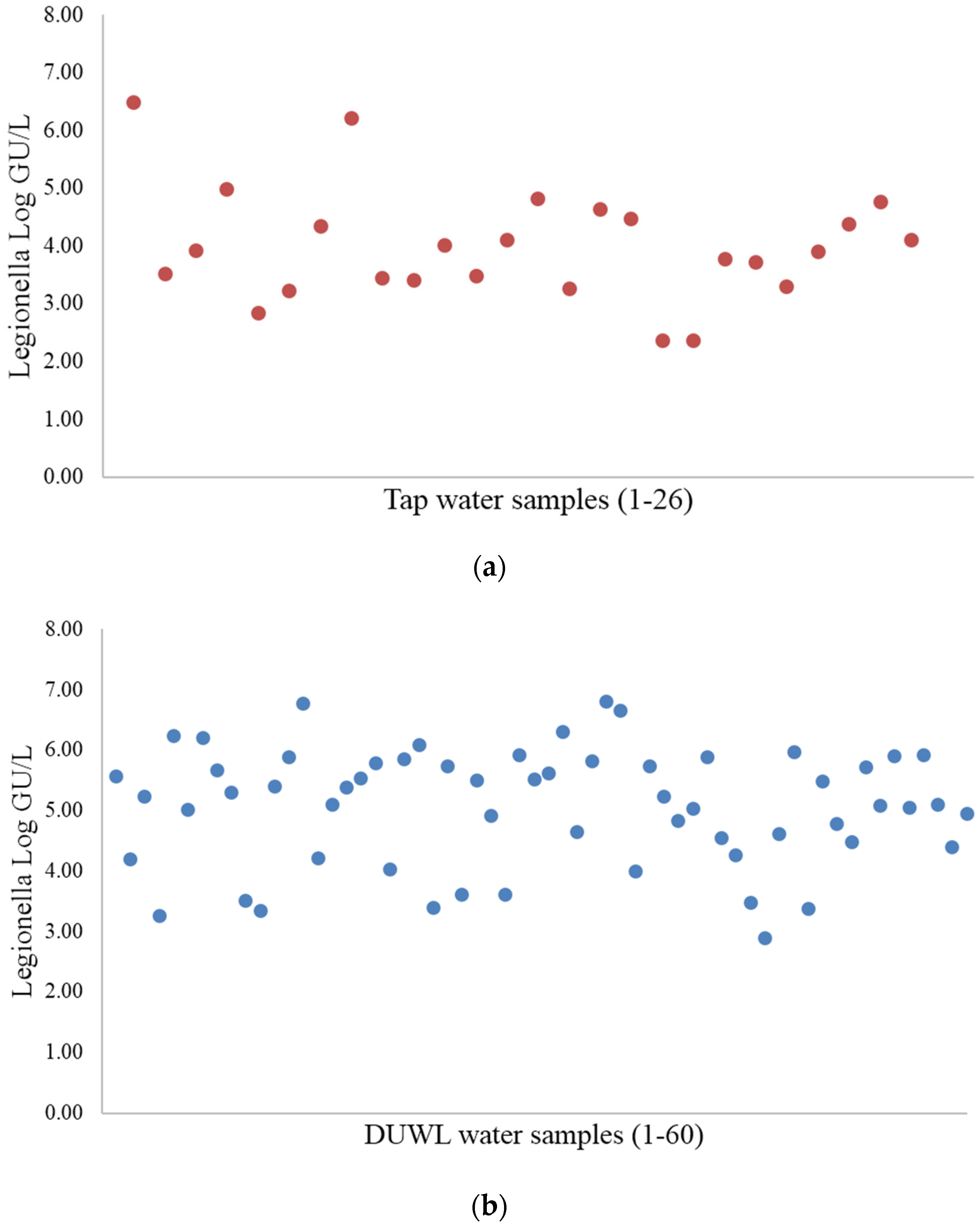
3.1. Legionella Quantification Using Culture and PMA-qPCR Methods

During the analysis, inhibition of PCR amplification (of the IC and the gene target) was observed in only one DUWL water sample. After this sample was diluted, the reaction was positive for the internal control of the amplification kit and the target template. For all of the other samples, the established range of the kit (28 < cycle threshold < 42) was always satisfied.
The results obtained using the culture and PMA-qPCR methods to evaluate 86 water samples were compared. Twenty-six samples were taken from tap water, and 60 were obtained from DUWLs.
Overall, Legionella spp. were detected in 100% (86/86) of the samples using the PMA-qPCR method, whereas they were detected in only 7% (6/86) of the samples using the culture method. Of these six culture-positive samples, four were collected from the output water of DUWLs (6.6%), and two were collected from tap water (7.7%). According to Fisher’s exact test, the difference was not significant (Table 1).
Table 1. Prevalence of Legionella spp. in water samples, as determined using PMA-qPCR and culture method.
Type of SampleCulture MethodPMA-qPCR
Positive n.Negative n.Positive n.Negative n.
Tap water224260
DUWL output456600
Total No. (%)6 (7%)80 (93%)86 (100%)(0%)
(Fisher’s test = 1, p < 0.05).
Non-pneumophila Legionella species were isolated from four of the DUWL output water samples and one tap water sample, and Legionella pneumophila sg. 2–14 was isolated from one tap water sample. Legionella spp. were isolated from both the tap water and DUWL output in only one of the 26 clinics. Furthermore one clinic was positive for Legionella spp. in both of the DUWLs installed.
The Legionella counts determined using PMA-qPCR ranged from 102 to 106 GU/L. The GU/L values for the 26 tap water samples and 60 DUWL samples determined using the PMA-qPCR method showed that the concentration of Legionella spp. was significantly lower (p = 0.000026) in the tap water samples, as determined the Mann-Whitney test (Figure 1).
Figure 1. Quantitative results for (a) the tap water and (b) DUWL samples, as determined using the PMA-qPCR method.
Ijerph 13 00211 g001
Statistical consistency (determined using the Mann-Whitney test): U-value = 326; p-value = 0.000026. Of the six samples that tested positive using the culture method, five were found to contain less than 103 CFU/mL of Legionella, and one contained >103 CFU/mL (Table 2).
Table 2. Quantification of Legionella in the six culture-positive environmental water samples: comparison of the results obtained using the PMA-qPCR and culture methods and the TVCs.
SampleLegionella PMA-qPCRLegionella CultureTVCs 22 °CTVCs 37 °C
(GU/L)(CFU/L)CFU/mLCFU/mL
DUWL output water6.7 × 1044.0 × 1023.0 × 1034.1 × 102
DUWL output water3.5 × 1041.0 × 1021.5 × 1022.6 × 102
DUWL output water1.1 × 1051.2 × 1031.4 × 1031.5 × 102
DUWL output water8.3 × 1059.0 × 1026.5 × 10270
Tap water4.2 × 1048.0 × 1024712
Tap water5.2 × 1034.0 × 10251

3.2. Waterborne Bacteria

The number of colony forming units (CFUs) of the TVCs determined for the DUWL and tap water samples differed, with the bacterial load of the tap water samples being significantly lower (Table 3).
Table 3. Quantitative results obtained using TVCs.
Type of SampleTVCs
22 °C37 °C
Tap water (geometric mean CFU/L ± SD)1.7 × 10² ± 5.8 × 1021.3 × 103 ± 5.8 × 10²
Dental unit (geometric mean CFU/L ± SD)1.5 × 10³ ± 1.4 × 1031.3 × 103 ± 2.0 × 10³
22 °C, U-value = 113.5; p-value = 0; 36 °C, U-value = 131; p-value = 0 (Mann-Whitney).
Comparing these results with the threshold values established by the European Council Directive 98/83/EC (20 CFU/mL at 36 °C and 100 CFU/mL at 22 °C) the counts were within the specified limits in only 5% (3/60) of the DUWL samples and in 77% (20/26) of the tap water samples.
Legionella spp. were isolated only from dental unit samples with bacterial loads ranging from 102 CFU/mL to 3 × 10³ CFU/mL, whereas they were not isolated from any of the samples with a high bioburden (>3 × 10³ CFU/mL); in addition, two of the 20 tap water samples that satisfied the required standards for drinking water were contaminated with Legionella spp. (Table 2 and Table 4).
Table 4. Relationship between Legionella-positive cultures and the TVCs.
TVCsDUWL Water Samples (n)Legionella-Positive Cultures (n)Tap Water Samples (n)Legionella-Positive Cultures (n)
Drinking water threshold *30202
102–3 × 10³ CFU/mL31450
>3 × 10³ CFU/mL26010
Total604262
* Threshold values established by the European Council Directive 98/83/E: <20 CFU/mL at 36 °C and <100 CFU/mL at 22 °C.
Overall, no significant association between the TVCs and the Legionella-positive culture results was demonstrated. We found that the TVC and PMA-qPCR detection methods had good accuracy, with 57 samples found to be Legionella-positive using both of these methods, yielding an accuracy rate of 95%.

4. Discussion

It has long been known that water samples collected at the outputs of dental unit waterlines are densely populated with microorganisms. For proliferation legionellae require the presence of other microorganisms (particularly amoebae), a supply of nutrients, and temperatures in the range of 20–45 °C [26]. As reported by many authors over the last 30 years, the rate of recovery of Legionella spp. from DUWLs ranges from 0% to 100%, and the counts of these bacteria in DUWL samples range from 0 to 106 CFU/mL [1,27,28,29,30].
The very large variability in the counts reported in the literature is likely due to several factors, such as the amount of available soluble organic compounds, the presence of heavy metals, the temperature and level of free chlorine in municipal water distribution systems, the TVCs, the sampling time, and the analytical methods used (culture, PCR, or immunofluorescence staining) [31]. Due to its capacity to detect low levels of target nucleic acids, including those of viable Legionella, non-cultivable Legionella and Legionella within amoebae, q-PCR may be considered the gold standard method for determining the concentrations of these bacteria in water.
This study is the first to quantify Legionella in DUWLs using PMA-qPCR. In most previously published reports, the prevalence of Legionella in DUWLs has been evaluated using culture methods, and only Atlas [1], Williams [32] and Dutil [33] used PCR methods. However, in contrast with these previous studies, our chosen method eliminated the background of dead cells by including a PMA treatment, and despite employing this strategy, all of the samples tested positive for Legionella.
The rate of recovery of Legionella from the DUWLs observed in our study greatly varied depending on the analytical method used. When we used the culture method to detect Legionella in the DUWL samples, we observed a low rate of contamination of 6.6% (also observed in a previous study that has not yet been reported, in which we analysed 40 DUWL water samples using the culture method and found that none of the dental units tested had Legionella-contaminated water); however, when we used PMA-qPCR for the same purpose, we found that 100% of the samples were positive. These results, which were obtained using cold water samples (at 18–20 °C), are very different from those obtained using environmental samples collected from hospital water systems, most likely due to differences in their aqueous matrices. In our previous study [21], we analysed samples of hot water, an ideal habitat for Legionella, and the level of agreement between the results obtained using the two methods (culture vs. PMA-qPCR) was 79%. Consistent with the results of other studies [31,34,35], we demonstrated that although a significant proportion of the DUWLs tested were disinfected, samples taken from them were more heavily contaminated with bacteria compared with tap water samples collected from the same offices (by approximately 10-fold) and that their bacterial levels were much higher than the standards set for drinking water. Only three of the dental units studied delivered water that met the accepted Italian standard for drinking water (<20 CFU/mL at 36 °C and <100 CFU/mL at 22 °C). During sample collection, two of these DUWLs were treated with high concentrations of disinfectant. Our results confirm the findings of other studies [5,36], which have demonstrated that the concentration of heterotrophic bacteria is not associated with the number of Legionella isolates obtained using culture methods. However, we observed a good level of accuracy between the TVC and the PMA-qPCR method.
The three samples that were found to be Legionella-positive using the culture method were those with total bacterial counts ranging from 102 to 103 CFU/mL. The presence of other bacteria in biofilms, particularly that of Gram-negative bacteria that produce bacteriocins may inhibit the growth of Legionella or at least stress Legionella cells, making it impossible for Legionella to be detected using culture assays. Toze et al. [37] examined the ability of heterotrophic bacterial strains isolated from chlorinated drinking water on low-nutrient media to inhibit the growth of Legionella species. Sixteen to 32% of these strains inhibited the growth of Legionella spp. on buffered charcoal yeast extract agar, and the exact proportion of the growth-inhibiting strain varied according to the particular Legionella species. Moreover, when the amino acid supply becomes growth-limiting, intracellular bacteria produce factors that lyse the spent host cells, allowing the bacteria to survive osmotic stress, disperse in the environment and re-establish intracellular niches protected from lysosomal degradation. Arrival in a rich intracellular environment stimulates a return to the replicative phenotype. When the nutrient levels and other conditions of the host cells are favourable, L. pneumophila cells express factors that promote maximal replication [38,39].
In addition to inhibition due to other microorganisms, it must be remembered that the understimation by plate count method depends on the presence of Legionella bacteria contained in amoebae that produce and excrete vesicles. Rowbotham [14] has hypothesized that inhaling vesicles containing tightly packed legionellae is dangerous when these bacteria are motile. Greater than 90% of the observed vesicles fell within the size range considered to be respirable (1 to 5 µm in diameter) [11]. The Legionella-positivity observed using the qPCR method in the 3 samples compliant with drinking water standards could be linked to the presence of amoebae that are resistant to disinfection treatments.

5. Conclusions

In summary, our results and the data obtained by Atlas [1] and Dutil [33] show high rates of Legionella contamination in DUWLs (from 68% to 93%), as determined by PCR analyses. Therefore, the presence of legionellae in dental units in which the effluent water is not treated with effective concentrations of chemical products, can be taken for granted because Legionella is an aquatic organism and the low temperatures of these units allow for its survival during starvation. Determining the Legionella concentrations in DUWL outputs using the culture method is not sufficient for predicting the exact size of the problem. We think that until the level of clinical risk is not expressed in GU/L, we cannot state that qPCR is the best method for the risk assessment. The concordance observed between the TVCs and PMA-qPCR results substantiates the assertion that the microbial load includes legionellae, regardless of their infectivity and pathogenicity. Therefore, we would like to share the suggestion of the CDC, which is that “… no rationale is seen for routine testing for such specific organisms” [40] and suggest that microbiological monitoring should not target individual organisms, such as Legionella, Pseudomonas, and Acinetobacter bacteria, but it should rather address the bioburden that encompasses them. Our final suggestion is that the cleanliness of dental units should be assessed by evaluating the concentrations of aerobic heterotrophic bacteria because contamination by these organisms are good indicators of the potential presence of Legionella.

Acknowledgments

We thank ANDI Torino (National Association of Italian Dentists) for providing access to the water samples from the dental units and American Journal Experts (AJE) for English-language editing.

Author Contributions

Savina Ditommaso conceived and designed the experiments and drafted the manuscript; Monica Giacomuzzi contributed to the analysis and interpretation of the data; Elisa Ricciardi contributed to the analysis and interpretation of the data; and Carla M. Zotti critically revised the manuscript.

Conflicts of Interest

The authors declare no conflict of interest.

References

  1. Atlas, R.M.; Williams, J.F.; Huntington, M.K. Legionella contamination of dental-unit waters. Appl. Environ. Microbiol. 1995, 61, 1208–1213. [Google Scholar] [PubMed]
  2. Barbeau, J.; Gauthier, C.; Payment, P. Biofilms, infectious agents, and dental unit waterlines: A review. Can. J. Microbiol. 1998, 44, 1019–1028. [Google Scholar] [CrossRef] [PubMed]
  3. Walker, J.T.; Bradshaw, D.J.; Finney, M.; Fulford, M.R.; Frandsen, E.; ØStergaard, E.; ten Cate, J.M.; Moorer, W.R.; Schel, A.J.; Mavridou, A.; et al. Microbiological evaluation of dental unit water systems in general dental practice in Europe. Eur. J. Oral Sci. 2004, 112, 412–418. [Google Scholar] [CrossRef] [PubMed]
  4. Dutil, S.; Veillette, M.; Mériaux, A.; Lazure, L.; Barbeau, J.; Duchaine, C. Aerosolization of Mycobacteria and legionellae during dental treatment: Low exposure despite dental unit contamination. Environ. Microbiol. 2007, 9, 2836–2843. [Google Scholar] [CrossRef] [PubMed]
  5. Bristela, M.; Skolka, A.; Schmid-Schwap, M.; Piehslinger, E.; Indra, A.; Wewalka, G.; Stauffer, F. Testing for aerobic heterotrophic bacteria allows no prediction of contamination with potentially pathogenic bacteria in the output water of dental chair units. GMS Krankenhaushyg. Interdiszip. 2012, 7. [Google Scholar] [CrossRef]
  6. Arvand, M.; Hack, A. Microbial contamination of dental unit waterlines in dental practices in Hesse, Germany: A cross-sectional study. Eur. J. Microbiol. Immunol. 2013, 3, 49–52. [Google Scholar] [CrossRef] [PubMed]
  7. Vincent-Houdek, M.; Muytjens, H.L.; Bongaerts, G.P.; van Ketel, R.J. Legionella monitoring: A continuing story of nosocomial infection prevention. J. Hosp. Infect. 1993, 25, 117–124. [Google Scholar] [CrossRef]
  8. Kool, J.L.; Bergmire-Sweat, D.; Butler, J.C.; Brown, E.W.; Peabody, D.J.; Massi, D.S.; Carpenter, J.C.; Pruckler, J.M.; Benson, R.F.; Fields, B.S. Hospital characteristics associated with colonization of water systems by Legionella and risk of nosocomial legionnaires’ disease: A cohort study of 15 hospitals. Infect. Control Hosp. Epidemiol. 1999, 20, 798–805. [Google Scholar] [CrossRef] [PubMed]
  9. Wellinghausen, N.; Frost, C.; Marre, R. Detection of legionellae in hospital water samples by quantitative real-time LightCycler PCR. Appl. Environ. Microbiol. 2001, 67, 3985–3993. [Google Scholar] [CrossRef] [PubMed]
  10. Ricci, M.L.; Fontana, S.; Pinci, F.; Fiumana, E.; Pedna, M.F.; Farolfi, P.; Sabattini, M.A.B.; Scaturro, M. Pneumonia associated with a dental unit waterline. Lancet 2012, 379. [Google Scholar] [CrossRef]
  11. Pankhurst, C.L. Risk assessment of dental unit waterline contamination. Prim. Dent. Care 2003, 10, 5–10. [Google Scholar] [CrossRef] [PubMed]
  12. Isituto Superiore di Sanità. Rapporto Annuale Sulla Legionellosi in Italia Nel 2013; Isituto Superiore di Sanità: Roma, Italy, 2014; pp. 3–9. (In Italian) [Google Scholar]
  13. Ministero della Salute. Linee Guida Per La Prevenzione ed Il Controllo Della Legionellosi; Ministero della Salute: Roma, Italy, 2015. Available online: http://www.salute.gov.it/imgs/C_17_pubblicazioni_2362_allegato.pdf (accessed on 4 February 2016). (In Italian)
  14. Rowbotham, T.J. Current views on the relationships between amoebae, legionellae and man. Isr. J. Med. Sci. 1986, 22, 678–689. [Google Scholar] [PubMed]
  15. Berk, S.G.; Ting, R.S.; Turner, G.W.; Ashburn, R.J. Production of respirable vesicles containing live Legionella pneumophila cells by two Acanthamoeba spp. Appl. Environ. Microbiol. 1998, 64, 279–286. [Google Scholar] [PubMed]
  16. Swanson, M.S.; Hammer, B.K. Legionella pneumophila pathogesesis: A fateful journey from amoebae to macrophages. Annu. Rev. Microbiol. 2000, 54, 567–613. [Google Scholar] [CrossRef] [PubMed]
  17. Michel, R.; Borneff, M. The significance of amoebae and other protozoa in water conduit systems in dental units. Zentralblatt Bakteriol. Mikrobiol. Hyg. Ser. B 1989, 187, 312–323. [Google Scholar]
  18. Villari, P.; Motti, E.; Farullo, C.; Torre, I. Comparison of conventional culture and PCR methods for the detection of Legionella pneumophila in water. Lett. Appl. Microbiol. 1998, 27, 106–110. [Google Scholar] [CrossRef] [PubMed]
  19. Ng, D.L.; Koh, B.B.; Tay, L.; Heng, B.H. Comparison of polymerase chain reaction and conventional culture for the detection of legionellae in cooling tower waters in Singapore. Lett. Appl. Microbiol. 1997, 24, 214–216. [Google Scholar] [CrossRef] [PubMed]
  20. Bates, M.N.; Maas, E.; Martin, T.; Harte, D.; Grubner, M.; Margolin, T. Investigation of the prevalence of Legionella species in domestic hot water systems. N. Z. Med. J. 2000, 113, 218–220. [Google Scholar] [PubMed]
  21. Ditommaso, S.; Ricciardi, E.; Giacomuzzi, M.; Arauco Rivera, S.R.; Ceccarelli, A.; Zotti, C.M. Overestimation of the Legionella spp. load in environmental samples by quantitative real-time PCR: Pretreatment with propidium monoazide as a tool for the assessment of an association between Legionella concentration and sanitary risk. Diagn. Microbiol. Infect. Dis. 2014, 80, 260–266. [Google Scholar] [CrossRef] [PubMed]
  22. Ditommaso, S.; Gentile, M.; Giacomuzzi, M.; Zotti, C.M. Recovery of Legionella species from water samples using an internal method based on ISO 11731: Suggestions for revision and implementation. Diagn. Microbiol. Infect. Dis. 2011, 70, 200–206. [Google Scholar] [CrossRef] [PubMed]
  23. Ditommaso, S.; Ricciardi, E.; Giacomuzzi, M.; Arauco Rivera, S.R.; Zotti, C.M. Legionella in water samples: How can you interpret the results obtained by quantitative PCR? Mol. Cell. Probes 2015, 29, 7–12. [Google Scholar] [CrossRef] [PubMed]
  24. International Standards Organization (ISO). Water Quality—Enumeration of Culturable Micro-organisms—Colony Count by Inoculation in A Nutrient Agar Culture Medium; ISO 6222; International Standards Organization: Geneva, Switzerland, 1999. [Google Scholar]
  25. Eaton, A.D.; Clesceri, L.S.; Greenberg, A.E.; American Public Health Association; American Water Works Association. Standard Methods for the Examination of Water and Wastewater; American Public Health Association: Washington, DC, USA, 1995. [Google Scholar]
  26. Wadowsky, R.M.; Wolford, R.; McNamara, A.M.; Yee, R.B. Effect of temperature, pH, and oxygen level on the multiplication of naturally occurring Legionella pneumophila in potable water. Appl. Environ. Microbiol. 1985, 49, 1197–1205. [Google Scholar] [PubMed]
  27. Walker, J.T.; Bradshaw, D.J.; Bennett, A.M.; Fulford, M.R.; Martin, M.V.; Marsh, P.D. Microbial biofilm formation and contamination of dental-unit water systems in general dental practice. Appl. Environ. Microbiol. 2000, 66, 3363–3367. [Google Scholar] [CrossRef] [PubMed]
  28. Mills, S.E. Waterborne pathogens and dental waterlines. Dent. Clin. N. Am. 2003, 47, 545–557. [Google Scholar] [CrossRef]
  29. Pankhurst, C.L.; Coulter, W.; Philpott-Howard, J.J.; Harrison, T.; Warburton, F.; Platt, S.; Surman, S.; Challacombe, S. Prevalence of Legionella waterline contamination and Legionella pneumophila antibodies in general dental practitioners in London and rural northern Ireland. Brit. Dent. J. 2003, 195, 591–594. [Google Scholar] [CrossRef] [PubMed]
  30. Ma’ayeh, S.Y.; Al-Hiyasat, A.S.; Hindiyeh, M.Y.; Khader, Y.S. Legionella pneumophila contamination of a dental unit water line system in a dental teaching centre. Int. J. Dent. Hyg. 2008, 6, 48–55. [Google Scholar] [CrossRef] [PubMed]
  31. Barbeau, J.; Tanguay, R.; Faucher, E.; Avezard, C.; Trudel, L.; Côté, L.; Prévost, A.P. Multiparametric analysis of waterline contamination in dental units. Appl. Environ. Microbiol. 1996, 62, 3954–3959. [Google Scholar] [PubMed]
  32. Williams, H.N.; Paszko-Kolva, C.; Shahamat, M.; Palmer, C.; Pettis, C.; Kelley, J. Molecular techniques reveal high prevalence of Legionella in dental units. J. Am. Dent. Assoc. 1996, 127, 1188–1193. [Google Scholar] [CrossRef] [PubMed]
  33. Dutil, S.; Tessier, S.; Veillette, M.; Laflamme, C.; Mériaux, A.; Leduc, A.; Barbeau, J.; Duchaine, C. Detection of Legionella spp. by fluorescent in situ hybridization in dental unit waterlines. J. Appl. Microbiol. 2006, 100, 955–963. [Google Scholar] [CrossRef] [PubMed]
  34. Noce, L.; di Giovanni, D.; Putnins, E.E. An evaluation of sampling and laboratory procedures for determination of heterotrophic plate counts in dental unit waterlines. J. Can. Dent. Assoc. 2000, 66, 262. [Google Scholar] [PubMed]
  35. Szymanska, J. Biofilm and dental unit waterlines. Ann. Agric. Environ. Med. 2003, 10, 151–157. [Google Scholar] [PubMed]
  36. Leoni, E.; Dallolio, L.; Stagni, F.; Sanna, T.; D’Alessandro, G.; Piana, G. Impact of a risk management plan on Legionella contamination of dental unit water. Int. J. Environ. Res. Public. Health 2015, 12, 2344–2358. [Google Scholar] [CrossRef] [PubMed]
  37. Toze, S.; Sly, L.I.; Macrae, I.C.; Fuerst, J.A. Inhibition of growth of Legionella species by heterotrophic plate count bacteria isolated from chlorinated drinking water. Curr. Microbiol. 1990, 21, 139–143. [Google Scholar] [CrossRef]
  38. Byrne, B.; Swanson, M.S. Expression of Legionella pneumophila virulence traits in response to growth conditions. Infect. Immun. 1998, 66, 3029–3034. [Google Scholar] [PubMed]
  39. Ohata, K.; Sugiyama, K.; Suzuki, M.; Shimogawara, R.; Izumiyama, S.; Yagita, K.; Endo, T. Growth of Legionella in nonsterilized, naturally contaminated bathing water in a system that circulates the water. In Legionella; ASM Press: Washington, DC, USA, 2006. [Google Scholar]
  40. Centers for Disease Control and Prevention. Guidelines for Infection Control in Dental Health-Care Settings—2003. Available online: http://www.cdc.gov/mmwr/preview/mmwrhtml/rr5217a1.htm (accessed on 4 February 2016).

