
Divergent Effects of Arsenic on NF-κB Signaling in Different Cells or Tissues: A Systematic Review and Meta-Analysis

Abstract
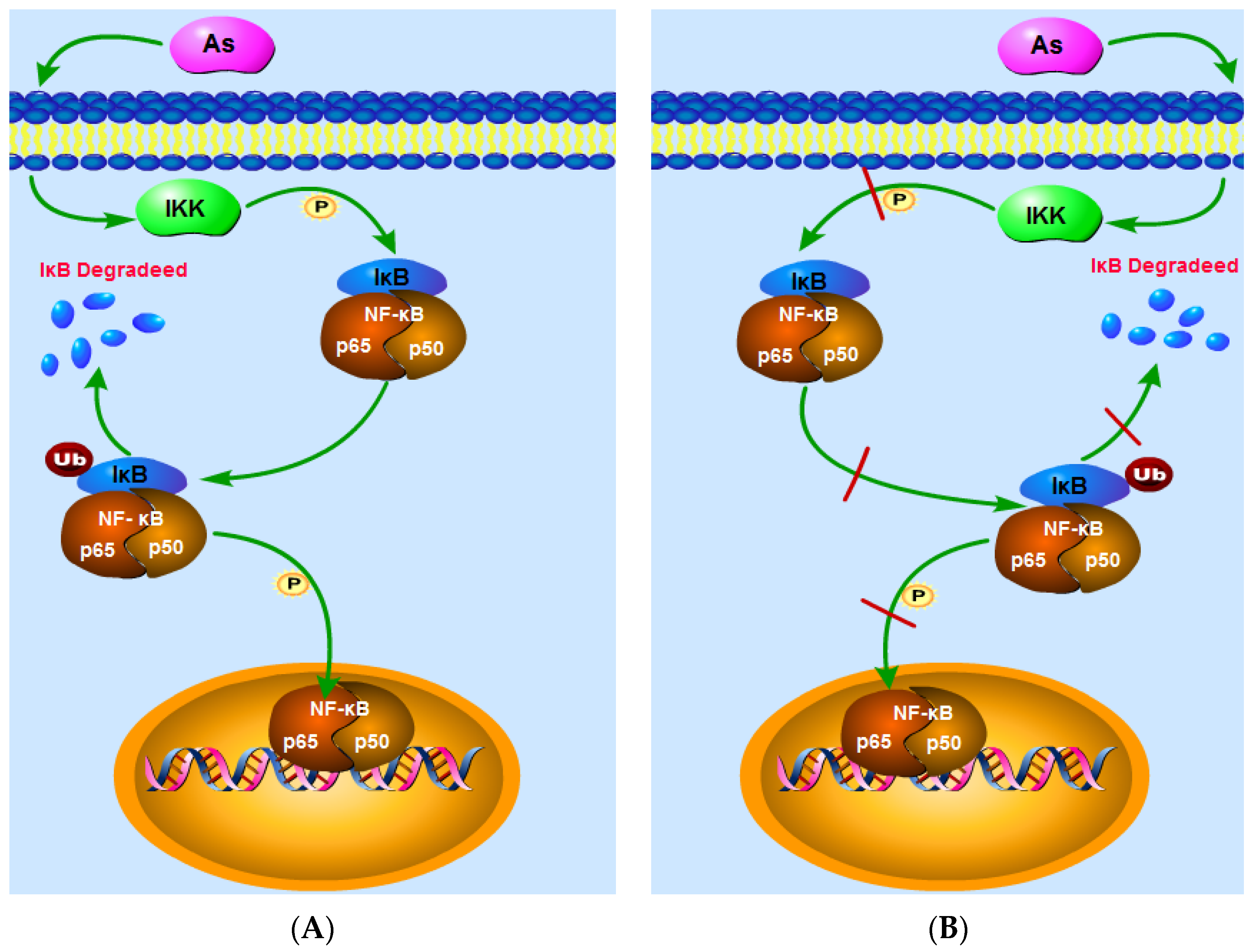
:1. Introduction
2. Materials and Methods
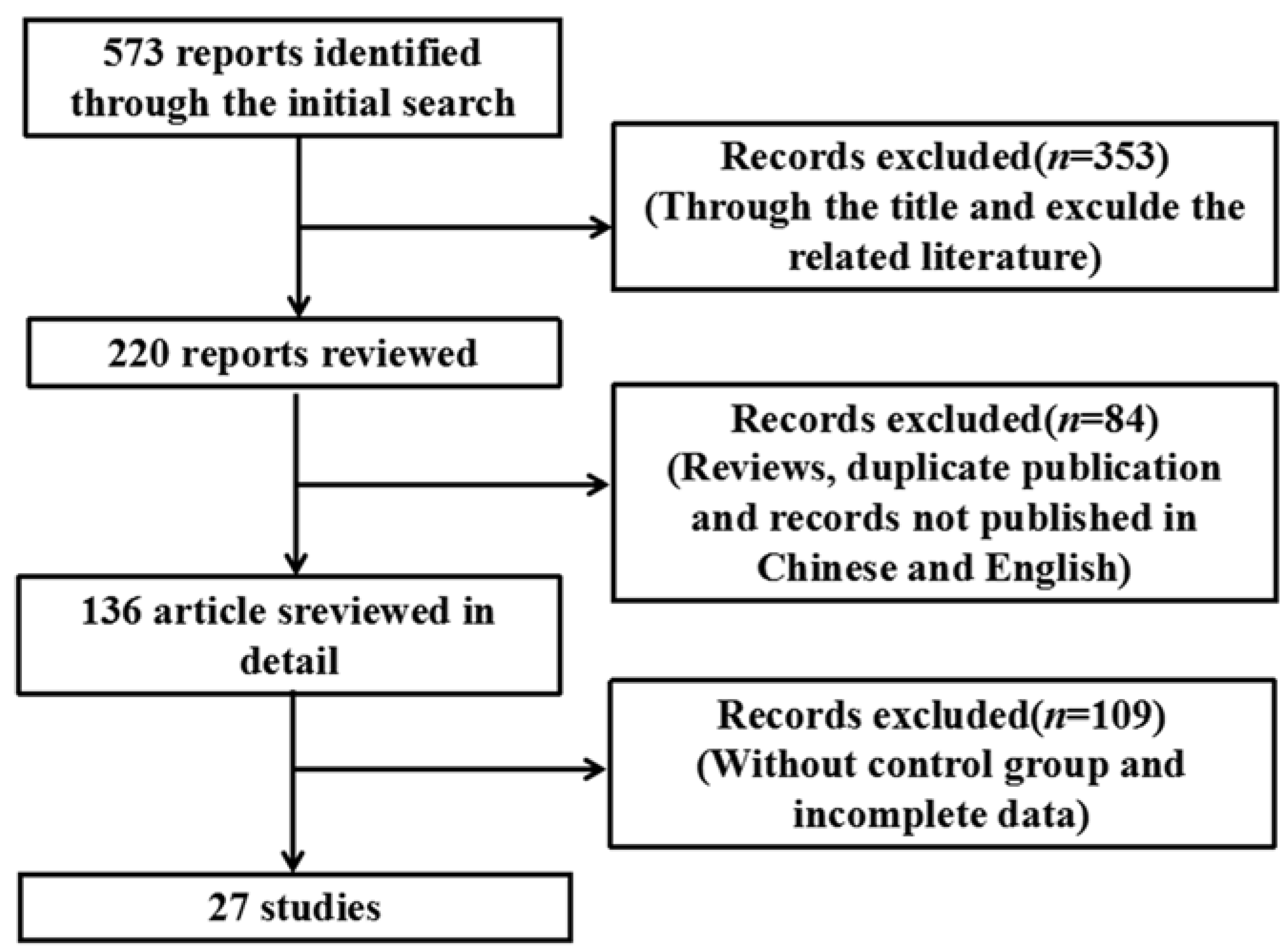
2.1. Search Strategy
2.2. Eligibility Criteria
2.3. Exclusion Criteria
2.4. Outcome Indicators
2.5. Data Extraction
2.6. Data Analysis
3. Results
3.1. Study Characteristics

| First Author (Year) | Language | n | Type of Cells or Tissue | Type of Arsenical Compounds | Period of Arsenic | Dose of Arsenic | Outcome Indicators |
|---|---|---|---|---|---|---|---|
| Jyotirmoy Ghosh 2009 [13] | English | 6 | Normal cells | NaAsO2 | ≤24 h | ≤5 μmol/L | 1,8 |
| Shi-Yi Liu 2014 [14] | Chinese | 3 | Normal cells | NaAsO2 | ˃24 h | ≤5 μmol/L | 2 |
| Daniella M. B. Kerbauy 2005 [15] | English | 3 | Cancer cells | As2O3 | ≤24 h | >5 μmol/L | 2,8 |
| Yi Hao 2012 [16] | Chinese | 3 | Cancer cells | As2O3 | ≤24 h | >5 μmol/L | 1,3,5,6 |
| François Binet 2010 [17] | English | 3 | Normal cells | As2O3 | ≤24 h | ≤5 μmol/L | 7 |
| Kumar Felix 2005 [18] | English | 3 | Normal cells | NaAsO2 | ≤24 h | >5 μmol/L | 3,7,8 |
| Xue-Zhong Gong 2015 [19] | English | 3 | Normal cells | NaAsO2 | ≤24 h | ≤5 μmol/L | 8 |
| Si-Qi Cao 2015 [20] | Chinese | 20 | Normal cells | DMAѴ | ˃24 h | >5 mg/kg | 1,4,5 |
| Xiao-Yan Qu 2012 [21] | English | 3 | Cancer cells | As2O3 | ˃24 h | ≤5 μmol/L | 1 |
| Yu Hu 2002 [22] | English | 3 | Normal cells | NaAsO2 | ≤24 h | ≤5 μmol/L | 7 |
| Lin-Fu Zhou 2006 [23] | English | 6 | Inflammatory tissue | As2O3 | ≤24 h | ≤5 mg/kg | 3,6 |
| Hye Ran Lee 2008 [24] | English | 3 | Cancer cells | As2O3 | ≤24 h | >5 μmol/L | 2,3,8 |
| Min Jeong Kim 2014 [25] | English | 3 | Cancer cells | As4O6 | ≤24 h | ≤5 μmol/L | 2,3,5 |
| De-Lin Wang 2007 [26] | Chinese | 15 | Cancer cells | As2O3 | ˃24 h | ≤5 μmol/L | 1,3,4,5,7 |
| Stephan Mathas 2003 [27] | English | 3 | Cancer cells | NaAsO2 | ˃24 h | >5 μmol/L | 7 |
| Jing Qiu 2008 [28] | Chinese | 9 | Cancer cells | As2O3 | ˃24 h | ≤5 mg/kg | 1,3,8 |
| Ruben Ruiz-Ramos 2009 [29] | English | 3 | Cancer cells | NaAsO2 | ≤24 h | >5 μmol/L | 2,8 |
| Yong-Shen Fan 2008 [30] | Chinese | 5 | Cancer cells | As2O3 | ˃24 h | ≤5 μmol/L | 6 |
| Shu-jian Wang 2008 [31] | Chinese | 3 | Cancer cells | As2O3 | ˃24 h | ≤5 μmol/L | 4 |
| Yi-Fang Mei 2006 [32] | Chinese | 4 | Inflammatory cells | As2O3 | ˃24 h | >5 μmol/L | 8 |
| Robert R. Roussel 2000 [33] | English | 3 | Normal cells | NaAsO2 | ≤24 h | ≤5 μmol/L | 2,3,6,8 |
| Ke-Xin Zhang 2015 [34] | English | 6 | Normal cells | As2O3 | ˃24 h | >5 μmol/L | 4 |
| Xiao-Wei Xu 2005 [35] | Chinese | 3 | Cancer cells | As2O3 | ≤24 h | ≤5 μmol/L | 8 |
| Xiao-Hong Zhang 2004 [36] | Chinese | 3 | Cancer cells | As2O3 | ≤24 h | ≤5 μmol/L | 3,5 |
| Yao Zhang 2008 [37] | Chinese | 3 | Cancer cells | As2O3 | ≤24 h | >5 μmol/L | 5 |
| P. B. Tchounwou 2002 [38] | English | 3 | Cancer cells | As2O3 | ˃24 h | ≤5 μg/mL | 8 |
| Yi-Fang Mei 2011 [39] | English | 3 | Inflammatory cells | As2O3 | ˃24 h | ≤5 μmol/L | 7 |
3.2. Meta-Analysis of Arsenic Exposure Effects on the NF-κB Signaling Pathway
3.2.1. Effects of Arsenic Exposure on p-IκB

3.2.2. Effects of Arsenic Exposure on IκB
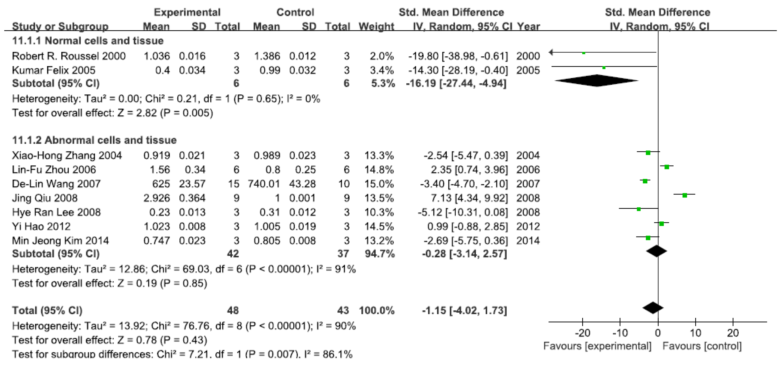
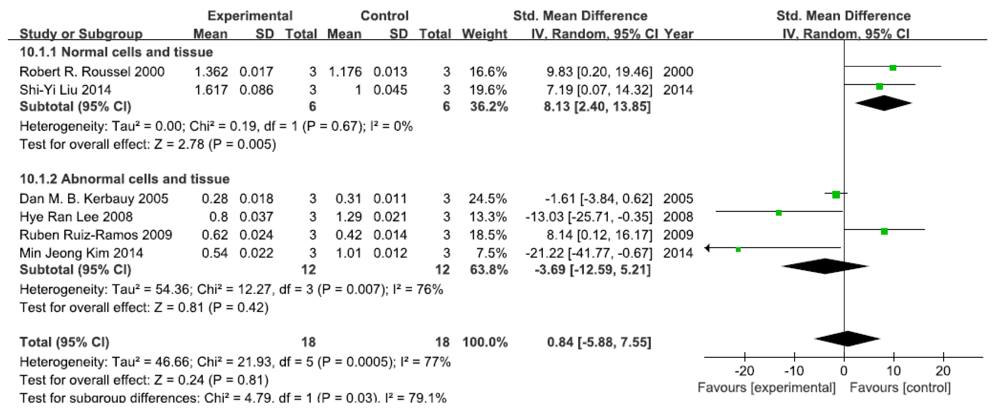
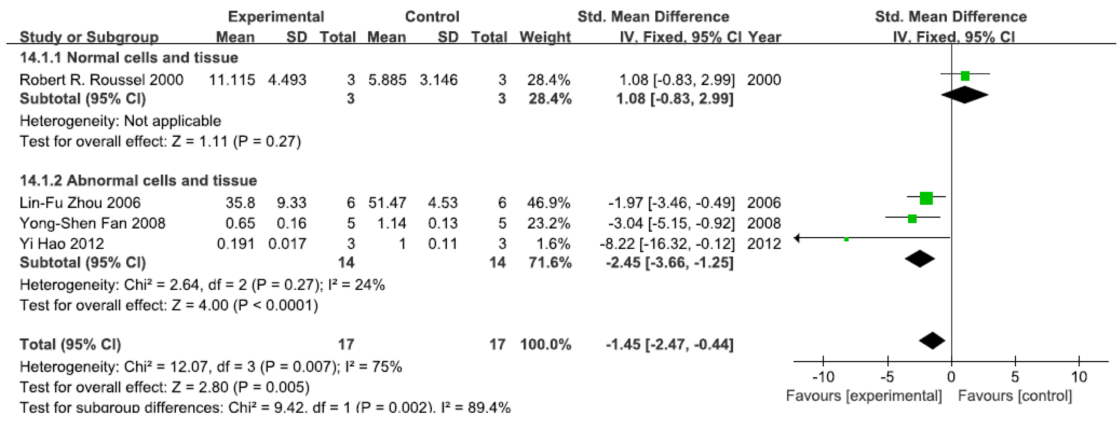
3.2.3. Effects of Arsenic Exposure on NF-κBp65
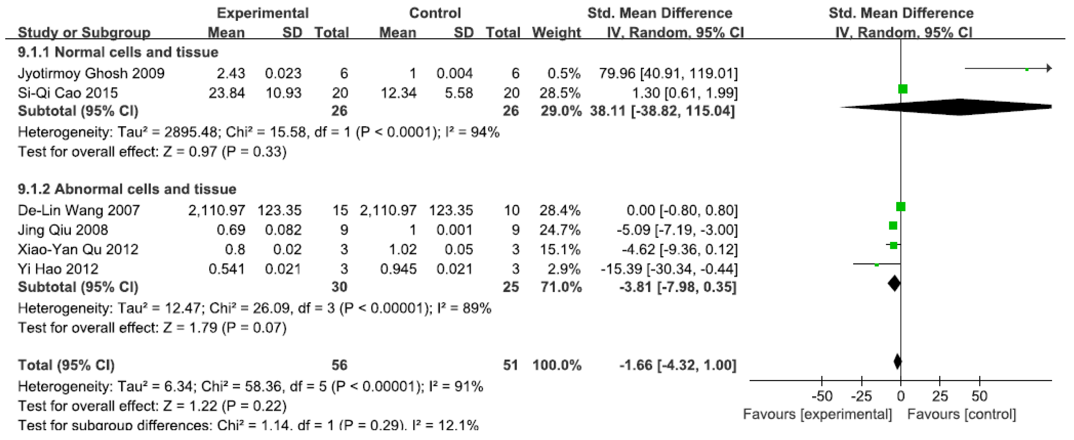
3.2.4. Effects of Arsenic Exposure on NF-κB

3.2.5. Effects of Arsenic Exposure on IKK

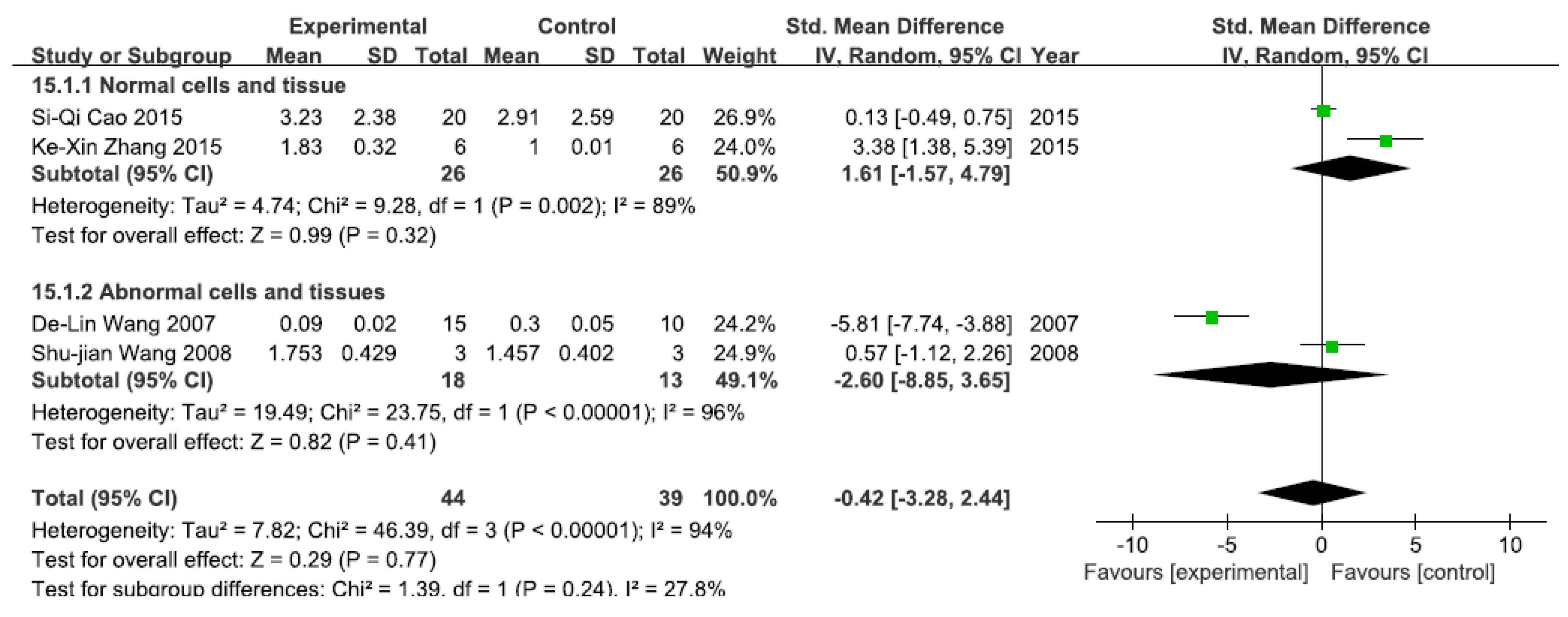
3.2.6. Effects of Arsenic Exposure on NF-κB Activity

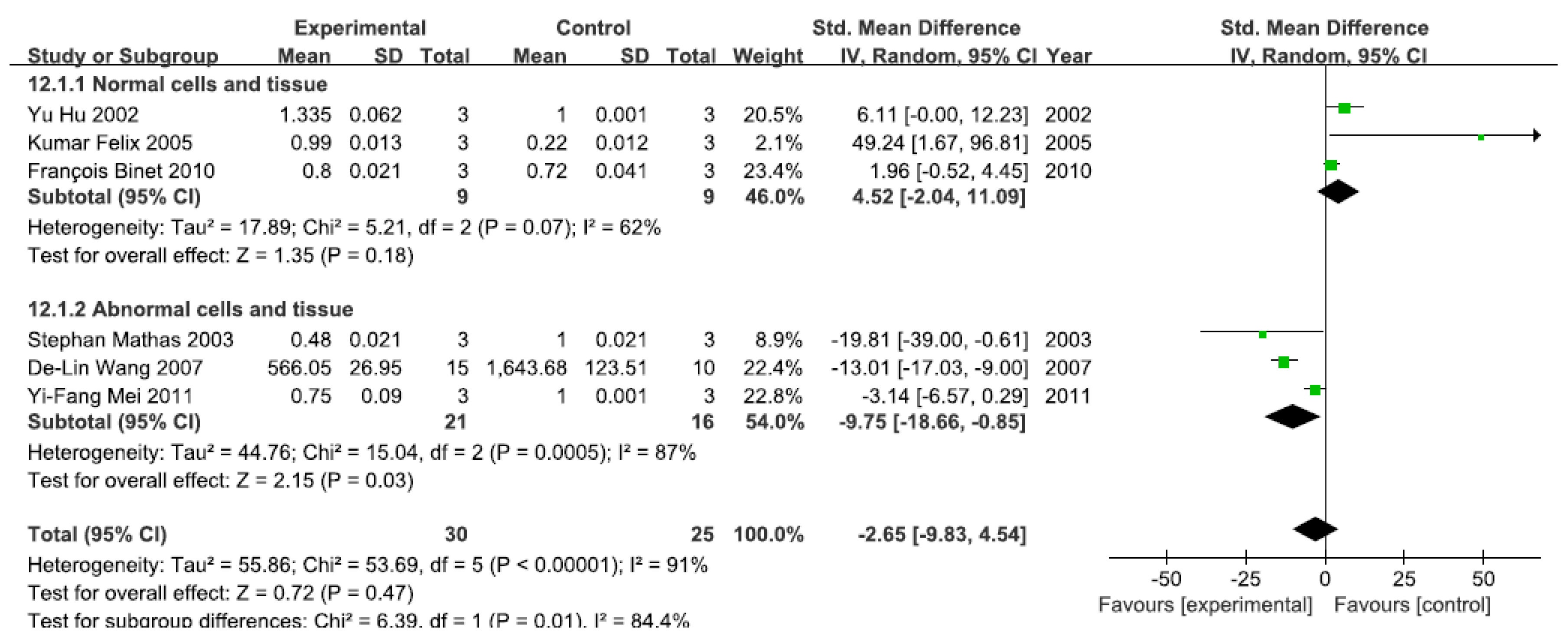
3.2.7. Effects of Arsenic Exposure on DNA-Binding Activity of NF-κB

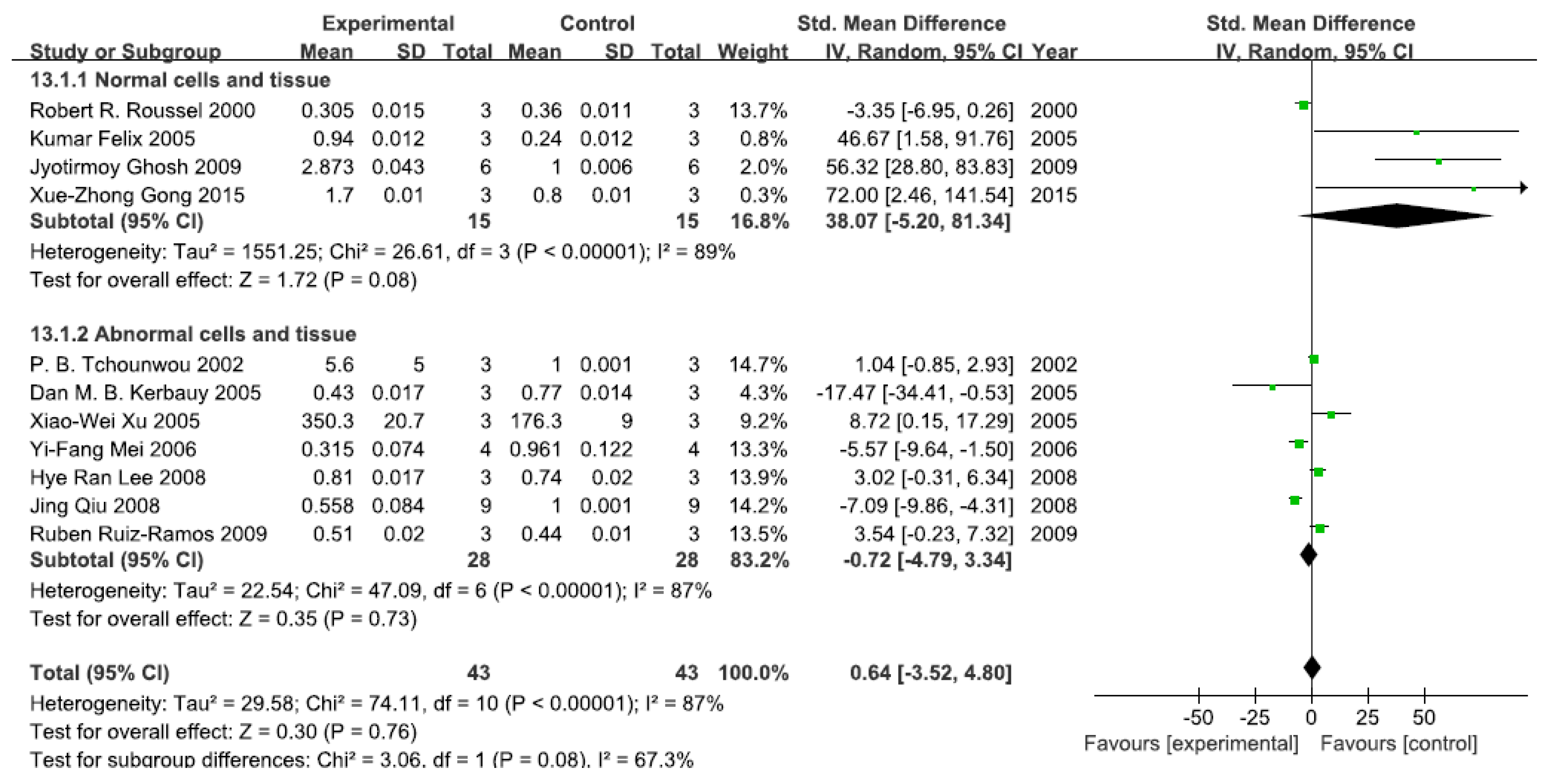
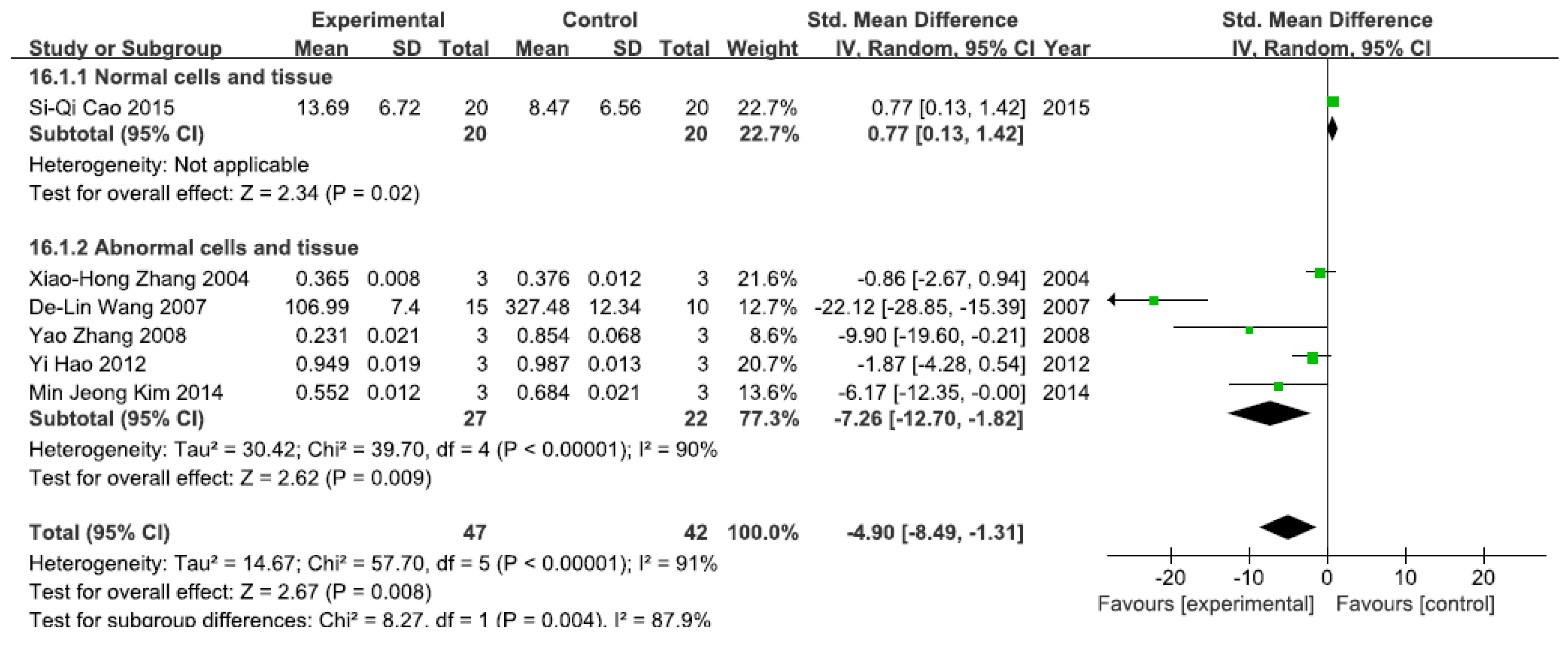
3.2.8. Effects of Arsenic Exposure on NF-κB mRNA
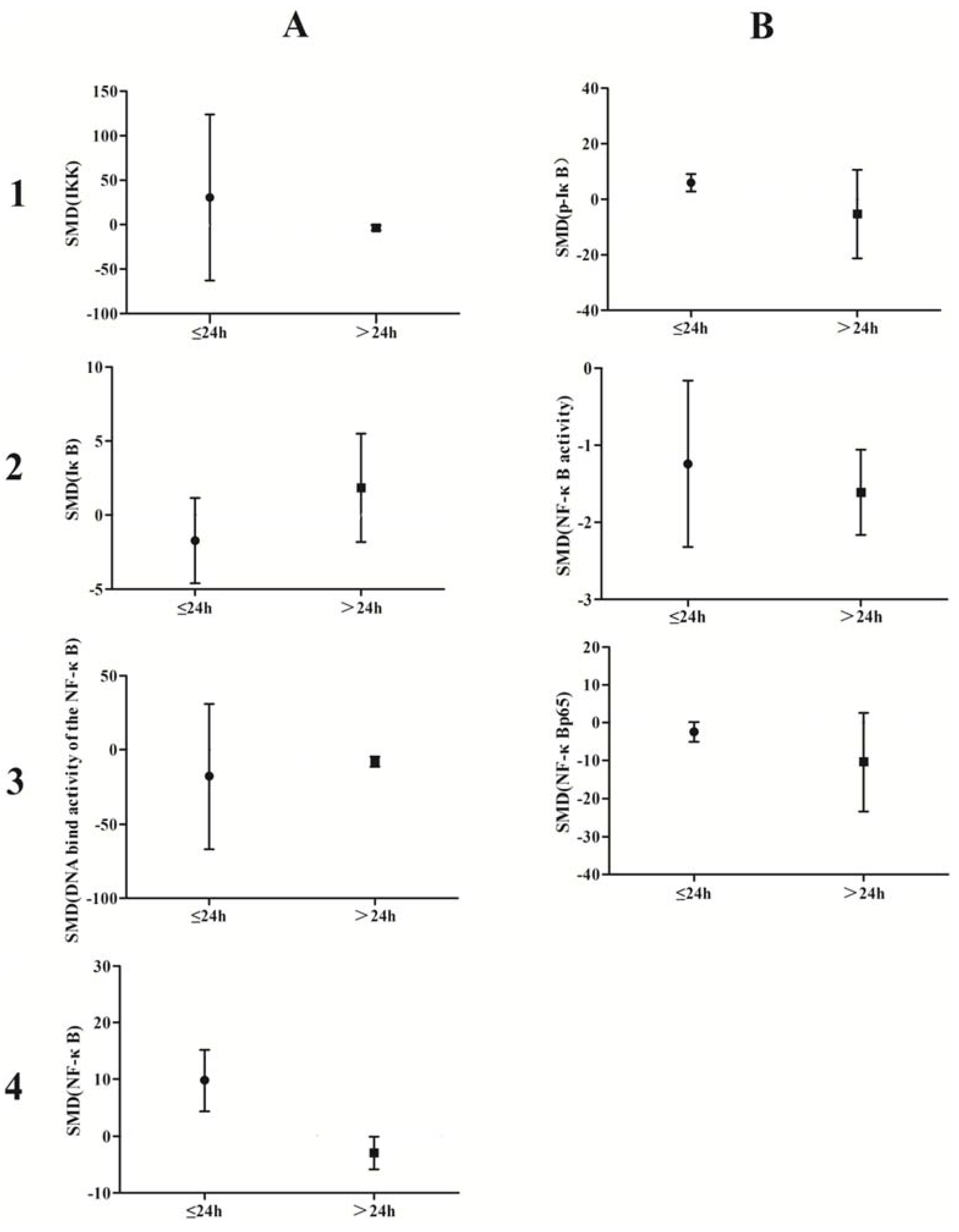
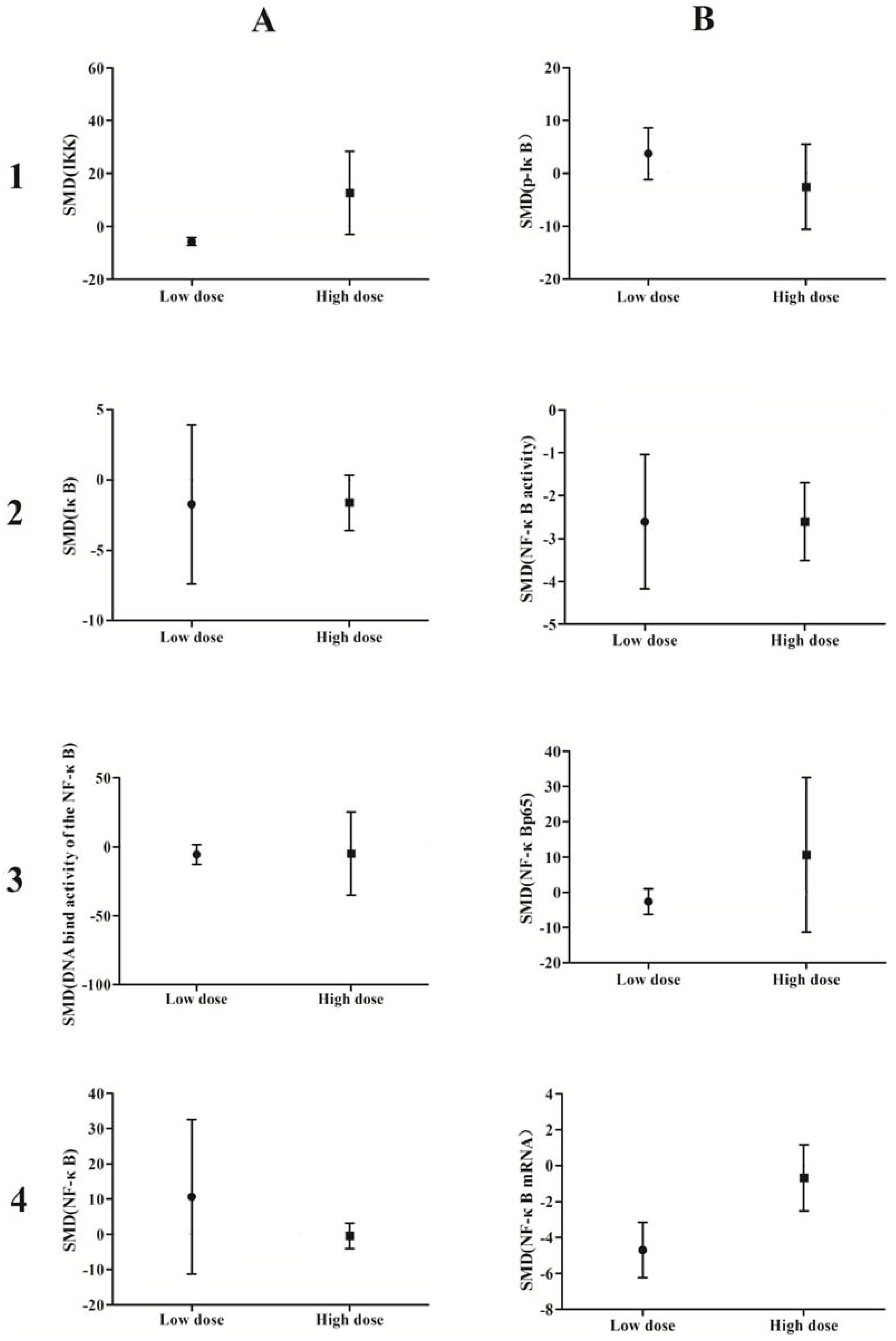
3.3. Subgroup Analyses of the Effects of Arsenic Exposure


3.4. Small-Study Effect Evaluation

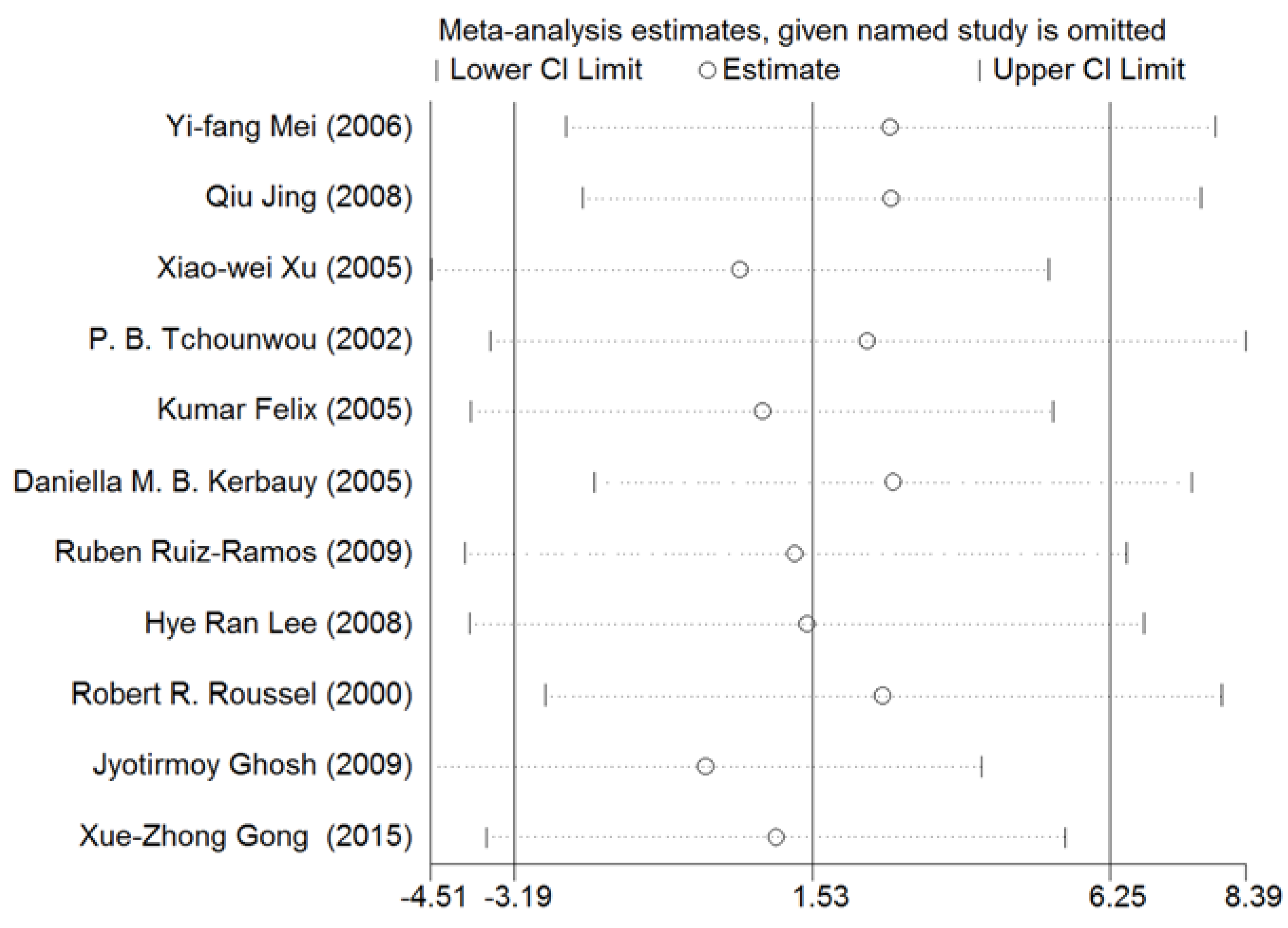
3.5. Sensitivity Analysis
3.6. Meta-Regression Analysis of Arsenic Exposure Effects
4. Discussion

5. Conclusions
Acknowledgments
Author Contributions
Conflicts of Interest
References
- Obiri, S.; Dodoo, D.K.; Essumang, D.K.; Amarsh, F.A. Cancer and non-cancer risk assessment from exposure to arsenic, copper, and cadmium in borehole, tap, and surface water in the Obuasi Municipality, Ghana. Hum. Ecol. Risk Assess. Int. J. 2010, 16, 651–665. [Google Scholar] [CrossRef]
- Navas-Acien, A.; Nachman, K.E. Public health responses to arsenic in rice and other foods. JAMA Intern. Med. 2013, 173, 1–2. [Google Scholar] [CrossRef] [PubMed]
- Jin, Y.L.; Liang, C.K.; He, G.L.; Cao, J.X. Study on distribution of endemic arsenism in China. J. Hyg. Res. 2003, 32, 519–540. [Google Scholar]
- Mink, P.J.; Alexander, D.D.; Barraj, L.M.; Kelsh, M.A. Low-level arsenic exposure in drinking water and bladder cancer: A review and meta-analysis. Regul. Toxicol. Pharmacol. Rtp. 2008, 52, 299–310. [Google Scholar] [CrossRef] [PubMed]
- Hopenhayn-Rich, C.; Biggs, M.L.; Smith, A.H. Lung and kidney cancer mortality associated with arsenic in drinking water in Cordoba, Argentina. Int. J. Epidemiol. 1998, 27, 561–569. [Google Scholar] [CrossRef] [PubMed]
- Liaw, J.; Marshall, G.; Yuan, Y.; Ferreccio, C.; Steinmaus, C.; Smith, A. Increased childhood liver cancer mortality and arsenic in drinking water in northern Chile. Cancer Epidemiol. Biomark. Prev. 2008, 17, 1982–1987. [Google Scholar] [CrossRef] [PubMed]
- Chen, Y.C.; Guo, Y.L.L.; Su, H.J.J.; Hsueh, Y.M.; Smith, T.J.; Ryan, L.M.; Lee, M.S.; Chao, S.C.; Lee, J.Y.; Christiani, D.C. Arsenic methylation and skin cancer risk in southwestern Taiwan. J. Occup. Environ. Med. 2003, 45, 241–248. [Google Scholar] [CrossRef] [PubMed]
- Benbrahim-Tallaa, L.; Waalkes, M.P. Inorganic arsenic and human prostate cancer. Environ. Health Perspect. 2008, 116, 307–318. [Google Scholar]
- Srivastava, S.; Chen, Y.; Barchowsky, A. Arsenic and cardiovascular disease. Toxicol. Sci. 2009, 107, 312–323. [Google Scholar]
- Abhyankar, L.N.; Jones, M.R.; Guallar, E.; Ana, N.A. Arsenic exposure and hypertension: A systematic review. Environ. Health Perspect. 2011, 120, 494–500. [Google Scholar] [CrossRef] [PubMed]
- Maull, E.A.; Ahsan, H.; Edwards, J.; Longnecker, M.P.; Ana, N.A.; Jingbo, P.; Ellen, K.; Silbergeld, M.S.; Tseng, C.H.; Kristina, A.; et al. Evaluation of the association between arsenic and diabetes: A National Toxicology Program workshop review. Environ. Health Perspect. 2012, 120, 1658–1667. [Google Scholar] [CrossRef] [PubMed]
- Beauchamp, E.M.; Kosciuczuk, E.M.; Serrano, R.; Nanavati, D.; Swindell, E.P.; Viollet, P.; O’Halloran, T.V.; Altman, K.; Platanias, L.C. Direct binding of arsenic trioxide to AMPK and generation of inhibitory effects on acute myeloid leukemia precursors. Mol. Cancer Ther. 2015, 14, 202–212. [Google Scholar] [CrossRef] [PubMed]
- Ghosh, J.; Das, J.; Manna, P.; Sil, P.C. Taurine prevents arsenic-induced cardiac oxidative stress and apoptotic damage: Role of NF-κB, p38 and JNK MAPK pathway. Toxicol. Appl. Pharmacol. 2009, 240, 73–87. [Google Scholar] [CrossRef] [PubMed]
- Liu, S.Y.; Yin, X.Y.; Cai, S.S.; Hu, N.N.; Yi, Q.Y.; Xin, L. Influence of low level and long-term arsenic exposure to the phosphorylated protein kinase B as well as its downstream signal factor IKK, I-ΚB and NF-κB in HaCat cells. Chin. J. Infect. Control 2014, 29, 12–14. [Google Scholar]
- Kerbauy, D.M.B.; Lesnikov, V.; Abbasi, N.; Sudeshna, S.; Bart, S.; Joachim, H.D. NF-kappaB and FLIP in arsenic trioxide (ATO)-induced apoptosis in myelodysplastic syndromes (MDSs). Blood 2006, 106, 3917–3925. [Google Scholar] [CrossRef] [PubMed]
- Hao, Y.; Li, Y.; Gao, M.; Dong, W.; Mei, R.H.U.; Song, L. Arsenic trioxide induces apoptosis in MCF7 human breast cancer cells by inhibiting IKK/NF-κB pathway activation. Mil. Med. 2012, 36, 263–266. [Google Scholar]
- Richardson, W.S.; Wilson, M.C.; Nishikawa, J.; Hayward, R.S. The well-built clinical question: A key to evidence-based decisions. Acp J. Club 1995, 123, A12–A13. [Google Scholar] [PubMed]
- Felix, K.; Manna, S.K.; Wise, K.; Barr, J.; Ramesh, G.T. Low levels of arsenite activates nuclear factor-κB and activator protein-1 in immortalized mesencephalic cells. J. Biochem. Mol. Toxicol. 2005, 19, 67–77. [Google Scholar] [CrossRef] [PubMed]
- Gong, X.; Ivanov, V.N.; Davidson, M.M.; Hei, T.K. Tetramethylpyrazine (TMP) protects against sodium arsenite-induced nephrotoxicity by suppressing ROS production, mitochondrial dysfunction, pro-inflammatory signaling pathways and programed cell death. Arch. Toxicol. 2014, 89, 1–14. [Google Scholar] [CrossRef] [PubMed]
- Cao, S.Q.; Hu, Y.H.; Zhang, L.; Liu, S.N.; Wang, F.; Shu-Hua, X.I. Effects of dimethylarsinic acid on expression levels of IKKα and p65 in bladder epithelial cells of rats. Chin. J. Ind. Med. 2015, 2, 95–97. [Google Scholar]
- Qu, X.; Du, J.; Zhang, C.; Fu, W.J.; Xi, J; Zou, J.F.; Hou, J. Arsenic trioxide exerts antimyeloma effects by inhibiting activity in the cytoplasmic substrates of histone deacetylase 6. PLoS ONE 2012, 7, 1–8. [Google Scholar] [CrossRef] [PubMed]
- Hu, Y.; Jin, X.; Snow, E.T. Effect of arsenic on transcription factor AP-1 and NF-κB DNA binding activity and related gene expression. Toxicol. Lett. 2002, 133, 33–45. [Google Scholar] [CrossRef]
- Zhou, L.F.; Zhu, Y.; Cui, X.F.; Xie, W.P.; Hu, A.H.; Yin, K.S. Arsenic trioxide, a potent inhibitor of NF-κB, abrogates allergen-induced airway hyperresponsiveness and inflammation. Respir. Res. 2006, 7, 1–12. [Google Scholar] [CrossRef] [PubMed]
- Lee, H.R.; Cheong, H.J.; Kim, S.J.; Nam, S.L.; Hee, S.P.; Jong, H.W. Sulindac enhances arsenic trioxide-mediated apoptosis by inhibition of NF-kappaB in HCT116 colon cancer cells. Oncol. Rep. 2008, 20, 41–47. [Google Scholar] [PubMed]
- Jeong, K.M.; Hyun, J.J.; Won, S.L.; Jeong, W.Y.; Nan, J.L.; Mi, S.Y.; Jung, H.K.; Hwan, S.C.; Sup, G.K.; Chan, S.H.; et al. Arsenic hexoxide enhances TNF-α-induced anticancer effects by inhibiting NF-κB activity at a safe dose in MCF-7 human breast cancer cells. Oncol. Rep. 2014, 31, 2305–2311. [Google Scholar]
- Wang, D.L.; Mi, C.; Chen, Z.X.; Wu, X.H.; Gou, X.; Peng, B.; Jiang, C.P. Expression of NF-K B signal fractions in renal cell carcinoma (RCC) tissue and influence of arsenic trioxide(As2O3)on NF-κB signal transduction in human RCC 786–0 cell. J. Chongqing Med. Univ. 2007, 5, 449–454. [Google Scholar]
- Stephan, M.; Andreas, L.; Martin, J.; Michael, J.; Franziska, J.; Claus, S.; Kurt, B.; Bernd, D. Inhibition of NF-κB essentially contributes to arsenic-induced apoptosis. Blood 2003, 102, 1028–1034. [Google Scholar]
- Qiu, J.; Wu, Y.P.; Wang, C.Q. Inhibitory effects of arsenic trioxide in combination with aspirin on the angiogenesis of human gastric carcinoma xenografts in nude mice. J. Clin. Rehabil. Tissue Eng. Res. 2008, 33, 6434–6438. [Google Scholar]
- Ruiz, R.R.; Lopez, C.L.; Rios, P.A.D.; De, V.R.A; Cebrian, M.E. Sodium arsenite induces ROS generation, DNA oxidative damage, HO-1 and c-Myc proteins, NF-kappaB activation and cell proliferation in human breast cancer MCF-7 cells. Mutat. Res. 2009, 674, 109–115. [Google Scholar] [CrossRef] [PubMed]
- Fan, Y.S.; Ren, Y.M.; Lin, C.F. Relationship between NF-κB expression and apoptosis induced by As2O3 on human gastric cancer cell line SGC-7901. Proc. Clin. Med. 2008, 8, 662–663. [Google Scholar]
- Wang, S.J.; Lu, Y.Z.; Ma, L. Effect of arsenic trioxide on HL-60 cell activity and NF- kappa B and its significanc. Shandong Med. J. 2008, 40, 80–81. [Google Scholar]
- Li, S.Z.; Sun, Z.J.; Wang, C. Effects of Mono-contamination of Cu and As on Coelomocytes of Earthworm Eisenia foetid. J. Agro-Environ. Sci. 2008, 27, 2382–2386. [Google Scholar]
- Roussel, R.R.; Barchowsky, A. Arsenic inhibits NF-kappa B mediated gene transcription by blocking I kappa B kinase activity and I kappa B alpha phosphorylation and degradation. Arch. Biochem. Biophys. 2000, 377, 204–212. [Google Scholar] [CrossRef] [PubMed]
- Zhang, K.; Zhao, P.; Guo, G.; Guo, Y.; Tian, L.; Sun, X.; Li, S.W.; He, Y.; Sun, Y.; Chai, H.L.; et al. Arsenic Trioxide Attenuates NF-κB and Cytokine mRNA Levels in the Livers of Cocks. Biol. Trace Element Res. 2015, 8, 1–6. [Google Scholar] [CrossRef] [PubMed]
- Liu, F.; Yan, X.Y.; Xue, Y.N. Study on the inhibition of NF-kappa B signaling pathway and induced apoptosis of human glioma U251 cells by p62. Chin. J. Pathophysiol. 2015, 10, 1840–1843. [Google Scholar]
- Zhang, X.H.; Su, M.D.; Wu, Q.H.; Liang, Z.; Zhao, X.Y. The process of K562 cell apoptosis induced by arsenic trioxide in I kappa B-alpha kappa B, expression of NF-protein. J. Pract. Oncol. 2004, 4, 292–295. [Google Scholar]
- Zhang, X.; Gao, Z.F.; Chen, C.J.; Yang, Q.L. Alteration of Reactive Oxygen Species, NF-Kappa B and C-IAP2 in Apoptosis of HL-60 Cell Induced by Arsenic Trioxide. Carcinog. Teratog. Mutagen. 2008, 5, 371–375. [Google Scholar]
- Liu, B.R.; Hu, L.H.; Guan, J.M.; Liu, D.; Jiang, H.C. A study On the apoptosis of gastric carcinoma cells induced by arsenic trioxide combined with Ad-IκBM. Chin. J. Dep. Intern. Med. 2007, 46, 569–572. [Google Scholar]
- Mei, Y.F.; Zhang, Z.Y.; Jin, H.; Zhao, Y.P.; Zheng, Y.N.; Jiang, H.Z.; Zhang, H.H.; Wang, H. The mechanism of arsenic trioxide on apoptosis of human fibroblast-Like synoviocytes of rheumatoid arthritis. Chin. J. Rheumatol. 2012, 10, 582–585. [Google Scholar]
- Wei, T.L.; Kee, L.C.; Chai, L.Y.; Gow, S.C.; Chang, L.W.; Hsin, S.Y. Arsenic induces human keratinocyte apoptosis by the fas/fas ligand pathway, which correlates with alterations in nuclear factor-kappa B and activator protein-1 activity. J. Investig. Dermatol. 2004, 122, 125–129. [Google Scholar]
- Stevenson, J.C. Arsenic and cardiovascular disease. Best Pract. Res. Clin. Obstet. Gynaecol. 2009, 107, 312–323. [Google Scholar]
- Chattopadhyay, B.P.; Mukherjee, A.K.; Gangopadhyay, P.K.; Alam, J.; Roychowdhury, A. Respiratory effect related to exposure of different concentrations of arsenic in drinking water in West Bengal, India. J. Environ. Sci. Eng. 2010, 52, 147–154. [Google Scholar] [PubMed]
- Alava, P.; Laing, G.D.; Tack, F.; Ryck, T.D.; Wiele, T.V.D. Westernized diets lower arsenic gastrointestinal bioaccessibility but increase microbial arsenic speciation changes in the colon. Chemosphere 2015, 119, 757–762. [Google Scholar] [CrossRef] [PubMed]
- Emadi, A.; Sadowska, M.; Carter, C.B.; Bhatnagar, V.; Merwe, I.; Levis, M.J.; Sausville, E.A.; Lapidus, R.A. Perturbation of cellular oxidative state induced by dichloroacetate and arsenic trioxide for treatment of acute myeloid leukemia. Leuk. Res. 2015, 39, 719–729. [Google Scholar] [CrossRef] [PubMed]
- Takashi, K.; Junko, O.K.; Nobuo, K.; Yoshio, H.; Shunichi, K. Cotylenin A and arsenic trioxide cooperatively suppress cell proliferation and cell invasion activity in human breast cancer cells. Int. J. Oncol. 2015, 46, 841–848. [Google Scholar]
© 2016 by the authors; licensee MDPI, Basel, Switzerland. This article is an open access article distributed under the terms and conditions of the Creative Commons by Attribution (CC-BY) license (http://creativecommons.org/licenses/by/4.0/).
Share and Cite
Wei, M.; Liu, J.; Xu, M.; Rui, D.; Xu, S.; Feng, G.; Ding, Y.; Li, S.; Guo, S. Divergent Effects of Arsenic on NF-κB Signaling in Different Cells or Tissues: A Systematic Review and Meta-Analysis. Int. J. Environ. Res. Public Health 2016, 13, 163. https://doi.org/10.3390/ijerph13020163
Wei M, Liu J, Xu M, Rui D, Xu S, Feng G, Ding Y, Li S, Guo S. Divergent Effects of Arsenic on NF-κB Signaling in Different Cells or Tissues: A Systematic Review and Meta-Analysis. International Journal of Environmental Research and Public Health. 2016; 13(2):163. https://doi.org/10.3390/ijerph13020163
Chicago/Turabian StyleWei, Meng, Jiaming Liu, Mengchuan Xu, Dongsheng Rui, Shangzhi Xu, Gangling Feng, Yusong Ding, Shugang Li, and Shuxia Guo. 2016. "Divergent Effects of Arsenic on NF-κB Signaling in Different Cells or Tissues: A Systematic Review and Meta-Analysis" International Journal of Environmental Research and Public Health 13, no. 2: 163. https://doi.org/10.3390/ijerph13020163