Article

An Investigation of Quorum Sensing Inhibitors against Bacillus cereus in The Endophytic Fungus Pithomyces sacchari of the Laurencia sp.

Key Laboratory of Tropical Biological Resources of Ministry of Education, School of Pharmaceutical Sciences, Hainan University, Haikou 570228, China
*
Author to whom correspondence should be addressed.
Mar. Drugs 2024, 22(4), 161; https://doi.org/10.3390/md22040161
Submission received: 21 February 2024 / Revised: 28 March 2024 / Accepted: 29 March 2024 / Published: 31 March 2024

Abstract

:
Bacillus cereus, a common food-borne pathogen, forms biofilms and generates virulence factors through a quorum sensing (QS) mechanism. In this study, six compounds (dankasterone A, demethylincisterol A3, zinnimidine, cyclo-(L-Val-L-Pro), cyclo-(L-Ile-L-Pro), and cyclo-(L-Leu-L-Pro)) were isolated from the endophytic fungus Pithomyces sacchari of the Laurencia sp. in the South China Sea. Among them, demethylincisterol A3, a sterol derivative, exhibited strong QS inhibitory activity against B. cereus. The QS inhibitory activity of demethylincisterol A3 was evaluated through experiments. The minimum inhibitory concentration (MIC) of demethylincisterol A3 against B. cereus was 6.25 μg/mL. At sub-MIC concentrations, it significantly decreased biofilm formation, hindered mobility, and diminished the production of protease and hemolysin activity. Moreover, RT-qPCR results demonstrated that demethylincisterol A3 markedly inhibited the expression of QS-related genes (plcR and papR) in B. cereus. The exposure to demethylincisterol A3 resulted in the downregulation of genes (comER, tasA, rpoN, sinR, codY, nheA, hblD, and cytK) associated with biofilm formation, mobility, and virulence factors. Hence, demethylincisterol A3 is a potentially effective compound in the pipeline of innovative antimicrobial therapies.

1. Introduction

Seaweeds, the largest group of oceanic plants, are capable of producing various metabolites, including polysaccharides, terpenes, and lectins [1]. Algal metabolites exhibit a range of beneficial effects, such as antibacterial, antiviral, antioxidant, and anticancer properties [1,2,3,4]. Endophytic fungi, residing within the plant body, coexist without harming the host and can synthesize compounds similar to those produced by the host plant [5]. For instance, the fungal endophyte Taxomyces andreanae, found within the inner bark of the Taxus brevifolia (Pacific yew tree), is capable of synthesizing paclitaxel derivatives [6]. Similarly, Diaporthe longicolla, an endophyte from the leaves of Saraca asoca, produces metabolites with noted antibacterial and antioxidant properties [7]. The ethyl acetate extract from Nigrospora sphaerica, another endophytic fungus, exhibits potent antioxidant activities, effectively combating free radicals [8]. Additionally, Hyllosticta capitalensis, an endophyte, is known for generating a variety of bioactive compounds that possess both antibacterial and antitumor potential [9]. Recent studies reveal that bioactive substances from endophytic fungi, known for their antioxidant, antibacterial, anti-inflammatory, and cancer cell inhibitory effects, have become a prominent focus for screening and research [10].
Foodborne diseases stemming from pathogens constitute a widespread global public health concern [11]. Bacillus cereus is a Gram-positive bacterium known for its ability to cause food poisoning [12]. Widely distributed in air, water, and soil, B. cereus may be present in various foods, including potatoes, rice, and beans [12]. B. cereus can produce enterotoxins and emetic toxins, leading to symptoms such as diarrhea and vomiting in affected individuals [13]. At the same time, it can form biofilms, enhancing its efficiency in producing harmful metabolites [14]. With the assistance of biofilms, B. cereus can evade the decomposition of digestive enzymes [15]. The flagella of B. cereus aid in movement to suitable locations and the formation of B. cereus biofilm is intricately connected to this locomotion process [16]. It can modulate gene expression through quorum sensing (QS), exercising control over biofilm formation, virulence factors, and spore production [17].
Given that the production of harmful toxins, as well as functions like mobility and membrane activity in B. cereus, rely on QS, addressing its impact on human health underscores the importance of developing treatments to disrupt its QS. Therefore, this study aimed to identify compounds from marine algal endophytic fungi that can inhibit the QS function of B. cereus, thereby attenuating its pathogenicity.

2. Results

2.1. Identification of Active Strains

Mycelium grown on potato dextrose agar (PDA) exhibited a white coloration and adopted a coarse and cotton-like texture. With the progression of growth, the central color could progressively transform to yellow (Figure 1a). The cells of the conidia exhibited microscopic features such as yellow coloration, oval or slightly oval shape, and 3–5 diaphragms (Figure 1b). The spore wall was smooth. The conidia were septate, elongated, and pale brown in color (Figure 1c). The strain was determined as Pithomyces by comparing its gene sequences to those in the gene library (Figure S9). The most closely related strain of W2-F1 was Pithomyces sacchari (99.00%) (Figure 1d).

2.2. The Elucidation of Compounds

The isolated six compounds were identified using nuclear magnetic resonance (NMR) and mass spectrum (MS) techniques, and their structures were determined by comparing them with the literature data (Figure 2). The NMR and MS spectra of the six compounds are shown in the Supporting Information (Figures S3–S8).
Compound 1 (4.8 mg) was identified as follows. Dankasterone A (C28H40O3, m/z = 425.3058 [M + H]+, cald. for 425.3050) [18]. 1H-NMR (400 MHz, MeOD) δ 6.16 (s, H-4, 1H), 5.35–5.31 (m, 2H), 2.87 (td, J = 8.9, 1.5 Hz, 1H), 2.69–2.60 (m, 2H), 2.60–2.53 (m, 2H), 2.51–2.46 (m, 1H), 2.41–2.39 (m, 1H), 2.38–2.34 (m, 1H), 2.09–1.99 (m, 3H), 1.95–1.84 (m, 3H), 1.78–1.68 (m, 3H), 1.56–1.38 (m, 2H), 1.29 (s, H-19, 3H), 1.13 (d, J = 7.1 Hz, H-21, 3H), 0.99 (s, H-18, 3H), 0.94 (d, J = 6.8 Hz, H-27, 3H), 0.86 (d, J = 6.7 Hz, H-26, 3H), 0.84 (d, J = 6.8 Hz, H-28, 3H). 13C-NMR (101 MHz, MeOD) δ 217.53 (q, C-14), 202.21 (q, C-6), 201.68 (q, C-3), 159.33 (q, C-5), 136.00 (CH, C-23), 134.28 (CH, C-22), 126.55 (CH, C-4), 64.05a (q, C-8), 55.68a (q, C-13), 50.96 (CH, C-9), 49.50 (CH, C-17), 44.70 (CH, C-24), 41.42 (CH2, C-7), 39.96 (CH2, C-1), 38.99 (CH2, C-12), 38.63 (CH2, C-15), 38.49 (CH, C-20), 37.39 (q, C-10), 35.17 (CH2, C-2), 34.37 (CH, C-25), 25.91 (CH2, C-11), 24.74 (CH3, C-19), 24.18 (CH3, C-21), 24.09 (CH2, C-16), 20.55 (CH3, C-27), 20.14 (CH3, C-26), 18.10 (CH3, C-28), 17.35 (CH3, C-18) (a assignments could be switched).
Compound 2 (8.7 mg) was identified as follows. Demethylincisterol A3 (C21H32O3, m/z = 331.2271 [M − H]-, cald. for 331.2279) [19,20]. 1H-NMR (400 MHz, CDCl3) δ 5.61 (d, J = 4.2 Hz, H-2 1H), 5.25 (dd, J = 15.2, 7.5 Hz, H-16, 1H), 5.16 (dd, J = 15.3, 8.2 Hz, H-15, 1H), 2.68–2.61 (m, H-8, 1H), 2.33–2.26 (m, 1H), 2.06–2.00 (m, 1H), 1.99–1.94 (m, 1H), 1.90–1.80 (m, 3H), 1.71–1.65 (m, 1H), 1.65–1.60 (m, 1H), 1.52–1.42 (m, 4H), 1.03 (d, J = 6.2 Hz, H-14, 3H), 0.91 (d, J = 6.4 Hz, H-21, 3H), 0.83 (d, J = 6.7 Hz, H-19, 3H), 0.82 (d, J = 6.6 Hz, H-20, 3H), 0.60 (s, H-12, 3H). 13C-NMR (101 MHz, CDCl3) δ 171.56 (q, C-1), 170.98 (q, C-3), 134.80 (CH, C-15), 132.96 (CH, C-16), 112.29 (CH, C-2), 105.39 (q, C-4), 55.46 (CH, C-11), 50.51 (CH, C-8), 48.98 (q, C-7), 42.96 (CH, C-17), 40.25 (CH, C-13), 35.37 (CH2, C-5), 35.19 (CH2, C-6), 33.17 (CH, C-18), 28.99 (CH2, C-10), 21.50 (CH2, C-9), 21.13 (CH3, C-14), 20.09 (CH3, C-20), 19.78 (CH3, C-19), 17.73 (CH3, C-21), 11.88 (CH3, C-12).
Compound 3 (3.2 mg) was identified as follows. Zinnimidine (C15H19NO3, m/z = 262.1435 [M + H]+, cald. for 262.1438) [21]. 1H-NMR (400 MHz, MeOD) δ 7.05 (s, H-6, 1H), 5.49 (t, J = 6.6 Hz, H-12, 1H), 4.60 (d, J = 6.1 Hz, H-11, 2H), 4.50 (s, H-8, 2H), 3.89 (s, H-9, 3H), 2.16 (s, H-10, 3H), 1.79 (s, H-14, 3H), 1.76 (s, H-15, 3H). 13C-NMR (101 MHz, MeOD) δ 173.70 (q, C-7), 159.86 (q, C-3), 155.15 (q, C-5), 138.94 (q, C-13), 132.43 (q, C-1), 127.47 (q, C-2), 124.43 (q, C-4), 120.93 (CH, C-12), 101.74 (CH, C-6), 66.59 (CH2, C-11), 60.11 (CH3, C-9), 45.06 (CH2, C-8), 25.87 (CH3, C-14), 18.26 (CH3, C-15), 9.68 (CH3, C-10).
Compound 4 (10.8 mg) was identified as follows. Cyclo-(L-Val-L-Pro) (C10H16N2O2, m/z = 197.1296 [M + H]+, cald. for 197.1285) [22,23,24]. 1H-NMR (400 MHz, MeOD) δ 4.20 (t, J = 7.2 Hz, H-2, 1H), 4.03 (s, H-4, 1H), 3.63–3.45 (m, H-5, 2H), 2.56–2.42 (m, 1H), 2.39–2.20 (m, 1H), 2.07–1.82 (m, 3H), 1.09 (d, J = 7.3 Hz, H-9, 3H), 0.93 (d, J = 6.9 Hz, H-10, 3H). 13C-NMR (101 MHz, MeOD) δ 172.60 (q, C-3), 167.57 (q, C-1), 61.51 (CH, C-4), 60.03 (CH, C-2), 46.18 (CH2, C-5), 29.87 (CH, C-8), 29.52 (CH2, C-7), 23.25 (CH2, C-6), 18.85 (CH3, C-9), 16.65 (CH3, C-10).
Compound 5 (7.4 mg) was identified as follows. Cyclo-(L-Ile-L-Pro) (C11H18N2O2, m/z = 211.1413 [M + H]+, cald. For 211.1441) [25,26]. 1H-NMR (400 MHz, MeOD) δ 4.19 (t, J = 7.0 Hz, H-2, 1H), 4.07 (s, H-4, 1H), 3.60–3.45 (m, H-5, 2H), 2.36–2.26 (m, H-8, 1H), 2.20–2.12 (m, H-7a, 1H), 2.05–1.98 (m, H-7b, 1H), 1.97–1.87 (m, H-6, 2H), 1.50–1.27 (m, H-9, 2H), 1.06 (d, J = 7.2 Hz, H-10, 3H), 0.93 (t, J = 7.4 Hz, H-11, 3H). 13C-NMR (101 MHz, MeOD) δ 172.45 (q, C-3), 167.59 (q, C-1), 61.30 (CH, C-4), 59.99 (CH, C-2), 46.17 (CH2, C-5), 37.06 (CH, C-8), 29.54 (CH2, C-7), 25.41 (CH2, C-9), 23.22 (CH2, C-6), 15.52 (CH3, C-10), 12.57 (CH3, C-11).
Compound 6 (7.5 mg) was identified as follows. Cyclo-(L-Leu-L-Pro) (C11H18N2O2, m/z = 211.1409 [M + H]+, cald. for 211.1441) [26,27]. 1H-NMR (400 MHz, MeOD) δ 4.25 (t, J = 7.1 Hz, H-2, 1H), 4.12 (dd, J = 6.8, 3.7 Hz, H-4, 1H), 3.56–3.44 (m, H-7, 2H), 2.36–2.23 (m, 1H), 2.10–1.78 (m, 5H), 1.58–1.44 (m, 1H), 0.95 (dd, J = 6.3, 2.5 Hz, H-10, 11, 6H). 13C-NMR (101 MHz, MeOD) δ 172.81 (q, C-3), 168.92 (q, C-1), 60.28 (CH, C-4), 54.63 (CH, C-2), 46.44 (CH2, C-5), 39.39 (CH2, C-8), 29.07 (CH2, C-7), 25.76 (CH, C-9), 23.65 (CH2, C-6), 23.30 (CH3, C-10), 22.20 (CH3, C-11).

2.3. The Growth Profiles of B. cereus at Sub-Minimum Inhibitory Concentrations of Demethylincisterol A3

After quorum sensing inhibitory (QSI)-screening the isolated compounds with B. cereus, it was found that only demethylincisterol A3 had antibacterial and QSI activities. Its minimum inhibitory concentration (MIC) was 6.25 μg/mL. At sub-MICs (3.12, 1.56, and 0.78 μg/mL), there was no effect on the growth of B. cereus (Figure 3).

2.4. Demethylincisterol A3 Inhibits the Biofilm Formation of B. cereus

The results of crystal violet staining indicated a significant reduction in biofilm formation in the demethylincisterol A3 treatment group when compared to the control group (Figure 4a). When the demethylincisterol A3 concentration was 3.12, 1.56, and 0.78 μg/mL, the biofilm decreased by 50.2%, 37.9%, and 15.2%, respectively. A substantial reduction in bacterial density after treatment with demethylincisterol A3 was observed using scanning electron microscopy (SEM) (Figure 4b).

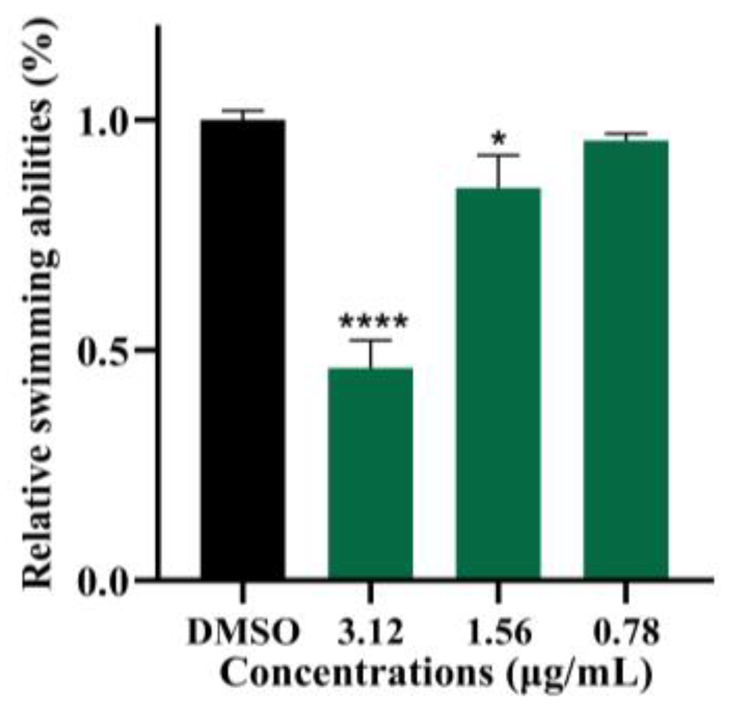
2.5. Demethylincisterol A3 Inhibits the Swimming of B. cereus

The swimming behavior of B. cereus was notably hindered after exposure to demethylincisterol A3 (Figure 5). Under the action of demethylincisterol A3 at 3.12, 1.56, and 0.78 μg/mL, the swimming ability of B. cereus was reduced by 53.9%, 14.7%, and 4.4%, respectively.

2.6. Demethylincisterol A3 Inhibits the Protease and Hemolytic Activity of B. cereus

After treatment with demethylincisterol A3, the protease of B. cereus was significantly inhibited (Figure 6a). Under the action of demethylincisterol A3 at 3.12, 1.56, and 0.78 μg/mL, the protease activities of B. cereus were reduced by 22.1%, 18.1%, and 10.7%, respectively. The hemolytic activity of B. cereus was significantly inhibited when treated with demethylincisterol A3 (Figure 6b). Under the action of demethylincisterol A3 at 3.12, 1.56, and 0.78 μg/mL, the hemolytic activities of B. cereus were reduced by 15.3%, 11.0%, and 7.6%, respectively.

2.7. Demethylincisterol A3 Inhibits the Expression of Key Virulence-Related Genes of B. cereus

The expression of virulence-related genes of B. cereus was significantly inhibited when treated with demethylincisterol A3 (Figure 7). Under the action of demethylincisterol A3 at 3.12 μg/mL, the expression of the sinR, tasA, papR, cytK, rpoN, hblD, comER, codY, plcR, and nheA genes in B. cereus was significantly reduced.

2.8. Demethylincisterol A3 Reduces the Mortality Rate of B. cereus

As shown in Figure 8, the supernatant of B. cereus treated with demethylincisterol A3 was injected into a Galleria mellonella larval model. Only 25% of the larvae in the DMSO group survived after 72 h. However, the survival rate of the experimental group treated with demethylincisterol A3 was significantly improved, with a survival rate of 88% for the larvae at a concentration of 3.12 μg/mL.

3. Discussion

In this study, demethylincisterol A3, along with five other compounds, was initially isolated from P. sacchari. As an incisterol-type steroid, demethylincisterol A3 has been identified in various fungi, possibly representing a common metabolite in fungal processes [28,29,30]. Notably, demethylincisterol A3 exhibits significant anti-tumor and anti-inflammatory effects [31,32]. Studies have demonstrated that the rate of inhibition exhibited by demethylincisterol A3 towards Helicobacter pylori is 11% greater than that of 100 μM quercetin [33]. Despite its recognized therapeutic potential, there has been limited research on its QS inhibitory effect against pathogens. Therefore, this study pioneers the investigation of demethylincisterol A3’s impact on QS, biofilm formation, and virulence factors in B. cereus.
At the MIC, B. cereus growth was inhibited, but at sub-MICs, there was no significant effect (Figure 3). B. cereus biofilms contribute to equipment adhesion, increased resistance, and toxicity [34]. Following demethylincisterol A3 treatment, biofilm formation was significantly inhibited, decreasing production by 50.2% at 3.12 μg/mL. Our research is consistent with that pertaining to Siphonocholin, which can effectively inhibit biofilm formation at 1/2 MIC [35]. At the same time, diallyl disulfide can also inhibit the biofilm formation of B. cereus at 1/2 MIC [36]. The downregulation of rpoN and comER, important regulators of B. cereus biofilms, is associated with reduced biofilm production [37,38]. Additionally, the decrease in tasA expression directly correlates with a reduction in biofilm matrix protein synthesis [39].
B. cereus utilizes its swimming ability to navigate and reach the target host [36], and this mobility is intertwined with its biofilm formation [16]. Upon the addition of demethylincisterol A3, both the swimming ability and the biofilm production of B. cereus decreased. Genes associated with flagella or mobility, including rpoN, codY, and sinR [38,40], showed significant downregulation after demethylincisterol A3 treatment.
Proteases and hemolysin serve as virulence factors produced by B. cereus, aiding in immune system evasion [41]. Hemolysins induce red blood cell rupture, leading to hemolysis [42]. Following demethylincisterol A3 treatment, B. cereus exhibited a significant reduction in protease and hemolysin production. In B. cereus, nhe represents non-hemolytic enterotoxin, hbl represents hemolysin BL, and cytK represents cytotoxin, regulated by plcR [42]. papR can produce PapR peptides to activate the PlcR protein [43]. Demethylincisterol A3 reduced the mortality rate of B. cereus in the G. mellonella larval model from 75% to 12%. Furthermore, demethylincisterol A3 diminished the transcription of the nheA, hblD, cytK, plcR, and papR genes in B. cereus. Therefore, demethylincisterol A3 may mitigate the production of virulence factors in B. cereus, weakening its pathogenicity by inhibiting the transcription of the nheA, hblD, cytK, plcR, and papR genes.
In the current study, cyclo-(L-Pro-L-Leu) was also extracted from P. sacchari. This cyclodipeptide was identified as a signaling molecule that facilitates communication between B. cereus and Cronobacter sakazakii [44]. It was already known whether or not it was a molecular signaling pathway in B. cereus; further research will be conducted to determine the above.

4. Materials and Methods

4.1. General

B. cereus (ATCC11778) was cultured using LB medium at 37 °C. Fresh Laurencia sp. was collected from the Nansha Islands area in the South China Sea (Figure S1), identified by Prof. X.L. Wang (Institute of Oceanology, CAS, China). The test compound was firstly dissolved in dimethyl sulfoxide (DMSO) to 1 mg/mL and sterilized using a filter membrane (0.22 μm) to obtain the stock solution. During all the experiments, an equal volume of DMSO was added as the negative control. The NMR was the Bruker Avance neo 400 (Bruker BioSpin AG, Ettlingen, Germany). The MS was ion trap time-of-flight mass spectrometry liquid chromatography-mass spectrometry (Shimadzu, Kyoto, Japan). All used chemicals in this work were analytical reagents.

4.2. Seaweed Sample Collection and Morphological Identification

The seaweed samples were collected by researcher Zhi-Kai Guo from the Institute of Biology, Chinese Academy of Tropical Agricultural Sciences, from the reef of Zhaoshu Island in the Xisha Xuande Islands. The algae species were identified by Professor Xu-Lei Wang from the Qingdao Institute of Oceanography, Chinese Academy of Sciences. The algal body was purplish red in appearance, with cylindrical branches—mostly forked or trifurcate branches, dense and clustered—with forked or threefold apical branchlets, and a concave apical center with trichonema. The tetrasporangium divides cruciform or conical. Identified as Laurencia sp. (Figure S1). Endophytes were extracted from algae. After placing fresh Laurencia sp. in a petri dish and disinfecting it three times with sterile water, it was rinsed with sterile water and alcohol for 30 seconds. It was then immersed in a 1% (w/w) NaClO solution for three minutes and then rinsed five times with sterile water. Once it was dried with filter paper, it was chopped into small segments. Mixed the small segments with sterile quartz sand. Then grinded the mixture in a sterile phosphate-buffered saline (PBS) environment. Diluted the grinding solution using a PBS gradient. A layer of 100 μL dilution solution (evenly distributed) was coated onto the culture plate containing PDA, Gause’s Synthetic Agar No.1 (GSA) and LB. LB plate was cultured at 37 °C, while PDA and GSA plates were cultured at 28 °C. Colonies with different morphologies were selected for purification and preservation during the cultivation process.

4.3. Identification of QSI Active-Strain W2-F1

The QSI activities of the crude extract of ethyl acetate from the isolated strain were evaluated by using the indicator strain Chromobacterium violaceum CV026. Among these 7 isolates, W2-F1 showed the best QSI activity (Figure S2). The morphological characteristics of strain W2-F1 cultivated in PDA medium (Hope Bio, Qingdao, China) were examined [45]. Further, the genomic DNA was extracted and amplified using ITS primers (5′-TCCGTAGGTGGAACCTGGG-3′ and 5′-TCTCCGCTTATTGCT-3′) and the obtained sequence was aligned in the NCBI. Based on the sequence similarities, the phylogenetic tree was constructed by the neighbor-joining method with the 1000-bootstrap value using MEGA 7.0 software [46]. The W2-F1 strain was identified as P. sacchari.

4.4. Scale-Up Fermentation of QSI Active-Strain P. sacchari and Isolation, Purification, and Elucidation of Compounds under Bio-Guided Screenings

P. sacchari was activated on a PDA plate, then inoculated into a solid PDA medium containing 3% sea salt and cultured at 28 °C for 14 days. After the culture was completed, the mycelium was broken together with the culture medium. Absolute ethanol was added to the mixture and treated with ultrasonic for 12 h and then filtered, repeated three times. The ethanol in the filtrate was removed by evaporation under reduced pressure. The residual liquid was extracted three times by equal ethyl acetate (EtOAc) and concentrated under reduced pressure to give a crude extract (110.1 g), which was subjected to column chromatography (CC) over a silica gel (100–200 mesh) eluted with a gradient of CH2Cl2/MeOH (100:0–0:100) to obtain fractions (R1-R11). The fraction R4 (5.26 g) showing QSI activity was further submitted to silica gel (200–300 mesh) column chromatography (CC) and eluted with a gradient system of petroleum ether/CH2Cl2 (20:1–1:1) and CH2Cl2/MeOH (100:1–10:1) to obtain twenty-two subfractions (R4–1–22). Fraction R4–9 (0.24 g) showing QSI activity was purified by reversed-phase C18 CC and semi-preparation HPLC equipped with a preparative column (5 μm, 21.2 × 250mm; Beijing, China) to obtain compounds 1 and 2 (eluting conditions: 100% MeOH, flow rate = 8 mL/min; yields: 4.8 and 8.7 mg). QSI-active R4–18 were eluted by silica gel column chromatography and semi-preparation HPLC to obtain compound 3 (eluting conditions: 90% MeOH, flow rate = 8 mL/min; yield: 3.2 mg) and R4–18–6. Compound 4 (yield: 10.8 mg) was obtained from R4–18–6 with a 60% MeOH eluting solution and a flow rate of 8 mL/min. Compounds 5 and 6 (yields: 7.4 and 7.5 mg) were obtained from R4–18–6 with a 50% MeOH elution solution and flow rate of 6 mL/min.

4.5. MIC of Demethylincisterol A3 against B. cereus

MIC of demethylincisterol A3 was measured by using the double dilution method [35]. The seed solution of B. cereus that was cultured overnight was added to LB medium at a rate of 1% (v/v). The compounds were added and the mixture was diluted and incubated at 37 °C and 180 rpm for 24 h. DMSO was used as the negative control. Growth profiles were measured at 620 nm.

4.6. Growth Measurement of B. cereus

The planktonic cell growth measurement was performed by the reported methods, with some modifications [47]. The B. cereus culture was inoculated into LB medium at a ratio of 1% (v/v). Demethylincisterol A3 was then added to achieve final concentrations of 3.12, 1.56, and 0.78 μg/mL. After 24 hours of cultivation at 37 °C and 180 rpm, OD620 was measured (Biotech Epoch 2, Santa Clara, CA, USA).

4.7. The Effects of Biofilm Formation

The biofilm formation assays were conducted following established procedures with slight modifications, as outlined in the report by [48]. B. cereus cultures that had been grown overnight were introduced into LB medium at a 1% (v/v) ratio, and demethylincisterol A3 was added. A 200 µL aliquot of this mixed culture was then transferred to a 96-well polystyrene microtiter plate. The final concentrations of demethylincisterol A3 in the wells were 3.12, 1.56, and 0.78 μg/mL. The plate was then incubated at 37 °C for 24 h. After this period, the culture medium was carefully removed and the wells were washed three times with sterile PBS to eliminate planktonic cells. The remaining water in the wells was evaporated at 60 °C. The biofilms that had adhered to the bottom of the wells were fixed with cool methanol for 15 min. The liquid was then removed and the plate was dried again at 60 °C. Each well was stained with 200 µL of a 0.05% (w/v) crystal violet solution. After 10 min staining, any excess crystal violet was removed and the wells were washed three times with PBS. Once the plate had dried at 60 °C, 200 µL of 95% (v/v) ethanol was added to each well and the plate was decolorized for 15 min on a shaker set at 37 °C and 180 rpm. A 150 µL sample of the decolorizing solution was then taken from each well and the absorbance was measured at 570 nm.
SEM measurement: The coating was fixed on the glass slide with 2.5% (v/v) glutaraldehyde for 12 h. After fixation, it was washed at once with ultrapure water and then gradient dehydration was performed with 50%, 60%, 70%, 80%, 90%, and 100% (v/v) ethanol in sequence. After drying, it was sprayed gold and observed under a SEMn microscope.

4.8. Swimming Motility Assay

The swimming motility assay was evaluated by established protocols and reports, as per the study by [46]. Swimming medium (10 g/L peptone, 5 g/L NaCl, 3 g/L agar) was added to demethylincisterol A3 to the final concentrations of 3.12, 1.56, and 0.78 μg/mL, respectively. It was mixed well and poured evenly into a petri dish. Sterile water served as a negative control. After solidification, 2 μL of B. cereus bacterial solution was inoculated in the center of the swimming medium. The migration diameter was recorded after 24 h of cultivation at 37 °C.

4.9. Measurement of Protease Production

Protease activities were measured by reported methods, with some modifications [49]. B. cereus cultures that had been grown overnight were introduced into LB medium at a 1% (v/v) ratio, and demethylincisterol A3 was added. The final concentrations of desmethylergosterol A3 were 3.12, 1.56, and 0.78 μg/mL, respectively. The solution was incubated at 37 °C and 180 rpm for 24 h. The fermentation broth was centrifuged (8000× g, 4 °C, 15 min) to collect the supernatant. The supernatant was filtered through a 0.22 μm membrane. Skim milk agar was prepared (1/4-strength LB broth with 4% (w/v) skim milk and 1.5% (w/v) agar). Added 50 μL of supernatant to the wells of a skim milk agar plate and incubated at 37 °C for 24 h. The protease activity was assessed by examining the clear zone on the skim milk agar plates.

4.10. Measurement of Hemolytic Activities

Hemolytic activities were measured by reported methods, with some modifications [50]. Hemolysis experiments were performed using a trypticase soy sheep blood agar plate (Huankai Microbial, Guangzhou, China). The supernatant was prepared according to the previous method. Then, 50 µL of cultured supernatant was added after drilling, incubated at 37 °C for 24 h, and the size of the hemolytic halo was observed.

4.11. Quantitative Real-Time PCR

Quantitative real-time PCR (RT-qPCR) was performed according to reported methods, with minor modifications [48]. The bacterial solution cultured overnight was diluted to a 0.5 Michaelis turbidimetric unit with sterile LB broth. Overnight cultured B. cereus was added in a 1% (v/v) ratio to LB medium. The experimental group was supplemented with demethylincisterol A3 to make its concentration 3.12 μg/mL, and the same volume of DMSO was used as the negative control group. The solution was incubated at 37 °C and 180 rpm for 24 h. The fermentation broth was centrifuged (8000× g, 4 °C, 15 min) to collect cells and washed thrice with diethylpyrocarbonate (DEPC)-treated water. The total RNA of B. cereus was extracted using an Eastern® Super Total RNA Extraction Kit (Promega, Beijing, China) and transformed into cDNA by a Reverse Transcription Kit (Biosharp, Beijing, China). The real-time PCR reaction was performed using a TB Green® Premix Ex Taq™ II FAST qPCR Mix (TaKaRa, Dalian, China) with a Bio-Rad CFX Connect System (Bio-Rad, Hercules, CA, USA). The primers used for real-time PCR are listed in Table 1. Gene 16S rRNA was used as an internal control [36].

4.12. Evaluating the Inhibitory Effect of Demethylincisterol A3 in a G. mellonella Model

A G. mellonella larval model was used to evaluate the toxic inhibitory effect of demethylincisterol A3 on B. cereus [51]. The supernatant was prepared as described above. The larvae were randomly divided into 15 groups (n = 10 per group) and 5 μL of supernatant was injected into the posterior part of the left abdominal foot. Then, the larvae were cultured at room temperature for 72 h, and their growth was recorded every 12 h. PBS served as a blank control.

4.13. Statistical Analysis

Unless otherwise stated, all experiments described were performed in triplicate independently. The data shown in the figures are the mean ± standard deviation (± SD) of the replicates. Statistical differences were determined using one-way ANOVA or t-test using GraphPad Prism 8, where p < 0.05 was considered statistically significant.

5. Conclusions

Demethylincisterol A3 exhibits significant inhibitory effects on B. cereus, encompassing reduced biofilm formation, diminished bacterial mobility, and lowered production of virulence factors. This multifaceted impact has the potential to attenuate the pathogenicity of B. cereus. Based on these results, it can be inferred that demethylincisterol A3 exhibits potential as a valuable compound in the development of novel antibacterial therapies.

Supplementary Materials

The following supporting information can be downloaded at https://www.mdpi.com/article/10.3390/md22040161/s1: Figure S1: Laurencia sp. was collected from the Nansha Islands area in the South China Sea; Figure S2: Evaluated QSI activities using the indicator strain of Chromobacterium violaceum CV026; Figure S3–S8: 1H, 13C NMR, and HRMS data of compounds 16; Figure S9: Sequence comparison results of strain W2-F1; Figure S10–S13: Preparation spectrum of compounds 16; Table S1: The chemical shifts of each hydrogen-bearing chiral carbon.

Author Contributions

Conceptualization, S.-L.X.; data curation, S.-L.X. and K.-Z.X.; formal analysis, S.-L.X. and K.-Z.X.; funding acquisition, A.-Q.J.; investigation, S.-L.X.; methodology, S.-L.X. and L.-J.Y.; project administration, A.-Q.J.; resources, A.-Q.J.; software, S.-L.X. and L.-J.Y.; supervision, A.-Q.J.; writing—original draft, S.-L.X., K.-Z.X. and L.-J.Y.; writing—review and editing, A.-Q.J. All authors have read and agreed to the published version of the manuscript.

Funding

This research was funded by the Hainan Province Science and Technology Special Fund (ZDYF2024SHFZ103), the National Natural Science Foundation of China (82160664), and Innovative Research Projects for Postgraduates in Hainan Province (Qhyb2021-38, Qhyb2022-46).

Institutional Review Board Statement

Not applicable.

Data Availability Statement

The data presented in this study are available from the corresponding author upon request.

Acknowledgments

We thank the Hainan University for its support.

Conflicts of Interest

The authors declare no conflicts of interest.

References

  1. Pagarete, A.; Ramos, A.S.; Puntervoll, P.; Allen, M.J.; Verdelho, V. Antiviral potential of slgal metabolites—A comprehensive review. Mar. Drugs 2021, 19, 94. [Google Scholar] [CrossRef] [PubMed]
  2. Nagamalla, L.; Shanmukha Kumar, J.V.; Sanjay, C.; Alsamhan, A.M.; Shaik, M.R. In-silico study of seaweed secondary metabolites as AXL kinase inhibitors. Saudi J. Biol. Sci. 2022, 29, 689–701. [Google Scholar] [CrossRef] [PubMed]
  3. Vairagkar, U.; Mirza, Y. Antagonistic activity of antimicrobial metabolites produced from seaweed-associated Bacillus amyloliquefaciens MTCC 10456 against Malassezia spp. Probiotics Antimicrob. Proteins 2021, 13, 1228–1237. [Google Scholar] [CrossRef] [PubMed]
  4. Catarino, M.D.; Silva-Reis, R.; Chouh, A.; Silva, S.; Braga, S.S.; Silva, A.M.S.; Cardoso, S.M. Applications of antioxidant secondary metabolites of Sargassum spp. Mar. Drugs 2023, 21, 172. [Google Scholar] [CrossRef]
  5. Digra, S.; Nonzom, S. An insight into endophytic antimicrobial compounds: An updated analysis. Plant Biotechnol. Rep. 2023, 17, 427–457. [Google Scholar] [CrossRef] [PubMed]
  6. Stierle, A.; Strobel, G.; Stierle, D. Taxol and taxane production by Taxomyces andreanae, an endophytic fungus of Pacific yew. Science 1993, 260, 214–216. [Google Scholar] [CrossRef] [PubMed]
  7. Nishad, J.H.; Singh, A.; Gautam, V.S.; Kumari, P.; Kumar, J.; Yadav, M.; Kharwar, R.N. Bioactive potential evaluation and purification of compounds from an endophytic fungus Diaporthe longicolla, a resident of Saraca asoca (Roxb.) Willd. Arch. Microbiol. 2021, 203, 4179–4188. [Google Scholar] [CrossRef] [PubMed]
  8. Gautam, V.S.; Singh, A.; Kumari, P.; Nishad, J.H.; Kumar, J.; Yadav, M.; Bharti, R.; Prajapati, P.; Kharwar, R.N. Phenolic and flavonoid contents and antioxidant activity of an endophytic fungus Nigrospora sphaerica (EHL2), inhabiting the medicinal plant Euphorbia hirta (dudhi) L. Arch. Microbiol. 2022, 204, 140. [Google Scholar] [CrossRef]
  9. Xu, Z.; Xiong, B.; Xu, J. Chemical investigation of secondary metabolites produced by mangrove endophytic fungus Phyllosticta capitalensis. Nat. Prod. Res. 2021, 35, 1561–1565. [Google Scholar] [CrossRef]
  10. Singh, V.K.; Kumar, A. Secondary metabolites from endophytic fungi: Production, methods of analysis, and diverse pharmaceutical potential. Symbiosis 2023, 90, 111–125. [Google Scholar] [CrossRef]
  11. Akinpelu, D.A.; Aiyegoro, O.A.; Akinpelu, O.F.; Okoh, A.I. Stem bark extract and fraction of Persea americana (Mill.) exhibits bactericidal activities against strains of Bacillus cereus associated with food poisoning. Molecules 2015, 20, 416–429. [Google Scholar] [CrossRef] [PubMed]
  12. Eglezos, S.; Huang, B.; Dykes, G.A.; Fegan, N. The prevalence and concentration of Bacillus cereus in retail food products in Brisbane, Australia. Foodborne Pathog. Dis. 2010, 7, 867–870. [Google Scholar] [CrossRef] [PubMed]
  13. Leong, S.S.; Korel, F.; King, J.H. Bacillus cereus: A review of “fried rice syndrome” causative agents. Microb. Pathog. 2023, 185, 106418. [Google Scholar] [CrossRef] [PubMed]
  14. Lin, Y.; Briandet, R.; Kovács, Á.T. Bacillus cereus sensu lato biofilm formation and its ecological importance. Biofilm 2022, 4, 100070. [Google Scholar] [CrossRef] [PubMed]
  15. Huang, Y.; Flint, S.H.; Palmer, J.S. Bacillus cereus spores and toxins–the potential role of biofilms. Food Microbiol. 2020, 90, 103493. [Google Scholar] [CrossRef] [PubMed]
  16. Zhao, L.; Duan, F.; Gong, M.; Tian, X.; Guo, Y.; Jia, L.; Deng, S. (+)-Terpinen-4-ol Inhibits Bacillus cereus biofilm formation by upregulating the interspecies quorum sensing signals diketopiperazines and diffusing signaling factors. J. Agric. Food Chem. 2021, 69, 3496–3510. [Google Scholar] [CrossRef] [PubMed]
  17. Gorgan, M.; Vanunu Ofri, S.; Engler, E.R.; Yehuda, A.; Hutnick, E.; Hayouka, Z.; Bertucci, M.A. The importance of the PapR7 C-terminus and amide protons in mediating quorum sensing in Bacillus cereus. Res. Microbiol. 2023, 174, 104139. [Google Scholar] [CrossRef] [PubMed]
  18. Amagata, T.; Tanaka, M.; Yamada, T.; Doi, M.; Minoura, K.; Ohishi, H.; Yamori, T.; Numata, A. Variation in cytostatic constituents of a sponge-derived Gymnascella dankaliensis by manipulating the carbon source. J. Nat. Prod. 2007, 70, 1731–1740. [Google Scholar] [CrossRef] [PubMed]
  19. Mansoor, T.A.; Hong, J.; Lee, C.-O.; Bae, S.-J.; Im, K.S.; Jung, J.H. Cytotoxic sterol derivatives from a marine sponge Homaxinella sp. J. Nat. Prod. 2005, 68, 331–336. [Google Scholar] [CrossRef]
  20. Yajima, A.; Kagohara, Y.; Shikai, K.; Katsuta, R.; Nukada, T. Synthesis of two osteoclast-forming suppressors, demethylincisterol A3 and chaxine A. Tetrahedron 2012, 68, 1729–1735. [Google Scholar] [CrossRef]
  21. Yang, X.-L.; Zhang, S.; Hu, Q.-B.; Luo, D.-Q.; Zhang, Y. Phthalide derivatives with antifungal activities against the plant pathogens isolated from the liquid culture of Pestalotiopsis photiniae. J. Antibiot. 2011, 64, 723–727. [Google Scholar] [CrossRef]
  22. Mehnaz, S.; Saleem, R.S.Z.; Yameen, B.; Pianet, I.; Schnakenburg, G.; Pietraszkiewicz, H.; Valeriote, F.; Josten, M.; Sahl, H.-G.; Franzblau, S.G.; et al. Lahorenoic acids A–C, ortho-dialkyl-substituted aromatic acids from the biocontrol strain Pseudomonas aurantiaca PB-St2. J. Nat. Prod. 2013, 76, 135–141. [Google Scholar] [CrossRef]
  23. Campbell, J.; Lin, Q.; Geske, G.D.; Blackwell, H.E. New and Unexpected Insights into the Modulation of LuxR-Type Quorum Sensing by Cyclic Dipeptides. ACS Chem. Bio. 2009, 4, 1051–1059. [Google Scholar] [CrossRef]
  24. Fdhila, F.; Vázquez, V.; Sánchez, J.L.; Riguera, R. DD-Diketopiperazines:  Antibiotics Active against Vibrio anguillarum Isolated from Marine Bacteria Associated with Cultures of Pecten maximus. J. Nat. Prod. 2003, 66, 1299–1301. [Google Scholar] [CrossRef]
  25. Song, S.; Fu, S.; Sun, X.; Li, P.; Wu, J.E.; Dong, T.; He, F.; Deng, Y. Identification of cyclic dipeptides from Escherichia coli as new antimicrobial agents against Ralstonia solanacearum. Molecules 2018, 23, 214. [Google Scholar] [CrossRef]
  26. Yang, B.; Dong, J.; Zhou, X.; Yang, X.; Lee, K.J.; Wang, L.; Zhang, S.; Liu, Y. Proline-containing dipeptides from a marine sponge of a Callyspongia Species. Helv. Chim. Acta 2009, 92, 1112–1117. [Google Scholar] [CrossRef]
  27. Han, Y.; Li, Y.Y.; Shen, Y.; Li, J.; Li, W.J.; Shen, Y.M. Oxoprothracarcin, a novel pyrrolo[1,4]benzodiazepine antibiotic from marine Streptomyces sp. M10946. Drug Discov. Ther. 2013, 7, 243–247. [Google Scholar] [CrossRef] [PubMed]
  28. Yang, X.; Wu, P.; Xue, J.; Li, H.; Wei, X. Seco-pimarane diterpenoids and androstane steroids from an endophytic Nodulisporium fungus derived from Cyclosorus parasiticus. Phytochemistry 2023, 210, 113679. [Google Scholar] [CrossRef]
  29. Li, D.X.; Cheng, X.; Ma, F.P.; Chen, J.Y.; Chen, Y.P.; Zhao, X.S.; Luo, Q. Identification of metabolites from edible mushroom Morchella sextelata and their biological evaluation. Nat. Prod. Res. 2023, 37, 1774–1781. [Google Scholar] [CrossRef] [PubMed]
  30. Jiang, C.; Ji, J.; Li, P.; Liu, W.; Yu, H.; Yang, X.; Xu, L.; Guo, L.; Fan, Y. New lanostane-type triterpenoids with proangiogenic activity from the fruiting body of Ganoderma applanatum. Nat. Prod. Res. 2022, 36, 1529–1535. [Google Scholar] [CrossRef]
  31. Su, J.-C.; Pan, Q.; Xu, X.; Wei, X.; Lei, X.; Zhang, P. Structurally diverse steroids from an endophyte of Aspergillus tennesseensis 1022LEF attenuates LPS-induced inflammatory response through the cholinergic anti-inflammatory pathway. Chem.-Biol. Interact. 2022, 362, 109998. [Google Scholar] [CrossRef] [PubMed]
  32. Liu, Y.; Fu, H.; Zuo, L. Synergistic cytotoxicity effect of 5-Fluorouracil and SHP2 Inhibitor demethylincisterol A3 on cervical cancer cell. Anti-Cancer Agents Med. Chem. 2022, 22, 1313–1319. [Google Scholar] [CrossRef] [PubMed]
  33. Na, M.W.; Lee, E.; Kang, D.-M.; Jeong, S.Y.; Ryoo, R.; Kim, C.-Y.; Ahn, M.-J.; Kang, K.B.; Kim, K.H. Identification of antibacterial sterols from Korean wild mushroom Daedaleopsis confragosa via bioactivity- and LC-MS/MS profile-guided fractionation. Molecules 2022, 27, 1865. [Google Scholar] [CrossRef] [PubMed]
  34. Huang, Y.; Flint, S.H.; Loo, T.S.; Palmer, J.S. Emetic toxin production of Bacillus cereus in a biofilm. LWT-Food Sci. Technol. 2022, 154, 112840. [Google Scholar] [CrossRef]
  35. Alam, P.; Alqahtani, A.S.; Mabood Husain, F.; Tabish Rehman, M.; Alajmi, M.F.; Noman, O.M.; El Gamal, A.A.; Al-Massarani, S.M.; Shavez Khan, M. Siphonocholin isolated from red sea sponge Siphonochalina siphonella attenuates quorum sensing controlled virulence and biofilm formation. Saudi Pharm. J. 2020, 28, 1383–1391. [Google Scholar] [CrossRef] [PubMed]
  36. Jin, Z.; Li, L.; Zheng, Y.; An, P. Diallyl disulfide, the antibacterial component of garlic essential oil, inhibits the toxicity of Bacillus cereus ATCC 14579 at sub-inhibitory concentrations. Food Control 2021, 126, 108090. [Google Scholar] [CrossRef]
  37. Hayrapetyan, H.; Tempelaars, M.; Nierop Groot, M.; Abee, T. Bacillus cereus ATCC 14579 RpoN (Sigma 54) Is a pleiotropic regulator of growth, carbohydrate metabolism, motility, biofilm formation and toxin production. PLoS ONE 2015, 10, e0134872. [Google Scholar] [CrossRef] [PubMed]
  38. Yan, F.; Yu, Y.; Wang, L.; Luo, Y.; Guo, J.-h.; Chai, Y. The comER gene plays an Important role in biofilm formation and sporulation in both Bacillus subtilis and Bacillus cereus. Front. Microbiol. 2016, 7, 1025. [Google Scholar] [CrossRef] [PubMed]
  39. Caro-Astorga, J.; Pérez-García, A.; de Vicente, A.; Romero, D. A genomic region involved in the formation of adhesin fibers in Bacillus cereus biofilms. Front. Microbiol. 2015, 5, 745. [Google Scholar] [CrossRef]
  40. Coburn, P.S.; Miller, F.C.; Enty, M.A.; Land, C.; LaGrow, A.L.; Mursalin, M.H.; Callegan, M.C. The Bacillus virulome in endophthalmitis. Microbiology 2021, 167, 001057. [Google Scholar] [CrossRef]
  41. Dietrich, R.; Jessberger, N.; Ehling-Schulz, M.; Märtlbauer, E.; Granum, P.E. The food poisoning toxins of Bacillus cereus. Toxins 2021, 13, 98. [Google Scholar] [CrossRef]
  42. Enosi Tuipulotu, D.; Mathur, A.; Ngo, C.; Man, S.M. Bacillus cereus: Epidemiology, virulence factors, and host-pathogen interactions. Trends Microbiol. 2021, 29, 458–471. [Google Scholar] [CrossRef]
  43. Huillet, E.; Bridoux, L.; Barboza, I.; Lemy, C.; André-Leroux, G.; Lereclus, D. The signaling peptide PapR is required for the activity of the quorum-sensor PlcRa in Bacillus thuringiensis. Microbiology 2020, 166, 398–410. [Google Scholar] [CrossRef]
  44. Bofinger, M.R.; de Sousa, L.S.; Fontes, J.E.N.; Marsaioli, A.J. Diketopiperazines as cross-communication quorum-sensing signals between Cronobacter sakazakii and Bacillus cereus. ACS Omega 2017, 2, 1003–1008. [Google Scholar] [CrossRef] [PubMed]
  45. Shafique, S.; Javed, A.; Shafique, S.; Hussain, A.; Rafique, R.; Mubarak, A. Isolation and identification of Pithomyces sacchari as a leaf spot pathogen of Helianthus annuus from Pakistan. Sci. Rep. 2022, 12, 22033. [Google Scholar] [CrossRef]
  46. Zeng, Y.X.; Liu, J.S.; Wang, Y.J.; Tang, S.; Wang, D.Y.; Deng, S.M.; Jia, A.Q. Actinomycin D: A novel Pseudomonas aeruginosa quorum sensing inhibitor from the endophyte Streptomyces cyaneochromogenes RC1. World J. Microbiol. Biotechnol. 2022, 38, 170. [Google Scholar] [CrossRef] [PubMed]
  47. Inaba, M.; Matsuda, N.; Banno, H.; Jin, W.; Wachino, J.-i.; Yamada, K.; Kimura, K.; Arakawa, Y. In vitro reduction of antibacterial activity of tigecycline against multidrug-resistant Acinetobacter baumannii with host stress hormone norepinephrine. Int. J. Antimicrob. Agents 2016, 48, 680–689. [Google Scholar] [CrossRef] [PubMed]
  48. Xu, K.-Z.; Xiang, S.-L.; Wang, Y.-J.; Wang, B.; Jia, A.-Q. Methyl gallate isolated from partridge tea (Mallotus oblongifolius (Miq.) Müll.Arg.) inhibits the biofilms and virulence factors of Burkholderia thailandensis. J. Ethnopharmacol. 2024, 320, 117422. [Google Scholar] [CrossRef]
  49. Chu, W.; Zhou, S.; Jiang, Y.; Zhu, W.; Zhuang, X.; Fu, J. Effect of traditional Chinese herbal medicine with antiquorum sensing activity on Pseudomonas aeruginosa. Evid.-Based Complement. Altern. Med. 2013, 2013, 648257. [Google Scholar] [CrossRef]
  50. Zhao, Y.; Chen, C.; Gu, H.-j.; Zhang, J.; Sun, L. Characterization of the genome feature and toxic capacity of a Bacillus wiedmannii isolate from the hydrothermal field in Okinawa Trough. Front. Cell. Infect. Microbiol. 2019, 9, 370. [Google Scholar] [CrossRef]
  51. Yin, L.; Wang, Y.; Xiang, S.; Xu, K.; Wang, B.; Jia, A.-Q. Tyramine, one quorum sensing inhibitor, reduces pathogenicity and restores tetracycline susceptibility in Burkholderia cenocepacia. Biochem. Pharmacol. 2023, 218, 115906. [Google Scholar] [CrossRef] [PubMed]
Figure 1. Morphological and phylogenetic tree of P. sacchari strain. (a) Colonies grown on a PDA. (b,c) The spores and mycelia were under a microscope with 40× and 100× magnification. (d) Phylogenetic tree.
Marinedrugs 22 00161 g001
Figure 2. The chemical structures of the isolated six compounds (16).
Marinedrugs 22 00161 g002
Figure 3. The growth of B. cereus at sub-MICs of demethylincisterol A3. B. cereus grew for 24 h in different sub-MICs (3.12, 1.56, and 0.78 μg/mL) of demethylincisterol A3; DMSO was used as the negative control. Error bars represent the standard deviation (n = 3).
Marinedrugs 22 00161 g003
Figure 4. The effect of demethylincisterol A3 on the biofilm formation of B. cereus. B. cereus was treated with different concentrations (3.12, 1.56, and 0.78 μg/mL) of demethylsterol A3 for 24 h, with DMSO as the negative control. (a) Biofilm formation. (b) SEM images of biofilms. Error bars represent the standard deviation (n = 3). * p < 0.05, **** p < 0.0001.
Marinedrugs 22 00161 g004
Figure 5. The effect of demethylincisterol A3 on the swimming of B. cereus was examined using a swimming medium. The swimming behavior of B. cereus for 24 h of treatment with demethylsterol A3 at various concentrations (3.12, 1.56, and 0.78 μg/mL) was tested. DMSO served as a negative control. After 24 h of incubation, the swimming diameter was observed and recorded. Values are represented as mean ± standard deviation (n = 3). Significant differences are denoted by * p < 0.05 and **** p < 0.0001, respectively.
Marinedrugs 22 00161 g005
Figure 6. The effect of demethylincisterol A3 on the virulence factors of B. cereus. The virulence factor produced by B. cereus after 24 h of treatment with different concentrations (3.12, 1.56, and 0.78 μg/mL) of demethylsterol A3 was evaluated. DMSO was used as a negative control. (a) The protease activity was assessed by examining the clear zone on the skim milk agar plates. (b) The hemolysin activities were analyzed by examining the hemolysis zone on the blood agar plates. Values are represented as mean ± standard deviation (n = 3). Significant differences are denoted by * p < 0.05, ** p < 0.01, and *** p < 0.001, respectively.
Marinedrugs 22 00161 g006
Figure 7. The effect of demethylincisterol A3 on B. cereus gene transcription was investigated. The expression of QS-related genes in B. cereus was examined after treatment with 3.12 μg/mL of demethylsterol A3 for 24 h. DMSO was the negative control. Three biological replicates were conducted and are represented as mean ± standard deviation (n = 3). Significant differences are denoted by * p < 0.05, ** p < 0.01, and *** p < 0.001, respectively.
Marinedrugs 22 00161 g007
Figure 8. The effect of demethylincisterol A3 on the survival rate of G. mellonella larval models was observed. PBS was the blank control. DMSO was used as a negative control. Statistical analyses were carried out using the log-rank (Mantel–Cox) test. Three biological replicates were conducted and are represented as mean ± standard deviation (n = 3). Significant differences are denoted by * p < 0.05, ** p < 0.01, and *** p < 0.001, respectively.
Marinedrugs 22 00161 g008
Table 1. PCR primers for RT-qPCR.
PrimerSequence (5′-3′)
16S rRNA_FGGAGGAAGGTGGGGATGAC
16S rRNA_RATGGTGTGACGGGCGGTGTG
sinR_FAGCGAGCGCCGATATGATAG
sinR_RTCGAGCGCATTCGTAACCAT
tasA_FACTCGCCACATGGAAACACA
tasA_RACGATTTGTTCGTTTTCTTCGT
papR_FTGTACCTCTTGATCACTGTGAGA
papR_RAACGTTAGCAATGGCATGGG
cytK_FCGATGACCCAAGCGCTGATA
ctyK_RGTTGCACTAGCACCAGGGAT
rpoN_FCACTTGAACGAGCTTTCGCC
rpoN_RGGGGCGCGTAATATTCAGGA
hblD_FGGTCCAGATGGGAAAGGTGG
hblD_RAAGTTGTGGGATCGTTGCCT
comER_FCAAGTTGCGGTCCTGCTTTC
comER_RAATTTCCCCATCCCCACGAC
codY_FCCACGACGGCTAACTACGAA
codY_RGCGTTATTACAGAGCGCAGC
plcR_FGGGTGATGCGGGGATTAACA
plcR_RGGCTCACTTCCGATTGGTGA
nheA_FTCTTGCAACAGCCAGACATT
nheA_RCTCTCGCACATTCGCCTTTG
Disclaimer/Publisher’s Note: The statements, opinions and data contained in all publications are solely those of the individual author(s) and contributor(s) and not of MDPI and/or the editor(s). MDPI and/or the editor(s) disclaim responsibility for any injury to people or property resulting from any ideas, methods, instructions or products referred to in the content.

