Article

High Structural Diversity of Aeruginosins in Bloom-Forming Cyanobacteria of the Genus Planktothrix as a Consequence of Multiple Recombination Events

by
Elisabeth Entfellner
1,
Kathrin B. L. Baumann
1,†,
Christine Edwards
2 and
Rainer Kurmayer
1,*
1
Research Department for Limnology, Universität Innsbruck, Mondseestrasse 9, 5310 Mondsee, Austria
2
CyanoSol Research Group, Pharmacy & Life Sciences, Robert Gordon University, Aberdeen AB10 7GJ, UK
*
Author to whom correspondence should be addressed.
Current address: Department of Surface Waters-Research and Management, Swiss Federal Institute for Aquatic Science and Technology (EAWAG), 6047 Kastanienbaum, Switzerland.
Mar. Drugs 2023, 21(12), 638; https://doi.org/10.3390/md21120638
Submission received: 29 October 2023 / Revised: 8 December 2023 / Accepted: 11 December 2023 / Published: 13 December 2023
(This article belongs to the Special Issue Bioactive Product from Marine Cyanobacteria)

Abstract

:
Many compounds produced by cyanobacteria act as serine protease inhibitors, such as the tetrapeptides aeruginosins (Aer), which are found widely distributed. The structural diversity of Aer is intriguingly high. However, the genetic basis of this remains elusive. In this study, we explored the genetic basis of Aer synthesis among the filamentous cyanobacteria Planktothrix spp. In total, 124 strains, isolated from diverse freshwater waterbodies, have been compared regarding variability within Aer biosynthesis genes and the consequences for structural diversity. The high structural variability could be explained by various recombination processes affecting Aer synthesis, above all, the acquisition of accessory enzymes involved in post synthesis modification of the Aer peptide (e.g., halogenases, glycosyltransferases, sulfotransferases) as well as a large-range recombination of Aer biosynthesis genes, probably transferred from the bloom-forming cyanobacterium Microcystis. The Aer structural composition differed between evolutionary Planktothrix lineages, adapted to either shallow or deep waterbodies of the temperate climatic zone. Thus, for the first time among bloom-forming cyanobacteria, chemical diversification of a peptide family related to eco-evolutionary diversification has been described. It is concluded that various Aer peptides resulting from the recombination event act in chemical defense, possibly as a replacement for microcystins.

Graphical Abstract

1. Introduction

The aeruginosins (Aer) constitute a bioactive peptide family that is found widely distributed among marine and freshwater cyanobacteria [1], and also among higher organisms such as marine sponges of the family Dysideidae [2,3]. The structural diversity of Aer is intriguingly high, and structure–function relationships have been used to understand the consequences for inhibition of serine proteases and other enzymes [4,5]. The bloom-forming cyanobacterium Planktothrix is a prolific producer of Aer, as well as other peptide families such as the toxic microcystins and bioactive anabaenopetins, cyanopeptolins, microginins, microviridins and prenylagaramides [6]. In particular, the Aer peptide family has the highest structural variability [7,8]. Other Aer peptide producers include the genera Microcystis, Nostoc, Nodularia [3,9] and Spaerocarvum [10]. Aer synthesis is encoded by aer genes, organized into a biosynthetic gene cluster (aer BGC) encoding several non-ribosomal peptide synthetases (NRPS), which are responsible for specific amino acid recognition and incorporation. The resulting Aer molecule is a linear tetrapeptide, containing an unusual 2-carboxy-6-hydroxyoctahydroindole (Choi) moiety. The characteristic amino acid Choi is synthesized via the enzymes AerD, E, F [11,12], modifying an amino acid that is activated by the first adenylation domain of the NRPS AerG [13]. In contrast to other peptides (i.e., microcystins, anabaenopeptins), numerous tailoring enzymes (e.g., sulfatases, halogenases, glycosyltransferases, methyltransferases, acetyltransferases) introduce high structural variability among the Aer peptide family [13,14].
Genome sequencing and metabolomics have shown that the Aer peptide family is more widely distributed than previously assumed [14,15]. Besides the structurally related spumigins and pseudoaeruginosins, also dysinosins [16], varlaxins produced by Nostoc sp. [17] and suomilide produced by Nodularia sphaerocarpa [14] might be considered as members of the Aer peptide family. In particular, the spuA-F genes encoding spumigin synthesis show a partial similarity to the aer gene cluster [18], while the synthesis of pseudoaeruginosins is encoded through a combination of spu and aer genes, as described from Nodularia spumigena [19]. Suomilide is characterized by the structurally related azabicyclononane (Abn) moiety [20], which is considered as a modification of Choi encoded by suoK and suoH [14].
The Aer synthesis pathway was first described from Planktothrix strain NIVA-CYA126/8 [13], which was found partially divergent from Microcystis [21]. Planktothrix NIES-204 and Microcystis NIES-843 demonstrate high homogeneity of aer genes [22]. By comparing the aerB, aerG, aerD, aerE and aerF genes, sequenced from 103 strains of cyanobacteria (representing twelve genera), a higher similarity of aer genes within Planktothrix, Microcystis and Aphanothece was observed, while aer genes from Nostoc, Nodularia or Anabaena were rather dissimilar [14]. Thus, in contrast to microcystin synthesis, but in accordance with anabaenopeptin synthesis [23], a rather high similarity between aer genes of P. agardhii and Microcystis sp. strains has been observed.
Since the aer genes have been detected most frequently among various Planktothrix species [6], we were interested in exploring the aer gene organization and the structural consequences of recombination events within this genus. It was the aim of this study to describe all the recombination events resulting in Aer structural modification in relation to Planktothrix spp. eco-evolutionary divergence. Thus, we quantified the variations within aer genes among 124 strains and correlated this genetic variability to the characterized Aer variants. The Planktothrix strains were isolated from 40 diverse waterbodies located on three different continents, occurring between the tropical and northern temperate climatic zones (Europe, North America and Africa), which were phylogenetically and ecologically assigned previously. In brief, three major lineages were identified: Lineage 1 and 2 included strains assigned to P. agardhii/P. rubescens, thriving either in the plankton of shallow or deeper lakes of the temperate climatic zone, while Lineage 3 consisted of strains representing more distantly related species such as P. pseudagardhii, P. mougeotii and P. tepida, occurring frequently in (sub)tropical regions [23,24].

2. Results and Discussion

2.1. Aeruginosin Biosynthesis Genes Show Multiple Recombinations and a Variable Evolutionary Origin

As previous studies reported high similarity of aer genes between Planktothrix and Microcystis [14,22,25], we compared the nucleotide sequence of the aer genes between these genera in detail (Figure 1 and Figures S1–S15). Regarding their similarity to Microcystis, two major groups of aer BGCs were observed in Planktothrix strains (Figure 1A). Overall, group 1 (e.g., Planktothrix NIES-204) showed a higher similarity to M. aeruginosa NIES-2481, i.e., the genetic distance on nucleotide level was <0.25. In contrast, group 2 consistently showed higher genetic distance > 0.2. The core aer gene composition and arrangement, including all genes encoding NRPS (i.e., aerA, aerB and aerG/G1 + G2/M), the genes encoding Choi synthesis (aerD-F), as well as the ABC transporter (aerN), were conserved. However, aerG (in group 2) was replaced by aerG1 and aerM (in group1) or aerG1 and aerG2 (in M. aeruginosa NIES-98). In addition, concerning aerB, a high nucleotide variation within Planktothrix and Microcystis was observed, resulting in distinct phylogenetic clades (Figure S2). Accordingly, four different specificity-conferring codes for the adenylation domain of AerB were identified using the webserver NRPS predictor2 [26]: Leu, Ile, Phe and Tyr (Figure S2). Notably, one (benthic) and distantly related Planktothrix strain PCC11201 [27] differed substantially in aer gene distance from all other Planktothrix strains (Figure 1B). Here, the aer genes were arranged similarly to those in Microcystis with aerI (encoding a putative glycosyltransferase), while aerL, aerK and ORF2 (open reading frame, encoding a putative acetyltransferase) were lacking. Consequently, in this specific—rather distantly related—Planktothrix strain PCC11201, the aer BGC might have its own origin from another unknown cyanobacterium.
The accessory genes aerC, aerH-L and ORF1-9 were found more irregularly distributed. The genes aerK, ORF1 (a putative oxidoreductase) and ORF2 (a putative acetyltransferase) were found among strains assigned to group 1 only. Notably, aerK, encoding a putative isomerase [14], had high similarity to Microcystis (genetic dissimilarity < 0.1), but was present in full length only in Microcystis and the Planktothrix strains No980 and No1020, whereas most other Planktothrix strains of group 1 had remnants of it. ORF1 was present in all strains of group 1 and showed high similarity (distance < 0.15; except for strain PCC11201). ORF2 was found in most strains of group 1; however, it was lacking in Planktothrix No1020 and PCC11201, and was found interrupted by a stop codon in Microcystis strain NIES-2549. The genes aerJ (encoding a halogenase) and aerL (encoding a sulfotransferase) occurred with highest frequency among strains carrying aer BGC group 1. Phylogenetic analysis of aerL sequences (372–1011 bp) revealed four clusters (Figure S11), possibly indicating sulfation at different moieties of the Aer molecule: Sulfation at Hpla (e.g., Microcystis NIES-843 [21]), sulfation at Choi (e.g., Microcystis NIES-98, [28]) and sulfation at an unknown position (e.g., Planktothrix No365). In contrast to group 1, the aerC, H, I and ORF3-9 genes were found frequently among strains carrying aer BGC group 2. However, only ORF8 (encoding an Aldo-/Keto-reductase, [14]) occurred consistently among strains that carried aerG instead of aerM/G2 (=strains carrying aer BGC group 1) and might be essential for Aer synthesis (Table 1).
Last but not least, Planktothrix strains No713 and PCC9214, assigned to Lineage 3, carried an unknown NRPS/PKS gene cluster, comprising aerD-F genes possibly encoding Choi synthesis (Figure 1B and Figures S4–S6). These strains may produce unknown peptides carrying a Choi moiety further extending the Aer peptide family.

2.2. Aer Gene Organization and Composition in Relation to Planktothrix Evolution

The aer gene composition was compared among 124 Planktothrix strains, which were phylogenetically assigned to three major lineages [23,24]: Lineage 1 (n = 42)/1A (n = 10) comprised strains isolated from plankton of shallow lakes mostly from the temperate climatic zone. In contrast, Lineage 2 (n = 45)/2A (n =16) comprised strains isolated from deeper physically stratified lakes of the temperate climatic zone, while Lineage 3 (n = 11) comprised strains isolated from the (sub)tropical climatic zone [23,24]. The presence/absence of aer genes was determined via PCR, amplifying 2 kbp-sized fragments of the aer gene cluster without interruption (Tables S1 and S2). All strains assigned to Lineage 1 or 2 showed at least the PCR products indicative of aerA (Table S2). Out of all 124 strains, the aer genes were detected for 115 strains (93%). The strains were grouped into three clusters via principal component analysis (PCA), (Figure 2): Cluster I contained 24 strains (assigned to Lineage 1 and 3), representing the genotypes carrying the aer BGC similar to Microcystis (i.e., strains carrying aer BGC group 1, Figure 1A). Cluster II comprised the majority of strains from all lineages (n = 91, i.e., strains carrying aer BGC group 2, Figure 1A), wherein a subgroup (n = 7) showed large gene cluster deletions. Cluster III contained nine strains from Lineage 3, all lacking the aer genes.

2.3. Aeruginosin Structural Variation and Chemical Diversification in Relation to Phylogenetic Lineages

We used HPLC-MSn to detect and characterize the Aer peptides, as extracted from biomass of the 124 Planktothrix strains [24]). In order to characterize the Aer structures in detail, the amino acid sequence of the tetrapeptide backbone, modifications (e.g., sulfates, chlorines, methyl, acetyl and prenyl moieties) and partly also the position of these additional groups were analyzed by specific MSn fragmentation (Tables S3–S5). A number of Aer peptide variants, sometimes even co-synthesized by the same strain, had identical protonated masses and fragmentation pattern, but eluted at different retention times. After re-isolation from chromatographic separation, the Aer peptide variants still eluted at different times, indicating isomers (e.g., Figure S16 strain No778).
An exact identification was possible only for a few known Aer peptides, such as Aer 126A/B from P. agardhii NIVA-CYA126/8 [13], Aer 828A from P. rubescens No91/1 [30] and Aer 89A/B from M. aeruginosa NIES-89 [31], i.e., from strain No1020 (Lineage 3). Some other Aer variants were detected among Planktothrix sp. previously, however, without structural elucidation. Furthermore, we detected many unknown structural Aer variants, partly variations of known Aer peptides carrying the same tetrapeptide backbone (Tables S3–S5).
We found 79 Planktothrix strains (64% of all strains) as Aer producers (Table S3). The proportion of Aer producers varied between lineages, i.e., strains assigned to Lineage 1/1A showed the highest proportion of Aer containing strains (48 out of 52 strains), while only 29 out of 61 strains (48%) assigned to Lineage 2/2A and 2 out of 11 strains (18%) assigned to Lineage 3 were found active as Aer producers (Figure 3A,B, Table S3). In total, 73 Aer variants were differentiated. Alltogether, strains contained between one and nine (median = 3) Aer variants (i.e., strain NIES-596 contained nine structural Aer variants). Strains assigned to Lineage 1 contained up to nine (median = 3) variants, while strains assigned to Lineage 2 contained less Aer structural variants (up to five). The majority of Aer variants (56 variants, 77%) occurred in more than one strain; however, only 27 Aer variants (37%) occurred in more than three strains. The most frequent Aer variants (occurring in 10–15 strains) constituted the putative Aer variants [M+H]+ 717.3, 715.3, 771.5 and 805.5 (Table S3) related to the large-range recombination event (aerJaerN, >20 kbp, aer BGC group 1 in Figure 1). Only seven variants were shared between Lineages 1 and 2 (Figure 3C). Typically, strains inactive in Aer synthesis showed either a large-range deletion of aer genes or the presence of an insertion sequence (IS) element (Table S2).
In summary, only strains assigned to Lineage 1 and 3 carried the large-range aer gene recombination resulting in the most frequent structural variant Aer 716 (partly co-produced with Aer 688) (Table S3). Interestingly, many of these strains of Lineage 1 and 3 have lost the genes encoding microcystin synthesis and have become nontoxic, as reported previously [6], e.g., only 11% of the strains of Lineage 1 carry mcy genes and produce MC (6 out of 52 strains). In contrast, strains of Lineage 2 were more frequently inactive in Aer production, however, all the strains carry the mcy genes and 70% produce MC (18 out of 61 strains are inactive), Table S6.

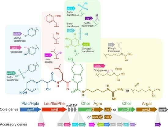
2.4. Relationships between Aeruginosin Structural Modification and Core/Accessory Aer Genes

Little variation was found in the tetrapeptide (backbone) structure, detecting five structural variants (without differentiating Leu from Ile), i.e., Plac–Leu(Ile)–Choi–Argal, Hpla/Plac–Leu(Ile)–Choi–Agm and Hpla/Plac–Phe–Choi–Agm (Figure 3C). We could not find a relationship between any accessory aer gene and the catalysis of the hydroxylation of Plac to Hpla; therefore, a relaxed specificity of the adenylation domain of AerA was assumed. In position 2, we found either Leu (e.g., Aer 126A/B) or Ile (which is indistinguishable from Leu by means of MSn fragmentation) or Phe (e.g., Microcin SF608). Among the Arg derivates at pos. 4, we found either Argal (e.g., Aer 89) or Agm (e.g., Aer 126B), though frequently Agm was found modified to Aeap (e.g., Aer 126A). Most strains showed a specific incorporation of only one Arg derivative, and only four strains produced Aer variants in parallel containing either argininal or Agm (e.g., NIVA-CYA116).
Increased structural diversity was generated by accessory modifications: chlorination at pos. 1 and/or 2, methylation at pos. 1, glycosylation at pos. 3, sulfation at pos. 1 or 3 and the conversion of Agm to Aeap at pos. 4. Moreover, we observed putative O-acetylation (plus 42 Da, C2H3O) of Choi and N-prenylation (plus 68 Da, C5H8) of Agm (e.g., Aer KB676 [32]).
No obvious difference in the amino acid specificity conferring code of the adenylation domain of AerA between strains incorporating either only Plac (30%) or Plac/Hpla (15%) or only Hpla (55%) at pos. 1 (Plac/Hpla) could be identified (i.e., for prediction of amino acid activation, no precedent in databases [26] predicting the substrate of adenylation domains could be found). The observed accessory modifications of Hpla at pos. 1 consisted of chlorination, methylation or sulfation. Chlorination was found catalyzed by the halogenase encoded in aerJ, as described for M. aeruginosa PCC7806 [21,29]. When compared to the aer genes from Planktothrix strain NIVA-CYA126/8, an additional ORF9 was located between ORF7 and 8 which was identified as a putative methyltransferase. Indeed, methylated Aer variants were detected in some of the strains carrying this additional ORF9 (Figure 4). Previously, in M. aeruginosa, the aerL gene was identified as a putative sulfotransferase, whereby in M. aeruginosa NIES-843, the hydroxyl group of Hpla at pos. 1 was found sulfated and in M. aeruginosa NIES-98, the Choi at pos. 3 was found sulfated [21]. Correspondingly, in this study, the presence of the aerL gene correlated with sulfated Aer. The sulfate group was not detected in initial LC-MSn analysis in the positive MS mode. However, nucleotide analysis of aerL revealed two aerL1 and aerL2 genotypes (Figure S11), most likely resulting in either sulfation of Choi (e.g., strain No365) or sulfation of Hpla (e.g., No66) at their hydroxyl group.
The sequence analysis of aerB (Figure S2) resulted in three different specificity-conferring codes of the catalytic domain of the adenylation domains: DAWFLGNV, predicted to activate Leu (e.g., No66, NIVA-CYA126/8); DAFFLGV, related to Ile (e.g., No790); and DAWTIAAV, related to Phe (e.g., No82). Indeed, we could confirm Phe in Aer structure among strains carrying AerB predicted to activate Phe (e.g., No82, PCC7821, NIVA-CYA98 and NIVA-CYA406).
In contrast to pos. 2, Choi was found conserved at pos. 3. Notably, aerDEF genes (encoding Choi synthesis) have a high sequence dissimilarity between aer BGC groups 1 and 2, suggesting a different origin (Figure 1). Glycosylation at the hydroxyl group of Choi was commonly detected. Previously, the aerI gene was described as a glycosyltransferase catalyzing the transfer of the xylose moiety to Choi resulting in aeruginosides [13]. In addition, a sulfate group was found linked to the xylose moiety by the same authors [13]. Correspondingly, many strains carrying a sulfate group shared the ORF7 encoding a putative sulfotransferase (Figure 4), [14].
The phylogenetic comparison of the homologous genes aerM, aerG2 and the second adenylation domain of aerG (Figure S7) overall corresponded to the taxonomy of the genus Planktothrix or Microcystis, with one exception, i.e., the aerG2 from M. aeruginosa NIES-98 was found related to the second adenylation domain of aerG from Planktothrix but not to aerM. As a result, all Planktothrix strains carrying aerM showed argininal (25%), whereas the other strains carrying aerG contained Agm (70%) at pos. 4 of the Aer peptide. Only four strains (NIVA-CYA116, No259, No281, No307) possibly produced Aer variants with both arginine derivatives. Agm was often found modified to Aeap, as already described for Aer 126A [13]. Taking all Aer-producing strains together, the presence of aerC correlated perfectly with this modification. Furthermore, we found some structural variants with an additional moiety at Agm (+68 Da), suggesting N-prenylation which, however, could not be related to an accessory enzyme encoded within the aer BGC.
In a previous study, a reductase encoded within aerM has been suggested to catalyze the cleavage of the Aer molecule from NRPS in M. aeruginosa NIES-843 and PCC7806 [21]. Such a reductase was missing in all Planktothrix strains carrying aerG only. Instead, ORF8 (a putative aldo-keto reductase [14]) has been detected among these strains (Figure 1) which might encode an alternative enzyme for cleavage of the Aer molecule.
According to Ahmed et al. [14], the aerK gene may encode an isomerase. This could explain why strain No980 (carrying aerK) produces Aer peptides with same mass but different retention time when compared with Aer peptides from strains e.g., No253 and NIVA-CYA116 carrying only aerK fragments. Analogously, ORF3 may encode an isomerase as well, since strain No108 produced Aer peptides with same mass but different retention time when compared with strains No97 and No91/1, sharing a similar aer gene organization but lacking aerC and ORF3.

2.5. High Resolution Mass Spectrometry (HRMS)

Six putative chlorinated Aer compounds related to the large-range aer gene recombination event (aerJaerN, >20 kbp) were isolated and purified from Planktothrix strains No66, NIVA-CYA116 and No1020. The HRMS results confirmed the presence of Choi ([M+H]+ 140 or 122) and chlorine in all six purified Aer compounds and showed typical Aer fragmentation patterns (Table S4). For Planktothrix strain No66, a plausible sum formula for purified Aer structure matching the obtained fragmentation pattern was calculated (Table 2). Consequently, it was concluded that the structure of Aer [M+H]+ 717 was possibly identical to the Aer structural variant already described from M. aeruginosa NIES-89, i.e., Aer 89A/B [31].

2.6. Toxicity of Aeruginosins Resulting from the Large-Range Recombination Events

In general, Aer peptides have been found bioactive because of proteolytic enzyme inhibition (i.e., serine proteases such as trypsin, thrombin, plasmin) effective in the nanomolar to lower micromolar range (e.g., [2,3,4,5]). We tested the toxicity of the variants Aer 716 ([M+H]+ 717.3 from No1020), Aer 688 ([M+H]+ 689.3 from NIVA-CYA116) and Aer 828A ([M+H]+ 829.3 from No91/1) using a standard toxicity assay (i.e., the anostracan crustacean Thamnocephalus platyurus). The observed toxicity was compared with that of [D-Asp3, (E)-Dhb7] microcystin (MC)-RR and other bioactive peptides including anabaenopeptin B and F (Figure S17). Among the tested peptides, the [D-Asp3, (E)-Dhb7] MC-RR showed highest toxicity followed by Aer 828A in a low µM range (e.g., [30], this study). The Aer 716 (resulting from the large-range aer gene recombination) had lower toxicity, comparable to anabaenopeptin B. Aer 688 had the lowest toxicity, comparable to anabaenopeptin F.
Notably, Aer 828A carries chlorine at Leu (pos. 2) of the molecule, while Aer 716 and Aer 688 carry a chlorine at Hpla (pos. 1) of the molecule. Scherer et al. [33] tested the effect of an additional chlorine and/or sulfate on Aer derivatives using the same acute toxicity T. platyurus assay as applied in this study. Their results indicated a comparable range in toxicity when a sulfate (at Xylose) or a chlorine (at pos. 2) or both were attached, while the same backbone structure lacking both moieties (i.e., Aer 126A) had lower toxicity. Overall, it is very likely that the presence of a chlorine or sulfate plays a role in modulating Aer toxicity [33]. However, by the same authors, conformational changes resulting from a chlorine towards both increasing and decreasing the bioactivity/toxicity also have been suggested. Analogously, since Aer 716 and Aer 688 only differ in Argal vs. Agm (pos. 4), steric effects in modulating the fit of the guanidinium moiety into the catalytic site of proteases might explain the reduced toxicity of Aer 688 [33].

3. Conclusions

By comparing the aer gene composition among Planktothrix, it was possible to show that strains (genotypes) carrying the large-range recombination of aer genes formed a more closely related aer genotype group that was found distinct from other strains. Very likely, this recombination stemmed from horizontal gene transfer (HGT) and the recombination was found to be integrated in frame and functional. Since the aerA and aerN genes located at the 5′ end or 3′ end of the aer BGC still showed high similarity with Planktothrix, but the other aer genes were found most similar to aer genes from Microcystis, an HGT event occurring between the two different genera is plausible.
The elucidation of the genetic basis of Aer synthesis improved our understanding of the evolution of the impressive Aer structural diversity: There was a perfect correlation between the occurrence of aerC (a putative oxygenase catalyzing oxidation of Agm to Aeap), aerI (a putative glycosyltransferase catalyzing glycosylation at Choi), aerL1 (a putative sulfotransferase catalyzing sulfation at Choi at pos. 3) and aerL2 (putatively catalyzing sulfation at Hpla at pos. 1) and the predicted structural modification of the Aer peptide as observed among the strains.
Interestingly, aer gene composition, as well as its activity and the resulting Aer structural diversity, correlated with Planktothrix speciation processes and ecological diversification. In particular, strains of Lineage 1 were found more active in Aer production, in contrast to strains of Lineage 2, which were found frequently inactive due to several partial gene cluster deletion events or IS elements. It is concluded that various Aer peptides resulting from recombination function as chemical defense, possibly in replacement of microcystins. Thus, the Aer peptides constitute the first example of a peptide family showing a chemical diversification in relation to the ecological and phylogenetic diversification within bloom-forming cyanobacteria. In summary, the genetic variability within aer genes, which has been found linked to structural variability, points to adaptive significance of the Aer peptide family in the course of Planktothrix evolution.

4. Materials and Methods

4.1. Organisms

In this study, 124 clonal Planktothrix strains were analyzed. The strains were clonally isolated, characterized and assigned phylogenetically, as described previously [6,24]. They were grown under sterile conditions in BG11 medium [34] at low light intensity (5–10 µmol m−2s−1 Osram Type L30W/77 Fluora, 16/8 h light-dark cycle) at 15 °C (Lineages 1 and 2) or 23 °C (Lineage 3), Table S6. Taxonomic affiliation was performed according to Suda et al. [35], Kurmayer et al. [24], Gaget et al. [36].

4.2. DNA Extraction, PCR and Sequencing

Cells from cultures were harvested by centrifugation and washed in Tris-EDTA (TE) buffer. Genomic DNA was extracted using the CTAB protocol as described previously [23]. DNA extracts were stored at −20 °C. PCR (Table S1, primer pairs a–x) was used to determine aer gene presence/absence via 2 kbp amplicons, covering the entire aer BGC amplifying overlapping fragments without interruption. Primers were designed according to the reference aer gene sequences from Planktothrix NIVA-CYA126/8 (NZ_ASAK00000000.1) and No66 (NZ_LR882963.1). PCR mixtures had a volume of 10 µL, containing 2 μL (5×) Phusion HF Buffer (Thermo Scientific, Vienna, Austria), 500 nM of each primer, 200 μM of each deoxynucleotide triphosphate (Thermo Scientific), 0.1 U of Phusion High-Fidelity DNA Polymerase (Thermo Scientific) and 10 ng of genomic DNA as a template. PCR amplification was performed by initial denaturation at 98 °C for 1 min; thereafter, 35 cycles of denaturation at 98 °C for 10 s, annealing at variable temperature for 15 s and elongation at 72 °C (Table S1) were performed. The PCR amplicon size was determined using standard gel electrophoresis (0.8% agarose gels in 0.5 × Tris-borate-EDTA (TBE) buffer) and visualized using Midori Green.
From the Planktothrix strains No63, No253, No790, No980 and No1020, aer gene fragments were sequenced from PCR products using standard Sanger sequencing at Eurofins Genomics (Ebersberg, Germany) via primers listed in Table S1. For this purpose, PCR mixtures had a higher volume of 30 µL and products were extracted from the agarose gel using a commercial gel extraction kit (QIAquick, Gel Extraction Kit, Qiagen, Hilden, Germany). All new sequences obtained during this study have been included in Table S7.

4.3. Sequence Comparison and Phylogenetic Analysis

The BLASTn algorithm was used to identify aer genes or homologs in Planktothrix and other cyanobacterial genera. Nucleotide sequence analysis was performed in Mega 7.0 [37]. Sequences were aligned using Clustal W (codons). To estimate the nucleotide similarity of the genes/ORFs (Figure 1), distance values were calculated using default adjustments (variance estimation: bootstrap method, 1000 replications; substitution model: nucleotide, Tamura-Nei model, transitions + transversions; uniform rates; pairwise deletions). Maximum likelihood trees (Figures S1–S15) were calculated in Mega 7.0 (phylogeny test: bootstrap method; 100 replications; substitution model: Tamura-Nei model; uniform rates; gaps included).
For Planktothrix, the following aer sequences were obtained from NCBI, including the strains CCAP1459/11A (NZ_BJCD01000048.1), NIES-204 (AP017991.1), NIVA-CYA15 (NZ_KE734694.1), NIVA-CYA34 (NZ_AVFT01000027.1), NIVA-CYA56/3 (NZ_KE734731.1), NIVA-CYA98 (AM990465.1), NIVA-CYA126/8 (LR882934.1), NIVA-CYA405 (NZ_KE734708.1), NIVA-CYA406 (NZ_KE734710.1), NIVA-CYA407 (NZ_KE734717.1), NIVA-CYA540 (NZ_KE734720.1), No2A (LR882938.1), No66 (LR882963.1), No82 (OW445656.1), No108 (LR882941.1), No365 (LR882944.1), No758 (LR882949.1), No976 (LR882952.1), PCC7805 (LR882950.1), PCC7811 (LR882969.1), PCC7821 (LR882958.1) and PCC11201 (NZ_LT797710.1). For M. aeruginosa, the aer sequences were obtained from strain FACHB-1757 (CP011339), FD4 (CP046973.1), NIES-98 (FJ609416.1), NIES-102 (AP019314.1), NIES-298 (CP046058.1), NIES-843 (AP009552.1), NIES-2481 (NZ_CP012375.1), NIES-2549 (CP011304.1) and PCC7806 (CP130696.1). In addition, Nodularia strain CCY9414 (NZ_CP007203.1) and Nostoc strain UIC10630 (NZ_JAAGOG010000094.1) were used as outgroups.

4.4. Multivariate Statistical Analysis

Principal Component Analysis (PCA) was performed to cluster all 124 Planktothrix strains according to aer BGC type, as inferred from PCR results on aer gene presence/absence (a binary (1/0) matrix was analyzed using the statistical package IBM SPSS Statistics 24.0) (Table S2).

4.5. Aeruginosin Peptide Identification and Fragmentation Using HPLC-MSn

Cyanobacterial cells were harvested by filtration on glass fiber filters (BMC, Ederol, Vienna, Austria), freeze-dried and stored at −20 °C. Peptides were extracted from biomass (2–5 mg dry weight) on ice with 50% aqueous methanol (v/v) according to [38]. Peptides were separated on a LiChroCART 250-4 cartridge system (LiChrospher 100 C18 (column dimensions 250 × 4 mm; 5 µm particle size; Merck, Darmstadt, Germany) by HPLC (HP 1100, Agilent) using a linear water/acetonitrile (0.05% trifluoroacetic acid) gradient from 20 to 50% acetonitrile in 45 min (1 mL min−1 flow rate, 30 °C column oven temperature). Peptide masses were determined using an ESI-MS ion trap (amazon SL, Bruker), operated in positive ion mode, with nitrogen as sheath gas (43 psi, 8 L min−1, 300 °C), helium as auxiliary gas and a capillary voltage of 5 kV.
In the first step, ESI-MS was coupled to HPLC for full mass scan (50–2000 m/z) and molecule automated fragmentation from the two most abundant ions and MS3 fragmentation from the most abundant ion of MS2 fragmentation spectra. Putative Aer peptides were identified by their fragmentation pattern, notably [M+H]+ 140 or 122, indicating the Choi-immonium ion. All putative Aer that contributed ≥5% of peak area compared to the most abundant peptide in the base peak chromatogram were recorded (Table S3). Some Aer variants had equal masses but different retention time, often co-occurring in one strain: collection and re-injection confirmed retention time for individual Aer variants with the same protonated mass and fragmentation pattern (e.g., Figure S16), indicating structural isomers. In a second step, putative Aer fractions were purified and directly injected into ESI-MSn for specific manual fragmentation (Table S4). For example, the Aer peptide of No1020 was identified via comparison to M. aeruginosa NIES-89: retention time 6.9 min, [M+H]+ 717.3, identical fragmentation pattern and the occurrence of a triple-peak due to tautomerization (Figure S18).

4.6. Aeruginosin Peptide Purification and Structure Identification Using HRMS

Six unknown chlorinated Aer variants were purified (approx. 100 µg) from biomass of Planktothrix strains No66, NIVA-CYA116, No1020. For this purpose, strains were cultivated at the mass culture facility at CyanoBiotech GmbH (Berlin, Germany). Biomass (approx. 3 g each) was extracted on ice in an Erlenmeyer flask (250 mL) with 50% aqueous methanol (v/v) for one hour. The crude extract was centrifuged (3700 g for 15 min; Thermo Scientific Multifuge X3R) and subsequently concentrated through solid phase extraction (SPE) via C18 cartridges (Sep-Pak ® tC18 cartridges; Waters, Vienna, Austria). The supernatant was filtered after centrifugation using glass microfiber filters (GF/F filters; 0.7 μm pore size; Whatman; 47 mm diameter). The filtrate was diluted with MilliQ water to 10% aqueous methanol and transferred to a phase-separation funnel. The SPE columns were activated with 100% methanol (3 × 1 mL) and equilibrated with 10% methanol (3 × 1 mL). The diluted crude extracts were applied after conditioning of the SPE columns under low pressure with a flow-through rate of 1 mL min−1 and eluted with 1 mL of 80% aqueous methanol (v/v).
Purified Aer were analyzed using an Ultra-High Performance Liquid Chromatography coupled to a Xevo quadrupole time-of-flight mass spectrometer (Waters, Wilmslow, UK). Peptides were separated on a BEH C18 column (100 × 2.1 mm; 1.7 μm particle size) which was maintained at 40 °C. The mobile phase was mixed by Milli-Q water plus 0.1% formic acid and acetonitrile plus 0.1% formic acid, with a linear gradient increasing from 20% to 70% acetonitrile within 10 min. Data were acquired by positive ion electrospray scanning from 50 to 2000 m/z with a scan time of 2 s and inter-scan delay of 0.1s. Ion source parameters, i.e., capillary and sampling cone, were 2.9 V and 25 V, respectively; desolvation temperature, 300 °C; and source temperature, 80 °C. Cone and desolvation gas flows were 50 L h−1 and 400 L h−1 respectively. Sodium iodide (2 μg μL−1 in 50/50 Propan-2-ol/Milli-Q) was used as calibrant with Leucine-enkephalin (0.5 mg mL−1 in 50/50 methanol/Milli-Q) as reference mass. Instrument control and processing were achieved using MassLynx v4.1. MS/MS spectra for the individual Aer molecule were obtained by targeting the protonated parent at 20–50 eV.

4.7. Toxicity Tests

To test the toxicity of the new peptides Aer 716 and Aer 688, several toxic/bioactive peptides produced by Planktothrix, i.e., [D-Asp3, (E)-Dhb7] MC-RR and anabaenopeptin B and F, as well as Aer 828A [30] were purified and tested using the Thamnotoxkit F (MicroBio Tests, Belgium). This test is a 24 h crustacean toxicity bioassay using larvae of the crustacean T. platyurus. The test was performed following the manufacturer’s instructions and a standardized operational procedure [39].
The concentration of the purified compounds was quantified via the molar extinction coefficient by measuring the absorbance using a spectrophotometer (UVmini-1240, Shimadzu). For the Aer peptides, the concentration was calculated as aeruginosin 103A equivalents (λmax = 224 nm; ε = 11,600) [40]. Anabaenopeptins B and F were quantified as anabaenopeptin B equivalents (λmax = 225 nm; ε = 8833) [41]. [D-Asp3, (E)-Dhb7] MC-RR was quantified using the extinction coefficient 50,400 at 239 nm (λmax) [42]. The compounds were dissolved in 1 mL 100% methanol. Each compound was tested in several concentrations in triplicates at least, with 10–20 animals per approach, i.e., [D-Asp3, (E)-Dhb7] MC-RR (1–10 µM), Aer 828A, Aer 716, Aer 688 (5–100 µM), anabaenopeptin B and F (5–120 µM). The final concentration of methanol did not exceed 1%. The mortality in the standard freshwater blanks and 1% methanol blanks was not allowed to exceed 10%.

Supplementary Materials

The following supporting information can be downloaded at: https://www.mdpi.com/article/10.3390/md21120638/s1, Figure S1: Maximum likelihood phylogenetic tree based on aerA (nucleotide) sequence (alignment: 4407 bp). P_, M_ indicate genera Planktothrix or Microcystis, each. Nostoc UIC10630 and Nodularia CCY9414 served as outgroups.; Figure S2: Maximum likelihood phylogenetic tree based on aerB (nucleotide) sequence (alignment: 4857 bp). P_, M_ indicate genera Planktothrix or Microcystis, each. Nostoc UIC10630 and Nodularia CCY9414 served as outgroups. Using the NRPS prediction tool [26] four different specificity-conferring codes for the adenylation domain of AerB were identified: Leu, Ile, Phe and Tyr.; Figure S3: Maximum likelihood phylogenetic tree based on aerD (nucleotide) sequence (alignment: 633 bp). P_, M_ indicate genera Planktothrix or Microcystis, each. Nostoc UIC10630 and Nodularia CCY9414 served as outgroups.; Figure S4: Maximum likelihood phylogenetic tree based on aerE (nucleotide) sequence (alignment: 756 bp). P_, M_ indicate genera Planktothrix or Microcystis, each. Nostoc UIC10630 and Nodularia CCY9414 served as outgroups.; Figure S5: Maximum likelihood phylogenetic tree based on aerF (nucleotide) sequence (alignment: 801 bp). P_, M_ indicate genera Planktothrix or Microcystis, each. Nostoc UIC10630 and Nodularia CCY9414 served as outgroups.; Figure S6: Maximum likelihood phylogenetic tree based on aerG (nucleotide) sequence (alignment: 3366 bp). P_, M_ indicate genera Planktothrix or Microcystis, each. Nostoc UIC10630 and Nodularia CCY9414 served as outgroups.; Figure S7: Maximum likelihood phylogenetic tree based on aerG_module2 and aerM (nucleotide) sequence (alignment: 4533 bp). P_, M_ indicate genera Planktothrix or Microcystis, each. Nostoc UIC10630 and Nodularia CCY9414 served as outgroups.; Figure S8: Maximum likelihood phylogenetic tree based on aerI (nucleotide) sequence (alignment: 1260 bp). P_, M_ indicate genera Planktothrix or Microcystis, each.; Figure S9: Maximum likelihood phylogenetic tree based on aerJ (nucleotide) sequence (alignment: 1905 bp). P_, M_ indicate genera Planktothrix or Microcystis, each.; Figure S10: Maximum likelihood phylogenetic tree based on aerK (nucleotide) sequence (alignment: 1044 bp). P_, M_ indicate genera Planktothrix or Microcystis, each.; Figure S11: Maximum likelihood phylogenetic tree based on aerL (nucleotide) sequence (alignment: 1011 bp). P_, M_ indicate genera Planktothrix or Microcystis, each. In addition, the putative sulfation reactions are indicated (Figure 4).; Figure S12: Maximum likelihood phylogenetic tree based on aerN (nucleotide) sequence (alignment: 2058 bp). P_, M_ indicate genera Planktothrix or Microcystis, each. Nodularia CCY9414 served as outgroup.; Figure S13: Maximum likelihood phylogenetic tree based on ORF1 (nucleotide) sequence (alignment: 786 bp). P_, M_ indicate genera Planktothrix or Microcystis, each.; Figure S14: Maximum likelihood phylogenetic tree based on ORF2 (nucleotide) sequence (alignment: 627 bp). P_, M_ indicate genera Planktothrix or Microcystis, each.; Figure S15: Maximum likelihood phylogenetic tree based on ORF8 (nucleotide) sequence (alignment: 1161 bp). P_ indicates genus Planktothrix.; Figure S16: (A) Base peak chromatograms (BPC) of methanolic peptide extracts from Planktothrix strain No778 with first injection (black), re-injection of peak 1 (red) and re-injection of peak 2 (blue). (B) Mass peak chromatograms [m/z] of peak 1 and 2 from the first injection (black), re-injection of peak 1 (red) and re-injection of peak 2 (blue); [m/z] 765.3.; Figure S17: Toxicity of the aeruginosin variants Aer 828A ([M+H]+ 829.3), Aer 89 ([M+H]+ 717.3), Aer 688 ([M+H]+ 689.3) as inferred using a standard toxicity assay (i.e., Thamnocephalus platyurus) and compared with that of [D-Asp3, (E)-Dhb7] MC-RR ([M+H]+ 1024), and anabaenopeptin B ([M+H]+ 837) and anabenopeptin F ([M+H]+ 851).; Figure S18: (A) Base peak chromatograms (BPC) of methanolic peptide extracts from Microcystis strain NIES-89 (black) and Planktothrix strain No1020 (red). (B) Mass peak chromatograms [m/z] of NIES-89_peak1 (black) and No1020_peak1 (red); [M-SO3+H]+ 637.3; [M+H]+ 717.3; [M+Na]+ 739.3; [M+K]+ 757.3. Table S1: PCR primers and conditions for testing aer gene presence/absence and used for aer gene sequencing from 124 Planktothrix strains.; Table S2: PCR results for testing aer gene presence/absence in 124 Planktothrix strains.; Table S3: HPLC-MSn results for Aer peptides listing retention time and m/z as detected from 124 Planktothrix strains.; Table S4: Manual MSn fragmentation of selected Aer peptides identified from various Planktothrix strains.; Table S5: List of Aer peptides and characterisation of Aer peptide structures as observed for 124 Planktothrix strains.; Table S6: List of all Planktothrix strains, pigmentation and origin, and presence of mcy genes and microcystin as described [6,24] and grouped according to the phylogenetic lineages as described [23]. Taxonomic affiliation according to Suda et al. [41], Kurmayer et al. [24], Gaget et al. [42].; Table S7: Sequences obtained from PCR products for various regions of aer BGC from Planktothrix strains No63, No253, No790, No980 and No1020 as well as some selected aer gene fragments.

Author Contributions

Conceptualization, E.E. and R.K.; methodology, E.E., C.E. and R.K.; investigation, E.E., C.E. (Aer peptide analysis and structure identification) and K.B.L.B., (toxicity tests) and R.K.; resources, E.E., C.E. and R.K.; writing—original draft, E.E., K.B.L.B. and R.K.; writing—review and editing, all authors. All authors have read and agreed to the published version of the manuscript.

Funding

This study was financed by the Austrian Science Fund (FWF) P24070 and P32193 to R.K. E.E. was supported by the Austrian Academy of Sciences (ÖAW) Ph.D. fellowship program.

Institutional Review Board Statement

Not applicable.

Data Availability Statement

The original data that support the findings of this study are available from the corresponding authors upon reasonable request.

Acknowledgments

We would like to thank Katharina Moosbrugger and Anneliese Wiedlroither for their excellent assistance in the laboratory. Many thanks to Dan Enke and Heike Enke (Cyano Biotech GmbH) in Berlin for analytical standards and the large-scale cultivation of Planktothrix strains No91/1, NIVA-CYA116 and No1020. The helpful suggestions of two anonymous reviewers improved the manuscript.

Conflicts of Interest

The authors declare no conflict of interest.

References

  1. Huang, I.S.; Pietrasiak, N.; Gobler, C.J.; Johansen, J.R.; Burkholder, J.M.; D’Antonio, S.; Zimba, P.V. Diversity of bioactive compound content across 71 genera of marine, freshwater, and terrestrial cyanobacteria. Harmful Algae 2021, 109, 102116. [Google Scholar] [CrossRef] [PubMed]
  2. Ersmark, K.; Del Valle, J.R.; Hanessian, S. Chemistry and biology of the aeruginosin family of serine protease inhibitors. Angew. Chem. Int. Ed. Engl. 2008, 47, 1202–1223. [Google Scholar] [CrossRef] [PubMed]
  3. Liu, J.; Zhang, M.; Huang, Z.; Fang, J.; Wang, Z.; Zhou, C.; Qiu, X. Diversity, biosynthesis and bioactivity of aeruginosins, a family of cyanobacteria-derived nonribosomal linear tetrapeptides. Mar. Drugs 2023, 21, 217. [Google Scholar] [CrossRef] [PubMed]
  4. Nagarajan, M.; Maruthanayagam, V.; Sundararaman, M. SAR analysis and bioactive potentials of freshwater and terrestrial cyanobacterial compounds: A review. J. Appl. Toxicol. 2013, 33, 313–349. [Google Scholar] [CrossRef]
  5. Wang, G.; Goyal, N. Aeruginosin analogs and other compounds with rigid bicyclic structure as potential antithrombotic agents. Cardiovasc. Hematol. Agents Med. Chem. 2009, 7, 147–165. [Google Scholar] [CrossRef]
  6. Kurmayer, R.; Deng, L.; Entfellner, E. Role of toxic and bioactive secondary metabolites in colonization and bloom formation by filamentous cyanobacteria Planktothrix. Harmful Algae 2016, 54, 69–86. [Google Scholar] [CrossRef] [PubMed]
  7. Grabowska, M.; Kobos, J.; Toruńska-Sitarz, A.; Mazur-Marzec, H. Non-ribosomal peptides produced by Planktothrix agardhii from Siemianówka Dam Reservoir SDR (northeast Poland). Arch. Microbiol. 2014, 196, 697–707. [Google Scholar] [CrossRef]
  8. Toporowska, M.; Mazur-Marzec, H.; Pawlik-Skowrońska, B. The effects of cyanobacterial bloom extracts on the biomass, Chl-a, MC and other oligopeptides contents in a natural Planktothrix agardhii Population. Int. J. Environ. Res. Public Health 2020, 17, 2881. [Google Scholar] [CrossRef]
  9. Welker, M.; von Döhren, H. Cyanobacterial peptides—Nature’s own combinatorial biosynthesis. FEMS Microbiol. Rev. 2006, 30, 530–563. [Google Scholar] [CrossRef]
  10. Stenico, E.; Rigonato, J.; Lorenzi, A.S.; Azevedo, M.T.; Sant’Anna, C.L.; Fiore, M.F. Bioactive cyanopeptides produced by Sphaerocavum brasiliense strains (Cyanobacteria). J. Braz. Chem. Soc. 2015, 26, 2088–2096. [Google Scholar] [CrossRef]
  11. Qiu, X.; Zhu, W.; Wang, W.; Jin, H.; Zhu, P.; Zhuang, R.; Yan, X. Structural and functional insights into the role of a cupin superfamily isomerase in the biosynthesis of Choi moiety of aeruginosin. J. Struct. Biol. 2019, 205, 44–52. [Google Scholar] [CrossRef]
  12. Qiu, X.; Wei, Y.; Zhu, W.; Fu, J.; Duan, X.; Jin, H.; Zhu, P.; Zhou, C.; Yan, X. Structural and functional investigation of AerF, a NADPH-dependent alkenal double bond reductase participating in the biosynthesis of Choi moiety of aeruginosin. J. Struct. Biol. 2020, 209, 107415. [Google Scholar] [CrossRef]
  13. Ishida, K.; Christiansen, G.; Yoshida, W.Y.; Kurmayer, R.; Welker, M.; Valls, N.; Bonjoch, J.; Hertweck, C.; Börner, T.; Hemscheidt, T.; et al. Biosynthesis and structure of aeruginoside 126A and 126B, cyanobacterial peptide glycosides bearing a 2-carboxy-6-hydroxyoctahydroindole moiety. Chem. Biol. 2007, 14, 565–576. [Google Scholar] [CrossRef]
  14. Ahmed, M.N.; Wahlsten, M.; Jokela, J.; Nees, M.; Stenman, U.H.; Alvarenga, D.O.; Strandin, T.; Sivonen, K.; Poso, A.; Permi, P.; et al. Potent inhibitor of human trypsins from the aeruginosin family of natural products. ACS Chem. Biol. 2021, 16, 2537–2546. [Google Scholar] [CrossRef]
  15. Calteau, A.; Fewer, D.P.; Latifi, A.; Coursin, T.; Laurent, T.; Jokela, J.; Kerfeld, C.A.; Sivonen, K.; Piel, J.; Gugger, M. Phylum-wide comparative genomics unravel the diversity of secondary metabolism in cyanobacteria. BMC Genom. 2014, 15, 977. [Google Scholar] [CrossRef]
  16. Schorn, M.A.; Jordan, P.A.; Podell, S.; Blanton, J.M.; Agarwal, V.; Biggs, J.S.; Allen, E.E.; Moore, B.S. Comparative genomics of cyanobacterial symbionts reveals distinct, specialized metabolism in tropical Dysideidae sponges. mBio 2019, 10, e00821-19. [Google Scholar] [CrossRef] [PubMed]
  17. Heinilä, L.M.P.; Jokela, J.; Ahmed, M.N.; Wahlsten, M.; Kumar, S.; Hrouzek, P.; Permi, P.; Koistinen, H.; Fewer, D.P.; Sivonen, K. Discovery of varlaxins, new aeruginosin-type inhibitors of human trypsins. Org. Biomol. Chem. 2022, 20, 2681–2692. [Google Scholar] [CrossRef]
  18. Fewer, D.P.; Jokela, J.; Paukku, E.; Österholm, J.; Wahlsten, M.; Permi, P.; Aitio, O.; Rouhiainen, L.; Gomez-Saez, G.V.; Sivonen, K. New structural variants of aeruginosin produced by the toxic bloom forming cyanobacterium Nodularia spumigena. PLoS ONE 2013, 8, e73618. [Google Scholar] [CrossRef] [PubMed]
  19. Liu, L.; Budnjo, A.; Jokela, J.; Haug, B.E.; Fewer, D.P.; Wahlsten, M.; Rouhiainen, L.; Permi, P.; Fossen, T.; Sivonen, K. Pseudoaeruginosins, nonribosomal peptides in Nodularia spumigena. ACS Chem. Biol. 2015, 10, 725–733. [Google Scholar] [CrossRef]
  20. Fujii, K.; Sivonen, K.; Adachi, K.; Noguchi, K.; Shimizu, Y.; Sano, H.; Hirayama, K.; Suzuki, M.; Harada, K.-I. Comparative study of toxic and non-toxic cyanobacterial products: A novel glycoside, suomilide, from non-toxic Nodularia spumigena HKVV. Tetrahedron Lett. 1997, 38, 5529–5532. [Google Scholar] [CrossRef]
  21. Ishida, K.; Welker, M.; Christiansen, G.; Cadel-Six, S.; Bouchier, C.; Dittmann, E.; Hertweck, C.; Tandeau de Marsac, N. Plasticity and evolution of aeruginosin biosynthesis in cyanobacteria. Appl. Environ. Microbiol. 2009, 75, 2017–2026. [Google Scholar] [CrossRef] [PubMed]
  22. Shimura, Y.; Fujisawa, T.; Hirose, Y.; Misawa, N.; Kanesaki, Y.; Nakamura, Y.; Kawachi, M. Complete sequence and structure of the genome of the harmful algal bloom-forming cyanobacterium Planktothrix agardhii NIES-204(T) and detailed analysis of secondary metabolite gene clusters. Harmful Algae 2021, 101, 101942. [Google Scholar] [CrossRef] [PubMed]
  23. Entfellner, E.; Frei, M.; Christiansen, G.; Deng, L.; Blom, J.; Kurmayer, R. Evolution of anabaenopeptin peptide structural variability in the cyanobacterium Planktothrix. Front. Microbiol. 2017, 8, 219. [Google Scholar] [CrossRef] [PubMed]
  24. Kurmayer, R.; Blom, J.F.; Deng, L.; Pernthaler, J. Integrating phylogeny, geographic niche partitioning and secondary metabolite synthesis in bloom-forming Planktothrix. ISME J. 2015, 9, 909–921. [Google Scholar] [CrossRef] [PubMed]
  25. McKindles, K.M.; McKay, R.M.; Bullerjahn, G.S. Genomic comparison of Planktothrix agardhii isolates from a Lake Erie embayment. PLoS ONE 2022, 17, e0273454. [Google Scholar] [CrossRef] [PubMed]
  26. Röttig, M.; Medema, M.H.; Blin, K.; Weber, T.; Rausch, C.; Kohlbacher, O. NRPSpredictor2—A web server for predicting NRPS adenylation domain specificity. Nucleic Acids Res. 2011, 39, W362–W367. [Google Scholar] [CrossRef] [PubMed]
  27. Pancrace, C.; Barny, M.A.; Ueoka, R.; Calteau, A.; Scalvenzi, T.; Pédron, J.; Barbe, V.; Piel, J.; Humbert, J.F.; Gugger, M. Insights into the Planktothrix genus: Genomic and metabolic comparison of benthic and planktic strains. Sci. Rep. 2017, 7, 41181. [Google Scholar] [CrossRef] [PubMed]
  28. Murakami, M.; Ishida, K.; Okino, T.; Okita, Y.; Matsuda, H.; Yamaguchi, K. Aeruginosins 98-A and B, trypsin inhibitors from the blue-green alga Microcystis aeruginosa (NIES-98). Tetrahedron Lett. 1995, 36, 2785–2788. [Google Scholar] [CrossRef]
  29. Cadel-Six, S.; Dauga, C.; Castets, A.M.; Rippka, R.; Bouchier, C.; Tandeau de Marsac, N.; Welker, M. Halogenase genes in nonribosomal peptide synthetase gene clusters of Microcystis (cyanobacteria): Sporadic distribution and evolution. Mol. Biol. Evol. 2008, 25, 2031–2041. [Google Scholar] [CrossRef]
  30. Kohler, E.; Grundler, V.; Häussinger, D.; Kurmayer, R.; Gademann, K.; Pernthaler, J.; Blom, J.F. The toxicity and enzyme activity of a chlorine and sulfate containing aeruginosin isolated from a non-microcystin-producing Planktothrix strain. Harmful Algae 2014, 39, 154–160. [Google Scholar] [CrossRef]
  31. Ishida, K.; Okita, Y.; Matsuda, H.; Okino, T.; Murakami, M. Aeruginosins, protease inhibitors from the cyanobacterium Microcystis aeruginosa. Tetrahedron 1999, 55, 10971–10988. [Google Scholar] [CrossRef]
  32. Elkobi-Peer, S.; Carmeli, S. New prenylated aeruginosin, microphycin, anabaenopeptin and micropeptin analogues from a Microcystis bloom material collected in Kibbutz Kfar Blum, Israel. Mar. Drugs 2015, 13, 2347–2375. [Google Scholar] [CrossRef]
  33. Scherer, M.; Bezold, D.; Gademann, K. Investigating the toxicity of the aeruginosin chlorosulfopeptides by chemical synthesis. Angew. Chem. Int. Ed. Engl. 2016, 55, 9427–9431. [Google Scholar] [CrossRef] [PubMed]
  34. Rippka, R. Isolation and purification of cyanobacteria. Methods Enzymol. 1988, 167, 3–27. [Google Scholar] [CrossRef] [PubMed]
  35. Suda, S.; Watanabe, M.M.; Otsuka, S.; Mahakahant, A.; Yongmanitchai, W.; Nopartnaraporn, N.; Liu, Y.; Day, J.G. Taxonomic revision of water-bloom-forming species of oscillatorioid cyanobacteria. Int. J. Syst. Evol. Microbiol. 2002, 52 Pt 5 Pt 5, 1577–1595. [Google Scholar] [CrossRef]
  36. Gaget, V.; Welker, M.; Rippka, R.; de Marsac, N.T. A polyphasic approach leading to the revision of the genus Planktothrix (Cyanobacteria) and its type species, P. agardhii, and proposal for integrating the emended valid botanical taxa, as well as three new species, Planktothrix paucivesiculata sp. nov. ICNP, Planktothrix tepida sp. nov. ICNP, and Planktothrix serta sp. nov. ICNP, as genus and species names with nomenclatural standing under the ICNP. Syst. Appl. Microbiol. 2015, 38, 141–158. [Google Scholar] [CrossRef] [PubMed]
  37. Kumar, S.; Stecher, G.; Tamura, K. MEGA7: Molecular evolutionary genetics analysis version 7.0 for bigger datasets. Mol. Biol. Evol. 2016, 33, 1870–1874. [Google Scholar] [CrossRef]
  38. Kosol, S.; Schmidt, J.; Kurmayer, R. Variation in peptide net production and growth among strains of the toxic cyanobacterium Planktothrix spp. Eur. J. Phycol. 2009, 44, 49–62. [Google Scholar] [CrossRef]
  39. Törökné, A. Thamnocephalus Test. In Handbook of Cyanobacterial Monitoring and Cyanotoxin Analysis; Meriluoto, J., Spoof, L., Codd, G., Eds.; John Wiley & Sons, Ltd.: Chichester, West Sussex, PO19 8SQ, UK, 2017; pp. 462–468. [Google Scholar]
  40. Kodani, S.; Ishida, K.; Murakami, M. Aeruginosin 103-A, a thrombin inhibitor from the cyanobacterium Microcystis viridis. J. Nat. Prod. 1998, 61, 1046–1048. [Google Scholar] [CrossRef]
  41. Harada, K.-I.; Fujii, K.; Shimada, T.; Suzuki, M.; Sano, H.; Adachi, K.; Carmichael, W.W. Two cyclic peptides, anabaenopeptins, a third group of bioactive compounds from the cyanobacterium Anabaena flos-aquae NRC 525-17. Tetrahedron Lett. 1995, 36, 1511–1514. [Google Scholar] [CrossRef]
  42. Blom, J.F.; Robinson, J.A.; Jüttner, F. High grazer toxicity of [D-Asp(3),(E)-Dhb(7)]microcystin-RR of Planktothrix rubescens as compared to different microcystins. Toxicon 2001, 39, 1923–1932. [Google Scholar] [CrossRef] [PubMed]
Figure 1. (A) Genetic basis for aeruginosin synthesis as compared between Microcystis and Planktothrix. The colors illustrate the genetic distance to Microcystis strain NIES-2549: lowest distance (dark blue) to high distance (in red). *, strains carrying aer genes but not producing aeruginosin. Additional sequences for Microcystis and Planktothrix strain PCC11201 have been included [27]). (B) Putative NRPS/PKS genes possibly encoding another Choi moiety similar to the aeruginosin peptide family.
Marinedrugs 21 00638 g001
Figure 2. Ordination of 124 Planktothrix strains according to aer gene presence/absence via principal component analysis (PCA). The aer gene (fragment) presence or absence was determined via PCR, amplifying 2 kbp-sized fragments of the aer gene cluster without interruption. Positive or negative PCR results were indicated as a binary (1/0) matrix.
Marinedrugs 21 00638 g002
Figure 3. (A) Phylogenetic distribution of aeruginosin-producing strains in Planktothrix spp. (n = 124). The number of strains assigned to Lineages 1, 2, 3 is indicated as described previously [23]. (B) The bar charts indicate the proportion of strains carrying aer genes (in grey) relative to analyzed strains (in white) and found active in Aer synthesis (in black). The width of the arrows is proportional to the number of strains carrying a specific aeruginosin variant. (C) Aeruginosin tetrapeptide structures and accessory modifications (yellow marked moieties highlight the structural differences in the backbone).
Marinedrugs 21 00638 g003
Figure 4. Frequency of occurrence of accessory gene functions and corresponding catalyzed structural modifications in aeruginosins as observed among 79 Planktothrix spp. strains. There was a perfect correlation between aerC, aerI, aerL1, aerL2 gene presence and the predicted structural modification.
Marinedrugs 21 00638 g004
Table 1. Functions of aer genes as published or indicated during this study. Abbreviations: NRPS, non-ribosomal peptide synthetase; PKS, polyketide synthase; Agm, agmatine; Aeap, 1-amino-2-(N-amidino-D3-pyrrolinyl)-ethyl; Plac, phenyllactic acid; Hpla, hydroxyphenyllactic acid; Xyl, xylose.
GeneFunctional DescriptionLiterature
aerANRPS/PKS hydrid (incorporation of Plac/Hpla at pos. 1)[13,21]
aerBNRPS (incorporation of Leu/Ile/Phe at
pos. 2)
[13,21]
aerCoxygenase (oxidation of Agm to Aeap)[14]; this study 1
aerD, E, FChoi synthesis (isomerase, reductase)[11,12,13]
aerG, G1 + G2, G1 + MNRPS (Choi at pos. 3 and Agm derivate at pos. 4)[13,21]
aerHhalogenase (chlorination at Leu at pos. 2) this study
aerIglycosyltransferase (Xyl at Choi)[13,21]
aerJhalogenase (chlorination at Hpla at pos. 1)[21,28,29]; this study
aerKisomerase[14]
aerL1sulfotransferase (sulfation at Choi at pos. 3)[21]; this study
aerL2sulfotransferase (sulfation at Hpla at pos. 1)[21]; this study
aerNABC transporter[13,21]
ORF1oxidoreductase (reduction of Agm; cleavage from synthesis operon)[14]; this study
ORF2hypothetical protein (O-acetylation at Choi)this study
ORF3hypothetical proteinthis study
ORF4sulfotransferase (sulfation at unknown pos.)[14]; this study
ORF5hypothetical protein
ORF6hypothetical protein
ORF7sulfotransferase (sulfation at Xyl)[14]; this study
ORF8aldo-/keto reductase (reduction of Agm; cleavage from synthesis operon)[14]; this study
ORF9hypothetical protein (O-methylation at Hpla at pos. 1) this study
1 putative function of accessory aer genes inferred from correlation between gene presence and corresponding structural modification (see main text).
Table 2. Overview of purified chlorinated aeruginosins, resulting from the large-range recombination event (aer BGC group 1), and the respective HRMS results. The presence of the Choi fragment and chlorine is indicated. The proposed sum formula for the chlorinated aeruginosin is indicated (nd, not detected).
Strain Retention
Time [min]
[M+H]+ Choi Fragment
[M+H]+
Chlorine Detected Sum Formula
NIVA-CYA1164.1717.25140.1Yesnd
5.9689.25140.1Yesnd
12.5759.26122.1Yesnd
16.5731.28122.1Yesnd
No10206.6717.25140.1Yesnd
No666.3717.20140.1YesC30H46N6O10SCl
Disclaimer/Publisher’s Note: The statements, opinions and data contained in all publications are solely those of the individual author(s) and contributor(s) and not of MDPI and/or the editor(s). MDPI and/or the editor(s) disclaim responsibility for any injury to people or property resulting from any ideas, methods, instructions or products referred to in the content.

