
Isolation of Scalimides A–L: β-Alanine-Bearing Scalarane Analogs from the Marine Sponge Spongia sp.


1
Korea Institute of Ocean Science & Technology (KIOST), Busan 49111, Republic of Korea
2
Department of Marine Biotechnology, University of Science & Technology, Daejeon 34113, Republic of Korea
*
Author to whom correspondence should be addressed.
Mar. Drugs 2022, 20(11), 726; https://doi.org/10.3390/md20110726
Submission received: 11 October 2022
/
Revised: 17 November 2022
/
Accepted: 17 November 2022
/
Published: 18 November 2022
(This article belongs to the Section Structural Studies on Marine Natural Products)
Abstract
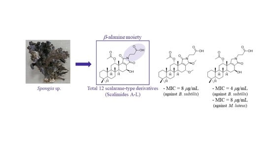
:A chemical investigation of a methanol extract of Spongia sp., a marine sponge collected from the Philippines, identified 12 unreported scalarane-type alkaloids—scalimides A–L (1–12)—together with two previously described scalarin derivatives. The elucidation of the structure of the scalaranes based on the interpretation of their NMR and HRMS data revealed that 1–12 featured a β-alanine-substituted E-ring but differed from each other through variations in their oxidation states and substitutions occurring at C16, C24, and C25. Evaluation of the antimicrobial activity of 1–12 against several Gram-positive and Gram-negative bacteria showed that 10 and 11 were active against Micrococcus luteus and Bacillus subtilis, respectively, with MIC values ranging from 4 to 16 μg/mL.
1. Introduction
Scalaranes are one of the most prevalent groups of sesterterpenes, and they are primarily found in marine invertebrates [1,2,3]. Since scalarin, the first reported natural scalarane-type marine product, was discovered in the marine sponge Cacospongia scalaris in 1972 [4], several hundreds of scalarane sesterterpenes have been identified from sponges belonging to the order Dicytoceratida [5]. This class of scalaranes is characterized as a trans-fused 6/6/6/6 carbocyclic ring system and, in many cases, contains an additional pentacyclic E ring [6]. The general structure of the E ring contains an oxygen atom in the form of a lactone or furan, but certain reports have shown that a nitrogen-rich environment can produce a pyrrole or lactam [7]. The first example of nitrogen-bearing scalarane was found in the case of molliorins that were reported from 1977 to 1979. A series of investigations on the marine sponge Cacospongia mollior resulted in consecutive isolations of five pyrrole-bearing scalaranes—molliorins A–E [8,9,10,11]. Plausible biosynthesis of pyrrole in molliorins that involved condensation reactions of scalaridial—which is a common intermediate—with the corresponding amines was proposed, and this could be proved by the synthesis of molliorins after the proposed condensation under acidic conditions.
Since the mid-2000s, scalaranes containing lactams or cyclic imides have been identified. Hyatelactam, which was isolated from the marine sponge Hyatella intestinalis in 2006, is the first example of a lactam-bearing scalarane [12]. Following this report, 17 additional scalaranes belonging to this class were isolated from the marine sponges Hyatella sp., Hyrtios sp., Petrosaspongia sp., and Spongia sp. [13,14,15,16,17]. In these cases, a maleimide or α,β-unsaturated γ-butyrolactam moiety appeared as a structural feature of the E ring, but only hyrtioscalarin A contained a linear amide terminus (Figure 1). Although the pharmacological activity of the scalarane alkaloids has not been thoroughly investigated, some interesting biological properties have been reported, such as the anticancer activities of hyatelactam against HT-29 (human colorectal adenocarcinoma cell line, GI50 = 8.1 μM) [7], hyrtioscalarin D against A549 (adenocarcinomic human alveolar basal epithelial cell, IC50 = 5.4 μM), and hyrtioscalarin G against A549 (IC50 = 6.9 μM) and PC3 (human prostate cancer cell, IC50 = 5.2 μM) [12], as well as the inhibitory effect of petrosaspongiolactam A on the binding of TDP-43 to bt-TAR-32 (IC50 = 0.6 μM) [9] and the antibacterial activity of the sodium form of hyrtioscalarin F against Bacillus subtilis (MIC = 6.25 μg/mL) and of hyrtioscalarin A against Klebsiella pneumoniae (MIC = 2 μg/mL) [8].
The marine sponge of the genus Spongia, which belongs to the order Dicytoceratida, is known as a rich source of scalarane sesterterpenes. To date, various scalarane sesterterpenes exhibiting cytotoxicity or antagonistic activity against FXR (farnesoid X receptor), such as scalarolide [18], isoscalarafurans A–B [19], scalalactams A–D [16], and 12-O-deacetyl-12-epi-19-deoxy-21-hydroxyscalarin [20], have been isolated from marine sponges [21,22]. Our preliminary biological evaluation of the methanol extract of Spongia sp. collected from the Bohol province in the Philippines revealed a mild antimicrobial activity, prompting an investigation of bioactive secondary metabolites of the sponge. In this study, we report the identification of 12 undescribed scalarane alkaloids—scalimides A–L (1–12)—that contain a β-alanine moiety and their antimicrobial activity against several strains of Gram-positive and Gram-negative bacteria (Figure 2).
2. Results
2.1. Elucidation of the Structure
Scalimide A (1) was isolated as an amorphous solid. Its molecular formula was determined to be C30H43NO7 by HRESIMS (m/z 552.2925 [M + Na]+, as calculated for C30H43NO7Na, 552.2932), corresponding to 10 degrees of unsaturation (DOUs). Inspection of the 1H and 13C NMR spectra (Table 1, Figures S4 and S5) in combination with the HSQC spectrum revealed the presence of four carbonyl carbons (δC 174.4, 172.1, 171.9, 170.4), one tetrasubstituted double bond (δC 150.0, 141.9), two sp3 oxymethines (δH 5.52/δC 76.3, δH 4.59/δC 65.6), three sp3 methines, nine sp3 methylenes, and six methyl singlets, including one acetyl group (δH 1.93/δC 21.0, δH 1.32/δC 21.5, δH 0.98/δC 17.4, δH 0.86/δC 33.7, δH 0.87/δC 16.4, δH 0.84/δC 21.7). The HMBC correlations from H3-19/20 (δH 0.84/0.87) to C-3 (δC 43.2)/C-4 (δC 34.2)/C-5 (δC 58.0), from H3-21 (δH 0.98) to C-8 (δC 38.6)/C-9 (δC 54.1)/C-14 (δC 51.3), from H3-22 (δH 0.87) to C-1 (δC 40.9)/C-5/C-9/C-10 (δC 38.1), and from H3-23 (δH 1.32) to C-12 (δC 76.3)/C-13 (δC 41.6)/C-14/C-18 (δC 150.0) identified the 6/6/6/6 fused cyclic system of the scalarane scaffold (Figure 3). The 12-acetoxy group was established by the HMBC correlations from H-12 (δH 5.52)/12-CH3CO (δH 1.93) to 12-CH3CO (δC 173.5). Another oxymethine was inferred at C-16, as was evident from the spin system for H-14 (δH 1.66)–H2-15 (δH 2.21/1.57)–H-16 (δH 4.59) in the 1H-1H COSY spectrum. The calculated DOU value and the preliminarily identified functional groups suggested that 1 contained an extra E ring in order to form a pentacyclic skeleton. The HMBC correlations from H3-23 to C-18 and from H-16 to C-17 (δC 141.9)/C-18/C-24 (δC 171.9) indicated the presence of a pyrrole-2,5-dione moiety that was found in scalalactam A [16]. The presence of the propionic acid side chain was corroborated by the 1H-1H COSY cross-peak from H2-1′ (δH 3.69) to H2-2′ (δH 2.56), the HMBC correlations from H2-1′/H2-2′ to C-3′ (δC 174.4), and a strong IR absorption at 1735 cm−1. Additionally, the HMBC correlations from H2-1′ to C-24/C-25 (δC 170.4) suggested that the propionic acid moiety was connected to the pyrrole-2,5-dione moiety through the nitrogen atom.
The trans-fused cyclic system of 1 was determined by the consecutive NOESY correlations observed for H-3α (δH 1.15)–H-5 (δH 0.87)–H-9 (δH 1.23)–H-14 and H-6β (δH 1.48)–H3-22–H-11β (δH 1.78)–H3-21–H3-23 (Figure 4). The 12-acetoxy group was assigned as α based on the NOESY correlation from H-12 to H3-23, which was confirmed by the coupling constant between H-12 and H2-11 (dd, JH-12-H-11 = 3.5, 2.7 Hz). Similarly, the β configuration of 16-OH was assigned based on the NOESY correlation between H-14 and H-16, as well as the relatively large coupling constant between H-15β (δH 1.57) and H-16 (dd, JH-16–H-15 = 9.4, 7.0 Hz). Based on the biosynthetic origin, the absolute configurations of C-8, C-9, C-10, C-13, and C-14 in 1 were deduced to be identical to the reported scalarane alkaloids (Figure 1).
Scalimide B (2) was isolated as an amorphous solid. Its molecular formula was determined to be C30H43NO7 by HRESIMS (m/z 552.2931 [M + Na]+, as calculated for C30H43NO7Na, 552.2932), corresponding to 10 degrees of unsaturation. At first glance, the 1H NMR spectrum of 2 was almost identical to that of 1, except for the smaller coupling constants observed for H-16 (δH 4.56, dd, J = 4.3, 1.5 Hz) and the appearance of H-15 (δH 1.93/1.83) at a lower frequency (Table 1), suggesting that 2 could be the 16-epimer of 1. The complete structure could be proven by observing the HMBC correlations from H-16 to C-17 (δC 140.5)/C-18 (δC 151.1) and from H-14 (δH 2.03) to C-16 (δC 60.2). Additionally, the configuration of C-16 was further supported by the absence of the NOESY correlation between H-14 and H-16 and the higher chemical shift value of H-14 (δH 2.03 vs. δH 1.66 in 1), which resulted from an increasing 1,3-diaxial interaction on the D ring due to an axial substitution of OH-16.
Scalimide C (3) was isolated as an amorphous solid. Its molecular formula was determined to be C31H45NO7 by HRESIMS (m/z 566.3087 [M + Na]+, as calculated for C31H45NO7Na, 566.3088), corresponding to 10 degrees of unsaturation. The 1H and 13C NMR spectra of 3 resembled those of 1, except for the presence of a methoxy group at δH 3.52/δC 58.0, suggesting that 3 was a methyl ether of 1 at C-16, which was consistent with its being 14 mass units higher than that of 1 in the HRESIMS spectrum (Table 1). The location of the methoxy group was confirmed by the HMBC correlation from the methyl singlet at δH 3.52 to C-16 (δH 4.29/δC 74.9) (Figure 3). The configuration of C-16 was assigned as β based on the NOESY correlation between H-14 and H-16, as well as the large coupling constant for H-16 (δH 4.29, JH-16–H-15 = 9.1, 7.0 Hz).
Scalimide D (4) was isolated as an amorphous solid. The molecular formula of 4 was not only identical to that of 3, but its 1H NMR spectrum also showed a high degree of similarity with that of 2, except for the presence of a methoxy group at δH 3.46/δC 57.9, suggesting that 4 was the OMe-16 analog of 2 (Table 1, Figure S25). The HMBC correlation from the methyl singlet at δH 3.46 to C-16 (δC 70.1) confirmed the substitution of OMe at C-16, and its α orientation of the methoxy group was determined by the coupling constants of H-16 (δH 4.18, dd, JH-16–H-15 = 4.0, 1.6 Hz).
Scalimide E (5) was isolated as an amorphous solid, and its molecular mass was 14 mass units higher than those of 3 and 4, as determined by HRESIMS (m/z 580.3240 [M + Na]+, as calculated for C32H47NO7Na, 580.3245). Compared with that of 4, the 1H NMR spectrum of 5 showed an additional methyl singlet at δH 3.63, suggesting that 5 is a methyl ester of 4 (Table 1, Figure S32). The HMBC correlation from the methyl group at δH 3.63 to C-3′ (δC 173.0) confirmed the presence of a methyl ester moiety in the side chain of 5 (Figure 3). Scalimide E (5) could be an artifact generated from 4 during the course of purification using MeOH and TFA, but this possibility was not investigated in this study.
Scalimide F (6) was isolated as an amorphous solid. Its molecular formula was determined to be C30H41NO6 by HRESIMS (m/z 534.2817 [M + Na]+, as calculated for C30H41NO6Na, 534.2826), corresponding to 11 degrees of unsaturation. The analysis of the 1H and 13C NMR spectra in combination with the HSQC data, revealed the presence of a disubstituted double bond at δH 6.46/δC 138.3 and δH 6.39/δC 117.7 instead of the 16-OH in 1 (Table 1, Figures S39–S41). The location of the double bond was determined to be Δ15 by the 1H-1H COSY cross-peaks from H-14 (δH 2.75) to H-15 (δH 6.46) and from H-15 to H-16 (δH 6.36), which was confirmed by the HMBC correlations from H-15 to C-14 (δC 54.9)/C-17 (δC 137.7) and from H-16 to C-15 (δC 138.3)/C-17/C-18 (δC 143.5).
Scalimide G (7) was isolated as an amorphous solid. Its molecular formula was determined to be C31H47NO7 by HRESIMS (m/z 568.3238 [M + Na]+, as calculated for C31H47NO7Na, 568.3245), corresponding to nine degrees of unsaturation. Analysis of the 1D and 2D NMR data revealed a similar pentacyclic framework to that of 4 (Table 2, Figures S46–S51). However, one additional sp3 oxymethine at δH 5.39/δC 81.7 was observed as a substitution for one of the carbonyl groups of a pyrrole-2,5-dione in 4. This information indicates the reduction of a carbonyl group in the pyrrole-2,5-dione of 4 to a hydroxyl group whose position was determined to be at C-24, as observed from the HMBC correlations from H-16 (δH 4.02)/H2-1′ (δH 3.68/3.53) to C-24 (δC 81.7) (Figures S2 and S50). Similarly, the small coupling constant of H-16 (dd, JH-16-H-15 = 4.4, 1.3 Hz) could be a sign of the α-orientation of OMe-16. Furthermore, the 24-OH configuration was assigned as a β-orientation based on the NOESY correlation between H-24 and 16α-OMe.
Scalimide H (8) was isolated as an amorphous solid. The 1H and 13C NMR data of 8 differed from those of 7 only in the presence of an additional methyl singlet at δH 3.04/δC 50.5, which was found to be in agreement with the HRESIMS result, indicating a molecular mass that was 14 units higher than that of 7 (Table 2, Figures S53, S54 and S60). The HMBC correlations from δH 3.04 to C-24 (δC 87.0) and from H-24 (δH 5.44) to δC 50.5 could be evidence of the OMe-24 substituent (Figure 5). Since OMe-24 exhibited a cross-peak with H3-23 in the NOESY spectrum, the OMe-24 was determined to be β-oriented (Figure S3).
Scalimide I (9) was isolated as an amorphous solid. Its molecular formula was determined to be C30H45NO6 by HRESIMS (m/z 538.3129 [M + Na]+, as calculated for C30H45NO6Na, 538.3139), corresponding to nine degrees of unsaturation. The 1H and 13C NMR data revealed that 9 had the same scaffold as 1, but with one methylene at δH 4.07/3.97 instead of a carbonyl group (Table 2, Figures S61 and S62). The HMBC correlations from δH 4.07/3.97 to C-17 (δC 153.9)/C-18 (δC 140.1)/C-25 (δC 171.7) indicated a γ-lactam moiety, and the NOESY correlation between one of the methylene protons at δH 3.97 and H-16 suggested the location of the methylene at C-24 to determine a 24-2H-pyrrol-25-one (Figure 5 and Figure S3). In addition, the β-configuration of OH-16 was confirmed by the large coupling constant of H-16 (dd, JH-16–H-15 = 9.8, 6.6 Hz).
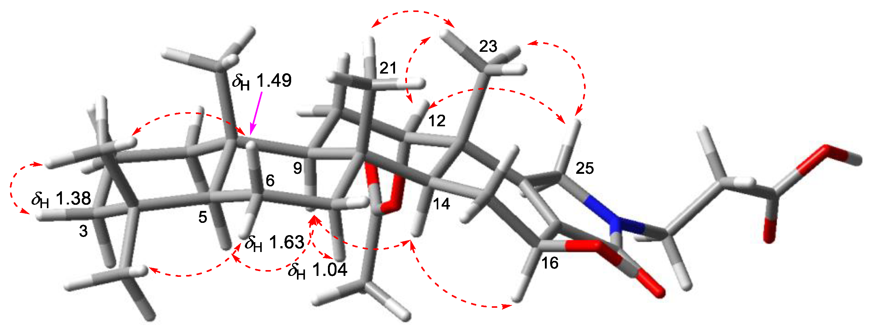
Scalimide J (10) was isolated as an amorphous solid. The HRESIMS analysis of 10 revealed an identical molecular formula to that of 9 (m/z 516.3317 [M + H]+, as calculated for C30H46NO6, 516.3320). The 1H NMR spectrum of 10 featured a methylene at δH 4.06/3.77 (Table 2, Figure S68), which exhibited HMBC correlations to C-17 (δC 133.4)/C-18 (δC 162.3), confirming the presence of the γ-lactam moiety (Figure 5). However, the inversion of the chemical shift between C-17 and C-18 (δC-18 > δC-17), in comparison with that of 9, suggested a 25-2H-pyrrol-24-one moiety, which was further evidenced by the NOESY correlations from H2-25 (δH 4.06/3.77) to H3-23 (δH 1.26). The β-orientation of OH-16 was determined by observing the consecutive NOESY correlations for H-5–H-9–H-14–H-16 (Figure 6).
Scalimide K (11) was isolated as an amorphous solid. Its molecular formula was determined to be C30H43NO5 by HRESIMS (m/z 520.3030 [M + Na]+, as calculated for C30H43NO5Na, 520.3033), corresponding to 10 degrees of unsaturation. The 1H NMR spectra of 11 in combination with the HSQC data showed olefinic protons at δH 6.25/6.03 and a methylene proton at δH 4.06 (Table 2, Figures S75–S77). The double bond between C-15 and C-16 was substantiated by the 1H-1H COSY correlations from H-14 (δH 2.68) to H-15 (δH 6.03) and from H-15 to H-16 (δH 6.25). Similarly to 10, the NOESY correlation between the methylene protons at δH 4.06 and H3-23 (δH 1.06) suggested the presence of a 25-2H-pyrrol-24-one moiety in 11.
Scalimide L (12) was isolated as an amorphous solid. The 1H NMR spectrum of 12 was almost identical to that of 11, except for an additional methyl singlet at δH 3.65, suggesting that 12 was a methyl ester of 11 (Table 2, Figure S82). The presence of an additional methyl group was consistent with the molecular formula of C31H45NO5, as determined in an HRESIMS analysis (m/z 512.3366 [M + H]+, as calculated for C31H46NO5, 512.3371). Therefore, the HMBC correlation from δH 3.65 to C-3′ (δC 174.4) confirmed the presence of the methyl propionate side chain, which may have been an artifact generated during the separation steps when using MeOH as an eluent.
2.2. Biological Activities
As the MeOH extract of Spongia sp. exhibited mild antimicrobial activity in our preliminary screening, the minimal inhibitory concentrations (MICs) of scalimides A–L (1–12) were evaluated by using several strains of Gram-positive and Gram-negative bacteria (Table 3). Interestingly, 1–12 were more potent against Gram-positive bacteria but were mostly inactive against Gram-negative bacteria. Among these compounds, 10 displayed the broadest spectrum of inhibitory effects, especially against Micrococcus luteus and Bacillus subtilis, with MIC values of 8 and 4 μg/mL, respectively. Although most of the isolated compounds displayed weak to moderate antibacterial activity, the structural diversity of the scalimides—arising from different substitutions on C-16, C-24, and C-25—provided useful information regarding the structure–activity relationship (SAR): (1) Regarding the MIC values of 1–4, the methyl ether at C-16 appeared to be more potent than the hydroxyl group for the activity toward B. subtilis; (2) when comparing 4 with 5 and 11 with 12, the carboxylic acid moiety at C-3′ was identified as a crucial factor for the antimicrobial activity; (3) reduction of the carbonyl group at C-24 to a hydroxyl group (4 to 7) and a methylene group (1 to 9) decreased antibacterial activity; (4) reduction of the carboxyl group of the imide at C-25 to a lactam (1 vs. 10 and 6 vs. 11) increased antibacterial activity at least four-fold. Additionally, the cytotoxicity of 1–12 against MCF7 (a breast cancer cell line) was evaluated to identify it as a potent anticancer agent, but only moderate anticancer effects were observed for all of the tested compounds.
3. Materials and Methods
3.1. General Experimental Procedures
Specific optical rotations were measured using a Rudolph Research Analytical (Autopol III) polarimeter (Rudolph Research Analytical, Hackettstown, NJ, USA). IR spectra were recorded using a JASCO FT/IR-4100 spectrophotometer (JASCO Corporation, Tokyo, Japan). The 1D (1H and 13C) and 2D (COSY, HSQC, HMBC, and NOESY) NMR spectra were recorded in CD3OD using a Bruker 600 MHz spectrometer (Bruker BioSpin GmbH, Rheinstetten, Germany) at 297.1 K. The 1H NMR spectra were collected from 32–64 scans, and the 13C NMR spectra were collected from 5000–15,000 scans, depending on the sample concentrations. The mixing time for the NOESY experiments was set to 0.33 s. Chemical shifts were reported in parts per million relative to CD3OD (δH 3.31, δC 49.0). High-resolution mass spectra were obtained using a Sciex X500R Q-TOF spectrometer (Framingham, MA, USA) equipped with an electrospray ionization (ESI) source. MPLC (medium-pressure liquid chromatography) was performed using a TELEDYNE ISCO CombiFlash Companion with a YMC-Dispopack AT ODS-25 40 g column (YMC Co. Ltd., Kyoto, Japan). HPLC (high-performance liquid chromatography) was performed using a PrimeLine Binary pump (Analytical Scientific Instruments, Inc., El Sobrante, CA, USA) equipped with a Shodex RI-101 (Shoko Scientific Co. Ltd., Yokohama, Japan) and UV-M201 using C18 columns (YMC-Triart C18, 250 × 10 mm I.D., or 250 × 4.6 mm I.D., 5 µm; YMC Co. Ltd., Kyoto, Japan).
3.2. Biological Material
Biological material was collected in March 2015 in the Philippines (9°45′32.31″ N 124°35′53.60″ E) at a depth of 15 m by scuba diving. The sponges were frozen at −20 °C until they were identified as Spongia sp. and chemically analyzed. A voucher sample (153PIL-209) was stored at the Marine Biotechnology Research Center, Korea Institute of Ocean Science and Technology (KIOST).
3.3. Extraction and Isolation
The specimens (wet wt.: 1.5 kg) were lyophilized and extracted repeatedly with MeOH (2.0 L × 2) and CH2Cl2 (2.0 L × 2) at room temperature. The extracts were combined and concentrated under reduced pressure. The residue (78.3 g) was partitioned with n-butanol (3.0 L) and water (3.0 L) to yield 35.5 g of an organic-soluble material. The n-butanol layer was further partitioned into n-hexane (2.0 L) and 15% aqueous methanol (2.0 L). The 15% aqueous methanol fraction (30.1 g) was concentrated and subjected to flash column chromatography over C18 (YMC Gel ODS-A, 60 Å, 230 mesh (YMC Co., Ltd., Kyoto, Japan)) with a stepwise gradient solvent system (50%, 60%, 70%, 80%, 90% aqueous-MeOH, and 100% MeOH, acetone, and EtOAc).
The 90% MeOH fraction (780.6 mg) was further separated using MPLC on a C18 column with a gradient solvent system from 70% aq-MeOH to 100% MeOH over 30 min to yield five fractions. The fourth fraction (566.9 mg) was separated using MPLC on a C18 column with a gradient solvent system from 60% MeOH to 100% MeOH over 40 min to yield four subfractions. The second subfraction (253.8 mg) was separated using HPLC (eluent 60% MeCN with 0.1% TFA) to yield 1 (3.5 mg, tR = 60 min), 2 (3.8 mg, tR = 62 min), 9 (1.4 mg, tR = 30 min), and 10 (1.9 mg, tR = 40 min). The third subfraction (197.9 mg) was separated using HPLC (eluent 70% MeCN with 0.1% TFA) to yield 3 (2.3 mg, tR = 66 min), 4 (6.7 mg, tR = 65 min), 5 (2.3 mg, tR = 67 min), 7 (4.0 mg, tR = 30 min), 8 (2.9 mg, tR = 58 min), 11 (4.6 mg, tR = 42 min), and 12 (1.1 mg, tR = 43 min). The fourth subfraction (37.8 mg) was separated using HPLC (eluent 80% MeCN with 0.1% TFA) to yield 6 (3.2 mg, tR = 58 min), scalarin (12.0 mg, tR = 50 min; the structure is shown in the Supporting Information), and 12α-acetoxy-19β-hydroxyscalara-15,17-dien-20,19-olide (6.1 mg, tR = 61 min).
1: Amorphous powder; +90 (c 0.1, MeOH); IR (ATR) νmax 3742, 2933, 2858, 1738, 1702, 1547, 1511, 1452, 1381, 1240, 1194 cm−1; 1H NMR and 13C NMR, see Table 1 and Figures S4 and S5; HRESIMS m/z 552.2925 [M + Na]+ (calculated for C30H43NO7Na, 552.2932).
2: Amorphous powder; +100 (c 0.1, MeOH); IR (ATR) νmax 3441, 2929, 2855, 1710, 1653, 1448, 1395, 1233, 1042 cm−1; 1H NMR and 13C NMR, see Table 1 and Figures S11 and S12; HRESIMS m/z 552.2931 [M + Na]+ (calculated for C30H43NO7Na, 552.2932).
3: Amorphous powder; +86.7 (c 0.1, MeOH); IR (ATR) νmax 2933, 2851, 1742, 1702, 1515, 1388, 1236, 1194 cm−1; 1H NMR and 13C NMR, see Table 1 and Figures S18 and S19; HRESIMS m/z 566.3087 [M + Na]+ (calculated for C31H45NO7Na, 566.3088).
4: Amorphous powder; +93.3 (c 0.2, MeOH); IR (ATR) νmax 2929, 1738, 1710, 1458, 1399, 1236, 1197, 1038 cm−1; 1H NMR and 13C NMR, see Table 1 and Figures S25 and S26; HRESIMS m/z 566.3083 [M + Na]+ (calculated for C31H45NO7Na, 566.3088).
5: Amorphous powder; +43.3 (c 0.1, MeOH); IR (ATR) νmax 2933, 2858, 1742, 1706, 1547, 1515, 1388, 1236, 1201 cm−1; 1H NMR and 13C NMR, see Table 1 and Figures S32 and S33; HRESIMS m/z 580.3240 [M + Na]+ (calculated for C32H47NO7Na, 580.3245).
6: Amorphous powder; +86.6 (c 0.1, MeOH); IR (ATR) νmax 2925, 1742, 1710, 1692, 1543, 1526, 1511, 1374, 1236, 1204 cm−1; 1H NMR and 13C NMR, see Table 1 and Figures S39 and S40; HRESIMS m/z 534.2817 [M + Na]+ (calculated for C30H41NO6Na, 534.2826).
7: Amorphous powder; +56.6 (c 0.1, MeOH); IR (ATR) νmax 3738, 2936, 2861, 1721, 1706, 1692, 1533, 1469, 1250 cm−1; 1H NMR and 13C NMR, see Table 2 and Figures S46 and S47; HRESIMS m/z 568.3238 [M + Na]+ (calculated for C31H47NO7Na, 568.3245).
8: Amorphous powder; +53.3 (c 0.1, MeOH); IR (ATR) νmax 3742, 2933, 2855, 1734, 1706, 1689, 1554, 1462, 1388, 1240 cm−1; 1H NMR and 13C NMR, see Table 2 and Figures S53 and S54; HRESIMS m/z 582.3395 [M + Na]+ (calculated for C32H49NO7Na, 582.3401).
9: Amorphous powder; +60 (c 0.1, MeOH); IR (ATR) νmax 3399, 2929, 2869, 1717, 1674, 1455, 1261, 1204, 1133 cm−1; 1H NMR and 13C NMR, see Table 2 and Figures S61 and S62; HRESIMS m/z 538.3129 [M + Na]+ (calculated for C30H45NO6Na, 538.3139).
10: Amorphous powder; +113.3 (c, 0.1, MeOH); IR (ATR) νmax 3420, 2925, 2855, 1717, 1674, 1466, 1374, 1246, 1194 cm−1; 1H NMR and 13C NMR, see Table 2 and Figures S68 and S69; HRESIMS m/z 516.3317 [M + H]+ (calculated for C30H46NO6, 516.3320).
11: Amorphous powder; +183.3 (c 0.2, MeOH); IR (ATR) νmax 2929, 2855, 1731, 1699, 1689, 1660, 1551, 1505, 1462, 1240 cm−1; 1H NMR and 13C NMR, see Table 2 and Figures S75 and S76; HRESIMS m/z 520.3030 [M + Na]+ (calculated for C30H43NO5Na, 520.3033).
12: Amorphous powder; +56.6 (c 0.1, MeOH); IR (ATR) νmax 2936, 1749, 1702, 1692, 1646, 1551, 1526, 1509 cm−1; 1H NMR and 13C NMR, see Table 2 and Figures S82 and S83; HRESIMS m/z 512.3366 [M + H]+ (calculated for C31H46NO5, 512.3371).
3.4. Assay
3.4.1. Antimicrobial Assays
The test of minimal inhibitory concentrations (MICs) for 1–12 was carried out by using six bacterial strains: Micrococcus luteus (KCTC-1915), Staphylococcus aureus (KCTC-1927), Bacillus subtilis (KCTC-1021), Escherichia coli (KCTC-2441), Salmonella typhimurium (KCTC-2515), and Klebsiella pneumoniae subsp. (KCTC-2690). Bacteria were streaked onto Mueller–Hinton agar (MHA) plates and incubated for 24 h at 37 °C. A single selected colony was transferred to Muller–Hinton broth (MHB), cultured for 24 h at 37 °C, and harvested through centrifugation at 250 rpm. The turbidity of the bacterial suspensions was adjusted with the MacFarland standard 0.5 to 1.5 × 10⁸ cfu/mL by adding sterile MHB. Pure 1–12 was dissolved in DMSO and dispensed into 96-well plates at a concentration of 128 μg/mL. These compounds were serially diluted to 0.25 μg/mL. Subsequently, the bacterial suspensions were inoculated in 96-well plates and incubated for 15–20 h at 37 °C. The lowest concentrations that inhibited bacterial growth were recorded as the MIC values. Kanamycin (K1377) was purchased from the Sigma Chemical Company (St. Louis, MO, USA) and used as a positive control.
3.4.2. Cytotoxicity Assays
An MTS assay was performed using the CellTiter 96®AQueousOne Solution Cell Proliferation Assay (MTS) (Promega, Madison, WI, USA). MCF-7 cells were plated in 384-well plates at a density of 3700 cells/well. The seeded cells were incubated for 24 h at 37 °C under a 5% (v/v) CO2 atmosphere, were treated with 1–12 at seven different concentrations (1.25, 2.5, 5.0, 10, 20, 40, and 80 μΜ), and then incubated for another 48 h. DMSO was used as a vehicle control and staurosporine was used as a positive control. Viable cell numbers were determined by the concentration of formazan resulting from the tetrazolium conversion, and the absorbance was measured at 490 nm using an EnVision Xcite Multilabel Reader (PerkinElmer, Waltham, MA, USA).
4. Conclusions
In summary, the investigation of the antibacterial components of the marine sponge Spongia sp. resulted in the isolation of 12 unreported β-alanine-bearing scalaranes (1–12), which were named scalimides A–L. The structures of 1–12 were unambiguously determined by using conventional spectroscopic methods with 1D and 2D NMR data and HRESIMS. The structural differences in 1–12 arose from the variations in the oxidation states at C-24 and C-25, producing an imide, aminal, or lactam, as well as from substitutions at C-16 and C-24. Evaluation of MIC values against selected Gram-positive and Gram-negative bacteria identified 10 as the most potent antimicrobial agent among the isolated compounds, providing insights into the structure–activity relationships. Further studies aimed at identifying potent antimicrobial natural products from the sponge Spongia sp. are currently underway.
Supplementary Materials
The following supporting information can be downloaded at: https://www.mdpi.com/article/10.3390/md20110726/s1, Tables S1–S12: 13C/1H chemical shifts and COSY, HMBC, and NOESY correlations for 1–12; Figure S1: Structures of known compounds isolated from Spongia sp.; Figures S2 and S3: Key 1H-1H COSY, HMBC, and NOESY correlations of 1–12. Figures S4–S88: 1H NMR, 13C NMR, COSY, HSQC, HMBC, NOESY, and HRMS spectra of 1–12.
Author Contributions
A.-Y.S. worked on the isolation and structural elucidation. H.-S.L. collected the marine sponges and conducted a preliminary biological evaluation. J.L. supervised the study. All authors have read and agreed to the published version of the manuscript.
Funding
This work was supported by grants from the Ministry of Ocean and Fisheries (PM62830) and the Korea Institute of Ocean Science and Technology (PEA0021).
Institutional Review Board Statement
Not applicable.
Informed Consent Statement
Not applicable.
Data Availability Statement
All data presented in this report are available with permission from the corresponding author upon request.
Acknowledgments
We are grateful to Young-A Kim at Hannam University for the helpful discussions regarding the identification of the marine sponges.
Conflicts of Interest
The authors declare no conflict of interest.
References
- Li, K.; Gustafson, K.R. Sesterterpenoids: Chemistry, biology, and biosynthesis. Nat. Prod. Res. 2021, 38, 1251–1281. [Google Scholar] [CrossRef] [PubMed]
- Yu, H.B.; Chen, H.Y.; Duan, S.; Zhu, Y.P.; Hu, B.; He, Y.; Cheng, S.T.; Jiao, B.H.; Liu, X.Y. Bioactive Scalarane—Type Sesterterpenoids from Marine Sources. Chem. Biodivers. 2022, 19, e202200049. [Google Scholar] [CrossRef] [PubMed]
- Máximo, P.; Lourenço, A. Marine sesterterpenes: An overview. Curr. Org. Chem. 2018, 22, 2381–2393. [Google Scholar] [CrossRef]
- Fattorusso, E.; Magno, S.; Santacroce, C.; Sica, D. Scalarin, a new pentacyclic C-25 terpenoid from the sponge Cacospongia scalaris. Tetrahedron 1972, 28, 5993–5997. [Google Scholar] [CrossRef]
- Abdelaleem, E.R.; Samy, M.N.; Desoukey, S.Y.; Liu, M.; Quinn, R.J.; Abdelmohsen, U.R. Marine natural products from sponges (Porifera) of the order Dictyoceratida (2013 to 2019); a promising source for drug discovery. RSC Adv. 2020, 10, 34959–34976. [Google Scholar] [CrossRef] [PubMed]
- Liu, Y.; Wang, L.; Jung, J.H.; Zhang, S. Sesterterpenoids. Nat. Prod. Rep. 2007, 24, 1401–1429. [Google Scholar] [CrossRef]
- Gonzalez, M.A. Scalarane sesterterpenoids. Curr. Bioact. Compd. 2010, 6, 178–206. [Google Scholar] [CrossRef] [Green Version]
- Cafieri, F.; De Napoli, L.; Fattorusso, E.; Santacroce, C. Molliorin-B, a second scalarin-like pyrroloterpene from the sponge Cacospongia mollior. Experientia 1977, 33, 994–995. [Google Scholar] [CrossRef]
- Cafieri, F.; De Napoli, L.; Iengo, A.; Santacroce, C. Molliorin-c, a further pyrroloterpene present in the sponge Cacospongia mollior. Experientia 1978, 34, 300–301. [Google Scholar] [CrossRef]
- Cafieri, F.; De Napoli, L.; Iengo, A.; Santacroce, C. Minor pyrroloterpenoids from the marine sponge Cacospongia mollior. Experientia 1979, 35, 157–158. [Google Scholar] [CrossRef]
- Cafieri, F.; De Napoli, L.; Fattorusso, E.; Santacroce, C.; Sica, D. Molliorin-A: A unique scalarin-like pyrroloterpene from the sponge Cacospongia mollior. Tetrahedron Lett. 1977, 18, 477–480. [Google Scholar] [CrossRef]
- Hernández-Guerrero, C.J.; Zubia, E.; Ortega, M.J.; Carballo, J.L. Sesterterpene metabolites from the sponge Hyatella intestinalis. Tetrahedron 2006, 62, 5392–5400. [Google Scholar] [CrossRef]
- Jeon, J.-E.; Bae, J.; Lee, K.J.; Oh, K.-B.; Shin, J. Scalarane sesterterpenes from the sponge Hyatella sp. J. Nat. Prod. 2011, 74, 847–851. [Google Scholar] [CrossRef] [PubMed]
- Festa, C.; Cassiano, C.; D’Auria, M.V.; Debitus, C.; Monti, M.C.; De Marino, S. Scalarane sesterterpenes from Thorectidae sponges as inhibitors of TDP-43 nuclear factor. Org. Biomol. Chem. 2014, 12, 8646–8655. [Google Scholar] [CrossRef] [PubMed]
- Elhady, S.S.; El-Halawany, A.M.; Alahdal, A.M.; Hassanean, H.A.; Ahmed, S.A. A new bioactive metabolite isolated from the Red Sea marine sponge Hyrtios erectus. Molecules 2016, 21, 82. [Google Scholar] [CrossRef] [Green Version]
- Yang, I.; Lee, J.; Lee, J.; Hahn, D.; Chin, J.; Won, D.H.; Ko, J.; Choi, H.; Hong, A.; Nam, S.-J. Scalalactams A–D, scalarane sesterterpenes with a γ-lactam moiety from a Korean Spongia sp. Marine sponge. Molecules 2018, 23, 3187. [Google Scholar] [CrossRef] [Green Version]
- Kwon, O.-S.; Kim, D.; Kim, C.-K.; Sun, J.; Sim, C.J.; Oh, D.-C.; Lee, S.K.; Oh, K.-B.; Shin, J. Cytotoxic scalarane sesterterpenes from the sponge Hyrtios erectus. Mar. Drugs 2020, 18, 253. [Google Scholar] [CrossRef]
- Phan, C.-S.; Kamada, T.; Hamada, T.; Vairappan, C.S. Cytotoxic sesterterpenoids from bornean sponge Spongia sp. Rec. Nat. Prod. 2018, 12, 643–647. [Google Scholar] [CrossRef]
- Davis, R.; Capon, R. Two new scalarane sesterterpenes: Isoscalarafuran-A and-B, epimeric alcohols from a southern Australian Marine Sponge, Spongia hispida. Aust. J. Chem. 1993, 46, 1295–1299. [Google Scholar] [CrossRef]
- Nam, S.-J.; Ko, H.; Ju, M.K.; Hwang, H.; Chin, J.; Ham, J.; Lee, B.; Lee, J.; Won, D.H.; Choi, H. Scalarane sesterterpenes from a marine sponge of the genus Spongia and their FXR antagonistic activity. J. Nat. Prod. 2007, 70, 1691–1695. [Google Scholar] [CrossRef]
- Yang, I.; Nam, S.-J.; Kang, H. Two New Scalaranes from a Korean Marine Sponge Spongia sp. Nat. Prod. Sci. 2015, 21, 289–292. [Google Scholar] [CrossRef] [Green Version]
- Nam, S.-J.; Ko, H.; Shin, M.; Ham, J.; Chin, J.; Kim, Y.; Kim, H.; Shin, K.; Choi, H.; Kang, H. Farnesoid X-activated receptor antagonists from a marine sponge Spongia sp. Bioorg. Med. Chem. Lett. 2006, 16, 5398–5402. [Google Scholar] [CrossRef] [PubMed]
Figure 1.
General structures of the E rings of scalarane alkaloids.

Figure 2.
Structures of scalimides A–L (1–12) isolated from the marine sponge Spongia sp.
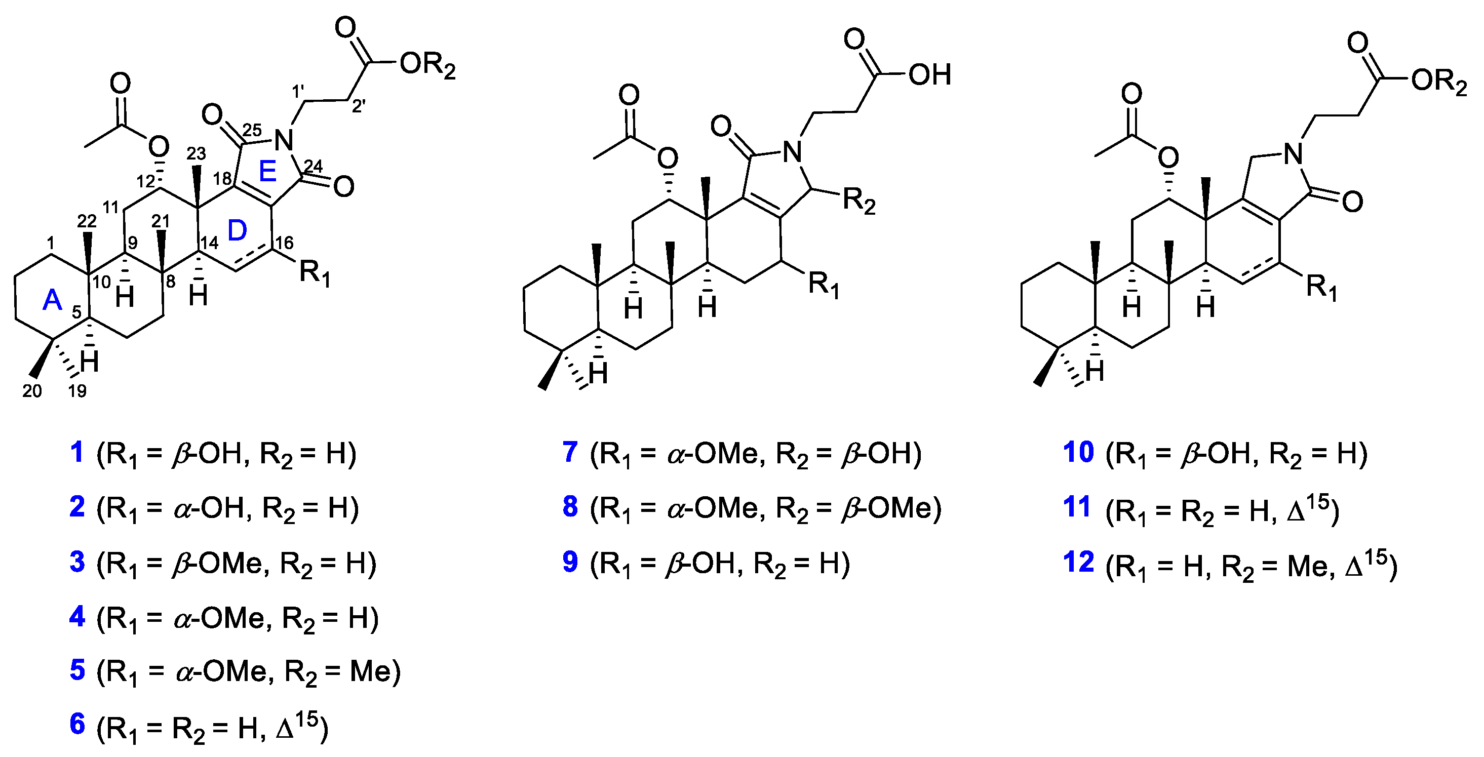
Figure 3.
Key COSY and HMBC correlations observed for 1, 3, and 5.
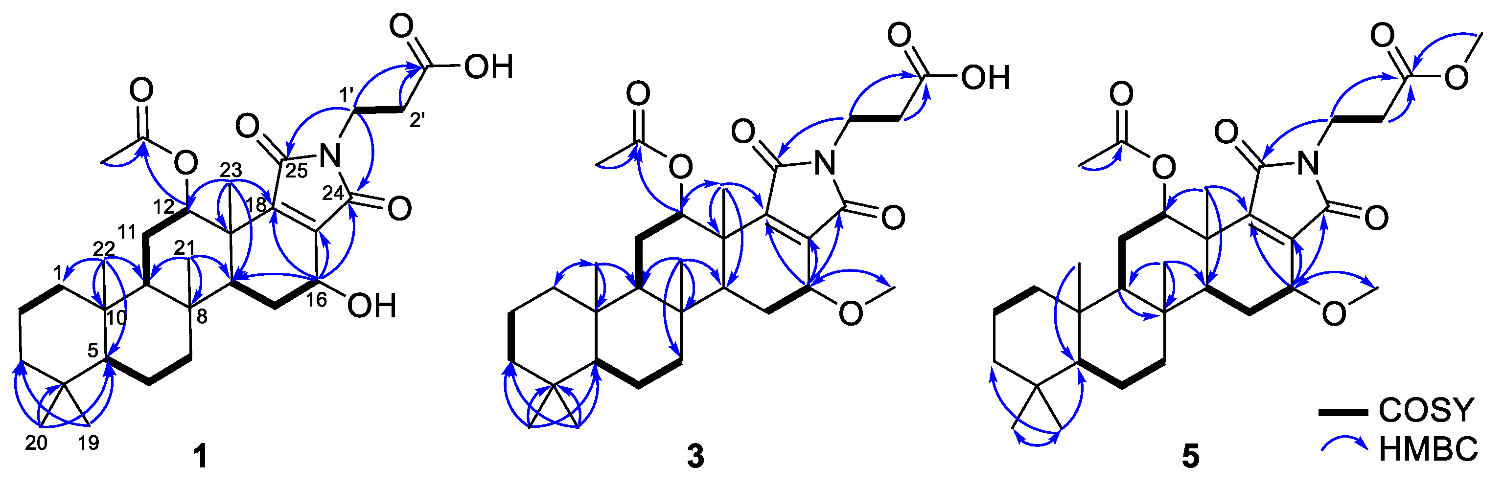
Figure 4.
Key NOESY correlations of 1.
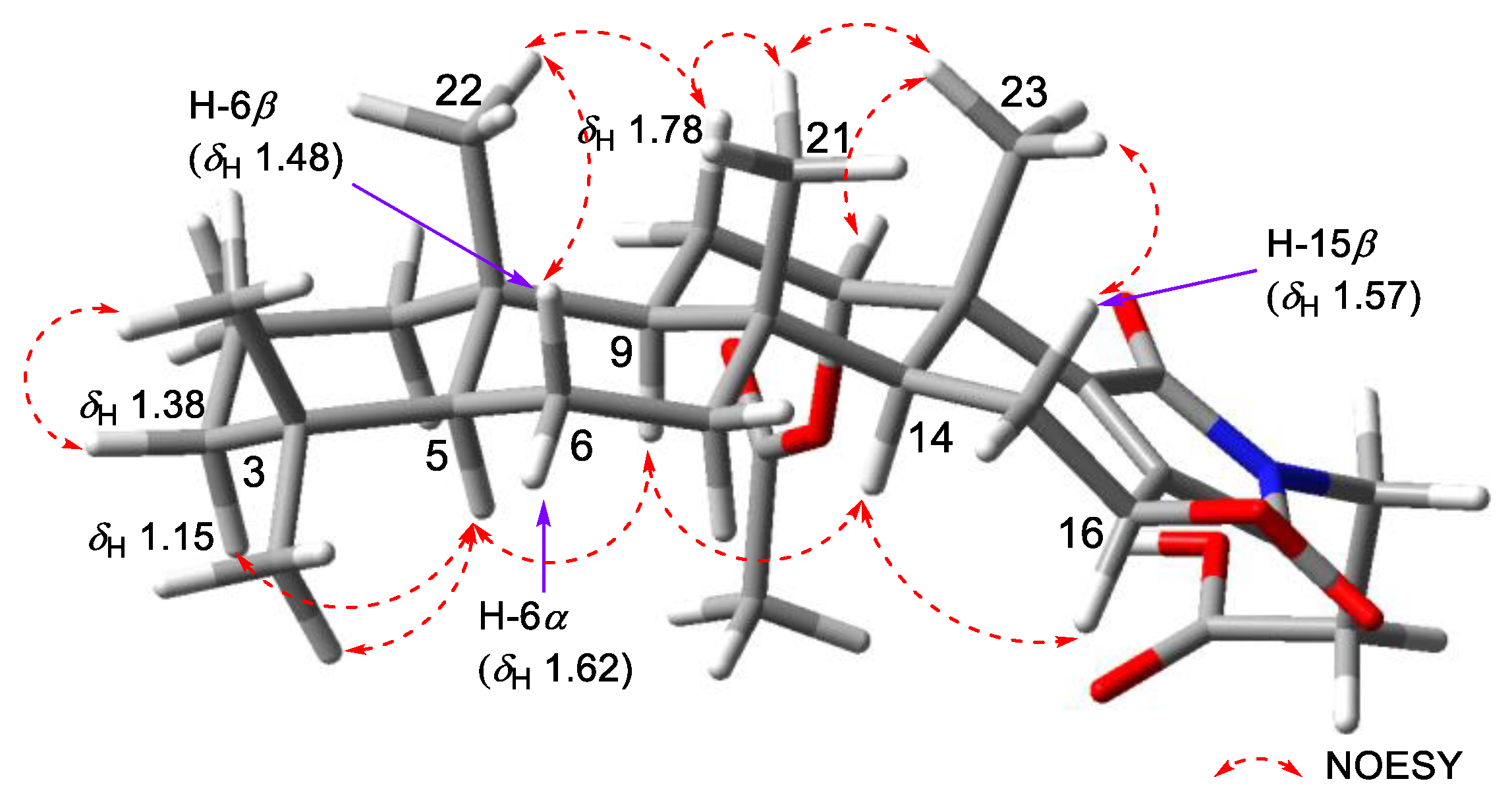
Figure 5.
Key COSY and HMBC correlations observed for 8, 9, and 10.
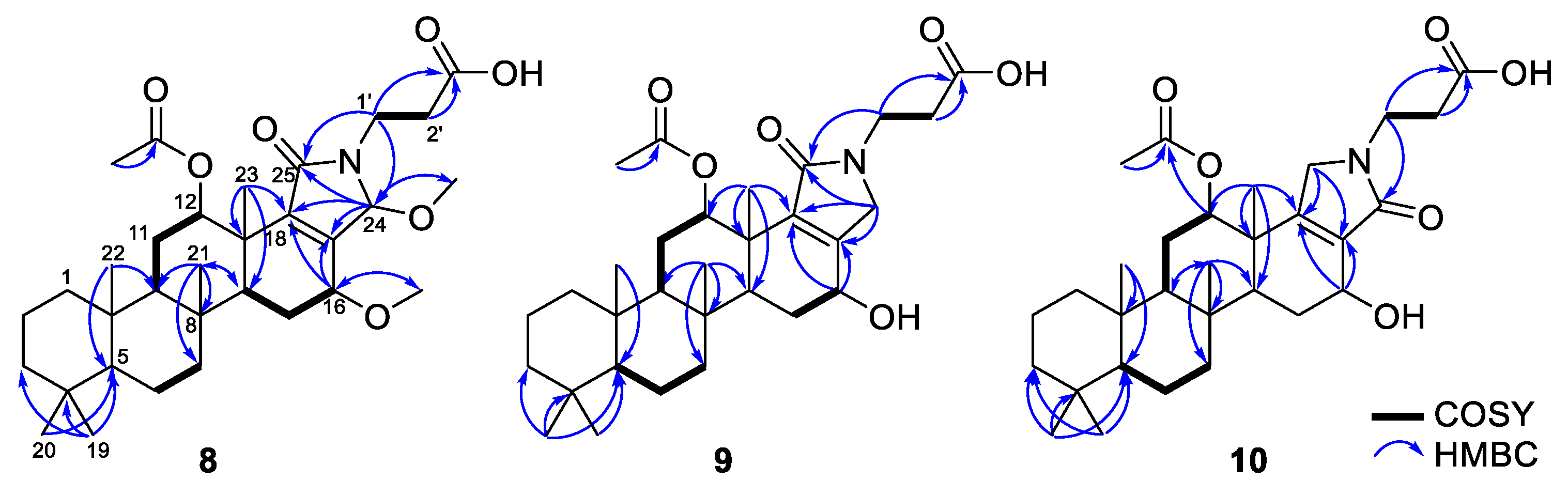
Figure 6.
NOESY correlations of 10.

Table 1.
The 13C (150 MHz) and 1H (600 MHz) NMR data for 1−6 a.
| Position | 1 | 2 | 3 | 4 | 5 | 6 | ||||||
|---|---|---|---|---|---|---|---|---|---|---|---|---|
| δc | δH (J in Hz) | δc | δH (J in Hz) | δc | δH (J in Hz) | δc | δH (J in Hz) | δc | δH (J in Hz) | δc | δH (J in Hz) | |
| 1 | 40.9 | 1.59, m 0.63, m | 40.9 | 1.61, m 0.65, m | 40.9 | 1.59, m 0.63, m | 40.9 | 1.59, m 0.65, td (12.8, 3.7) | 40.9 | 1.60, m 0.65, m | 40.9 | 1.61, m 0.68, td (12.7, 3.8) |
| 2 | 19.5 | 1.65, m 1.41, m | 19.5 | 1.64, m 1.43, m | 19.5 | 1.63, m 1.40, m | 19.5 | 1.63, m 1.41, m | 19.5 | 1.65, m 1.42, m | 19.5 | 1.66, m 1.42, m |
| 3 | 43.2 | 1.38, m 1.15, m | 43.2 | 1.38, m 1.17, m | 43.2 | 1.38, m 1.15, td (13.1, 3.9) | 43.2 | 1.37, m 1.16, td (13.4, 4.0) | 43.2 | 1.38, m 1.17, td (13.1, 3.9) | 43.2 | 1.40, m 1.17, td (14.0, 13.5, 4.2) |
| 4 | 34.2 | 34.2 | 34.2 | 34.6 | 34.2 | 34.3 | ||||||
| 5 | 58.0 | 0.87, m | 58.0 | 0.92, m | 57.9 | 0.87, m | 58.0 | 0.88, m | 58.0 | 0.88, m | 58.0 | 0.91, m |
| 6 | 19.2 | 1.62, m 1.48, m | 19.2 | 1.63, m 1.48, m | 19.2 | 1.63, m 1.49, m | 19.2 | 1.63, m 1.49, m | 19.2 | 1.62, m 1.49, qd (13.2, 3.4) | 18.9 | 1.65, m 1.53, m |
| 7 | 42.4 | 1.88, m 1.03, m | 42.3 | 1.84, m 1.11, m | 42.3 | 1.89, dt (12.6, 3.5) 1.05, m | 42.4 | 1.88, dt (12.6, 3.0) 1.03, m | 42.4 | 1.88, dt (12.7, 3.5) 1.03, td (12.7, 3.9) | 41.9 | 2.03, m 1.03, m |
| 8 | 38.6 | 38.3 | 38.7 | 38.3 | 38.3 | 38.5 | ||||||
| 9 | 54.1 | 1.23, m | 54.3 | 1.32, m | 54.1 | 1.22, m | 54.3 | 1.25, dd (13.3, 2.3) | 54.3 | 1.26, dd (13.4, 2.3) | 53.4 | 1.31, m |
| 10 | 38.1 | 38.1 | 38.1 | 38.1 | 38.1 | 38.0 | ||||||
| 11 | 21.9 | 1.99, dd (12.6, 6.9) 1.78, ddd (15.2, 13.2, 2.4) | 21.9 | 2.04, m 1.82, m | 21.7 | 1.99, m 1.77, ddd (15.2, 13.2, 2.4) | 21.9 | 2.01, dt (15.1, 3.0) 1.78, ddd (15.2, 13.3, 2.5) | 21.9 | 2.01, dt (14.8, 2.8) 1.78, ddd (15.2, 13.2, 2.5) | 22.1 | 2.06, m 1.76, ddd (15.2, 13.2, 2.4) |
| 12 | 76.3 | 5.48, dd (3.5, 2.7) | 76.3 | 5.52, t (2.9) | 76.1 | 5.48, dd (3.2, 2.3) | 76.2 | 5.49, dd (3.5, 2.3) | 76.1 | 5.49, t (2.9) | 74.7 | 5.44, dd (3.5, 2.3) |
| 13 | 41.6 | 41.7 | 41.5 | 41.8 | 41.8 | 41.9 | ||||||
| 14 | 51.3 | 1.66, m | 46.6 | 2.03, m | 51.0 | 1.65, m | 46.9 | 1.92, m | 46.9 | 1.94, dd (12.9, 1.6) | 54.9 | 2.75, t (3.0) |
| 15 | 28.6 | 2.21, dd (12.8, 6.9) 1.57, m | 28.0 | 1.93, m 1.83, m | 25.5 | 2.32, dd (12.8, 7.1) 1.59, m | 23.5 | 2.11, dd (14.1, 1.6) 1.63, m | 23.5 | 2.11, m 1.62, m | 138.3 | 6.46, dd (9.7, 2.7) |
| 16 | 65.6 | 4.59, dd (9.4, 7.0) | 60.2 | 4.56, dd (4.3, 1.5) | 74.9 | 4.29, dd (9.1, 7.0) | 70.1 | 4.18, dd (4.0, 1.6) | 70.0 | 4.12, dd (4.0, 1.6) | 117.7 | 6.39, dd (9.7, 3.2) |
| 17 | 141.9 | 140.5 | 140.6 | 139.1 | 139.2 | 137.7 | ||||||
| 18 | 150.0 | 151.1 | 151.3 | 151.7 | 151.7 | 143.5 | ||||||
| 19 | 33.7 | 0.87, s | 33.8 | 0.87, s | 33.7 | 0.87, s | 33.7 | 0.87, s | 33.8 | 0.87, s | 33.8 | 0.88, s |
| 20 | 21.7 | 0.84, s | 21.7 | 0.85, s | 21.7 | 0.85, s | 21.8 | 0.85, s | 21.7 | 0.85, s | 21.8 | 0.85, s |
| 21 | 17.4 | 0.98, s | 17.6 | 0.96, s | 17.4 | 0.98, s | 17.6 | 0.97, s | 17.6 | 0.97, s | 19.3 | 1.09, s |
| 22 | 16.5 | 0.87, s | 16.4 | 0.88, s | 16.5 | 0.87, s | 16.4 | 0.88, s | 16.4 | 0.88, s | 16.4 | 0.89, s |
| 23 | 21.5 | 1.32, s | 20.1 | 1.21, s | 21.4 | 1.31, s | 19.9 | 1.22, s | 19.9 | 1.22, s | 17.0 | 1.11, s |
| 24 | 171.9 | 171.0 | 170.2 | 171.0 | 170.9 | 170.1 | ||||||
| 25 | 170.4 | 170.6 | 170.3 | 170.5 | 170.5 | 170.5 | ||||||
| 12-CH3CO | 172.1 | 172.1 | 171.8 | 172.0 | 171.9 | 171.9 | ||||||
| 12-CH3CO | 21.1 | 1.94, s | 21.1 | 1.94, s | 21.1 | 1.92, s | 21.1 | 1.94, s | 21.1 | 1.96, s | 21.2 | 1.99, s |
| 16-OCH3 | 58.0 | 3.52, s | 57.9 | 3.46, s | 57.9 | 3.46, s | ||||||
| 1′ | 34.5 | 3.69, m | 34.5 | 3.70, t (7.0) | 34.6 | 3.70, td (6.9, 1.7) | 34.6 | 3.70, td (6.9, 2.1) | 34.6 | 3.72, t (6.7) | 34.6 | 3.72, t (6.9) |
| 2′ | 33.5 | 2.56, td (6.9, 4.8) | 33.5 | 2.56, td (6.9, 1.5) | 33.4 | 2.56, td (6.9, 3.1) | 33.5 | 2.56, td (7.0, 5.0) | 33.8 | 2.56, td (6.7, 5.1) | 33.5 | 2.57, td (6.9, 3.1) |
| 3′ | 174.4 | 174.4 | 174.4 | 174.3 | 173.0 | 174.5 | ||||||
| 4′ | 52.3 | 3.63, s | ||||||||||
a Data obtained in CD3OD. The assignments were aided by the COSY, NOESY, HSQC, and HMBC spectra.
Table 2.
The 13C (150 MHz) and 1H (600 MHz) NMR data for 7−12 a.
| Position | 7 | 8 | 9 | 10 | 11 | 12 | ||||||
|---|---|---|---|---|---|---|---|---|---|---|---|---|
| δc | δH (J in Hz) | δc | δH (J in Hz) | δc | δH (J in Hz) | δc | δH (J in Hz) | δc | δH (J in Hz) | δc | δH (J in Hz) | |
| 1 | 40.9 | 1.60, m 0.64, m | 40.9 | 1.60, m 0.64, m | 40.9 | 1.61, m 0.65, m | 40.9 | 1.59, m 0.64, m | 40.9 | 1.60, m 0.68, m | 40.9 | 1.60, m 0.68, m |
| 2 | 19.5 | 1.64, m 1.40, m | 19.5 | 1.64, m 1.40, m | 19.5 | 1.66, m 1.42, m | 19.5 | 1.65, m 1.41, m | 19.6 | 1.66, m 1.42, m | 19.5 | 1.66, m 1.42, m |
| 3 | 43.2 | 1.38, m 1.16, m | 43.2 | 1.38, m 1.15, m | 43.2 | 1.40, m 1.18, m | 43.2 | 1.38, m 1.16, td (13.2, 4.0) | 43.2 | 1.39, dt (13.4, 3.8) 1.18, td (13.4, 4.1) | 43.2 | 1.39, m 1.18, td (13.2, 3.9) |
| 4 | 34.2 | 34.2 | 34.2 | 34.2 | 34.3 | 34.3 | ||||||
| 5 | 58.1 | 0.87, m | 58.0 | 0.88, m | 58.0 | 0.89, m | 58.0 | 0.88, m | 58.1 | 0.92, m | 58.1 | 0.91, m |
| 6 | 19.2 | 1.60, m 1.49, qd (13.0, 3.3) | 19.2 | 1.62, m 1.49, qd (13.0, 3.3) | 19.2 | 1.64, m 1.51, m | 19.2 | 1.63, m 1.49, m | 18.9 | 1.64, m 1.52, qd (12.8, 3.3) | 18.9 | 1.64, m 1.53, m |
| 7 | 42.4 | 1.88, m 1.03, td (12.8, 3.9) | 42.5 | 1.88, m 1.01, td (12.9, 4.1) | 42.6 | 1.90, m 1.03, dt (13.1, 7.0) | 42.5 | 1.89, m 1.04, m | 42.0 | 2.02, td (12.6, 3.4) 1.03, m | 41.9 | 2.02, m 1.03, m |
| 8 | 38.3 | 38.3 | 38.5 | 38.6 | 38.5 | 38.5 | ||||||
| 9 | 54.4 | 1.23, m | 54.4 | 1.23, m | 54.3 | 1.23, dd (13.3, 2.3) | 54.3 | 1.28, m | 53.5 | 1.35, dd (13.1, 2.4) | 53.5 | 1.34, m |
| 10 | 38.1 | 38.1 | 38.1 | 38.1 | 38.1 | 38.1 | ||||||
| 11 | 21.9 | 1.98, m 1.75 ddd (15.2, 13.2, 2.4) | 21.8 | 1.99, dt (14.8, 3.0) 1.75, ddd (15.2, 13.2, 2.5) | 21.8 | 1.99, m 1.76, ddd (15.2, 13.2, 2.5) | 22.3 | 1.91, m 1.79, m | 22.6 | 1.97, m 1.77, ddd (15.2, 13.0, 2.2) | 22.6 | 1.96, dd (14.8, 3.3) 1.77, m |
| 12 | 75.9 | 5.56, t (2.8) | 75.8 | 5.57, t (2.8) | 75.9 | 5.60, t (2.8) | 76.6 | 4.96, t (2.8) | 75.1 | 5.05, dd (3.7, 2.1) | 75.0 | 5.06, s |
| 13 | 40.7 | 41.1 | 40.9 | 42.3 | 43.4 | 43.4 | ||||||
| 14 | 47.5 | 1.79, m | 47.3 | 1.81, dd (13.0, 7.1) | 51.7 | 1.67, m | 50.9 | 1.68, m | 54.1 | 2.68, d (3.0) | 54.1 | 2.68, m |
| 15 | 22.8 | 2.12, d (14.4) 1.60, m | 22.5 | 2.13, d (14.5) 1.64, m | 28.9 | 2.14, dd (12.2, 6.6) 1.57, m | 28.6 | 2.15, dd (12.8, 7.0) 1.53, m | 130.3 | 6.03, dd (9.8, 2.6) | 130.3 | 6.03, dd (9.7, 2.6) |
| 16 | 71.3 | 4.02, dd (4.4, 1.3) | 71.7 | 3.91, dd (4.4, 1.4) | 68.6 | 4.44, dd (9.8, 6.6) | 66.4 | 4.49, dd (9.8, 6.7) | 118.8 | 6.25, dd (9.7, 3.3) | 118.8 | 6.24, dd (9.7, 3.3) |
| 17 | 151.4 | 148.8 | 153.9 | 133.4 | 130.3 | 130.3 | ||||||
| 18 | 142.8 | 145.6 | 140.1 | 162.3 | 159.8 | 159.8 | ||||||
| 19 | 33.8 | 0.88, s | 33.8 | 0.88, s | 33.7 | 0.88, s | 33.7 | 0.88, s | 33.8 | 0.88, s | 33.8 | 0.88, s |
| 20 | 21.8 | 0.85, s | 21.8 | 0.85, s | 21.7 | 0.86, s | 21.7 | 0.85, s | 21.8 | 0.86, s | 21.8 | 0.86, s |
| 21 | 17.5 | 0.96, s | 17.5 | 0.97, s | 17.6 | 0.98, s | 17.7 | 0.99, s | 19.2 | 1.10, s | 19.2 | 1.10, s |
| 22 | 16.5 | 0.88, s | 16.5 | 0.88, s | 16.4 | 0.88, s | 16.6 | 0.88, s | 16.4 | 0.89, s | 16.4 | 0.89, s |
| 23 | 19.9 | 1.16, s | 20.1 | 1.18, s | 21.6 | 1.26, s | 21.6 | 1.26, s | 17.5 | 1.06, s | 17.5 | 1.06, s |
| 24 | 81.7 | 5.39, d (1.9) | 87.0 | 5.44, m | 52.0 | 4.07, d (19.7) 3.97, d (19.8) | 172.1 | 171.6 | 173.9 | |||
| 25 | 170.0 | 170.3 | 171.7 | 50.4 | 4.06, d (19.6) 3.77, d (19.6) | 50.5 | 4.06, m | 50.4 | 4.09, m 4.01, m | |||
| 12-CH3CO | 172.2 | 172.1 | 172.3 | 172.5 | 172.0 | 172.4 | ||||||
| 12-CH3CO | 21.1 | 1.90, s | 21.1 | 1.90, s | 21.2 | 1.90, s | 21.2 | 2.01, s | 21.3 | 2.11, s | 21.3 | 2.13, s |
| 16-OCH3 | 57.4 | 3.43, s | 57.4 | 3.43, s | ||||||||
| 24-OCH3 | 50.5 | 3.04, s | ||||||||||
| 1′ | 36.2 | 3.68, m 3.53, dq (14.3, 7.2) | 36.6 | 3.69, m 3.40, m | 39.4 | 3.75, dt (13.4, 6.4) 3.58, dt (14.0, 6.7) | 39.3 | 3.74, m 3.58, dt (13.7, 6.4) | 39.5 | 3.73, dt (14.1, 6.3) 3.65, m | 39.4 | 3.74, m 3.66, m |
| 2′ | 34.2 | 2.63, dt (16.4, 7.3) 2.53, dt (16.4, 6.4) | 33.5 | 2.64, ddd (16.4, 8.0, 6.4) 2.52, dt (16.4, 6.1) | 34.1 | 2.60, dt (13.5, 6.6) | 34.0 | 2.59, m | 34.1 | 2.61, td (6.8, 6.3, 5.1) | 33.9 | 2.64, m |
| 3′ | 175.0 | 174.9 | 175.3 | 175.1 | 175.3 | 174.4 | ||||||
| 4′ | 52.3 | 3.65, s | ||||||||||
a Data obtained in CD3OD. The assignments were aided by the COSY, NOESY, HSQC, and HMBC spectra.
Table 3.
MIC values and cytotoxicity of scalimides A–L (1–12).
| Compounds | MIC (μg/mL) | MCF7 b | |||||
|---|---|---|---|---|---|---|---|
| Gram (+) Bacteria a | Gram (−) Bacteria a | ||||||
| A | B | C | D | E | F | EC50 (μM) | |
| 1 | 32 | 64 | 16 | >128 | >128 | >128 | 25.6 |
| 2 | >128 | >128 | 128 | >128 | >128 | >128 | 73.9 |
| 3 | 32 | 64 | 8 | >128 | >128 | >128 | 13.7 |
| 4 | 32 | 128 | 4 | >128 | >128 | >128 | 16.0 |
| 5 | >128 | >128 | >128 | >128 | >128 | >128 | 26.5 |
| 6 | 64 | 128 | 32 | >128 | >128 | >128 | 38.7 |
| 7 | 32 | 128 | 32 | >128 | >128 | >128 | 31.5 |
| 8 | 32 | 128 | 8 | >128 | >128 | >128 | 20.7 |
| 9 | >128 | >128 | >128 | >128 | >128 | >128 | 42.7 |
| 10 | 8 | 16 | 4 | 32 | 64 | 64 | 23.3 |
| 11 | 16 | 32 | 4 | >128 | >128 | >128 | 20.6 |
| 12 | >128 | >128 | >128 | >128 | >128 | >128 | 42.9 |
| Kanamycin | 2 | 0.06 | 0.5 | 2 | 1 | 2 | |
| Staurosporine | 0.13 | ||||||
a A: Micrococcus luteus (KCTC-1915); B: Staphylococcus aureus (KCTC-1927); C: Bacillus subtilis (KCTC-1021); D: Salmonella typhimurium (KCTC-2515); E: Klebsiella pneumoniae (KCTC-2690); F: Escherichia coli (KCTC-2441); b MCF7: breast cancer cell line.
Publisher’s Note: MDPI stays neutral with regard to jurisdictional claims in published maps and institutional affiliations. |
© 2022 by the authors. Licensee MDPI, Basel, Switzerland. This article is an open access article distributed under the terms and conditions of the Creative Commons Attribution (CC BY) license (https://creativecommons.org/licenses/by/4.0/).
Share and Cite
MDPI and ACS Style
Shin, A.-Y.; Lee, H.-S.; Lee, J. Isolation of Scalimides A–L: β-Alanine-Bearing Scalarane Analogs from the Marine Sponge Spongia sp. Mar. Drugs 2022, 20, 726. https://doi.org/10.3390/md20110726
AMA Style
Shin A-Y, Lee H-S, Lee J. Isolation of Scalimides A–L: β-Alanine-Bearing Scalarane Analogs from the Marine Sponge Spongia sp. Marine Drugs. 2022; 20(11):726. https://doi.org/10.3390/md20110726
Chicago/Turabian StyleShin, A-Young, Hyi-Seung Lee, and Jihoon Lee. 2022. "Isolation of Scalimides A–L: β-Alanine-Bearing Scalarane Analogs from the Marine Sponge Spongia sp." Marine Drugs 20, no. 11: 726. https://doi.org/10.3390/md20110726
Note that from the first issue of 2016, this journal uses article numbers instead of page numbers. See further details here.