Marine Bioactive Peptides—An Overview of Generation, Structure and Application with a Focus on Food Sources


1
Institute for Food Technology and Biochemistry, Faculty of Agriculture, University of Belgrade, 11070 Belgrade, Serbia
2
Department of Chemistry, Life Sciences and Environmental Sustainability, and SITEIA.PARMA, University of Parma, 42123 Parma, Italy
3
Consorzio Italbiotec, Via Fantoli 16/15, 20138 Milan, Italy
*
Author to whom correspondence should be addressed.
Mar. Drugs 2020, 18(8), 424; https://doi.org/10.3390/md18080424
Submission received: 4 July 2020
/
Revised: 10 August 2020
/
Accepted: 11 August 2020
/
Published: 13 August 2020
(This article belongs to the Special Issue Marine Bioactive Peptides II: Structure, Function, and Therapeutic Potential)
Abstract
:The biggest obstacles in the application of marine peptides are two-fold, as in the case of non-marine plant and animal-derived bioactive peptides: elucidating correlation between the peptide structure and its effect and demonstrating its stability in vivo. The structures of marine bioactive peptides are highly variable and complex and dependent on the sources from which they are isolated. They can be cyclical, in the form of depsipeptides, and often contain secondary structures. Because of steric factors, marine-derived peptides can be resistant to proteolysis by gastrointestinal proteases, which presents an advantage over other peptide sources. Because of heterogeneity, amino acid sequences as well as preferred mechanisms of peptides showing specific bioactivities differ compared to their animal-derived counterparts. This review offers insights on the extreme diversity of bioactivities, effects, and structural features, analyzing 253 peptides, mainly from marine food sources. Similar to peptides in food of non-marine animal origin, a significant percentage (52.7%) of the examined sequences contain one or more proline residues, implying that proline might play a significant role in the stability of bioactive peptides. Additional problems with analyzing marine-derived bioactive peptides include their accessibility, extraction, and purification; this review considers the challenges and proposes possible solutions.
1. Introduction
Although marine peptides have only fairly recently garnered deserved attention (especially compared to peptides from other plant/animal sources), their potential to generate classes of peptides with interesting properties such as antiaging, antituberculosis, anticoagulant, and antidiabetic makes them promising agents not only in medicine and pharmacy [1,2,3], but also in the cosmetic industry [4,5,6]. Because of the beneficial interactions of the marine peptides with phenolic compounds [7], and to the improved emulsifying and foaming properties [8], their usage in the food industry has also proven to be valuable. Attempts have also been made to employ peptides derived from seaweed in prebiotics and nutraceuticals [9]. The use of marine waste for peptide generation is not only useful from economic and ecological standpoints, but can also produce peptides with proven ACE (angiotensin-converting enzyme) inhibitory, antioxidant, and immunomodulatory activity [10,11,12,13,14].
However, several problems regarding wide-spread use of marine-derived peptides still need to be solved: finding optimal conditions for the isolation of peptides from their sources and creating uniform conditions for their production from particular sources; establishing correlations between structure and bioactivity; demonstrating the peptide’s stability and effectiveness in vivo; and in certain cases, improving their accessibility and extraction yields. For example, in the case of marine peptides derived from seaweed, both the accessibility of source material and the inefficiency of peptide extraction present problems. Extraction of seaweed-derived peptides is additionally hindered by the presence of polysaccharides in cell walls of the seaweed [15].
Similarly to peptides of other animal and plant origin, the structures of marine bioactive peptides are highly dependent on the source from which they are isolated [16,17]. But such variations in activities and structures of marine peptides are even more pronounced because of the high taxonomic diversity among and within the five major groups of marine organisms used as food, fish, algae, bivalves, cephalopods, and crustaceans, spanning four kingdoms of living organisms.
However, unlike peptides from other sources, many marine-derived peptides are more resistant to proteolysis by gastrointestinal proteases, because of the steric factors derived from their unusual structures (branched, cyclic and possessing both D and L amino acids) [18,19].
Thus, the aim of this work is to present the current understanding of marine peptides’ production, structures, and applications, and also to compare and contrast them with production, structures, and applications of peptides from other animal food sources.
In order to achieve that, we comprised a list of 253 peptides from all five groups of marine organisms used as food (Table S1), using the BIOPEP database [20] and our own survey of literature. Reason why BIOPEP was chosen for peptide selection is two-fold: first, it is currently the most inclusive database, since it encompasses all bioactive peptides, regardless of their origin, effect, or length, with 4031 entries as of August 2020. Second, it provides additional information, such as in cases of ACE inhibitors, EC50 value, and type of organism from which peptide(s) were extracted. Although other databases for marine products exist, they are either too broad, such as the marine natural product database [21] or too narrow, focusing only on a specific type of organism and class of peptides (such as PenBase [22]) or a specific length (such as PepBank [23]) or a particular bioactivity [24,25,26,27,28,29]. Thus, we decided to use BIOPEP as the database most closely appropriate and add any missing sequences found during our literature survey. Our decision to include peptides from freshwater algae [15,16,17] in our analysis rests on the fact that a large number of them possess interesting medicinal potential (including peptides with very efficient antithrombotic and antidiabetic properties). Decision to include peptides from freshwater algae was also driven by two additional factors: the number of marine algae-derived peptides with known sequence is relatively low and freshwater algae are often used as food.
We included peptides ranging from 2 to 40 amino acid (AA) residues in length, reporting their sequence, the source from which they were isolated, their bioactive effect, and their EC50 value (where available) (Table S1). The complete set of peptides derives from eight different phyla, and represents 14 classes, from Eubacteria, Chromista, Plantae, and Animalia. To perform statistical analysis, we followed the flow chart given in Figure 1. First, we classified peptides according to their bioactivity (ACE inhibitors, antioxidative, immunomodulatory, antimicrobial, antithrombotic, and antidiabetic). Second, we classified AA residues, as in our previous paper [30], either as aliphatic (glycine (G) alanine (A), leucine (L), isoleucine (I), proline (P), and valine (V)); as aromatic (tryptophan (W), phenylalanine (F), and tyrosine (Y)); as polar noncharged (asparagine (N), glutamine (Q), methionine (M), cysteine (C), serine (S), and threonine (T)); as positively charged (histidine (H), arginine (R), and lysine (K)); as negatively charged (aspartic acid (D) and glutamic acid (E)). Third, we calculated the percentages of each class of amino acid present in peptides of a specific activity. Then, statistical tests were performed. One-way analysis of variance (one-way ANOVA) was used to assess if there is a significant influence of amino acid composition of peptide on its activity. The effect of the type of AA residue on peptide activity was assessed using the chi-square (χ2) test [31]. Additionally, we compared results for marine peptides with results for peptides from food of non-marine animal origin (taken from 30) to see if there are differences between sequences of peptides exhibiting the same effect in different types of food sources.
2. Isolation and Purification of Marine-Derived Peptides
The bioactive peptides may already be present in some food samples. They can also be generated either by processing (during preparation of hydrolyzates or technological operations) or released from parent protein during digestion (Figure 2). Peptides already present in products can be either ribosomal or non-ribosomal. Non-ribosomal peptides are those peptides that are synthesized by non-ribosomal peptide synthetase (NRPS) enzymes [32] rather than on ribosomes. Among the marine bioactive peptides, a significant number of the pharmacologically attractive and the most researched peptides are non-ribosomal. In marine organisms non-ribosomal peptides are present mostly in marine bacteria and sponges [33,34]. All peptides listed in Table S1 are of ribosomal origin.
As in the case of peptides from other animal and plant sources, isolation and production of marine-derived peptides face a few challenges. Such challenges primarily arise from (i) the lack of standardization of hydrolysis and/or extraction processes, (ii) usage of the whole hydrolyzate (instead of individual peptides) to determine activity, (iii) technological limitations in methods for purification and analysis, and (iv) as stated previously, variation in composition and primary structure of peptides between different groups of marine organisms.
Two major reasons could be identified as causes for hydrolysis standardization problems:
- Usage of insufficiently characterized, crude enzyme preparations obtained from another organism [35,36,37]. Although the efficiency of this method of extraction might be high, the presence of large numbers of proteases with different specificities [38] can make reproducibility of the results questionable and hinder standardization of this extraction method.
- Change in ratio of enzyme(s) to substrate, conditions of hydrolysis, and in ratio of proteases (if more than one protease is used) (Table 1).
Because of their different Km and specificities, such changes in enzyme/substrate ratios and/or temperature and pH will have a significant impact on the number and type of peptides produced [42,60,61,62].
Additionally, prior to hydrolysis, extraction is usually done by organic solvents, buffers, or water. The extract is further purified by application of often multiple types of chromatographic methods [66,67,68,69,70,71,72,73,74]. However, as with the often variable conditions for hydrolysis, extraction of marine peptides, even from the same class of organisms, is often done with buffers of variable compositions [72,73,74] or different combinations of organic solvents [75,76,77]. Such practice, although allowing the extraction of new peptides with different bioactivities, is adding to variability and complexity of the results.
Although hydrolyzate is usually further fractionated and individual peptides are separated, sometimes the extract as a whole is purified and analyzed further [42,67,69,70,78]. This prevents the assessment of individual peptides, and in turn the establishment of correlation between each peptide’s structure and its bioactivity.
Technological limitations present another challenge when it comes to correlating structure and bioactivity of individual peptides. Those technological limitations can be broadly classified as problems with separation of bioactive peptides and problems with their analysis. Most separations are performed using either ultrafiltration or chromatographic techniques that separate peptides based on their charge, size, or hydrophobicity. Considering that peptides are often of very similar size, charge, and/or hydrophobicity, and that most of these fractionation techniques take into account only one of the mentioned characteristics, isolation of individual peptides is hindered. Proposed solutions to this problem are two-fold: employment of orthogonal or multidimensional separation systems or inclusion of additional steps, such as electrodiffusion [79]. In regard to the challenges of analyzing individual peptides, which is usually done by mass spectrometry, a solution might be found in using more sophisticated analyzers such as ion traps or triple quadrupoles [80].
As stated before, the isolation of peptides from certain classes of marine organisms requires additional steps, such as the addition of enzymes (cellulase and/or xylanase) for extractions of peptides from marine algae [15].
Because of the great variety of marine organisms, it is hard to give recommendations for one-fits-all solution that can act for extraction/hydrolysis, purification, and identification of marine peptides. Especially since, as stated before, certain organisms require additional steps in procedure or specific conditions (e.g., disruption of cell wall in algae and bacteria, multiple washing steps during organic extraction of peptides from fish via surimi process [81], etc.). However, the main question that dictates both extraction/hydrolysis step as well as purification step is whether we are performing only an exploratory search (e.g., accessing all possible activities) or whether we are looking for peptides with specific activities. This is a question of crucial importance because it determines the selection of an appropriate method. For example, both extraction with organic solvents and extraction with acids/alkaline solutions (followed by isoelectric precipitation) can lead to the production of variety of peptides, but both of these methods are time-consuming, environmentally dangerous, and require longer purification process [81,82]. Additionally, because of the harsh condition and/or changes in pH both these methods can damage proteins and could result in loss of amino acids such as tryptophan [81,82]. Fermentation can also be applied for extraction of marine peptides [41], but depending on the strain(s) used and required experimental conditions (namely, temperature and pH) this can also lead to additional reactions of amino acids and changes in hydrophobicity [83]. Application of certain techniques, such as ultrasound-assisted extraction or microwave-assisted extraction was proven to result in high number of bioactive peptides, which was especially useful for extraction of peptides from algae, since it also leads to the rupture of cell wall [82,84]. One of the problems with those methods is variable yield [82,84]. Enzymatic hydrolysis is a highly specific and reproducible technique, but as stated before, because of the complexities of marine organisms and interactions of their proteins with other components within the cell, will not produce the same peptides in all marine organisms [15,41,84]. Additionally, as discussed above, hydrolysis with multiple proteases results simultaneously in high number of peptides with different bioactivities, while application of single proteases produces fewer peptides, but with similar effects. Another important factor to consider during enzymatic hydrolysis is the level of stability of peptides. Since, as would be discussed in following sections, the presence of proline enhances the stability of peptides, the usage of trypsin for hydrolysis might be beneficial.
As discussed, purification of extract/hydrolyzate is usually done by combination of filtration and chromatographic techniques. Such procedure also carries its challenges: ultrafiltration, which is commonly used to separate peptides from larger components in the extract/hydrolyzate, is relatively a cheap and fast method (especially if used in reactors), but usually requires several repetitions (because of decrease in selectivity and accumulation of peptides on membrane surface) and does not eliminate salts and other soluble components that can interfere with chromatographic separation. Certain improvements of filtration technique, such as application of electric field, pressure gradient, and/or electrodialysis cell, have been employed (with different level of success) [85]. Chromatographic techniques use characteristics such as polarity, charge, or affinity for separation of peptides. These methods all have several major drawbacks: first, the separation efficiency is impacted by number of peptides in the hydrolyzate; second, the separation condition (especially temperature) impacts greatly on both speed and efficiency of separation (which can greatly impact reproducibility of results); third, individual types of chromatography separate peptides based on only one characteristic, so multiple different chromatographic methods must be applied for separation of complex mixtures [79,86]. A potential solution for at least one of these drawbacks is the application of multi-dimensional liquid chromatography (MD-LC) in which peptides could be simultaneously separated based on two characteristics [86].
3. Comparison of the Structures of Marine-Sourced Peptides with Other Animal-Sourced Peptides and the Impacts of Protein Structure on Their Activity
Although different classifications of effects of bioactive peptides can be found in literature, the most often mentioned ones include: ACE inhibitors, antioxidative, antimicrobial, immunomodulatory, antithrombotic, and antidiabetic peptides. Table S2 summarizes the main bioactivities and mechanisms of actions. ACE inhibitors act as inhibitors for ACE, resulting in decrease of blood pressure. Antioxidative peptides help prevent or reduce damage caused by free radicals, using different mechanisms [30] (Table S2). Antimicrobial peptides prevent growth and reproduction of microorganisms. Immunomodulatory peptides help boost adaptive immune response, and are the most heterogenous group in terms of mechanisms employed [30]. Antithrombotic peptides help prevent platelet aggregation, thus decreasing the risk of diseases like stroke, arterial sclerosis, etc. Antidiabetic peptides help decrease the concentration of glucose in the bloodstream, also by using multiple mechanisms (Table S2).
As with the peptides of non-marine animal origin [30], the percentages of bioactive marine peptides isolated from different organisms vary depending on their effect (Figure 3). Over-representation of ACE inhibitors and antioxidative peptides in the literature can be partly explained by economic incentives driving research on drugs [87,88] to fight the rise of cardiovascular and antioxidant diseases [89,90].
Often peptides can be multifunctional, exhibiting more than one effect [91,92,93,94]. There are three important reasons for such occurrence: the peptides might act like signaling molecules at a systemic level [95], or there might be connections between metabolic pathways [93,94,96,97,98,99], or peptides may utilize more than one mode of action [30,100]. For peptides of certain bioactivity, such as antioxidative or antithrombotic peptides, the mode of action is well-known and relatively simple [30,56,101,102] (Table S2). On the other hand, for some categories, such as immunomodulatory peptides, different modes of actions are exhibited simultaneously and to a different extent in different source organisms [103,104,105,106,107] (Table S2). Variability and complexity of marine peptides has slowed the elucidation of the mechanism behind some activities. More intriguingly, several of the proposed mechanisms for marine-derived peptide activities are quite unique and different compared to other animal-derived peptides [30]. Examples include the interaction between cytotoxic peptides from sea anemones and voltage-dependent membrane channels [108], and the antimalarial effect of C Phycocyanin (isolated from marine cyanobacteria), exhibited through binding to ferriprotoporphyrin IX [2]. These findings could show that either specificities among organism classes dictate the particular mode of action, or that the structure of the peptides themselves is different.
Given these different modes of actions, different tests are used to determine specific effects of a given peptide (Table S2). This fact is especially problematic when it comes to immunomodulatory peptides, since if all aspects of immune response (cytotoxicity, activation of macrophages and neutrophils, up-regulation of caspase genes and propagation of apoptosis, etc.) were to be tested that would be both time consuming and highly costly. Additionally, even when testing for activities, such as antimicrobial, ACE inhibitory or antioxidative, for which few, well-defined assays are commonly employed, such assays often have different detection limits, specificity, reproducibility, and/or show preferential detection of compounds with given characteristic (such as polarity in the case of antioxidative tests) (Table S2). A further point of confusion is that different tests use different units to demonstrate effect (Table S2), thus making difficult any comparison of activity of different peptides. Therefore, efforts should be made to define which test should be used to monitor specific effects (and consequently in which units effects are measured) in order to obtain clear insight into peptides bioactivities.
As expected, the percentage of AA residue types in marine-derived peptides varies depending on the peptides’ bioactivity (Figure 4). To confirm this, we compared observed and expected frequencies for each type of AA residues (aliphatic, aromatic, noncharged polar, and positively and negatively charged) using the χ2 test. As in our previous paper [30], the expected frequencies were calculated using the following formula:
Expected frequency = (the number of amino acids in a given group × the total number of peptides with a particular effect)/20
The χ2 test [31] showed significant differences for observed and expected frequencies of AA residues in specific classes of bioactive peptides (χ2 (1,8) = 2150.07; p = 2.52 × 10⁻¹⁵).
Difference in percentages of different AA residues in peptides with particular bioactivity can be explained through correlations between structure and function. For example, a high percentage of hydrophobic (aliphatic and aromatic) AA residues in antidiabetic peptides (Figure 4) [109,110,111] is necessary for inhibition of dipeptidyl peptidase IV, a key enzyme involved in degradation of incretins [110,112,113], and for recognition of receptors for glucagon-like peptides [114,115]. A high percentage of negatively charged AA residues (Figure 4) has been reported in antithrombotic peptides showing both direct thrombin inhibition [116,117] and inhibition of factors in intrinsic pathways [72,118]. A high frequency of positively charged AA residues in so-called cationic antimicrobial peptides (Figure 4) [119,120,121] is proven to be necessary for interactions with anionic microbe membranes.
Similar to the results obtained for non-marine animal peptides [30], a significant percentage (52.7%) of examined sequences contain one or more proline residues (Table S1). This could imply that proline plays a significant role in peptide stability [18]. Therefore, the use of trypsin to generate stable peptides might be a valuable consideration [122].
When data for percentage of AA residues per category for animal-derived peptides described in our previous work [30] and marine-derived peptides (Figure 4) are compared, dependency of peptide’s structure from source becomes visible and suggestive of possible peculiarities. To estimate these differences, we used the one-way ANOVA test to compare the percentages of polar noncharged, aliphatic, aromatic, and positively and negatively charged AA residues per activity (antithrombotic, antimicrobial, immunomodulating, antioxidative, and ACE inhibitors) in non-marine animal and marine sources. As previously stated, the source organisms for the marine peptides are taxonomically diversified, ranging from bacteria to plants and animals. Antidiabetic marine-derived peptides were not included because of the insufficient data for antidiabetic peptides from other animal sources. Results of one-way ANOVA showed that for each bioactivity, with the exception of the antithrombotic peptides, the percentages of AA residues of a given category were statistically different at p = 0.05 (ACE inhibitors: F(4,5) = 137.26; p = 2.69 × 10⁻⁵; antioxidative: F(4,5) = 253.87; p = 5.86 × 10⁻⁶; antimicrobial: F(4,5) = 36.32; p = 0.00069; immunomodulatory: F(4,5) = 18.40; p = 0.0034; antithrombotic: F(4,5) = 5.1; p = 0.052).
There are three possible explanations for such occurrence. First, as previously stated, marine peptides come from taxonomically diverse groups of highly specific organisms, and this can have great impact on both peptide structure and function: the sample of peptides of animal origin described in [30] derives from few species in the phylum Chordata, comprised of classes Aves and Mammalia. On the contrary, the sample of marine peptides encompasses 14 different classes in 4 kingdoms. Additionally, even if not considered in this analysis, this is especially prominent in the case of peptides isolated from sponges and mollusks, which can be cyclical or composed of “unusual” amino acid residues [19,41,84,123,124,125]. Second, this opens the possibility that other mechanisms exist beyond those found and described in non-marine-derived peptides [30]. For example, Kouno et al. [126] found that dried bonito hydrolyzate contains peptides that regulate in vivo blood pressure by directly acting on vascular smooth muscle, instead of acting through ACE inhibition. Additionally, certain marine-derived antimicrobial peptides can also act as K+ blockers [127] (Table S2). However, further studies showing the modes of action of different classes of marine peptides are needed. Third, as shown in Figure 3, distribution of peptides showing specific bioactivities is not uniform across all marine organisms, with a prevalence of fish, algae, and bivalves. Moreover, some categories of peptides are derived from few species (see Figure 3 and Table S1).
Because of the above mentioned factors, combined with overall differences in the literature concerning marine-derived vs. animal-derived peptides, comparisons between specific groups (for example terrestrial Chordata vs. fish) are not possible at this point. Hence, in this analysis, we chose to compare all animal-derived peptides showing particular bioactivity with all marine-derived peptides exhibiting the same effect. As stated, results of such comparison would be skewed and should be taken with caution.
Further point to be taken into account when discussing amino acid composition of peptides showing a specific effect is whether any indication exists that the mechanism of action of the marine peptide depends on the type of organism from which it is isolated [105]. Since, as we discussed, difference in preferred mechanism stems from difference in amino acid composition, this implies that inclusion of all groups of marine organisms into analysis can skew the results. For example, peptides from fish showing both antimicrobial and immunomodulatory activities, as well as immunomodulatory peptides from crustaceans, are rich in positively charged and polar amino acids (Figure 4). High percentage of polar and positively charged amino residues in marine organisms can be a consequence of certain specificities of their mechanisms of action, such as interaction with antibodies and complement system, prevention of formation of biofilms, induction of cytokine synthesis, suppression of NO production (Table S2) [105,128,129,130]. Since the majority of antimicrobial peptides included in this analysis are isolated from fish and bivalves (Figure 3) this could be an additional factor accounting for a large difference in content of positively charged and polar amino acids among antimicrobial peptides from marine and animal sources. Given that ACE inhibitors isolated from marine fish and algae (that made up the biggest portion of ACE inhibitory peptides in this analysis; Figure 3) also show non-competitive inhibition as a mechanism of action [131,132] this could explain a slightly higher ratio of aromatic and aliphatic amino acid residues in peptides isolated from marine sources compared to animal sources. The majority of antioxidative peptides in this analysis was isolated from marine fish and bivalves (Figure 3). Slightly higher ratio of positively and negatively charged amino acid residues in antioxidative peptides of marine origin compared to antioxidative peptides of animal origin (in which higher ratio of polar amino residues was observed, [30]) is harder to explain, since seemingly they do not show differences in mechanism of action (Table S2). One possible explanation for such discrepancy could be overrepresentation of peptides from certain marine species in the literature and in this analysis.
4. Stability of Marine-Derived Peptides In Vivo
The stability of peptides in vivo is determined by several factors, most importantly the peptides’ size and structure [30,133]. Besides influencing the speed of a peptide’s transport from lumen to enterocytes, the size of the peptide will also influence the mode of transport (paracellular, transcellular or via peptide transporters-PEPT1 and PEPT2) [134,135,136]. The type of amino acid residues, as well as the shape of the peptide, greatly influences its susceptibility to degradation by gastrointestinal proteases [137,138]. This could explain the high percentage of proline residues in examined sequences, since the presence of proline promotes increased stability toward protease degradation [18,139,140,141].
Marine-derived peptides have several other characteristics as well that can increase their in vivo stability compared to other animal and plant sourced peptides. These properties contribute to “unusual” structures and make peptides less likely to be recognized by digestive enzymes, thus preventing the formation of enzyme-peptide complex. These characteristics can be broadly classified as the following:
- Presence of unusual AA residues, such as bromotryptophan [84,159,160]. Bromotryptophan, as brominated tryptophan, is non-coded amino acid that is usually found in non-ribosomal proteins and peptides, although some ribosomal proteins of marine origin were shown to contain 6-bromo-l-tryptophan [160,161].
- Presence of secondary structures [41,127,162,163,164,165]. Given that formation of α helix requires only 4 amino acid residues per turn it can be formed in shorter peptides. Therefore, α helix is the most common secondary structure in marine peptides, although in longer peptides β sheets can also be present [163].
Not all groups of marine organisms produce peptides with all of these characteristics, and some of these characteristics are more common in certain groups. Cyclic peptides are often found in marine sponges and mollusks [19,84,138,145,146], while cyclic depsipeptides are often isolated from marine bacteria [153,155,157,158]. Secondary structures, especially α helix, are common in antimicrobial marine peptides [162,163,164,165], because the presence of α helix allows penetration through a microbe’s membrane [163,164].
However, although all these factors contribute to the peptides’ stability in vivo, bioactivities are usually tested during in vitro experiments. Correlating results obtained in in vitro experiments with the conditions existing in the human gastrointestinal tract presents a major challenge in determining the in vivo stability. Certain factors that can influence both the stability and the digestibility of peptides in vivo are often not considered during in vitro generation. These factors include: Using enzymes that are very different from digestive enzymes in their structures/activities or using them in ratios different than physiological ones; not accounting for the effects of technological processing (effects of temperature, pressure or fermentation); not considering changes in pH between different parts of the gastrointestinal tract; not analyzing the “matrix effect”(the interaction with other components in food or the body); and not counting the effect of peristaltics. Some attempts to use gastrointestinal proteases for peptide generation in the same ratio present in the human duodenum have been made [166,167]. But changes in pH, the “matrix effect” and the effect of peristaltics still cannot be properly assessed, especially with the static digestion models that are commonly being employed.
Fortunately, because of the significant number of antitumor, antidiabetic, and antihypertensive peptides isolated from marine sources [41,84,168,169,170,171,172,173,174], epidemiological studies and clinical trials are now under way. Therefore, at least for these types of peptides, the relative influence of different factors affecting peptides’ activity and correlation between in vitro and in vivo effects will be determined in the near future.
5. Usage of Whole Hydrolyzate vs. Purified Peptides vs. Synthetic Peptides in Production of Food and Marine Drugs
Marine peptides and hydrolyzates can be utilized in two principal ways: as food/feed supplements and as drugs [2,41,82,84,177,178,179,180,181,182,183,184,185,186,187]. Whole hydrolyzates of marine-derived proteins are usually applied in food/feed preparations, where they serve different functions, as antioxidants [178], hypolipidemic agents [181,182], enhancing physical performance in elderly people [177], and preventing hair loss [180]. Although all these activities have been verified by clinical trials, there are several problems with using whole protein hydrolyzates. First, the composition of the hydrolyzates (the type and content of individual amino acids) is greatly dependent on the type and ratio of enzymes used for hydrolysis as well as on the pH during hydrolysis [188,189]. Second, in whole hydrolyzates it is hard to determine which peptides are biologically active. Third, because of complex compositions, it becomes hard to predict their behaviors in vivo. However, there are two significant advantages in using whole hydrolyzates: cost and time. The methods and chemicals necessary to isolate and characterize individual peptides (such as the use of chromatographic techniques and mass spectrometry) both prolong and add expenses to the creation of products able to be tested for in vivo activity. Research to improve the extraction step can enhance the number of peptides with more pronounced and/or different bioactivities in hydrolyzate. For example, microwave assisted extraction [82] has proven to be a good method for enhancing peptide’s bioactivity.
Individual peptides require identification, purification, and analysis before being used in food preparation or drugs. Therefore, their cost is higher compared to hydrolyzates. This is a particular problem with antimicrobial peptides that are more expensive to produce than “commercial” antibiotics [84], and thus a less attractive option. Another problem is when the mode of action of an individual peptide is unknown [183,185], as it leads to unexpected results due to metabolic pathway interconnections [95]. Such is the case with antidiabetic peptides from salmon skin which, although they caused decrease in blood level glucose, also increased levels of HDL (high-density lipoproteins) [171]. Furthermore, certain peptides exhibit their effect preferentially in specific tissues [190]. Because of the “matrix effect” and problematic correlations with animal models, peptides that showed particular bioactivity in vitro or in animal models can show variable results in clinical trials [41]. Since such variations in activity can be partially ascribed to decreased stability, encapsulation and the use of different carriers are methods being considered and employed [191,192,193,194]. Conversely, these methods can lead to difficulty in a peptide’s release and therefore hinder its activity [193,194].
Because of the prevalence of cancer and diabetes mellitus type 2, antidiabetic and anticancer peptides are the most interesting targets for synthesis [41,84,183,184,185]. Since most of the peptides exhibiting anticancer effects have cyclical structures [29,84,145,154,167,170,173,174], this makes production of synthetic anticancer peptides difficult. Also, as said previously, although the stability of such cyclic peptides is high, their digestibility is lower than linear peptides. This can prevent their transport into enterocytes and from enterocytes into capillaries, thus hindering their activity.
In all of these cases, one important point has not received the attention it deserves. A large number of in vivo studies are still performed on rats and mice [36,62,84,126,169,171,181]. Given the clear differences in anatomy and physiology, correlation between animal models and humans is questionable [195,196]. Additionally, since age, health, nutrition, and hormonal status [197,198,199] of the subject can all influence the peptides’/hydrolyzates’ effects, precaution should be taken during selection of subjects for clinical trials.
6. Conclusions
Although there are still many challenges to be solved, mainly harmonization of extraction methods and hydrolysis, as well as correlation between in vitro and in vivo results, marine organisms have proven to be invaluable sources of peptides with unique structures and diverse bioactivities. Because marine peptide research has only recently gotten its fair share of attention, many of their mechanisms of action are still unknown. In future, as more marine-derived peptides are isolated and characterized, comparisons of structures and activities with counterparts from other organisms will become more informative. However, the construction of databases, such as StraPep [200] (http://isyslab.info/StraPep/, accessed August 2020), that correlate the structures of bioactive peptides with their functions will help solve this problem. Additionally, correlating structures with mechanism of action will help not only with constructing proper peptide carriers and predicting their release and possible undesired effects in clinical trials, but can also provide useful guidance in searching for peptides with the same potential.
Supplementary Materials
The following are available online at https://www.mdpi.com/1660-3397/18/8/424/s1: Table S1: Sequences, activity, source material, EC50 (where applicable), and length of the 253 marine-derived peptides used for analysis in this paper. Table S2: Description of main bioactivities of peptides, mechanisms of action and bioassays for testing.
Author Contributions
Authors M.P. and E.M. researched literature, wrote the manuscripts, and performed the statistical analysis. Author M.M. researched literature and critically revised the manuscript. All authors have read and agreed to the published version of the manuscript.
Funding
This research was funded by EU FP7 project, Grant Agreement No.316004 (REGPOT-AREA) and by POR FESR 2014-2020 Region Emilia-Romagna, project BIOWAFER.
Acknowledgments
The authors wish to acknowledge Nelson Marmiroli (Univ. Parma) and Renee Stoops (Plant Allies) for critical reading of the manuscript.
Conflicts of Interest
The authors declare no conflict of interest.
References
- Rangel, M.; de Santana, C.J.C.; Pinheiro, A.; dos Anjos, L.; Barth, T.; Rodrigues Pires Júnior, O.; Fontes, W.; Castro, M.S. Marine depsipeptides as promising pharmacotherapeutic agents. Curr. Protein Pept. Sci. 2017, 18, 72–91. [Google Scholar] [CrossRef] [PubMed]
- Anjum, K.; Abbas, S.Q.; Akhter, N.; Shagufta, B.I.; Shah, S.A.A.; Hassan, S.S.U. Emerging biopharmaceuticals from bioactive peptides derived from marine organisms. Chem. Biol. Drug Des. 2017, 90, 12–30. [Google Scholar] [CrossRef] [PubMed]
- Mayer, A.M.S.; Rodríguez, A.D.; Taglialatela-Scafati, O.; Fusetani, N. Marine pharmacology in 2009–2011: Marine compounds with antibacterial, antidiabetic, antifungal, anti-inflammatory, antiprotozoal, antituberculosis, and antiviral activities; affecting the immune and nervous systems, and other miscellaneous mechanisms of action. Mar. Drugs 2013, 11, 2510–2573. [Google Scholar] [CrossRef]
- Kim, S.W.; Wijesekara, I. Development and biological activities of marine-derived bioactive peptides: A review. J. Funct. Food 2010, 2, 1–9. [Google Scholar] [CrossRef]
- Venketasan, J.; Anil, S.; Kim, S.W.; Shim, M.S. Marine fish proteins and peptides for cosmeceuticals: A review. Mar. Drugs 2017, 15, 143. [Google Scholar] [CrossRef]
- Corinaldesi, C.; Barone, G.; Marcellini, F.; Dell’Anno, A.; Danovaro, R. Marine microbial-derived molecules and their potential use in cosmeceutical and cosmetic products. Mar. Drugs 2017, 15, 118. [Google Scholar] [CrossRef]
- Wang, H.M.D.; Li, X.C.; Lee, D.J.; Chang, J.S. Potential biomedical applications of marine algae. Bioresour. Technol. 2017, 244, 1407–1415. [Google Scholar] [CrossRef]
- Halim, N.R.A.; Yusof, H.M.; Sarbon, N.M. Functional and bioactive properties of fish protein hydolysates and peptides: A comprehensive review. Trends Food Sci. Techol. 2016, 51, 24–33. [Google Scholar] [CrossRef]
- Charoensiddhi, S.; Conlon, M.A.; Franco, C.M.M.; Zhang, W. The development of seaweed-derived bioactive compounds for use as prebiotics and nutraceuticals using enzyme technologies. Trends Food Sci. Techol. 2017, 70, 20–33. [Google Scholar] [CrossRef] [Green Version]
- Lemes, A.C.; Sala, L.; da Costa Ores, J.; Cavalcante Braga, A.R.; Buranelo Egea, M.; Fernandes, K.F. A review of the latest advances in encrypted bioactive peptides from protein-rich waste. Int. J. Mol. Sci. 2016, 17, 950. [Google Scholar] [CrossRef] [Green Version]
- Sheih, I.C.; Wu, T.K.; Fang, T.J. Antioxidant properties of a new antioxidative peptide from algae protein waste hydrolysate in different oxidation systems. Bioresour. Technol. 2009, 100, 3419–3425. [Google Scholar] [CrossRef] [PubMed]
- Cian, R.E.; Alaiz, M.; Vioque, J.; Drago, S.R. Enzyme proteolysis enhanced extraction of ACE inhibitory and antioxidant compounds (peptides and polyphenols) from Porphyra columbina residual cake. J. Appl. Phycol. 2013, 25, 1197–1206. [Google Scholar] [CrossRef]
- Gajanan, P.G.; Elavarasan, K.; Shamasundar, B.A. Bioactive and functional properties of protein hydrolysates from fish frame processing waste using plant proteases. Environ. Sci. Pollut. Res. Int. 2016, 23, 24901–24911. [Google Scholar] [CrossRef] [PubMed]
- Gildberg, A. Enzymes and bioactive peptides from fish waste related to fish silage, fish feed and fish sauce production. J. Aquat. Food Prod. Technol. 2008, 13, 3–11. [Google Scholar] [CrossRef]
- Admassu, H.; Gasmalla, M.A.A.; Yang, R.; Zhao, W. Bioactive peptides derived from seaweed protein and their health benefits: Antihypertensive, antioxidant, and antidiabetic properties. J. Food Sci. 2018, 83, 6–16. [Google Scholar] [CrossRef] [Green Version]
- Mayer, A.M.S.; Rodríguez, A.D.; Taglialatela-Scafati, O.; Fusetani, N. Marine pharmacology in 2012–2013: Marine compounds with antibacterial, antidiabetic, antifungal, anti-inflammatory, antiprotozoal, antituberculosis, and antiviral activities; affecting the immune and nervous systems, and other miscellaneous mechanisms of action. Mar. Drugs 2017, 15, 273. [Google Scholar] [CrossRef]
- Blunt, J.W.; Carroll, A.R.; Copp, B.R.; Davis, R.A.; Keyzers, R.A.; Prinsep, M.R. Marine natural products. Nat. Prod. Rep. 2018, 35, 8–53. [Google Scholar] [CrossRef] [Green Version]
- Shinnar, A.E.; Butler, K.L.; Park, H.J. Cathelicidin family of antimicrobial peptides: Proteolytic processing and protease resistance. Bioorg. Chem. 2003, 31, 425–436. [Google Scholar] [CrossRef]
- Phyo, Y.Z.; Ribeiro, J.; Fernandes, C.; Kijjoa, A.; Pinto, M.M.M. Marine natural peptides: Determination of absolute configuration using liquid chromatography methods and evaluation of bioactivities. Molecules 2018, 23, 306. [Google Scholar] [CrossRef] [Green Version]
- Minkiewicz, P.; Dziuba, J.; Iwaniak, A.; Dzuiba, M.; Darewicz, M. BIOPEP database and other programs for processing bioactive peptide sequences. J. Aoac Int. 2008, 91, 965–980. [Google Scholar] [CrossRef] [Green Version]
- Lei, J.; Zhou, J. A marine natural product database. J. Chem. Inf. Comput. Sci. 2002, 42, 742–748. [Google Scholar] [CrossRef] [PubMed]
- Gueguen, Y.; Garnier, J.; Robert, L.; Lefranc, M.P.; Mougenot, I.; de Lorgeril, J.; Janech, M.; Gross, P.S.; Warr, G.W.; Cuthbertson, B.; et al. PenBase, the shrimp antimicrobial peptide penaeidin database: Sequence-based classification and recommended nomenclature. Dev. Comp. Immunol. 2006, 30, 283–288. [Google Scholar] [CrossRef] [PubMed] [Green Version]
- Shtatland, T.; Guettler, D.; Kossodo, M.; Pivovarov, M.; Weissleder, R. PepBank—A database of peptides based on sequence text mining and public peptide data sources. Bmc Bioinform. 2007, 8, 280. [Google Scholar] [CrossRef] [PubMed] [Green Version]
- Kapoor, P.; Singh, H.; Gautam, A.; Chaudhary, K.; Kumar, R.; Raghava, G.P.S. TumorHoPe: A database of tumor homing peptides. PLoS ONE 2012, 7, e35187. [Google Scholar] [CrossRef] [Green Version]
- Kumar, R.; Chaudhary, K.; Sharma, M.; Nagpal, G.; Chauhan, S.; Singh, S.; Gautam, A.; Raghava, G.P.S. AHTPDB: A comprehensive platform for analysis and presentation of antihypertensive peptides. Nucleic Acids Res. 2014, 43, 956–962. [Google Scholar] [CrossRef] [PubMed] [Green Version]
- Gautam, A.; Chaudhary, K.; Singh, S.; Joshi, A.; Anand, P.; Tuknait, A.; Mathur, D.; Varshney, G.; Raghava, G.P.S. Hemolytik: A database of experimentally determined hemolytic and non-hemolytic peptides. Nucleic Acid Res. 2013, 42, 444–449. [Google Scholar] [CrossRef] [Green Version]
- Piotto, S.; Sessa, L.; Concilio, S.; Iannelli, P. YADAMP: Yet another database of antimicrobial peptides. Int. J. Antimicrob. Agents 2012, 39, 346–351. [Google Scholar] [CrossRef]
- Quareshi, A.; Thakur, N.; Kumar, M. HIPdb: A database of experimentally validated HIV inhibiting peptides. PLoS ONE 2013, 8, e54908. [Google Scholar] [CrossRef] [Green Version]
- Tyagi, A.; Tuknait, A.; Anand, P.; Gupta, S.; Sharma, M.; Mathur, D.; Joshi, A.; Singh, S.; Gautam, A.; Raghava, G.P.S. CancerPPD: A database of anticancer peptides and proteins. Nucleic Acids Res. 2015, 43, D837–D843. [Google Scholar] [CrossRef] [Green Version]
- Maestri, E.; Pavlicevic, M.; Montorsi, M.; Marmiroli, N. Meta-analysis for correlating structure of bioactive peptides in foods of animal origin with regard to effect and stability. Compr. Rev. Food Sci. Food Saf. 2019, 18, 3–30. [Google Scholar] [CrossRef] [Green Version]
- McHugh, M.L. The Chi-square test of independence. Biochem. Med. 2013, 23, 143–149. [Google Scholar] [CrossRef] [Green Version]
- Martínez-Núñez, M.A.; López, V.E.L. Nonribosomal peptides synthetases and their applications in industry. Sustain. Chem. Process. 2016, 4, 13. [Google Scholar] [CrossRef]
- Matsunaga, S.; Fusetani, N. Nonribosomal peptides from marine sponges. Curr. Org. Chem. 2003, 7, 945–966. [Google Scholar] [CrossRef]
- Agrawal, S.; Acharya, D.; Adholeya, A.; Barrow, C.J.; Deshmukh, S.K. Nonribosomal peptides from marine microbes and their antimicrobial and anticancer potential. Front. Pharmacol. 2017, 8, 828. [Google Scholar] [CrossRef]
- Aissaoui, N.; Abidi, F.; Hardouin, J. ACE inhibitory and antioxidant activities of novel peptides from Scorpaena notata by-product protein hydrolysate. Int. J. Pept. Res. Ther. 2017, 23, 13–23. [Google Scholar] [CrossRef]
- Balti, R.; Bougatef, A.; Sila, A.; Guillochon, D.; Dhulster, P.; Nedjar-Arroume, N. Nine novel angiotensin I-converting enzyme (ACE) inhibitory peptides from cuttlefish (Sepia officinalis) muscle protein hydrolysates and antihypertensive effect of the potent active peptide in spontaneously hypertensive rats. Food Chem. 2015, 170, 519–525. [Google Scholar] [CrossRef] [PubMed]
- Wu, R.; Chen, L.; Liu, D.; Huang, J.; Zhang, J.; Xiao, X.; Lei, M.; Chen, Y.; He, H. Preparation of antioxidant peptides from salmon byproducts with bacterial extracellular proteases. Mar. Drugs 2017, 15, 4. [Google Scholar] [CrossRef] [Green Version]
- Shahidi, F.; Zhong, Y. Bioactive peptides. J. Aoac Int. 2008, 91, 914–931. [Google Scholar] [CrossRef] [Green Version]
- Aleman, A.; Gimenez, B.; Perez-Santin, E.; Gomez-Guillen, M.C.; Montero, P. Contribution of Leu and Hyp residues to antioxidant and ACE-inhibitory activities of peptide sequences isolated from squid gelatin hydrolysate. Food Chem. 2011, 125, 334–341. [Google Scholar] [CrossRef] [Green Version]
- Kong, Y.Y.; Chen, S.S.; Wei, J.Q.; Chen, Y.P.; Lan, W.T.; Yang, Q.W.; Huang, G.R. Preparation of antioxidative peptides from Spanish mackerel (Scomberomorus niphonius) processing byproducts by enzymatic hydrolysis. Biotechnology 2015, 14, 188–193. [Google Scholar] [CrossRef] [Green Version]
- Cheung, R.C.; Ng, T.B.; Wong, J.H. Marine peptides: Bioactivities and applications. Mar. Drugs 2015, 13, 4006–4043. [Google Scholar] [CrossRef] [PubMed]
- Auwal, M.S.; Zarei, M.; Abdul-Hamid, A.; Saari, N. Optimization of bromelain-aided production of angiotensin I-converting enzyme inhibitory hydrolysates from stone fish using response surface methodology. Mar. Drugs 2017, 15, 104. [Google Scholar] [CrossRef] [PubMed]
- Salampessy, J.; Phillips, M.; Seneweera, S.; Kailasapathy, K. Release of antimicrobial peptides through bromelain hydrolysis of leatherjacket (Meuchenia sp.) insoluble proteins. Food Chem. 2010, 120, 556–560. [Google Scholar] [CrossRef]
- Ghassem, M.; Arihara, K.; Salam, A.; Said, M.; Ibrahim, S. Purification and identification of ACE inhibitory peptides from Haruan (Channa striatus) myofibrillar protein hydrolysate using HPLC–ESI-TOF MS/MS. Food Chem. 2011, 129, 1770–1777. [Google Scholar] [CrossRef]
- Sánchez, A.; Vázquez, A. Bioactive peptides: A review. Food Qual. Saf. 2017, 1, 29–46. [Google Scholar] [CrossRef]
- Ryan, J.T.; Ross, R.P.; Bolton, D.; Fitzgerald, G.F.; Stanton, C. Bioactive peptides from muscle sources: Meat and fish. Nutrients 2011, 3, 765–791. [Google Scholar] [CrossRef] [Green Version]
- Fujita, H.; Yoshikawa, M. LKPNM: A prodrug-type ACE-inhibitory peptide derived from fish protein. Int. J. Immunopharmacol. 1999, 44, 123–127. [Google Scholar] [CrossRef]
- Ono, S.; Hosokawa, M.; Miyashita, K.; Takahashi, K. Isolation of angiotensin-I converting enzyme inhibitory effect derived from hydrolysate of upstream chum salmon muscle. J. Food Sci. 2003, 68, 1611–1614. [Google Scholar] [CrossRef]
- Lee, S.Y.; Hur, S.J. Antihypertensive peptides from animal products, marine organisms, and plants. Food Chem. 2017, 228, 506–517. [Google Scholar] [CrossRef]
- Panjaitan, F.C.A.; Gomez, H.L.R.; Chang, Y.W. In silico analysis of bioactive peptides released from Giant grouper (Epinephelus lanceolatus) roe proteins identified by proteomics approach. Molecules 2018, 23, 2910. [Google Scholar] [CrossRef] [Green Version]
- Je, J.Y.; Park, P.J.; Kwon, J.Y.; Kim, S.K. A novel angiotensin-I converting enzyme inhibitory peptide from Allaska pollack (Theragra chalcogramma) frame protein hydrolysate. J. Agric. Food Chem. 2004, 52, 7842–7845. [Google Scholar] [CrossRef] [PubMed]
- Lee, S.H.; Qian, Z.J.; Kim, S.K. A novel angiotensin-I converting enzyme inhibitory peptide from tuna frame protein hydrolysate and its antihypertensive effect in spontaneously hypertensive rats. Food Chem. 2010, 118, 96–102. [Google Scholar] [CrossRef]
- Qian, Z.J.; Je, J.Y.; Kim, S.K. Antihypertensive effect of angiotensin-I converting enzyme-inhibitory peptide from hydrolysates of bigeye tuna dark muscle, Thunnus obesus. J. Agric. Food Chem. 2007, 55, 8398–8403. [Google Scholar] [CrossRef] [PubMed]
- Bougatef, A.; Nedjar-Arroume, N.; Ravallec-Ple, R.; Leroy, Y.; Guillochon, D.; Barkia, A.; Nasri, M. Angiotensin I-converting enzyme (ACE) inhibitory activities of sardinelle (Sardinella aurita) by-products protein hydrolysates obtained by treatment with microbial and visceral fish serine proteases. Food Chem. 2008, 111, 350–356. [Google Scholar] [CrossRef]
- Wu, H.; He, H.L.; Chen, X.L.; Sun, C.Y.; Zhang, Y.Z.; Zhou, B.C. Purification and identification of novel angiotensin-I-converting enzyme inhibitory peptides from shark meat hydrolysate. Process. Biochem. 2008, 43, 457–461. [Google Scholar] [CrossRef]
- He, S.; Zhang, Y.; Sun, H.; Du, M.; Qiu, J.; Tang, M.; Sun, X.; Zhu, B. Antioxidative peptides from proteolytic hydrolysates of false abalone (Volutharpa ampullacea perryi): Characterization, identification, and molecular docking. Mar. Drugs 2019, 17, 116. [Google Scholar] [CrossRef] [Green Version]
- Kim, S.K.; Ravichandran, Y.D.; Khan, S.B.; Kim, Y.T. Prospective of the cosmeceuticals derived from marine organisms. Biotechnol. Bioprocess. Eng. 2008, 13, 511–523. [Google Scholar] [CrossRef]
- Thomas, N.V.; Kim, S.K. Beneficial effects of marine algal compounds in cosmeceuticals. Mar. Drugs 2013, 11, 146–164. [Google Scholar] [CrossRef] [Green Version]
- Kim, S.K. Marine cosmeceuticals. J. Cosmet. Dermatol. 2014, 13, 56–67. [Google Scholar] [CrossRef]
- Jun, S.Y.; Park, P.J.; Jung, W.K.; Kim, S.K. Purification and characterization of an antioxidative peptide from enzymatic hydrolysate of yellowfin sole (Limanda aspera) frame protein. Eur. Food Res. Technol. 2004, 219, 20–26. [Google Scholar] [CrossRef]
- Qian, Z.-J.; Heo, S.-J.; Oh, C.H.; Kang, D.-H.; Jeong, S.H.; Park, W.S.; Choi, I.-W.; Jeon, Y.-J.; Jung, W.K. Angiotensin I-converting enzyme (ACE) inhibitory peptide isolated from biodiesel byproducts of marine microalgae, Nannochloropsis Oculata. J. Biobased Mater. Bioenergy 2013, 7, 135–142. [Google Scholar] [CrossRef]
- Ko, S.-C.; Kang, N.; Kim, E.-A.; Kang, M.C.; Lee, S.-H.; Kang, S.-M.; Lee, J.-B.; Jeon, B.-T.; Kim, S.-K.; Park, S.-J.; et al. A novel angiotensin I-converting enzyme (ACE) inhibitory peptide from a marine Chlorella ellipsoidea and its antihypertensive effect in spontaneously hypertensive rats. Process. Biochem. 2012, 47, 2005–2011. [Google Scholar] [CrossRef]
- Barkia, I.; Al-Haj, L.; Hamid, A.A.; Zakaria, M.; Saari, N.; Zadjali, F. Indigenous marine diatoms as novel sources of bioactive peptides with antihypertensive and antioxidant properties. Int. J. Food Sci. Technol. 2019, 54, 1514–1522. [Google Scholar] [CrossRef]
- Ngo, D.H.; Kim, S.H. Marine bioactive peptides as potential antioxidants. Curr. Protein Pept. Sci. 2013, 14, 189–198. [Google Scholar] [CrossRef] [PubMed]
- Hu, Z.; Yang, P.; Zhou, C.; Li, S.; Hong, P. Marine collagen peptides from the skin of Nile Tilapia (Oreochromis niloticus): Characterization and wound healing evaluation. Mar. Drugs 2017, 15, 102. [Google Scholar] [CrossRef]
- Michael, P.; Hansen, K.Ø.; Isaksson, J.; Andersen, J.H.; Hansen, E. A novel brominated alkaloid Securidine A, isolated from the marine Bryozoan Securiflustra securifrons. Molecules 2017, 22, 1236. [Google Scholar] [CrossRef] [Green Version]
- Samarakoon, K.W.; Ko, J.Y.; Shah, M.R.; Lee, J.H.; O-Nam, K.; Lee, J.B.; Jeon, Y.J. In vitro studies of anti-inflammatory and anticancer activities of organic solvent extracts from cultured marine microalgae. Algae 2013, 28, 1–9. [Google Scholar] [CrossRef]
- Liaset, B.; Madsen, L.; Hao, Q.; Criales, G.; Mellgren, G.; Marschall, H.U.; Hallenborg, P.; Espe, M.; Frøyland, L.; Kristiansen, K. Fish protein hydrolysate elevates plasma bile acids and reduces visceral adipose tissue mass in rats. Biochim. Biophys. Acta 2009, 1791, 254–262. [Google Scholar] [CrossRef]
- Mendonça, S.; Saldiva, P.H.; Cruz, R.J.; Arêas, J.A.G. Amaranth protein presents cholesterol-lowering effect. Food Chem. 2009, 116, 738–742. [Google Scholar] [CrossRef]
- Wergedahl, H.; Liaset, B.; Gudbrandsen, O.A.; Lied, E.; Espe, M.; Muna, Z.; Mørk, S.; Berge, R.K. Fish protein hydrolysate reduces plasma total cholesterol, increases the proportion of HDL cholesterol, and lowers acyl-CoA:cholesterol acyltransferase activity in liver of Zucker rats. J. Nutr. 2004, 134, 1320–1327. [Google Scholar] [CrossRef] [Green Version]
- Mitta, G.; Hubert, F.; Noël, T.; Roch, P. Myticin, a novel cysteine-rich antimicrobial peptide isolated from haemocytes and plasma of the mussel Mytilus galloprovincialis. Eur. J. Biochem. 1999, 265, 71–78. [Google Scholar] [CrossRef] [PubMed]
- Jung, W.K.; Kim, S.K. Isolation and characterisation of an anticoagulant oligopeptide from blue mussel, Mytilus Edulis. Food Chem. 2009, 117, 687–692. [Google Scholar] [CrossRef]
- Jung, W.K.; Je, J.Y.; Kim, H.J.; Kim, S.K. A novel anticoagulant protein from Scapharca broughtonii. J. Biochem. Mol. Biol. 2002, 35, 199–205. [Google Scholar] [CrossRef] [PubMed] [Green Version]
- Tao, J.; Zhao, Y.Q.; Chi, C.F.; Wang, B. Bioactive peptides from cartilage protein hydrolysate of spotless smoothhound and their antioxidant activity in vitro. Mar. Drugs 2018, 16, 100. [Google Scholar] [CrossRef] [PubMed] [Green Version]
- Park, C.B.; Lee, J.H.; Park, I.Y.; Kim, M.S.; Kim, S.C. A novel antimicrobial peptide from the loach, Misgurnus anguillicaudatus. FEBS Lett. 1997, 411, 173–178. [Google Scholar] [CrossRef] [Green Version]
- Sepčić, K.; Kauferstein, S.; Mebs, D.; Turk, T. Biological activities of aqueous and organic extracts from tropical marine sponges. Mar. Drugs 2010, 8, 1550–1566. [Google Scholar] [CrossRef] [Green Version]
- Purushottama, G.B.; Venkateshvaran, K.; Pani Prasad, K.; Nalini, P. Bioactivities of extracts from marine sponge Halichondria panicea. J. Venom. Anim. Toxins Incl. Trop. Dis. 2009, 15, 444–459. [Google Scholar] [CrossRef] [Green Version]
- Samarakoon, K.W.; O-Nam, K.; Ko, J.Y.; Lee, J.H.; Kang, M.C.; Kim, D.; Lee, J.B.; Lee, J.S.; Jeon, Y.J. Purification and identification of novel angiotensin-I converting enzyme (ACE) inhibitory peptides from cultured marine microalgae (Nannochloropsis oculata) protein hydrolysate. J. Appl. Phycol. 2013, 25, 1595–1606. [Google Scholar] [CrossRef]
- Acquah, C.; Chan, Y.W.; Pan, S.; Agyei, D.; Udenigwe, C.C. Structure-informed separation of bioactive peptides. J. Food Biochem. 2019, 43, e12765. [Google Scholar] [CrossRef] [Green Version]
- Mora, L.; Gallego, M.; Reig, M.; Toldrà, F. Challenges in the quantitation of naturally generated bioactive peptides in processed meats. Trends Food Sci. Technol. 2017, 69, 306–314. [Google Scholar] [CrossRef]
- Le Gouic, A.V.; Harnedy, P.A.; FitzGerald, R.J. Bioactive peptides from fish protein by-products. In Bioactive Molecules in Food; Mérillon, J.-M., Ramawat, K., Eds.; Springer International Publishing AG: Cham, Switzerland, 2019; pp. 355–388. [Google Scholar] [CrossRef]
- Wang, X.; Yu, H.; Xing, R.; Li, P. Characterization, preparation, and purification of marine bioactive peptides. Biomed. Res. Int. 2017, 2017, 9746720. [Google Scholar] [CrossRef] [PubMed] [Green Version]
- Wolfenden, R.; Lewis, C.A.; Yuan, Y.; Carter, C.W. Temperature dependence of amino acid hydrophobicities. Proc. Natl. Acad. Sci. USA 2015, 112, 7484–7488. [Google Scholar] [CrossRef] [PubMed] [Green Version]
- Sable, R.; Parajuli, P.; Joi, S. Peptides, peptidomimetics, and polypeptides from marine sources: A wealth of natural sources for pharmaceutical applications. Mar. Drugs 2017, 15, 124. [Google Scholar] [CrossRef] [PubMed] [Green Version]
- Bazinet, L.; Firdaous, L. Membrane processes and devices for separation of bioactive peptides. Recent Pat. Biotechnol. 2009, 3, 61–72. [Google Scholar] [CrossRef] [PubMed]
- Cavaliere, C.; Capriotti, A.L.; La Barbera, G.; Montone, C.M.; Piovesana, S.; Laganà, A. Liquid chromatographic strategies for separation of bioactive compounds in food matrices. Molecules 2018, 23, 3091. [Google Scholar] [CrossRef] [PubMed] [Green Version]
- Haggag, Y.A.; Donia, A.A.; Osman, M.A.; El-Gizawy, S.A. Peptides as drug candidates: Limitations and recent development perspectives. Biomed. J. Sci. Technol. Res. 2018, 6659–6662. [Google Scholar] [CrossRef]
- Lau, J.L.; Dunn, M.K. Therapeutic peptides: Historical perspectives, current development trends, and future directions. Bioorg. Med. Chem. 2018, 26, 2700–2707. [Google Scholar] [CrossRef]
- Luepker, R.V. Cardiovascular disease: Rise, fall, and future prospects. Annu. Rev. Public Health 2011, 32, 1–3. [Google Scholar] [CrossRef] [Green Version]
- Liu, Z.; Ren, Z.; Zhang, J.; Chuang, C.C.; Kandaswamy, E.; Zhou, T.; Zuo, L. Role of ROS and nutritional antioxidants in human diseases. Front. Physiol. 2018, 9, 477. [Google Scholar] [CrossRef] [Green Version]
- Meisel, H. Multifunctional peptides encrypted in milk proteins. Biofactors 2004, 21, 55–61. [Google Scholar] [CrossRef]
- Aguilar-Toalá, J.E.; Santiago-López, L.; Peres, C.M.; Peres, C.; Garcia, H.S.; Vallejo-Cordoba, B.; González-Córdova, A.F.; Hernández-Mendoza, A. Assessment of multifunctional activity of bioactive peptides derived from fermented milk by specific Lactobacillus plantarum strains. J. Dairy Sci. 2017, 100, 65–75. [Google Scholar] [CrossRef] [PubMed]
- Bali, A.; Randhawa, P.K.; Jagg, A.S. Interplay between RAS and opioids: Opening the Pandora of complexities. Neuropeptides 2014, 48, 249–256. [Google Scholar] [CrossRef] [PubMed]
- Udenigwe, C.C.; Aluko, R.E. Food protein-derived bioactive peptides: Production, processing, and potential health benefits. J. Food Sci. 2012, 77, R11–R24. [Google Scholar] [CrossRef] [PubMed]
- Boonen, K.; Creemers, J.W.; Schoofs, L. Bioactive peptides, networks and systems biology. Bioessays 2009, 31, 300–314. [Google Scholar] [CrossRef]
- Shou, I.; Wang, L.N.; Suzuki, S.; Fukui, M.; Tomino, Y. Effects of antihypertensive drugs on antioxidant enzyme activities and renal function in stroke-prone spontaneously hypertensive rats. Am. J. Med. Sci. 1997, 314, 377–384. [Google Scholar] [CrossRef]
- Godsel, L.M.; Leon, J.S.; Engman, D.M. Angiotensin converting enzyme inhibitors and angiotensin II receptor antagonists in experimental myocarditis. Curr. Pharm. Des. 2003, 9, 723–735. [Google Scholar] [CrossRef] [Green Version]
- Kucharewicz, I.; Pawlak, R.; Matys, T.; Pawlak, D.; Buczko, W. Antithrombotic effect of captopril and losartan is mediated by angiotensin-(1-7). Hypertension 2002, 40, 774–779. [Google Scholar] [CrossRef] [Green Version]
- Ruiz-Ortega, M.; Lorenzo, O.; Egido, J. Angiotensin III increases MCP-1 and activates NF-κB and AP-1 in cultured mesangial and mononuclear cells. Kidney Int. 2000, 57, 2285–2298. [Google Scholar] [CrossRef] [Green Version]
- Izadpanah, A.; Gallo, R.L. Antimicrobial peptides. J. Am. Acad. Derm. 2005, 52, 381–390. [Google Scholar] [CrossRef]
- Zou, T.B.; He, T.P.; Li, H.B.; Tang, H.W.; Xia, E.Q. The structure-activity relationship of the antioxidant peptides from natural proteins. Molecules 2016, 21, 72. [Google Scholar] [CrossRef]
- Xiong, J.; Fang, W.; Fang, W.; Bai, L.; Huo, J.; Kong, Y.; Yunman, L. Anticoagulant and antithrombotic activity of a new peptide pENW (pGlu-Asn-Trp). J. Pharm. Pharmacol. 2009, 61, 89–94. [Google Scholar] [CrossRef] [PubMed]
- Haney, E.F.; Hancock, R.E. Peptide design for antimicrobial and immunomodulatory applications. Biopolymers 2013, 100, 572–583. [Google Scholar] [CrossRef] [PubMed]
- Bowdish, D.M.; Davidson, D.J.; Scott, M.G.; Hancock, R.E. Immunomodulatory activities of small host defense peptides. Antimicrob. Agents Chemother. 2005, 49, 1727–1732. [Google Scholar] [CrossRef] [PubMed] [Green Version]
- Kang, H.K.; Lee, H.H.; Seo, C.H.; Park, Y. Antimicrobial and immunomodulatory properties and applications of marine-derived proteins and peptides. Mar. Drugs 2019, 17, 350. [Google Scholar] [CrossRef] [PubMed] [Green Version]
- Lombardi, V.R.M.; Corzo, L.; Carrera, I.; Cacabelos, R. The search for biomarine-derived compounds with immunomodulatory activity. J. Explor. Res. Pharmacol. 2018, 3, 30–41. [Google Scholar] [CrossRef] [Green Version]
- Zheng, L.H.; Wang, Y.J.; Sheng, J.; Wang, F.; Zheng, Y.; Lin, X.H.; Sun, M. Antitumor peptides from marine organisms. Mar. Drugs 2011, 9, 1840–1859. [Google Scholar] [CrossRef] [Green Version]
- Aneiros, A.; Garateix, A. Bioactive peptides from marine sources: Pharmacological properties and isolation procedures. J. Chromatogr. B Anal. Technol. Biomed. Life Sci. 2004, 803, 41–53. [Google Scholar] [CrossRef]
- Ramadhan, A.H.; Nawas, T.; Zhang, X.; Pembe, W.M.; Wenshui, X.; Xu, Y. Purification and identification of a novel antidiabetic peptide from Chinese giant salamander (Andrias davidianus) protein hydrolysate against α-amylase and α-glucosidase. Int. J. Food Prop. 2017, 20, S3360–S3372. [Google Scholar] [CrossRef] [Green Version]
- Xia, E.Q.; Zhu, S.S.; He, M.J.; Luo, F.; Fu, C.Z.; Zou, T.B. Marine peptides as potential agents for the management of type 2 diabetes mellitus-a prospect. Mar. Drugs 2017, 15, 88. [Google Scholar] [CrossRef] [Green Version]
- Marthandam Asokan, S.; Wang, T.; Su, W.T.; Lin, W.T. Antidiabetic effects of a short peptide of potato protein hydrolysate in STZ-induced diabetic mice. Nutrients 2019, 11, 779. [Google Scholar] [CrossRef] [Green Version]
- McIntosh, C.H.; Demuth, H.U.; Pospisilik, J.A.; Pederson, R. Dipeptidyl peptidase IV inhibitors: How do they work as new antidiabetic agents? Regul. Pept. 2005, 128, 159–165. [Google Scholar] [CrossRef] [PubMed]
- Kehinde, B.A.; Sharma, P. Recently isolated antidiabetic hydrolysates and peptides from multiple food sources: A review. Crit. Rev. Food Sci. Nutr. 2020, 60, 322–340. [Google Scholar] [CrossRef] [PubMed]
- Nauck, M. Incretin therapies: Highlighting common features and differences in the modes of action of glucagon-like peptide-1 receptor agonists and dipeptidyl peptidase-4 inhibitors. Diabet. Obes. Metab. 2016, 18, 203–216. [Google Scholar] [CrossRef] [PubMed] [Green Version]
- Bhat, V.K.; Kerr, B.D.; Flatt, P.R.; Gault, V.A. A novel GIP-oxyntomodulin hybrid peptide acting through GIP, glucagon and GLP-1 receptors exhibits weight reducing and anti-diabetic properties. Biochem. Pharmacol. 2013, 85, 1655–1662. [Google Scholar] [CrossRef] [PubMed]
- Azoulay, Z.; Rapaport, H. The assembly state and charge of amphiphilic β-sheet peptides affect blood clotting. J. Mater. Chem. B 2016, 4, 3859–3867. [Google Scholar] [CrossRef] [PubMed]
- Qiao, M.; Tu, M.; Wang, Z.; Mao, F.; Chen, H.; Qin, L.; Du, M. Identification and antithrombotic activity of peptides from Blue Mussel (Mytilus edulis) protein. Int. J. Mol. Sci. 2018, 19, 138. [Google Scholar] [CrossRef] [Green Version]
- Rajapakse, N.; Jung, W.K.; Mendis, E.; Moon, S.H.; Kim, S.K. A novel anticoagulant purified from fish protein hydrolysate inhibits factor XIIa and platelet aggregation. Life Sci. 2005, 76, 2607–2619. [Google Scholar] [CrossRef]
- Leoni, G.; De Poli, A.; Mardirossian, M.; Gambato, S.; Florian, F.; Venier, P.; Wilson, D.N.; Tossi, A.; Pallavicini, A.; Gerdol, M. Myticalins: A novel multigenic family of linear, cationic antimicrobial peptides from marine mussels (Mytilus spp.). Mar. Drugs 2017, 15, 261. [Google Scholar] [CrossRef] [Green Version]
- Omardien, S.; Brul, S.; Zaat, S.A. Antimicrobial activity of cationic antimicrobial peptides against gram-positives: Current progress made in understanding the mode of action and the response of bacteria. Front. Cell Dev. Biol. 2016, 4, 111. [Google Scholar] [CrossRef]
- Jiang, Z.; Vasil, A.I.; Hale, J.D.; Hancock, R.E.W.; Vasil, M.L.; Hodges, R.S. Effects of net charge and the number of positively charged residues on the biological activity of amphipathic α-helical cationic antimicrobial peptides. Biopolymers 2008, 90, 369–383. [Google Scholar] [CrossRef]
- Rodriguez, J.; Gupta, N.; Smith, R.D.; Pevzner, P.A. Does trypsin cut before proline? J. Proteome Res. 2008, 7, 300–305. [Google Scholar] [CrossRef] [PubMed]
- Di Cosmo, A.; Polese, G. Molluscan Bioactive Peptides. In Handbook of Biologically Active Peptides, 2nd ed.; Kastin, A.J., Ed.; Academic Press Inc.: Cambridge, MA, USA, 2013; pp. 276–286. [Google Scholar] [CrossRef]
- Pangestuti, R.; Kim, S.K. Bioactive peptide of marine origin for the prevention and treatment of non-communicable diseases. Mar. Drugs 2017, 15, 67. [Google Scholar] [CrossRef]
- Lee, Y.; Phat, C.; Hong, S.C. Structural diversity of marine cyclic peptides and their molecular mechanisms for anticancer, antibacterial, antifungal, and other clinical applications. Peptides 2017, 95, 94–105. [Google Scholar] [CrossRef] [PubMed]
- Kouno, K.; Hirano, S.; Kuboki, H.; Kasai, M.; Hatae, K. Effects of dried bonito (katsuobushi) and captopril, an angiotensin I-converting enzyme inhibitor, on rat isolated aorta: A possible mechanism of antihypertensive action. Biosci. Biotechnol. Biochem. 2005, 69, 911–915. [Google Scholar] [CrossRef] [Green Version]
- Semreen, M.H.; El-Gamal, M.I.; Abdin, S.; Alkhazraji, H.; Kamal, L.; Hammad, S.; El-Awady, F.; Waleed, D.; Kourbaj, L. Recent updates of marine antimicrobial peptides. Saudi Pharm. J. 2018, 26, 396–409. [Google Scholar] [CrossRef]
- Masso-Silva, J.A.; Diamond, G. Antimicrobial peptides from fish. Pharmaceuticals 2014, 7, 265–310. [Google Scholar] [CrossRef] [Green Version]
- Valero, Y.; Saraiva-Fraga, M.; Costas, B.; Guardiola, F.A. Antimicrobial peptides from fish: Beyond the fight against pathogens. Rev. Aquacult. 2020, 12, 224–253. [Google Scholar] [CrossRef]
- Raheem, N.; Straus, S.K. Mechanisms of action for antimicrobial peptides with antibacterial and antibiofilm functions. Front. Microbiol. 2019, 10, 2866. [Google Scholar] [CrossRef] [Green Version]
- Pujiastuti, D.Y.; Ghoyatul Amin, M.N.; Alamsjah, M.A.; Hsu, J.-L. Marine organisms as potential sources of bioactive peptides that inhibit the activity of angiotensin I-converting enzyme: A review. Molecules 2019, 24, 2541. [Google Scholar] [CrossRef] [Green Version]
- Deng, Z.; Liu, Y.; Wang, J.; Wu, S.; Geng, L.; Sui, Z.; Zhang, Q. Antihypertensive effects of two novel angiotensin I-converting enzyme (ACE) inhibitory peptides from Gracilariopsis lemaneiformis (Rhodophyta) in spontaneously hypertensive rats (SHRs). Mar. Drugs 2018, 16, 299. [Google Scholar] [CrossRef] [Green Version]
- Wang, B.; Xie, N.; Li, B. Influence of peptide characteristics on their stability, intestinal transport, and in vitro bioavailability: A review. J. Food Biochem. 2019, 43, 12571. [Google Scholar] [CrossRef] [Green Version]
- Assimakopoulos, S.F.; Papageorgiou, I.; Charonis, A. Enterocytes’ tight junctions: From molecules to diseases. World J. Gastrointest. Pathophysiol. 2011, 2, 123–137. [Google Scholar] [CrossRef] [PubMed]
- Brodin, B.; Nielsen, C.U.; Steffansen, B.; Frokjaer, S. Transport of peptidomimetic drugs by the intestinal di/tri-peptide transporter, PepT1. Pharmacol. Toxicol. 2002, 90, 285–296. [Google Scholar] [CrossRef] [PubMed]
- Segura-Campos, M.; Chel-Guerrero, L.; Betancur-Ancona, D.; Hernandez-Escalante, V.M. Bioavailability of bioactive peptides. Food Rev. Int. 2011, 27, 213–226. [Google Scholar] [CrossRef]
- FitzGerald, R.J.; Meisel, H. Milk protein-derived peptide inhibitors of angiotensin-I-converting enzyme. Br. J. Nutr. 2000, 84, S33–S37. [Google Scholar] [CrossRef] [PubMed] [Green Version]
- Van der Pijl, P.C.; Kies, A.K.; Ten Have, G.A.M.; Duchateau, G.S.M.J.E.; Deutz, N.E.P. Pharmacokinetics of proline-rich tripeptides in the pig. Peptides 2008, 29, 2196–2202. [Google Scholar] [CrossRef]
- Foltz, M.; Meynen, E.E.; Bianco, V.; van Platerink, C.; Koning, T.M.M.G.; Kloek, J. Angiotensin converting enzyme inhibitory peptides from a lactotripeptide-enriched milk beverage are absorbed intact into the circulation. J. Nutr. 2007, 137, 953–958. [Google Scholar] [CrossRef] [Green Version]
- Jambunathan, K.; Galande, A.K. Design of a serum stability tag for bioactive peptides. Protein Pept. Lett. 2014, 21, 32–38. [Google Scholar] [CrossRef]
- Apostolovic, D.; Stanic-Vucinic, D.; de Jongh, H.H.J.; de Jong, G.A.H.; Mihailovic, J.; Radosavljevic, J.; Radibratovic, M.; Nordlee, J.A.; Baumert, J.L.; Milcic, M.; et al. Conformational stability of digestion-resistant peptides of peanut conglutins reveals the molecular basis of their allergenicity. Sci. Rep. 2016, 6, 29249. [Google Scholar] [CrossRef]
- Arai, M.; Yamano, Y.; Fujita, M.; Setiawan, A.; Kobayashi, M. Stylissamide X, a new proline-rich cyclic octapeptide as an inhibitor of cell migration, from an Indonesian marine sponge of Stylissa sp. Bioorg. Med. Chem. Lett. 2012, 22, 1818–1821. [Google Scholar] [CrossRef]
- Zagon, J.; Dehne, L.I.; Bögl, K.W. D-amino acids in organisms and food. Nutr. Res. 1994, 14, 445–463. [Google Scholar] [CrossRef]
- Schultz, A.W.; Oh, D.C.; Carney, J.R.; Williamson, R.T.; Udwary, D.W.; Jensen, P.R.; Gould, S.J.; Fenical, W.; Moore, B.S. Biosynthesis and structures of cyclomarins and cyclomarazines, prenylated cyclic peptides of marine actinobacterial origin. J. Am. Chem. Soc. 2008, 130, 4507–4516. [Google Scholar] [CrossRef] [PubMed]
- Sparidan, R.W.; Stokvis, E.; Jimeno, J.M.; López-Lázaro, L.; Schellens, J.H.; Beijnen, J.H. Chemical and enzymatic stability of a cyclic depsipeptide, the novel, marine-derived, anti-cancer agent kahalalideF. Anticancer Drugs 2001, 12, 575–582. [Google Scholar] [CrossRef]
- Guan, L.L.; Sera, Y.; Adachi, K.; Nishida, F.; Shizuri, Y. Isolation and evaluation of nonsiderophore cyclic peptides from marine sponges. Biochem. Biophys. Res. Commun. 2001, 283, 976–981. [Google Scholar] [CrossRef] [PubMed]
- Ibrahim, A.H.; Attia, Z.E.; Hajjar, D.; Anany, M.A.; Desoukey, S.Y.; Fouad, M.A.; Kamel, S.M.; Wajant, H.; Gulder, T.A.M.; Abdelmohsen, U.R. New cytotoxic cyclic peptide from the marine sponge-associated Nocardiopsis sp. UR67. Mar. Drugs 2018, 16, 290. [Google Scholar] [CrossRef] [Green Version]
- Gang, D.; Kim, D.W.; Park, H.-S. Cyclic peptides: Promising scaffolds for biopharmaceuticals. Genes 2018, 9, 557. [Google Scholar] [CrossRef] [Green Version]
- Joo, S.-H. Cyclic peptides as therapeutic agents and biochemical tools. Biomol. Ther. 2012, 20, 19–26. [Google Scholar] [CrossRef] [Green Version]
- Sharma, M.; Singhvi, I.; Ali, Z.M.; Kumar, M.; Dev, S.K. Synthesis and biological evaluation of natural cyclic peptide. Future J. Pharm. Sci. 2018, 4, 220–228. [Google Scholar] [CrossRef]
- Stawikowski, M.; Cudic, P. Depsipeptide Synthesis. In Peptide Characterization and Application Protocols; Fields, G.B., Ed.; Humana Press: New York, NY, USA, 2007; Volume 386, pp. 321–339. [Google Scholar] [CrossRef]
- Nguyen, M.M.; Ong, N.; Suggs, L. A general solid phase method for the synthesis of depsipeptides. Org. Biomol. Chem. 2013, 11, 1167–1170. [Google Scholar] [CrossRef] [Green Version]
- Vining, O.B.; Medina, R.A.; Mitchell, E.A.; Videau, P.; Li, D.; Serrill, J.D.; Kelly, J.X.; Gerwick, W.H.; Proteau, P.J.; Ishmael, J.E.; et al. Depsipeptide companeramides from a Panamanian marine cyanobacterium associated with the coibamide producer. J. Nat. Prod. 2015, 78, 413–420. [Google Scholar] [CrossRef]
- Kitagaki, J.; Shi, G.; Miyauchi, S.; Murakami, S.; Yang, Y. Cyclic depsipeptides as potential cancer therapeutics. Anti-Cancer Drug 2015, 26, 259–271. [Google Scholar] [CrossRef] [PubMed]
- Ding, C.Y.G.; Ong, J.F.M.; Goh, H.C.; Coffill, C.R.; Tan, L.T. Benderamide A, a cyclic depsipeptide from a Singapore collection of marine cyanobacterium cf. Lyngbya sp. Mar. Drugs 2018, 16, 409. [Google Scholar] [CrossRef] [Green Version]
- Pelay-Gimeno, M.; Tulla-Puche, J.; Albericio, F. “Head-to-side-chain” cyclodepsipeptides of marine origin. Mar. Drug 2013, 11, 1693–1717. [Google Scholar] [CrossRef] [Green Version]
- Oku, N.; Kawabata, K.; Adachi, K.; Katsuta, A.; Shizuri, Y. Unnarmicins A and C, new antibacterial depsipeptides produced by marine bacterium Photobacterium sp. MBIC06485. J. Antibiot. 2008, 61, 11–17. [Google Scholar] [CrossRef] [PubMed] [Green Version]
- Ratnayake, A.S.; Bugni, T.S.; Feng, X.; Harper, M.K.; Skalicky, J.J.; Mohammed, K.A.; Andjelic, C.D.; Barrows, L.R.; Ireland, C.M. Theopapuamide, a cyclic depsipeptide from a Papua New Guinea lithistid sponge Theonella swinhoei. J. Nat. Prod. 2006, 69, 1582–1586. [Google Scholar] [CrossRef] [Green Version]
- Nguyen, B.; Caer, J.P.; Mourier, G.; Thai, R.; Lamthanh, H.; Servent, D.; Benoit, E.; Molgó, J. Characterization of a novel Conus bandanus conopeptide belonging to the M-superfamily containing bromotryptophan. Mar. Drugs 2014, 12, 3449–3465. [Google Scholar] [CrossRef] [Green Version]
- Jimenez, E.C. Bromotryptophan and its analogs in peptides from marine animals. Protein Pept. Lett. 2019, 26, 251–260. [Google Scholar] [CrossRef]
- Bittner, S.; Scherzer, R.; Harlev, E. The five bromotryptophans. Amino Acids 2007, 33, 19. [Google Scholar] [CrossRef]
- Zhang, S.K.; Song, J.W.; Gong, F.; Li, S.B.; Chang, H.Y.; Xie, H.M.; Gao, H.W.; Tan, Y.X.; Ji, S.P. Design of an α-helical antimicrobial peptide with improved cell-selective and potent anti-biofilm activity. Sci. Rep. 2016, 6, 27394. [Google Scholar] [CrossRef] [Green Version]
- Ravichandran, S.; Kumaravel, K.; Rameshkumar, G.; AjithKumar, T.T. Antimicrobial peptides from the marine fishes. Res. J. Immunol. 2010, 3, 146–156. [Google Scholar] [CrossRef]
- Partridge, A.W.; Kaan, H.Y.K.; Juang, Y.C.; Sadruddin, A.; Lim, S.; Brown, C.J.; Ng, S.; Thean, D.; Ferrer, F.; Johannes, C.; et al. Incorporation of putative helix-breaking amino acids in the design of novel stapled peptides: Exploring biophysical and cellular permeability properties. Molecules 2019, 24, 2292. [Google Scholar] [CrossRef] [Green Version]
- Falanga, A.; Lombardi, L.; Franci, G.; Vitiello, M.; Iovene, M.R.; Morelli, G.; Galdiero, M.; Galdiero, S. Marine antimicrobial peptides: Nature provides templates for the design of novel compounds against pathogenic bacteria. Int. J. Mol. Sci. 2016, 17, 785. [Google Scholar] [CrossRef] [PubMed] [Green Version]
- Bougatef, A.; Hajji, M.; Balti, R.; Lassoued, I.; Triki-Ellouz, Y.; Nasri, M. Antioxidant and free radical-scavenging activities of smooth hound (Mustelus mustelus) muscle protein hydrolysates obtained by gastrointestinal proteases. Food Chem. 2018, 114, 1198–1205. [Google Scholar] [CrossRef]
- Suarez-Jimenez, G.M.; Burgos-Hernandez, A.; Ezquerra-Brauer, J.M. Bioactive peptides and depsipeptides with anticancer potential: Sources from marine animals. Mar. Drugs 2012, 10, 963–986. [Google Scholar] [CrossRef] [PubMed]
- Zhu, C.F.; Li, G.Z.; Peng, H.B.; Zhang, F.; Chen, Y.; Li, Y. Treatment with marine collagen peptides modulates glucose and lipid metabolism in Chinese patients with type 2 diabetes mellitus. Appl. Physiol. Nutr. Metab. 2010, 35, 797–804. [Google Scholar] [CrossRef] [PubMed]
- Tu, A.T. Sea snake venoms and neurotoxins. J. Agric. Food Chem. 1974, 22, 36–43. [Google Scholar] [CrossRef] [PubMed]
- Negi, B.; Kumar, D.; Rawat, D.S. Marine peptides as anticancer agents: A remedy to mankind by nature. Curr. Protein Pept. Sci. 2017, 18, 885–904. [Google Scholar] [CrossRef]
- Zhu, C.F.; Peng, H.B.; Liu, G.Q.; Zhang, F.; Li, Y. Beneficial effects of oligopeptides from marine salmon skin in a rat model of type 2 diabetes. Nutrition 2010, 26, 1014–1020. [Google Scholar] [CrossRef]
- Marr, A.K.; Gooderham, W.J.; Hancock, R.E. Antibacterial peptides for therapeutic use: Obstacles and realistic outlook. Curr. Opin. Pharmacol. 2006, 6, 468–472. [Google Scholar] [CrossRef] [PubMed]
- Dyshlovoy, S.A.; Honecker, F. Marine compounds and cancer: 2017 updates. Mar. Drugs 2018, 16, 41. [Google Scholar] [CrossRef] [Green Version]
- Simmons, T.L.; Andrianasolo, E.; McPhail, K.; Flatt, P.; Gerwick, W.H. Marine natural products as anticancer drugs. Mol. Cancer Ther. 2005, 4, 333–342. [Google Scholar] [CrossRef] [PubMed] [Green Version]
- Bøgh, K.L.; Madsen, C.B. Food allergens: Is there a correlation between stability to digestion and allergenicity? Crit. Rev. Food Sci. Nutr. 2016, 56, 1545–1567. [Google Scholar] [CrossRef] [PubMed] [Green Version]
- Foster, E.S.; Kimber, I.; Dearman, R.J. Relationship between protein digestibility and allergenicity: Comparisons of pepsin and cathepsin. Toxicology 2013, 309, 30–38. [Google Scholar] [CrossRef] [PubMed]
- Nygård, L.A.K.; Mundal, I.; Dahl, L.; Šaltytė Benth, J.; Rokstad, A.M.M. Nutrition and physical performance in older people-effects of marine protein hydrolysates to prevent decline in physical performance: A randomised controlled trial protocol. Bmj Open 2018, 8, 023845. [Google Scholar] [CrossRef] [PubMed] [Green Version]
- Guerard, F.; Sumaya-Martinez, M.T.; Linard, B.; Dufosse, L. Marine protein hydrolysates with antioxidant properties. Agro Food Ind. Hi Tech. 2005, 16, 16–18. [Google Scholar]
- Delcroix, J.; Gatesoupe, F.-J.; Desbruyères, E.; Huelvan, C.; Le Delliou, H.; Le Gall, M.-M.; Quazuguel, P.; Mazurais, D.; Zambonino-Infante, J.L. The effects of dietary marine protein hydrolysates on the development of sea bass larvae, Dicentrarchus labrax, and associated microbiota. Aquac. Nutr. 2015, 21, 98–104. [Google Scholar] [CrossRef] [Green Version]
- Rizer, R.L.; Stephens, T.J.; Herndon, J.H.; Sperber, B.R.; Murphy, J.; Ablon, G.R. A marine protein-based dietary supplement for subclinical hair thinning/loss: Results of a multisite, double-blind, placebo-controlled clinical trial. Int. J. Trichol. 2015, 7, 156–166. [Google Scholar] [CrossRef] [Green Version]
- Drotningsvik, A.; Vikøren, L.A.; Mjøs, S.A.; Oterhals, Å.; Pampanin, D.; Flesland, O.; Gudbrandsen, O.A. Water-soluble fish protein intake led to lower serum and liver cholesterol concentrations in obese Zucker fa/fa rats. Mar. Drugs 2018, 16, 149. [Google Scholar] [CrossRef] [Green Version]
- Vildmyren, I.; Cao, H.J.V.; Haug, L.B.; Valand, I.U.; Eng, Ø.; Oterhals, Å.; Austgulen, M.H.; Halstensen, A.; Mellgren, G.; Gudbrandsen, O.A. Daily intake of protein from cod residual material lowers serum concentrations of nonesterified fatty acids in overweight healthy adults: A randomized double-blind pilot study. Mar. Drugs 2018, 16, 197. [Google Scholar] [CrossRef] [Green Version]
- Ruiz-Ruiz, F.; Mancera-Andrade, E.I.; Iqbal, H.M. Marine-derived bioactive peptides for biomedical sectors: A review. Protein Pept. Lett. 2017, 24, 109–117. [Google Scholar] [CrossRef]
- Barbie, P.; Kazmaier, U. Total synthesis of cyclomarins A, C and D, marine cyclic peptides with interesting anti-tuberculosis and anti-malaria activities. Org. Biomol. Chem. 2016, 14, 6036–6054. [Google Scholar] [CrossRef] [PubMed]
- Reen, F.J.; Gutiérrez-Barranquero, J.A.; Dobson, A.D.; Adams, C.; O’Gara, F. Emerging concepts promising new horizons for marine biodiscovery and synthetic biology. Mar. Drugs 2015, 13, 2924–2954. [Google Scholar] [CrossRef] [Green Version]
- Kang, H.K.; Choi, M.C.; Seo, C.H.; Park, Y. Therapeutic properties and biological benefits of marine-derived anticancer peptides. Int. J. Mol. Sci. 2018, 19, 919. [Google Scholar] [CrossRef] [Green Version]
- Andrade, L.M.; Andrade, C.J.; Dias, M.; Nascimento, C.A.O.; Mendes, M.A. Chlorella and Spirulina microalgae as sources of functional foods, nutraceuticals, and food supplements; an overview. Moj Food Process. Technol. 2018, 6, 00144. [Google Scholar] [CrossRef] [Green Version]
- Je, J.-Y.; Qian, Z.-J.; Byun, H.-G.; Kim, S.-K. Purification and characterization of an antioxidant peptide obtained from tuna backbone protein by enzymatic hydrolysis. Process. Biochem. 2007, 42, 840–846. [Google Scholar] [CrossRef]
- Kim, G.; Jang, H.; Kim, C. Antioxidant capacity of caseinophosphopeptides prepared from sodium caseinate using Alcalase. Food Chem. 2007, 104, 1359–1365. [Google Scholar] [CrossRef]
- Matsui, T.; Imamura, M.; Oka, H.; Osajima, K.; Kimoto, K.-I.; Kawasaki, T.; Matsumoto, K. Tissue distribution of antihypertensive dipeptide, Val-Tyr, after its single oral administration to spontaneously hypertensive rats. J. Pept. Sci. 2004, 10, 535–545. [Google Scholar] [CrossRef]
- Mohan, A.; Rajendran, S.R.C.K.; He, Q.S.; Bazinet, L.; Udenigwe, C.C. Encapsulation of food protein hydrolysates and peptides: A review. RSC Adv. 2015, 5, 79270–79278. [Google Scholar] [CrossRef]
- Chakrabarti, S.; Guha, S.; Majumder, K. Food-derived bioactive peptides in human health: Challenges and opportunities. Nutrients 2018, 10, 1738. [Google Scholar] [CrossRef] [Green Version]
- Ochnio, M.E.; Martínez, J.H.; Allievi, M.C.; Palavecino, M.; Martínez, K.D.; Pérez, O.E. Proteins as nano-carriers for bioactive compounds. The case of 7S and 11S soy globulins and folic acid complexation. Polymers 2018, 10, 149. [Google Scholar] [CrossRef] [Green Version]
- Keservani, R.K.; Sharma, A.K.; Jarouliya, U. Protein and peptide in drug targeting and its therapeutic approach. Ars. Pharm. 2015, 56, 165–177. [Google Scholar] [CrossRef]
- Musther, H.; Olivares-Morales, A.; Hatley, O.J.; Liu, B.; Rostami Hodjegan, A. Animal versus human oral drug bioavailability: Do they correlate? Eur. J. Pharm. Sci. 2014, 57, 280–291. [Google Scholar] [CrossRef] [PubMed]
- Shanks, N.; Greek, R.; Greek, J. Are animal models predictive for humans? Philos. Ethics Hum. Med. 2009, 4, 2. [Google Scholar] [CrossRef] [PubMed] [Green Version]
- Bauer, J.; Biolo, G.; Cederholm, T.; Cesari, M.; Cruz-Jentoft, A.J.; Morley, J.E.; Phillips, S.; Sieber, C.; Stehle, P.; Teta, D.; et al. Evidence-based recommendations for optimal dietary protein intake in older people: A position paper from the PROT-AGE study group. J. Am. Med. Dir. Assoc. 2013, 14, 542–559. [Google Scholar] [CrossRef] [PubMed]
- Chen, H.; Pan, Y.; Wong, E.A.; Webb, K.E. Dietary protein level and stage of development affect expression of an intestinal peptide transporter (cPepT1) in chickens. J. Nutr. 2005, 135, 193–198. [Google Scholar] [CrossRef]
- Sharkey, K.A.; Mawe, G.M. Neurohormonal signalling in the gastrointestinal tract: New frontiers. J. Physiol. 2014, 592, 2923–2925. [Google Scholar] [CrossRef]
- Wang, J.; Yin, T.; Xiao, X.; He, D.; Xue, Z.; Jiang, X.; Wang, Y. StraPep: A structure database of bioactive peptides. Database 2018, 2018, 038. [Google Scholar] [CrossRef]
Figure 1.
Flow chart for obtaining and analyzing data.

Figure 2.
Generation of marine bioactive peptides.

Figure 3.
Distribution of bioactive peptides in the different groups of marine organisms. Peptides with multiple activities have been counted in each category.
Figure 3.
Distribution of bioactive peptides in the different groups of marine organisms. Peptides with multiple activities have been counted in each category.

Figure 4.
Comparison of amino acid distribution in bioactive peptides from different sources. Bioactive peptides from animal (non-marine) sources have been described in Maestri et al. 2019 [30]. Bioactive peptides from marine organisms are described in this paper. The five main categories of amino acids are as described in the text. Deviation from 100% can be explained by the presence of multifunctional peptides, unusual amino acid residues, etc.


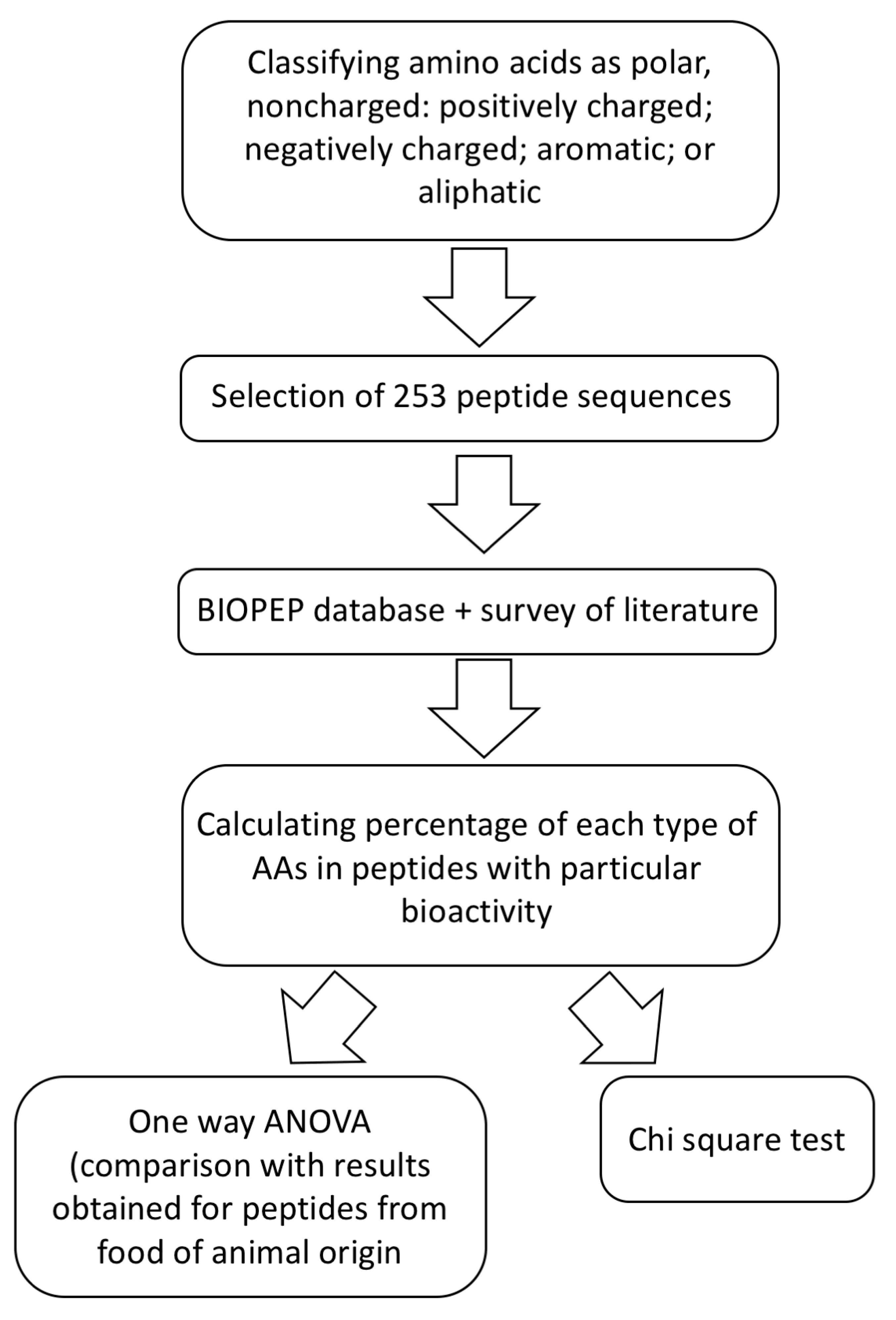
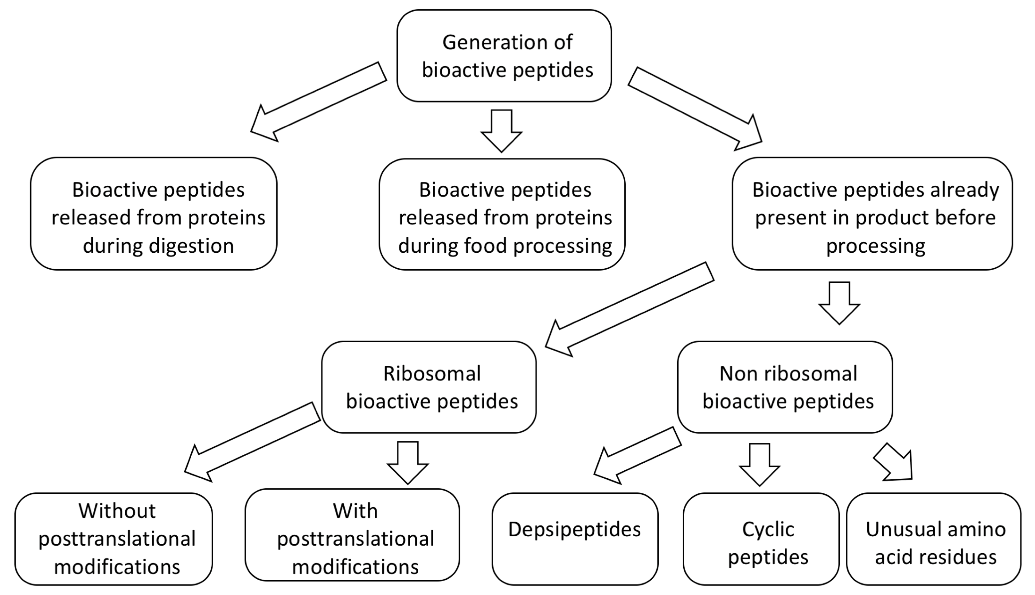
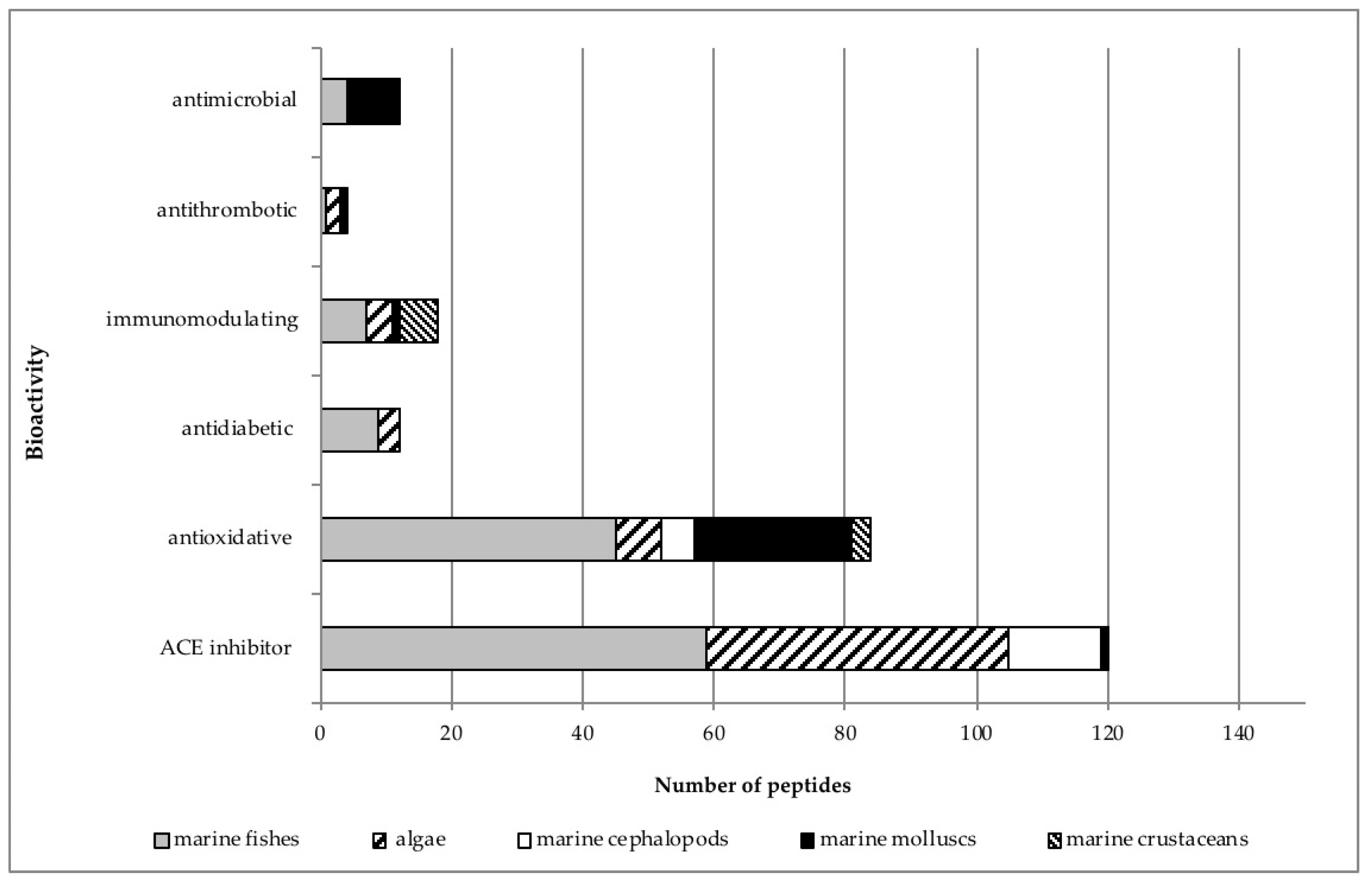
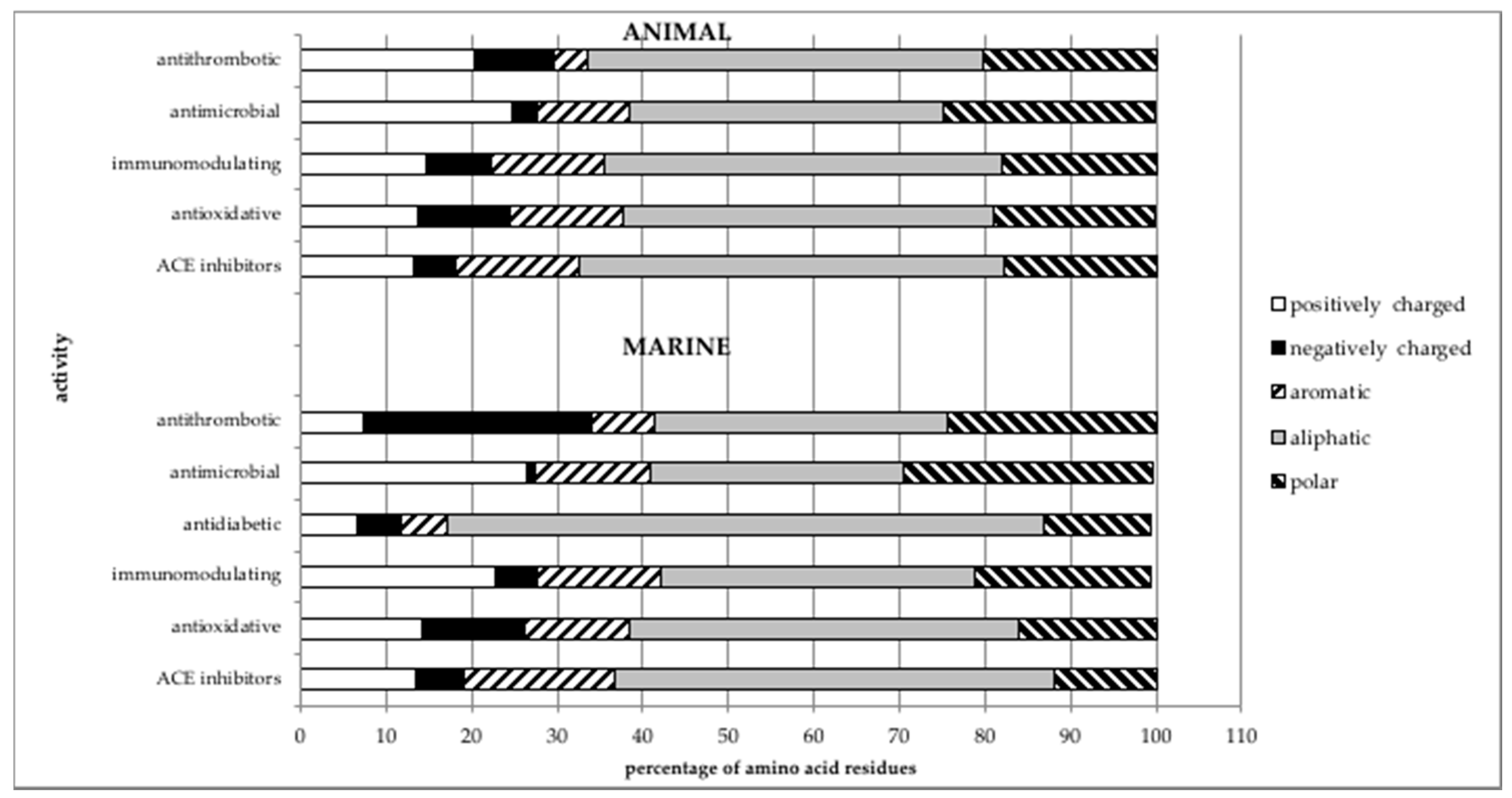
Table 1.
Commonly used proteases for production of marine-derived peptides.
| Protease/Proteases Combination | References |
|---|---|
| Alcalase | [39,40,41] |
| Bromelain | [7,42,43] |
| Proteinase K + thermolysin | [44,45,46] |
| Thermolysin | [47,48,49,50] |
| Pepsin | [51,52,53] |
| Proteases | [54,55,56] |
| Alcalase + pepsin + chymotrypsin | [57,58,59] |
| Neutrase | [53,60,61] |
| Papain | [62,63,64] |
| Neutral protease + papain | [65] |
© 2020 by the authors. Licensee MDPI, Basel, Switzerland. This article is an open access article distributed under the terms and conditions of the Creative Commons Attribution (CC BY) license (http://creativecommons.org/licenses/by/4.0/).
Share and Cite
MDPI and ACS Style
Pavlicevic, M.; Maestri, E.; Marmiroli, M. Marine Bioactive Peptides—An Overview of Generation, Structure and Application with a Focus on Food Sources. Mar. Drugs 2020, 18, 424. https://doi.org/10.3390/md18080424
AMA Style
Pavlicevic M, Maestri E, Marmiroli M. Marine Bioactive Peptides—An Overview of Generation, Structure and Application with a Focus on Food Sources. Marine Drugs. 2020; 18(8):424. https://doi.org/10.3390/md18080424
Chicago/Turabian StylePavlicevic, Milica, Elena Maestri, and Marta Marmiroli. 2020. "Marine Bioactive Peptides—An Overview of Generation, Structure and Application with a Focus on Food Sources" Marine Drugs 18, no. 8: 424. https://doi.org/10.3390/md18080424
Note that from the first issue of 2016, this journal uses article numbers instead of page numbers. See further details here.