Article

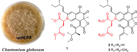
New Glutamine-Containing Azaphilone Alkaloids from Deep-Sea-Derived Fungus Chaetomium globosum HDN151398

1
Key Laboratory of Marine Drugs, Chinese Ministry of Education, School of Medicine and Pharmacy, Ocean University of China, Qingdao 266003, China
2
Laboratory for Marine Drugs and Bioproducts of Qingdao National Laboratory for Marine Science and Technology, Qingdao 266237, China
3
Key Laboratory of Testing and Evaluation for Aquatic Product Safety and Quality, Ministry of Agriculture and Rural Affairs, P. R. China; Yellow Sea Fisheries Research Institute, Chinese Academy of Fishery Sciences, Qingdao 266071, China
4
Open Studio for Druggability Research of Marine Natural Products, Pilot National Laboratory for Marine Science and Technology, Qingdao 266237, China
*
Authors to whom correspondence should be addressed.
Mar. Drugs 2019, 17(5), 253; https://doi.org/10.3390/md17050253
Submission received: 2 April 2019 / Revised: 18 April 2019 / Accepted: 25 April 2019 / Published: 28 April 2019
(This article belongs to the Special Issue Deep-Sea Natural Products II)

Abstract

:
Three new azaphilone alkaloids containing glutamine residues, namely N-glutarylchaetoviridins A–C (13), together with two related compounds (4 and 5) were isolated from the extract of Chaetomium globosum HDN151398, a fungus isolated from a deep-sea sediment sample collected in South China Sea. Their structures were elucidated on the basis of extensive 1D and 2D NMR as well as HRESIMS spectroscopic data and chemical analysis. N-glutarylchaetoviridins A–C (13) represent the first class of chaetoviridins characterized by embedded glutamate residues. Amino acids incubation experiments produced five azaphilone laden different amino acids residues (610) which indicated that this method can enhanced the structural diversity of this strain by culturing with amino acids. Cytotoxicity of the isolated compounds were evaluated against a panel of human cancer cell lines.

Graphical Abstract

1. Introduction

Deep-sea-derived microorganisms have proven to be a prolific source of secondary metabolites with an ample variety of captivating chemical structures and diverse pharmacological properties [1,2]. In our recent search for bioactive secondary metabolites from marine-derived fungi, a fungal strain Chaetomium globosum HDN151398, isolated from a deep-sea sediment sample (depth 2476 m) collected from South China Sea, was selected for its intriguing HPLC-UV profile (Figure S1) and significant crude extract cytotoxic activity (69% inhibition of K562 cells at the concentration of 100 μg/mL). Chemical investigation of the organic extract of the fungus led to the isolation of three new glutamine-containing azaphilone alkaloids, N-glutarylchaetoviridins A–C (13) together with two known chaetoviridins (4 and 5).
Azaphilones are a family of structurally erratic fungal pigmented polyketides with pyrone-quinone structures containing a highly oxygenated bicyclic core and a chiral quaternary center [3,4]. The oxygen atom in the pyran chromophore of the azaphilones could be biosynthetically replaced by nitrogen atom in the presence of primary amines and the colour of the pigment shifted to red accordingly [4]. Recently, azaphilones have been recognized as a unique family of secondary metabolites with diverse bioactivities including antimicrobial [5], cytotoxic [6], anti-inflammatory [7] and other activities [8,9,10], which have provoked enormous attention of scientists for biosynthesis [11] and chemical synthesis studies [12]. In the present work, we report the isolation, structure elucidation and biological activities of the previously unreported azaphilones (13) (Figure 1) from the strain Chaetomium globosum HDN151398 as well as the incubation experiments with different amino acids to produce more diverse analogues.

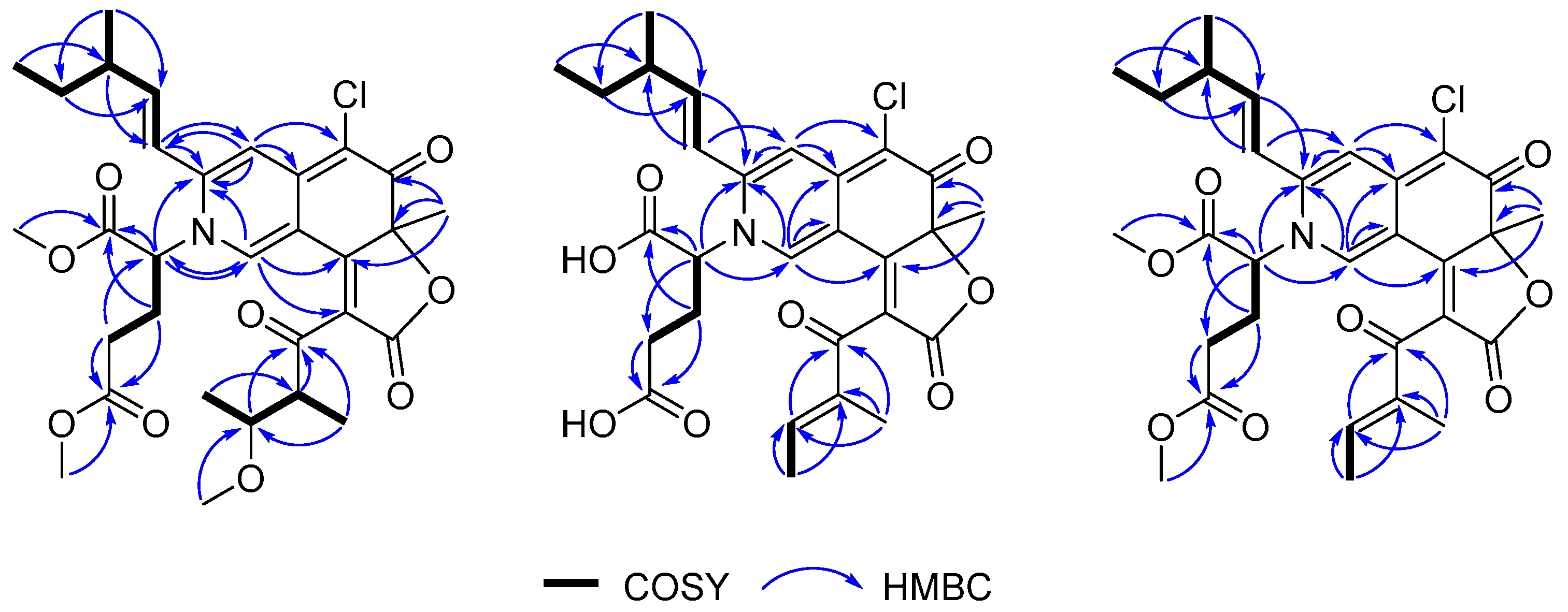
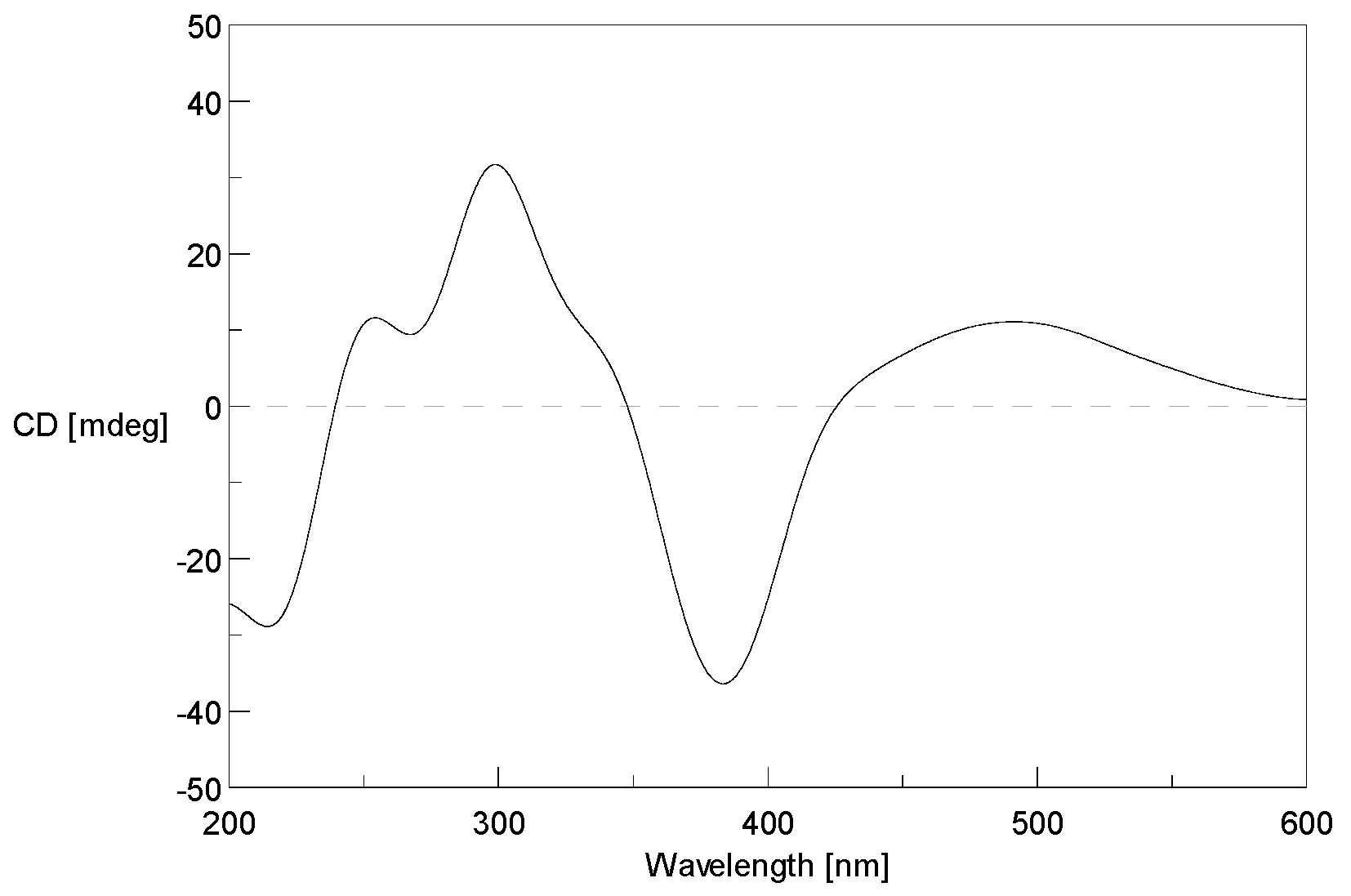
2. Results and Discussion

Compound 1 was isolated as a dark red powder with the molecular formula C31H38O9NCl determined by the (+)-HRESIMS peak at m/z 604.2308 [M + H]+ (calcd. for C31H39O9NCl, 604.2308), indicating 13 degrees of unsaturation. An isotopic peak ratio of 3:1 for [M + H]+:[M + H + 2]+ was observed, indicating the presence of a single chlorine atom in the molecule. The infrared (IR) absorption at 1717 cm−1 indicated the presence of the carbonyl functionality. The 1H NMR data (Table 1) of 1 showed eight methyls [δH 0.95 (3H, t, J = 7.4 Hz, H-13), 1.13 (3H, d, J = 6.7 Hz, H-14), 1.73 (3H, s, H-15), 3.72 (3H, s, H-6′), 3.83 (3H, s, H-7′), 1.04 (3H, d, J = 6.3 Hz, H-6″), 1.07 (3H, d, J = 6.8 Hz, H-7″), 2.93 (3H, s, H-8″)], and four olefinic protons [δH 8.48 (1H, s, H-1), 6.84 (1H, s, H-4), 6.25 (1H, d, J = 15.4 Hz, H-9), 6.40 (1H, dd, J = 7.5, 15.4 Hz, H-10)]. The 13C NMR data (Table 1), assigned by the aid of DEPT and HSQC spectra, displayed the resonances of eight methyl (δC 8.4, 11.7, 16.1, 16.1, 26.6, 52.1, 53.6, 56.2), three methylene (δC 27.0, 29.0, 29.2), eight methine (δC 39.0, 49.3, 61.9, 77.7, 111.1, 119.0, 136.9, 150.3), and twelve nonprotonated carbons (δC 88.5, 101.2, 112.4, 125.0, 144.5, 148.4, 165.4, 167.9, 168.4, 172.2, 182.1, 199.3). Careful comparison of the 13C NMR data of 1 and those of chaetoviridin A [13] revealed that they share a similar pyrone-quinone-containing skeleton. The main differences between 1 and chaetoviridin A were the chemical shifts at C-1 (δC 136.9 versus δC 151.5) and C-3 (δC 148.4 versus δC 157.1), and a group of extra resonances in 1 which were attributed to a methylated glutamic acid moiety. The pyrone-quinone core structure was further verified by the 1H-1H COSY cross peaks from H-6″ to H-5″, from H-5″ to H-4″, from H-4″ to H-7″, from H-13 to H-12, from H-12 to H-11, from H-11 to H-10, from H-10 to H-9 and from H-14 to H-11 as well as HMBC correlations from H-1 to C-8 and C-3, from H-9 to C-3 and C-4, from H-4 to C-5 and C-4a, from H-15 to C-6, C-7, and C-8 and from H-4″ to C-3″and C-2″(Figure 2). The methylated glutamic acid moiety was deduced by the COSY correlations from H-2′ to H-3′and from H-3′ to H-4′ as well as the HMBC correlations from H-6′ and H-4′ to C-5′, from H-7′ and H-2′ to C-1′. Based on the variation between the chemical shifts at C-1 and C-3 and taking the molecular formula into account, a nitrogen atom, instead of an oxygen atom, was placed in position 2. Further HMBC cross peaks from H-2′ to C-1 (δ 136.9) and C-3 (δ 148.4) (Figure 2) attached the dimethylglutarate moiety to N-2 of 1,4-hydropyridine-quinone scaffold moiety. As this compound has never been previously reported, it was named N-glutarylchaetoviridin A.
The geometrical configuration of the double bond between C-9 and C-10 was inferred to be trans from the coupling constants of the olefinic protons (J9,10 = 15.5 Hz). The relative configuration of 1 was determined based on a combination of NOESY correlations and comparison of its NMR data with those of chaetoviridin A. The stereochemistry of 1 established based on the NOESY correlations from H-1 to H-4″, from H-1 to H-6″, and from H-8″ to H-7″, the similar electronic circular dichroism (ECD) curves of 1 (Figure 3) and chaetoviridin A [13,14], and the co-isolation of biogenetically related compounds 4 and 5 which also shared the same chiral centres. The absolute configuration of C-7 and C-11 were further confirmed by Steyn and Vleggaar’s method [15] and the degradation of 1 [16], respectively. The ECD spectrum of 1ε387 −10.1; Figure 3) revealed that the absolute configuration of C-7 is S according to Steyn and Vleggaar’s CD method [15]. Compound 1 was degraded by 5% potassium hydroxide to afford a carboxylic acid (Figure 4) which was identified as (4S)-2E-4-methylhex-2-enoic acid by comparison of spectral data and specific optical rotation with the authentic sample. The configuration of the L-glutamate moiety in 1 was determined by the advanced Marfey’s method [17] by comparison of the retention time and mass data of the hydrolysis product with those of d/l-glutamate standards by HPLC (Figure S30). Accordingly, the absolute configuration of 1 was concluded to be 7S, 11S, 2′S, 4″S, and 5″R, respectively.
Compound 2 was isolated as a dark red powder with the molecular formula C28H30O8NCl determined by the (+)-HRESIMS m/z 544.1731 [M + H]+ (calcd. for C28H31O8NCl, 544.1733), requiring 14 degrees of unsaturation. The isotopic peak [M + H]+:[M + H + 2]+=3:1 was observed, indicating the presence of a single chlorine atom in the molecule. The IR spectrum displayed absorption bands for carbonyl (1684 and 1761 cm−1) functionalities. The 1H NMR data (Table 1) of 2 showed five methyls [δH 0.96 (3H, t, J = 7.5 Hz, H-13), 1.13 (3H, d, J = 6.7 Hz, H-14), 1.69 (3H, s, H-15), 1.89 (3H, d, J = 7.0 Hz, H-6″), 1.87 (3H, s, H-7″)], and five olefinic protons [δH 7.76 (1H, s, H-1), 6.90 (1H, s, H-4), 6.46 (1H, d, J = 14.2 Hz, H-9), 6.41 (1H, m, H-10), 6.64 (1H, q, J = 6.9 Hz, H-5″)]. The 13C NMR (Table 2), in combination with DEPT and HSQC spectra, displayed the resonances of five methyl (δC 9.2, 10.6, 14.0, 17.9, 25.0), three methylene (δC 26.8, 28.6, 29.2), seven methine (δC 38.9, 62.7, 110.6, 119.7, 136.3, 146.7, 150.1), thirteen quaternary (δC 88.3, 99.4, 112.4, 124.4, 138.0, 145.9, 150.4, 161.6, 168.3, 169.4, 173.8, 181.8, 190.6) carbons. The 1H and 13C NMR data of 2 were very similar to those of 1, while the main differences were the absence of three methyl and one proton signals (δH 3.83, 3.72, 2.93, and 3.71, respectively) and the downfield shift of H-7″ (δ 1.87), H-6″ (δ 1.89) and H-5″ (δ 6.64), suggesting that the single bond between C-4″ (δ 138.0) and C-5″ (δ 146.7) was oxidized to a double bond. This postulation was confirmed by COSY correlations from H-5″ to H-6″ and HMBC cross peaks from H-7″to C-3″and C-4″, and from H-5″ to C-3″. Further 2D NMR analysis (Figure 2) verified the planar structure as shown in Figure 1 and we named it N-glutarylchaetoviridin B.
The coupling constants of the olefinic protons (J9,10 = 14.2 Hz) indicated the trans configuration of the double bond (Δ9,10). The NOESY correlations between H-6″/H-7″ demonstrated that the double bond between C-4″ and C-5″ was in E configuration. The amino acid residue in 2 was identified as l-glutamate by the advanced Marfey’s method [17]. The CD spectrum of 2 (Δε387 −10.8; Figure 5) revealed the 7S absolute configuration according to Steyn and Vleggaar’s CD method [15]. The absolute configuration at C-11 was determined as S by the degradation of 2. Thus, the absolute configurations of C-7, 11 and 2′ of 2 were assigned as 7S, 11S, and 2′S.
Compound 3 was isolated as a dark red powder with the molecular formula C30H34O8NCl determined by the (+)-HRESIMS m/z 572.2050 [M + H]+ (calcd. for C30H35O8NCl, 572.2046), indicating 14 degrees of unsaturation. Comparison of the 1D NMR data of 3 with those of 2, it was found that there are two methoxy groups [δH 3.67 (3H, s, H-6′), 3.77 (3H, s, H-7′); δC 52.1, 53.4] in 3. Mass spectrometric data as well as the key HMBC correlations from H-6′ (δ 3.67) to C-5′ (δ 172.1) and H-7′ (δ 3.77) to C-1′ (δ 168.3) confirmed that 3 is a 6′,7′-dimethoxyl analogue of 2, and was named N-glutarylchaetoviridin C. As the ECD curve of 3 is very similar to that of 2 (Figure 5) it was concluded that 3 has the same stereochemistry as 2.
Two previously reported compounds, chaetomugilin A (4) [18] and chaetomugilin C (5) [18] were also isolated and their identity was proved by comparison of their NMR and MS data with those reported in the literature.
Compounds 15 were evaluated for their cytotoxic activity against twelve human cancer cell lines including human hepatocellular carcinoma cell line (BEL-7402), human colon cancer cell line (HCT-116), human cervix cancer cell line (HeLa), human hepatic cancer cell line (L-02), human gastric cancer cell line (MGC-803), human ovarian cancer cell line (HO8910), human neuroblastoma cell line (SH-SY5Y), human lung adenocarcinoma cell line (NCI-H1975), human glioblastoma cell line (U87), human breast cancer cell line (MDA-MB-231), human kidney cancer cell line (K562) and human promyelocytic leukemia cell line (HL-60) (Table S1). Compounds 3, 4, and 5 showed a broad spectrum of cytotoxic activity. Among them, 3 showed significant cytotoxic activity against MGC-803 and HO8910 with IC50 values of 6.6 and 9.7 µM, respectively.
Inspired by the fact that azaphilones have a capacity to incorporate amino acids, five different amino acids (l-tryptophan, l-tyrosine, l-histidine, l-alanine, l-glycine) were added to the culture medium in order to produce more diverse analogues. All the molecular ion peaks of the proposed structures could be easily detected by LC-MS (Figure 6). Furthermore, the structures of 610 (Figure 7) were further confirmed by both (+)-HRESIMS and NMR data (Table 3, Figures S35–S44). Consequently, these results further validate the property of azaphilones to combine with amino acids and to produce more diverse compounds.

3. Materials and Methods

3.1. General Experimental Procedures

Optical rotations were obtained on a JASCO P-1020 (JASCO Corporation, Tokyo, Japan) digital polarimeter. UV spectra were recorded on Waters 2487 (Waters Corporation, Milford, MA, USA), while the ECD spectrum were recorded on JASCO J-815 spectropolarimeter (JASCO Corporation, Tokyo, Japan). 1H NMR, 13C NMR, DEPT and 2D NMR spectra were recorded on an Agilent 500 MHz DD2 spectrometer (Agilent Technologies Inc., Santa Clara, CA, USA). HRESIMS and ESIMS spectra were obtained using a Thermo Scientific LTQ Orbitrap XL mass spectrometer (Thermo Fisher Scientific, Waltham, MA, USA) on positive ionisation mode. Column chromatography (CC) was performed with silica gel (200-300 mesh, Qingdao Marine Chemical Inc., Qingdao, China) and Sephadex LH-20 (Amersham Biosciences, San Francisco, CA, USA). MPLC was performed on a Bona-Agela CHEETAHTM HP100 (Beijing Agela Technologies Co., Ltd., Beijing, China). RP-HPLC was performed on an ODS column (HPLC (YMC-Pack ODS-A, 10 × 250 mm, 5 µm, 3 mL/min)) (YMC Co., Ltd., Kyoto, Japan). LC-MS was performed using an Acquity UPLC H-Class coupled to a SQ Detector 2 mass spectrometer using a BEH C18 column (1.7 μm, 2.1 × 50 mm, 1mL/min) (Waters Corporation, Milford, MA, USA).

3.2. Fungal Material

The fungal strain was isolated from the sediment sample collected from South China Sea (depth 2476 m, E 117.3957°, N 19.9778°, collected in May, 2017) and identified as Chaetomium globosum based on sequencing of the ITS region (GenBank no. MH828376) with 100% similarity. The strain was deposited at the Key Laboratory of Marine Drugs, the Ministry of Education of China, School of Medicine and Pharmacy, Ocean University of China, Qingdao, China.

3.3. Fermentation

The fungus was cultured under static condition at room temperature for 30 days in 1 L Erlenmeyer flasks each containing 300 mL of liquid culture medium, composed of glucose (1%), maltose (2%), mannitol (2%), monosodium glutamate (1%), KH2PO4 (0.05%), MgSO4·7H2O (0.03%), corn steep liquor (0.1%) and yeast extract (0.3%) after adjusting its pH to 6.5 in natural sea water (collected from JiaoZhou Bay, Qingdao, China).

3.4. Isolation

The whole fermentation broth (40 L) was filtered through muslin cloth to separate the supernatant from the mycelia. The supernatant was extracted with EtOAc (3 × 40 L), and the mycelia were homogenized and extracted with MeOH (3 × 10 L). The EtOAc and MeOH solutions of the supernatant and mycelia were combined and evaporated under reduced pressure to give a crude. The extract (30.0 g) was fractioned by VLC of silica gel using a step gradient elution DCM-MeOH (100:0 to 0:100) to give ten fractions (Fr.1 to Fr.10). Fr.6 was further fractioned by MPLC (C-18 ODS) using a step gradient elution of MeOH-H2O (5:95 to 100:0) to yield 12 subfractions (Fr.6-1 to Fr.6-12). Fr.6-3, Fr.6-4 and Fr.6-5 were further fractioned by a Sephadex LH-20 column with MeOH to provide five subfractions (Fr.6-3-1 to Fr.6-3-5), six fractions (Fr.6-4-1 to Fr.6-4-6) and four fractions (Fr.6-5-1 to Fr.6-5-4) respectively. Fr.6-4-4, Fr.6-3-3 and Fr.6-5-3 were separated by semi-preparative HPLC eluted with MeOH-H2O (65:35) to obtain 1 (9.7 mg, tR = 32 min) and 4 (10.0 mg, tR = 34 min), MeOH-H2O (45:55) to obtain 2 (10.0 mg, tR = 40 min), and MeOH-H2O (70:30) to obtain 5 (15.0 mg, tR = 26 min) and 3 (35.0 mg, tR = 27 min), respectively.

3.5. Absolute Configuration of Amino Acids

Compounds 13 were hydrolyzed in 6 N HCl at 60 °C overnight. The solution was dried under a stream of N2 and dissolved in H2O (100 μL). The acid hydrolysates of 13 were dissolved in H2O (50 μL) separately, and then 0.25 μM FDAA in 100 μL of acetone was added, followed by 1 N NaHCO3 (25 μL). The mixtures were heated for 1 h at 43 °C. After cooling to room temperature, the reaction was quenched by the addition of 2 N HCl (25 μL). Finally, the resulting solution was filtered through a small 4.5 μm filter and stored in the freezer until ready for HPLC analysis. Amino acid standards were derivatized with FDAA in a similar manner. The resulting FDAA derivatives of compounds 13, l- and d-glutamate were separately analyzed by reversed-phase HPLC (5 × 250 mm YMC C18 column, 5 μm, with a linear gradient of MeCN (A) and 0.05% aqueous TFA (B) from 5% to 55% A over 55 min at a flow rate of 1 mL/min, UV detection at 320 nm). Each chromatographic peak was identified by comparing its retention time with the FDAA derivatives of the l- and d- amino acid standards. The standards gave the following retention times (in min): 40.20 for l-FDAA, 38.61 for d-FDAA, 41.32 for l-Me-FDAA, 39.41 for d-Me-FDAA. The analysis gave retention time (in min) of 41.32, 40.20, and 41.32 (Figures S30 and S31), establishing the S configuration for all the glutamic acid residues [17,19].
N-glutarylchaetoviridin A (1): dark red powder; [α] D 20 +50 (c 0.03, MeOH); IR (KBr) νmax 3724, 3649, 2924, 2361, 1717, 1652, 1196, 1027, 669 cm−1; UV (MeOH) λmax (log ε): 215 (3.25), 299 (2.10), 389 (2.04) nm; ECD (2.5 mM, MeOH) λmax (Δε) 240 (+9.26), 300 (+9.33), 390 (−10.10), 490 (+3.56) nm; 1H and 13C NMR data see Table 1 and Table 2; (+)-HRESIMS m/z 604.2308 [M + H]+ (calcd. for C31H39O9NCl, 604.2308).
N-glutarylchaetoviridin B (2): dark red powder; [α] D 20 +332 (c 0.09, MeOH); IR (KBr) νmax 3751, 3420, 1761, 1684, 1485, 1190, 1020, 723 cm−1; UV (MeOH) λmax (log ε): 238 (3.20), 298 (2.04), 391 (1.98) nm; ECD (2.5 mM, MeOH) λmax (Δε) 220 (+9.24), 245 (+4.32), 300 (+10.31), 390 (−10.79), 490 (+3.96) nm; 1H and 13C NMR data see Table 1 and Table 2; (+)-HRESIMS m/z 544.1731 [M + H]+ (calcd. for C28H31O8NCl, 544.1733).
N-glutarylchaetoviridin C (3): dark red powder; [α] D 20 +456 (c 0.07, MeOH); IR (KBr) νmax 3676, 2960, 2362, 1759, 1605, 1489, 1193, 1018, 705 cm−1; UV (MeOH) λmax (log ε): 230 (3.15), 295 (2.02), 393 (2.01) nm; ECD (2.5 mM, MeOH) λmax (Δε) 220 (+9.24), 245 (+4.12), 300 (+10.25), 390 (−10.66), 490 (+3.62) nm; 1H and 13C NMR data see Table 1 and Table 2; (+)-HRESIMS m/z 572.2050 [M + H]+ (calcd. for C30H35O8NCl, 572.2046).

3.6. Degradation of 1–3 by Potassium Hydroxide

Compounds 13 (3.0 mg) were separately dissolved in 5% aq. potassium hydroxide (5 mL) and the reaction mixture was stirred for 3 h at 100 °C. Then, the reaction mixture was extracted with CHCl3 (5 mL). The water layer was adjusted to pH 3.0 with 9% sulfuric acid and re-extracted with petroleum ether (5 mL). The organic extract was concentrated to dryness in vacuo. The residue was purified by HPLC using MeCN–H2O gradient (30:70 to 100:0 in 45 min) as the eluent to afford (4S)-2E-4-methylhex-2-enoic acid (0.1 mg, tR = 15 min). The physicochemical properties of this carboxylic acid were identical to the authentic sample [16].

3.7. Cytotoxicity Assay

Cytotoxic activity of 15 were evaluated against BEL-7402, HCT-116, HeLa, L-02, MGC-803, HO8910, SH-SY5Y, NCI-H1975, U87, MDA-MB-231 cancer cells by SRB method, K562 and HL-60 by MTT method using adriamycin (ADM) as a positive control. The detailed methodologies for biological testing have been described in our previous report [20]. All of the experiments were carried out in triplicate.

3.8. Amino Acid Incubation Experiment

The fungus was cultured and subjected to a large-scale fermentation under the same protocol, stated above in the fermentation section. The only difference is that monosodium glutamate was replaced by five different amino acids. Dried extracts were dissolved in 1 mL of MeOH and analyzed by UPLC-MS (MeCN-H2O, 1 mL/min; 0–15 min, 5%–95%; 15–18 min, 100%; 18–20 min, 5%) and further separated by semi-preparative HPLC, eluted with MeCN-H2O (40:60) to obtain 6 (2.5 mg, tR = 24 min), 7 (2.0 mg, tR = 30 min) and 8 (1.0 mg, tR = 32 min), MeCN-H2O (30:70) to obtain 9 (2.5 mg, tR = 18 min), and MeCN-H2O (25:75) to obtain 10 (1.5 mg, tR = 22 min), respectively.

4. Conclusions

In summary, a series of azaphilones (15) were isolated from the deep-sea-derived fungus C. globosum HDN151398. Distinguished from the previously reported azaphilone derivatives, 13 belong to a new class of chaetoviridins which is linked to a glutamate residue, indicating that unique geographical features of deep-sea environment may promote the unique biogenetic and metabolic pathways of the microorganisms in which they inhabit. Compounds 3, 4, and 5 showed a broad spectrum of cytotoxicity, among which, 3 was active against MGC-803 and HO8910 with the IC50 values of 6.6 and 9.7 µM, respectively. Amino acids feeding experiment showed that it is an effective method to increase structural diversity of azaphilones.

Supplementary Materials

The following are available online at https://www.mdpi.com/1660-3397/17/5/253/s1, Figure S1: HPLC analysis of the crude of HDN151398; Figure S2: The 18S rRNA sequences data of HDN151398; Figures S3–S32: 1D and 2D NMR spectra, IR spectra, HRESIMS spectra, and UV spectra of 13; Figures S33–S34: HPLC analysis of the FDAA derivatives of the compounds 13 and l-Me-glutamate and d-Me-glutamate; Table S1: Cytotoxicities of compounds 15 against twelve cancer cell lines; Figures S35–S44: 1H NMR spectra and HRESIMS spectra of 610.

Author Contributions

The contributions of the respective authors are as follows: C.S. drafted the work. C.S., X.G., S.M., and L.Z. performed the fermentation, extraction, isolation, and structural elucidation of the constituents. J.P. was performed the biological evaluations. G.Y., Q.C., Q.G., T.Z., G.Z., and D.L. contributed to checking and confirming all of the procedures of the isolation and the structural elucidation. D.L. and T.Z. designed the study, supervised the laboratory work, and contributed to the critical reading of the manuscript. All the authors have read the final manuscript and approved the submission.

Funding

This work was financially supported by the Scientific and Technological Innovation Project Financially Supported by Qingdao National Laboratory for Marine Science and Technology (No.2016ASKJ05-04, 2016ASKJ08-02), the National Natural Science Foundation of China (No.41606166), National Science and Technology Major Project for Significant New Drugs Development(2018ZX09735004), the Shandong Provincial Natural Science Fund (No. ZR2016BQ37), the NSFC-Shandong Joint Fund for Marine Science Research Centers (No. U1606403), the Fundamental Research Funds for the Central Universities (201941001), the Marine S&T Fund of Shandong Province for Pilot National Laboratory for Marine Science and Technology (Qingdao) (No. 2018SDKJ0401-2), Taishan Scholar Youth Expert Program in Shandong Province (tsqn201812021), China Postdoctoral Science Foundation (2017M622286) and Qingdao Postdoctoral Applied Research Project Financially Supported by Qingdao Municipal Bureau of Human Resource and Social Security.

Conflicts of Interest

The authors declare no conflict of interest.

References

  1. Wang, Y.T.; Xue, Y.R.; Liu, C.H. A Brief Review of Bioactive Metabolites Derived from Deep-Sea Fungi. Mar. Drugs 2015, 13, 4594–4616. [Google Scholar] [CrossRef] [PubMed] [Green Version]
  2. Tortorella, E.; Tedesco, P.; Palma Esposito, F.; January, G.G.; Fani, R.; Jaspars, M.; de Pascale, D. Antibiotics from Deep-Sea Microorganisms: Current Discoveries and Perspectives. Mar. Drugs 2018, 16, 355. [Google Scholar] [CrossRef]
  3. Luo, X.; Lin, X.; Tao, H.; Wang, J.; Li, J.; Yang, B.; Zhou, X.; Liu, Y. Isochromophilones A-F, Cytotoxic Chloroazaphilones from the Marine Mangrove Endophytic Fungus Diaporthe sp. SCSIO 41011. J. Nat. Prod. 2018, 81, 934–941. [Google Scholar] [CrossRef]
  4. Gao, J.M.; Yang, S.X.; Qin, J.C. Azaphilones: chemistry and biology. Chem. Rev. 2013, 113, 4755–4811. [Google Scholar] [CrossRef]
  5. Chen, M.; Shen, N.X.; Chen, Z.Q.; Zhang, F.M.; Chen, Y. Penicilones A-D, Anti-MRSA Azaphilones from the Marine-Derived Fungus Penicillium janthinellum HK1-6. J. Nat. Prod. 2017, 80, 1081–1086. [Google Scholar] [CrossRef] [PubMed]
  6. Li, X.; Tian, Y.; Yang, S.X.; Zhang, Y.M.; Qin, J.C. Cytotoxic azaphilone alkaloids from Chaetomium globosum TY1. Bioorg. Med. Chem. Lett. 2013, 23, 2945–2947. [Google Scholar] [CrossRef] [PubMed]
  7. Wang, T.H.; Lin, T.F. Monascus Rice Products. Adv. Food Nutr. Res. 2007, 53, 123–159. [Google Scholar] [PubMed]
  8. Nam, J.Y.; Kim, H.K.; Kwon, J.Y.; Han, M.Y.; Son, K.H.; Lee, U.C.; Choi, J.D.; Kwon, B.M. 8-O-Methylsclerotiorinamine, antagonist of the Grb2-SH2 domain, isolated from Penicillium multicolor. J. Nat. Prod. 2000, 63, 1303–1305. [Google Scholar] [CrossRef] [PubMed]
  9. Clark, R.C.; Lee, S.Y.; Searcey, M.; Boger, D.L. The isolation, total synthesis and structure elucidation of chlorofusin, a natural product inhibitor of the p53–MDM2 protein–protein interaction. Nat. Prod. Rep. 2009, 26, 465–477. [Google Scholar] [CrossRef] [PubMed]
  10. Musso, L.; Dallavalle, S.; Merlini, L.; Bava, A.; Nasini, G.; Penco, S.; Giannini, G.; Giommarelli, C.; De Cesare, A.; Zuco, V.; et al. Natural and semisynthetic azaphilones as a new scaffold for Hsp90 inhibitors. Bioorg. Med. Chem. 2010, 18, 6031–6043. [Google Scholar] [CrossRef] [PubMed]
  11. Bai, J.; Lu, Y.; Xu, Y.M.; Zhang, W.; Chen, M.; Lin, M.; Gunatilaka, A.A.; Xu, Y.; Molnar, I. Diversity-Oriented Combinatorial Biosynthesis of Hybrid Polyketide Scaffolds from Azaphilone and Benzenediol Lactone Biosynthons. Org. Lett. 2016, 18, 1262–1265. [Google Scholar] [CrossRef] [PubMed] [Green Version]
  12. Makrerougras, M.; Coffinier, R.; Oger, S.; Chevalier, A.; Sabot, C.; Franck, X. Total Synthesis and Structural Revision of Chaetoviridins, A. Org. Lett. 2017, 19, 4146–4149. [Google Scholar] [CrossRef] [PubMed]
  13. Takahashi, M.; Koyama, K.; Natori, S. Four new azaphilones from Chaetomium globosum var. flavo-viridae. Chem. Pharm. Bull. 1990, 38, 625–628. [Google Scholar] [CrossRef]
  14. Wang, W.; Liao, Y.; Chen, R.; Hou, Y.; Ke, W.; Zhang, B.; Gao, M.; Shao, Z.; Chen, J.; Li, F. Chlorinated Azaphilone Pigments with Antimicrobial and Cytotoxic Activities Isolated from the Deep Sea Derived Fungus Chaetomium sp. NA-S01-R1. Mar. Drugs 2018, 16, 61. [Google Scholar] [CrossRef] [PubMed]
  15. Steyn, P.S.; Vleggaar, R. The Structure of Dihydrodeoxy-8-epi-austdiol and the Absolute Configuration of the Azaphilones. J. Chem. Soc. Perkin Transactions 1976, 7, 204–206. [Google Scholar] [CrossRef]
  16. Yamada, T.; Muroga, Y.; Tanaka, R. New azaphilones, seco-chaetomugilins A and D, produced by a marine-fish-derived Chaetomium globosum. Mar. Drugs 2009, 7, 249–257. [Google Scholar] [CrossRef] [PubMed]
  17. Fujii, K.; Ikai, Y.; Mayumi, T.; Oka, H.; Suzuki, M.; Harada, K.I. A Nonempirical Method Using LC/MS for Determination of the Absolute Configuration of Constituent Amino Acids in a Peptide:  Elucidation of Limitations of Marfey’s Method and of Its Separation Mechanism. Anal. Chem. 1997, 69, 549–557. [Google Scholar] [CrossRef]
  18. Yamada, T.; Doi, M.; Shigeta, H.; Muroga, Y.; Hosoe, S.; Numata, A.; Tanaka, R. Absolute stereostructures of cytotoxic metabolites, chaetomugilins A–C, produced by a Chaetomium species separated from a marine fish. Tetrahedron Lett. 2008, 49, 4192–4195. [Google Scholar] [CrossRef]
  19. Peng, J.; Gao, H.; Zhang, X.; Wang, S.; Wu, C.; Gu, Q.; Guo, P.; Zhu, T.; Li, D. Psychrophilins E–H and Versicotide C, Cyclic Peptides from the Marine-Derived Fungus Aspergillus versicolor ZLN-60. J. Nat. Prod. 2014, 77, 2218–2223. [Google Scholar] [CrossRef] [PubMed]
  20. Zhang, Z.; He, X.; Liu, C.; Che, Q.; Zhu, T.; Gu, Q.; Li, D. Clindanones A and B and cladosporols F and G, polyketides from the deep-sea derived fungus Cladosporium cladosporioides HDN14-342. RSC Adv. 2016, 6, 76498–76504. [Google Scholar] [CrossRef]
Figure 1. Structures of 15.
Marinedrugs 17 00253 g001
Figure 2. Key HMBC and 1H-1H COSY correlations for 13.
Marinedrugs 17 00253 g002
Figure 3. Experimental electronic circular dichroism (ECD) spectrum of 1 in methanol.
Marinedrugs 17 00253 g003
Figure 4. Alkaline degradation of 13.
Marinedrugs 17 00253 g004
Figure 5. Experimental ECD spectra of 2 and 3 in methanol.
Marinedrugs 17 00253 g005
Figure 6. LC/MS analysis of the metabolic extracts from the HDN151398 by incubation with different amino acids.
Marinedrugs 17 00253 g006
Figure 7. Structures of 610.
Marinedrugs 17 00253 g007
Table 1. 1H NMR data for 13 at 500 MHz (δ in ppm, J in Hz).
No.1 a2 b3 a
18.48, s7.76, s7.81, s
46.84, s6.90, s6.72
96.25, d (15.4)6.46, d (14.2)6.17, d (15.4)
106.40, dd (7.5, 15.4)6.41, m6.32, dd (7.6, 15.4)
112.33, m2.35, m2.28, m
121.49, m1.51, m1.44, m
130.95, t (7.4)0.96, t (7.5)0.91, t (7.5)
141.13, d (6.7)1.13, d (6.7)1.08, d (6.7)
151.73, s1.69, s1.69, s
2′5.13, t (7.9)5.26, m5.05, t (7.3)
3′2.37, m2.29, m2.47, m
2.68, m2.57, m2.30, m
4′2.47, m2.44, t (6.6)2.42, m
2.57, m2.49, m2.56, m
6′3.72, s 3.67, s
7′3.83, s 3.77, s
4″3.71, m
5″3.56, m6.64, q (6.9)6.55, q (6.3)
6″1.04, d (6.3)1.89, d (7.0)1.87, d (6.9)
7″1.07, d (6.8)1.87, s1.83, s
8″2.93, s
a Measured in CDCl3; b Measured in methanol-d4.
Table 2. 13C NMR Data for 13 at 125 MHz (δ in ppm).
No.1 a2 b3 a
1136.9136.3135.4
2
3148.4150.4148.2
4111.1110.6110.7
4a144.5145.9143.8
5101.299.4101.6
6182.1181.8182.1
788.588.388.5
8165.4161.6162.9
8a112.4112.4112.3
9119.0119.7119.2
10150.3150.1149.9
1139.038.938.9
1229.028.629.0
1311.710.611.6
1416.117.919.0
1526.625.026.2
1′167.9169.4168.3
2′61.962.761.7
3′27.026.827.2
4′29.229.229.3
5′172.2173.8172.1
6′52.1 52.1
7′53.6 53.4
1″168.4168.3168.1
2″125.0124.4125.1
3″199.3190.6190.8
4″49.3138.0137.6
5″77.7146.7146.5
6″16.114.015.4
7″8.49.210.7
8″56.2
a Measured in CDCl3. b Measured in methanol-d4.
Table 3. 1H NMR data for 610 (δ in ppm, J in Hz).
No.6 b,d7 a,e8 a,e9 a,e10 a,e
19.08, s8.61, s8.53, s8.53, s8.99, s
46.87, s6.42, s6.49, s6.78, s6.58, s
96.26, d (15.0)6.14, d (12.8)6.28, d (12.8)6.40, d (15.6)6.34, d (15.7)
106.43, m6.01, dd (5.9, 12.8)6.14, m6.45, dd (7.1, 15.6)6.25, m
112.35, m2.06, m2.24, m2.26, m2.26, m
121.59, m1.26, m1.39, m1.39, m1.40, m
130.96, t (6.6)0.76, t (6.2)0.86, t (6.2)0.83, t (7.4)0.85, t (7.4)
141.14, ov. c0.98, d (5.6)0.89, d (5.0)1.02, d (6.7)1.03, d (6.7)
151.93, s1.43, s1.54, s1.56, s1.60, s
2′5.08, m5.53, dd (4.1, 8.5)5.49, dd (4.3, 8.9)4.97, d (18.1)5.48, t (7.7)
5.10, d (18.1)
3′1.50, t (6.3)3.48, m3.17, t (9.4) 3.49, m
3.68, dd (4.1, 12.8)3.47, dd (4.1,12.3)
4′
5′ 6.96, d (7.1) 7.34, s
6′ 7.49, d (6.6)6.58, d (7.1)
7′ 6.91, t (6.2) 7.41, s
8′ 7.03, t (6.1)6.58, d (7.1)
9′ 7.29, d (6.8)6.96, d (7.1)
11′ 10.94, s
12′ 7.14, d (1.9)
4″3.56, m3.56, m3.56, m3.49, m
5″3.81, m3.62, m3.61, m3.62, m6.65, d (6.8)
6″1.07, m0.89, d (5.2)0.98, d (5.4)0.92, d (6.2)1.82, d (6.8)
7″1.14, ov. c0.92, d (5.6)1.01, d (5.6)0.96, d (6.7)1.77, s
a Measured in DMSO-d6; b Measured in CDCl3; c ov.: overlapped signal; d Measured at 500 MHz; e Measured at 600 MHz.

