
Polyketides from the Mangrove-Derived Endophytic Fungus Nectria sp. HN001 and Their α-Glucosidase Inhibitory Activity

Abstract
:
1. Introduction
2. Results
3. Experimental Section
3.1. General
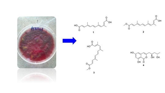
3.2. Fungal Material
3.3. Fermentation, Extraction, and Isolation
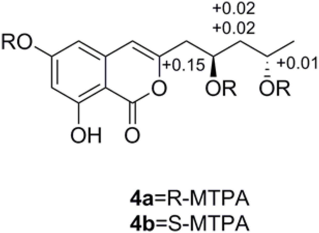
3.4. Preparation of (R)- and (S)-MTPA Esters of 4
3.5. In Vitro Inhibition Studies on α-Glucosidase
4. Conclusions
Acknowledgments
Author Contributions
Conflicts of Interest
References
- Kato, A.; Hayashi, E.; Miyauchi, S.; Adachi, I.; Imahori, T.; Natori, Y.; Yoshimura, Y.; Nash, R.J.; Shimaoka, H.; Nakagome, I.; et al. Alpha-1-C-butyl-1,4-dideoxy-1,4-imino-l-arabinitol as a second-generation iminosugar-based oral alpha-glucosidase inhibitor for improving postprandial hyperglycemia. J. Med. Chem. 2012, 55, 10347–10362. [Google Scholar] [CrossRef] [PubMed]
- Association, A.D. Diagnosis and classification of diabetes mellitus. Diabetes Care 2010, 33, 62–69. [Google Scholar] [CrossRef] [PubMed]
- International Diabetes Federation. IDF Diabetes Atlas, 7th ed. Revision 2015. Available online: http://www.Idf.Org/diabetesatlas/ (accessed on 1 December 2015).
- Zhang, H.W.; Song, Y.C.; Tan, R.X. Biology and chemistry of endophytes. Nat. Prod. Rep. 2006, 23, 753–771. [Google Scholar] [CrossRef] [PubMed]
- Ebel, R.; Rateb, M.E. Secondary metabolites of fungi from marine habitats. Nat. Prod. Rep. 2011, 28, 290. [Google Scholar]
- Li, H.; Jiang, J.; Liu, Z.; Lin, S.; Xia, G.; Xia, X.; Ding, B.; He, L.; Lu, Y.; She, Z. Peniphenones A–D from the mangrove fungus Penicillium dipodomyicola HN4–3A as inhibitors of Mycobacterium tuberculosis Phosphatase Mptpb. J. Nat. Prod. 2014, 77, 800–806. [Google Scholar] [CrossRef] [PubMed]
- Wen, L.; Cai, X.; Xu, F.; She, Z.; Chan, W.L.; Vrijmoed, L.; Jones, E.G.; Lin, Y. Three metabolites from the mangrove endophytic fungus Sporothrix sp.(# 4335) from the South China Sea. J. Org. Chem. 2009, 74, 1093–1098. [Google Scholar] [PubMed]
- Xiao, Z.; Huang, H.; Shao, C.; Xia, X.; Ma, L.; Huang, X.; Lu, Y.; Lin, Y.; Long, Y.; She, Z. Asperterpenols A and B, new sesterterpenoids isolated from a mangrove endophytic fungus Aspergillus sp. 085242. Org. Lett. 2013, 15, 2522–2525. [Google Scholar] [CrossRef] [PubMed]
- Huang, X.; Huang, H.; Li, H.; Sun, X.; Huang, H.; Lu, Y.; Lin, Y.; Long, Y.; She, Z. Asperterpenoid A, a new sesterterpenoid as an inhibitor of Mycobacterium tuberculosis protein tyrosine phosphatase B from the culture of Aspergillus sp. 16–5c. Org. Lett. 2013, 15, 721–723. [Google Scholar] [CrossRef] [PubMed]
- Liu, Y.; Yang, Q.; Xia, G.; Huang, H.; Li, H.; Ma, L.; Lu, Y.; He, L.; Xia, X.; She, Z. Polyketides with α-glucosidase inhibitory activity from a mangrove endophytic fungus Penicillium sp. Hn29–3b1. J. Nat. Prod. 2015, 78, 1816–1822. [Google Scholar] [CrossRef] [PubMed]
- Liu, Z.; Xia, G.; Chen, S.; Liu, Y.; Li, H.; She, Z. Eurothiocin A and B, sulfur-containing benzofurans from a soft coral-derived fungus Eurotium rubrum SH-823. Mar. Drugs 2014, 12, 3669–3680. [Google Scholar] [CrossRef] [PubMed]
- Xiao, Z.E.; Lin, S.E.; Tan, C.; Lu, Y.; He, L.; Huang, X.; She, Z. Asperlones A and B, dinaphthalenone derivatives from a mangrove endophytic fungus Aspergillus sp. 16–5C. Mar. Drugs 2015, 13, 366–378. [Google Scholar] [CrossRef] [PubMed]
- Nam, K.N.; Park, Y.M.; Jung, H.J.; Lee, J.Y.; Min, B.D.; Park, S.U.; Jung, W.S.; Cho, K.H.; Park, J.H.; Kang, I.; et al. Anti-inflammatory effects of crocin and crocetin in rat brain microglial cells. Eur. J. Pharmacol. 2010, 648, 110–116. [Google Scholar] [CrossRef] [PubMed]
- Mancini, A.; Serrano-Diaz, J.; Nava, E.; D’Alessandro, A.M.; Alonso, G.L.; Carmona, M.; Lorens, S. Crocetin, a carotenoid derived from saffron (Crocus Sativus L.), improves acetylcholine-induced vascular relaxation in hypertension. J. Vasc Res. 2014, 51, 393–404. [Google Scholar] [CrossRef] [PubMed]
- Bae, M.; Kim, H.; Shin, Y.; Kim, B.Y.; Lee, S.K.; Oh, K.B.; Shin, J.; Oh, D.C. Separacenes A–D, novel polyene polyols from the marine actinomycete, Streptomyces sp. Mar. Drugs 2013, 11, 2882–2893. [Google Scholar] [CrossRef] [PubMed]
- Kundim, B.A.; Itou, Y.; Sakagami, Y.; Fudou, R.; Yamanaka, S.; Ojika, M. Novel antifungal polyene amides from the myxobacterium Cystobacter fuscus: Isolation, antifungal activity and absolute structure determination. Tetrahedron 2004, 60, 10217–10221. [Google Scholar] [CrossRef]
- Klingner, A.; Bothe, H.; Wray, V.; Marner, F.-J. Identification of a yellow pigment formed in maize roots upon mycorrhizal colonization. Phytochemistry 1995, 38, 53–55. [Google Scholar] [CrossRef]
- Liu, D.; Li, X.M.; Meng, L.; Li, C.S.; Gao, S.S.; Shang, Z.; Proksch, P.; Huang, C.G.; Wang, B.G. Nigerapyrones A–H, alpha-pyrone derivatives from the marine mangrove-derived endophytic fungus Aspergillus niger MA-132. J. Nat. Prod. 2011, 74, 1787–1791. [Google Scholar] [CrossRef] [PubMed]
- Ola, A.; Thomy, D.; Lai, D.; Oesterhelt, H.; Proksch, P. Inducing secondary metabolite production by the endophytic fungus Fusarium tricinctum through coculture with Bacillus subtilis. J. Nat. Prod. 2013, 76, 2094–2099. [Google Scholar] [CrossRef] [PubMed]
- Seco, J.M.; Quinoá, E.; Riguera, R. The assignment of absolute configuration by NMR. Chem. Rev. 2004, 104, 17–118. [Google Scholar] [CrossRef]
- Seco, J.M.; Martino, M.; Quiñoá, E.; Riguera, R. Absolute configuration of 1,n-diols by NMR: The importance of the combined anisotropic effects in bis-arylmethoxyacetates. Org. Lett. 2000, 2, 3261–3264. [Google Scholar] [CrossRef] [PubMed]
- Li, R.; Chen, S.; Niu, S.; Guo, L.; Yin, J.; Che, Y. Exserolides A–F, new isocoumarin derivatives from the plant endophytic fungus Exserohilum sp. Fitoterapia 2014, 96, 88–94. [Google Scholar] [CrossRef] [PubMed]
- Cui, H.; Xu, B.; Wu, T.; Xu, J.; Yuan, Y.; Gu, Q. Potential antiviral lignans from the roots of Saururus chinensis with activity against Epstein–Barr virus lytic replication. J. Nat. Prod. 2014, 77, 100–110. [Google Scholar] [CrossRef] [PubMed]
- Parisot, D.; Devys, M.; Barbier, M. 6-O-demethyl-5-deoxyfusarubin and its anhydro derivative produced by a mutant of the fungus nectria haematococca blocked in fusarubin biosynthesis. J. Antibiot. 1991, 44, 103–107. [Google Scholar] [CrossRef] [PubMed]
- Gutiérrez, M.; Theoduloz, C.; Rodríguez, J.; Lolas, M.; Schmeda-Hirschmann, G. Bioactive metabolites from the fungus Nectria galligena, the main apple canker agent in chile. J. Agric. Food Chem. 2005, 53, 7701–7708. [Google Scholar] [CrossRef] [PubMed]
- Irvine, N.M.; Yerkes, C.N.; Graupner, P.R.; Roberts, R.E.; Hahn, D.R.; Pearce, C.; Gerwick, B.C. Synthesis and characterization of synthetic analogs of cinnacidin, a novel phytotoxin from Nectria sp. Pest. Manag. Sci. 2008, 64, 891–899. [Google Scholar] [CrossRef] [PubMed]
- Barber, M.; Hardisson, A.; Jackman, L.; Weedon, B. 316. Studies in nuclear magnetic resonance. Part IV. Stereochemistry of the bixins. J. Chem. Soc. 1961, 1625–1630. [Google Scholar] [CrossRef]
- Pfander, H.; Wittwer, F. Carotinoid-glykoside 2. Mitteilung untersuchungen zur carotinoid-zusammensetzung im safran. Helv. Chim. Acta 1975, 58, 1608–1620. [Google Scholar] [CrossRef] [PubMed]
- Eschenmoser, W.; Hans Eugster, C. Absolute konfiguration von azafrin. Helv. Chim. Acta 1975, 58, 1722–1727. [Google Scholar] [CrossRef]
- Schwenk, D.; Nett, M.; Dahse, H.M.; Horn, U.; Blanchette, R.A.; Hoffmeister, D. Injury-induced biosynthesis of methyl-branched polyene pigments in a white-rotting basidiomycete. J. Nat. Prod. 2014, 77, 2658–2663. [Google Scholar] [CrossRef] [PubMed]
- Lai, S.; Shizuri, Y.; Yamamura, S.; Kawai, K.; Furukawa, H. Three new phenolic metalolites from Penicillium species. Heterocycles 1991, 32, 297–305. [Google Scholar]


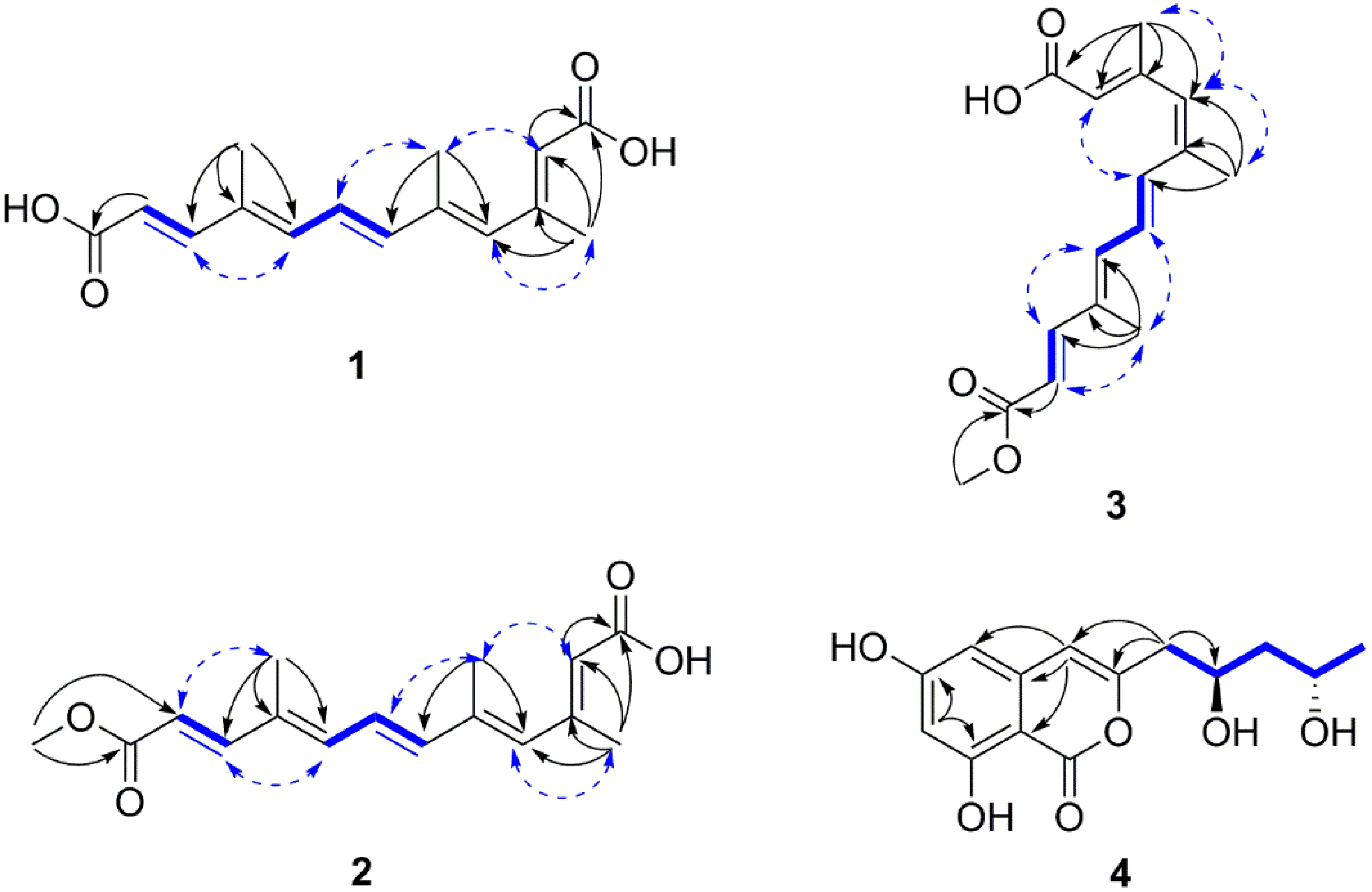
| Position | 1 | 2 | 3 | |||
|---|---|---|---|---|---|---|
| δH (J in Hz) | δC | δH (J in Hz) | δC | δH (J in Hz) | δC | |
| 1 | 167.7, C | 171.6, C | 170.9, C | |||
| 2 | 5.76, s | 120.2, CH | 5.78, s | 118.8, CH | 5.76, s | 118.9, CH |
| 3 | 151.6, C | 155.8, C | 155.7, C | |||
| 4 | 6.20, s | 136.1, CH | 6.08, s | 136.3, CH | 5.97, s | 134.3, CH |
| 5 | 138.5, C | 134.6, C | 137.7, C | |||
| 6 | 6.60, d (15.1) | 141.8, CH | 6.44, d (15.2) | 141.8, CH | 6.91, d (15.1) | 134.6, CH |
| 7 | 6.76, dd (15.1, 11.2) | 126.2, CH | 6.70, dd (15.2, 11.2) | 126.6, CH | 6.73, dd (15.1,11.4) | 127.8, CH |
| 8 | 6.57, d (11.2) | 138.4, CH | 6.43, d (11.2) | 138.8, CH | 6.49, d (11.4) | 139.0, CH |
| 9 | 134.4, C | 135.1, C | 135.1, C | |||
| 10 | 7.27, d (15.6) | 148.1, CH | 7.38, d (15.6) | 149.7, CH | 7.38, d (15.6) | 149.0, CH |
| 11 | 5.86, d (15.6) | 117.9, CH | 5.92, d (15.6) | 116.6, CH | 5.92, d (15.6) | 117.1, CH |
| 12 | 167.4, C | 168.0, C | 167.9, C | |||
| 13 | 2.24, s | 18.5, CH3 | 2.30, s | 20.3, CH3 | 2.26, s | 20.1, CH3 |
| 14 | 2.02, s | 14.2, CH3 | 2.04, s | 14.8, CH3 | 2.00, s | 21.4, CH3 |
| 15 | 1.93, s | 12.5, CH3 | 1.93, s | 12.8, CH3 | 1.93, s | 12.9, CH3 |
| -OCH3 | 3.74, s | 51.8, CH3 | 3.74, s | 51.7, CH3 |
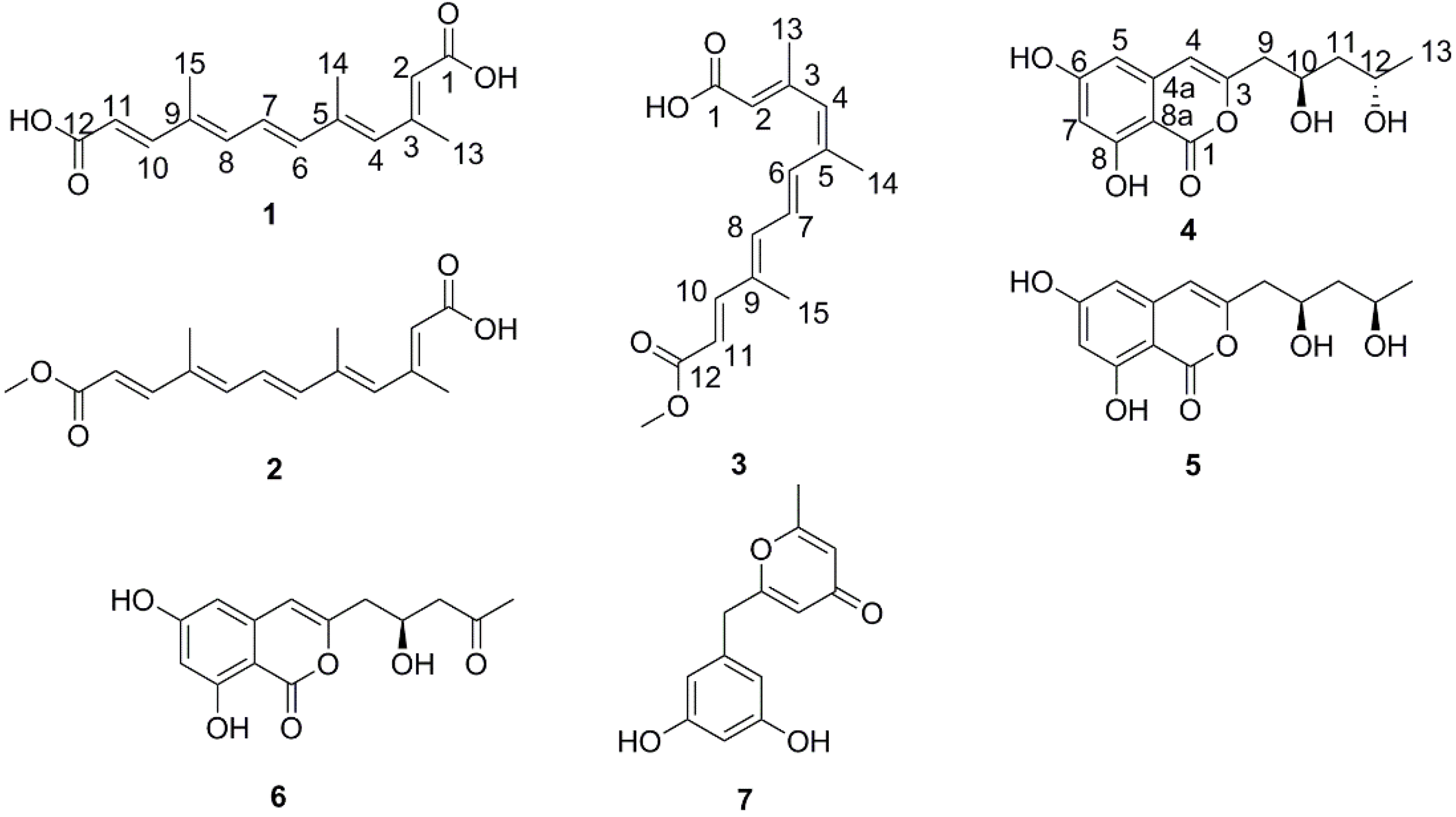
| 4 | 5 | |||
|---|---|---|---|---|
| Position | δH (J in Hz) | δC | δH (J in Hz) | δC |
| 1 | 168.0, C | 168.0, C | ||
| 3 | 156.3, C | 156.1, C | ||
| 4 | 6.37, s | 107.2, CH | 6.37, s | 107.3, CH |
| 4a | 141.4, C | 141.4, C | ||
| 5 | 6.31, s | 103.8, CH | 6.31, s | 103.8, CH |
| 6 | 167.5, C | 167.5, C | ||
| 7 | 6.31, s | 102.8, CH | 6.31, s | 102.8, CH |
| 8 | 165.0, C | 165.0, C | ||
| 8a | 100.0, C | 100.0, C | ||
| 9 | 2.59, dd (14.5, 8.0) | 43.1, CH2 | 2.57, dd (14.5, 8.3) | 42.5, CH2 |
| 2.65, dd (14.5, 4.9) | 2.70, dd (14.5, 4.4) | |||
| 10 | 4.21, m | 67.2, CH | 4.14, m | 68.6, CH |
| 11 | 1.60, ddd (14.3, 10.6, 3.4) | 47.1, CH2 | 1.71, ddd (13.9, 8.8, 7.6) | 46.4, CH2 |
| 1.55, ddd (14.3,10.6, 3.4) | 1.61, ddd (13.9, 5.4, 4.3) | |||
| 12 | 4.02, m | 65.4, CH | 4.00, m | 67.0, CH |
| 13 | 1.21, d (6.3) | 24.5, CH3 | 1.20, d (6.2) | 23.5, CH3 |
| Compounds | 1 | 2 | 3 | 4 | 5 | 6 | 7 | Acarbose a |
|---|---|---|---|---|---|---|---|---|
| IC50 (μM) b | 121.8 ± 0.4 | 23.5 ± 0.3 | 42.3 ± 0.2 | 343.7 ± 1.0 | 392.5 ± 1.7 | 538.7 ± 4.3 | >900 | 815.3 ± 3.8 |
© 2016 by the authors; licensee MDPI, Basel, Switzerland. This article is an open access article distributed under the terms and conditions of the Creative Commons Attribution (CC-BY) license (http://creativecommons.org/licenses/by/4.0/).
Share and Cite
Cui, H.; Liu, Y.; Nie, Y.; Liu, Z.; Chen, S.; Zhang, Z.; Lu, Y.; He, L.; Huang, X.; She, Z. Polyketides from the Mangrove-Derived Endophytic Fungus Nectria sp. HN001 and Their α-Glucosidase Inhibitory Activity. Mar. Drugs 2016, 14, 86. https://doi.org/10.3390/md14050086
Cui H, Liu Y, Nie Y, Liu Z, Chen S, Zhang Z, Lu Y, He L, Huang X, She Z. Polyketides from the Mangrove-Derived Endophytic Fungus Nectria sp. HN001 and Their α-Glucosidase Inhibitory Activity. Marine Drugs. 2016; 14(5):86. https://doi.org/10.3390/md14050086
Chicago/Turabian StyleCui, Hui, Yayue Liu, Yang Nie, Zhaoming Liu, Senhua Chen, Zhengrui Zhang, Yongjun Lu, Lei He, Xishan Huang, and Zhigang She. 2016. "Polyketides from the Mangrove-Derived Endophytic Fungus Nectria sp. HN001 and Their α-Glucosidase Inhibitory Activity" Marine Drugs 14, no. 5: 86. https://doi.org/10.3390/md14050086
APA StyleCui, H., Liu, Y., Nie, Y., Liu, Z., Chen, S., Zhang, Z., Lu, Y., He, L., Huang, X., & She, Z. (2016). Polyketides from the Mangrove-Derived Endophytic Fungus Nectria sp. HN001 and Their α-Glucosidase Inhibitory Activity. Marine Drugs, 14(5), 86. https://doi.org/10.3390/md14050086