Article

Exploring the Chemodiversity and Biological Activities of the Secondary Metabolites from the Marine Fungus Neosartorya pseudofischeri

1
School of Pharmaceutical Sciences, Sun Yat-sen University, Guangzhou 510006, China
2
Guangdong Technology Research Center for Advanced Chinese Medicine, Guangzhou 510006, China
3
School of Chemistry and Chemical Engineering, Sun Yat-sen University, Guangzhou 510275, China
4
Guangdong Institute of Gastroenterology, Guangzhou 510655, China
5
Guangdong Key Laboratory of Colorectal and Pelvic Floor Diseases, Guangzhou 510655, China
6
The Sixth Affiliated Hospital, Sun Yat-sen University, Guangzhou 510655, China
7
Institute of Chinese Medical Sciences, Guangdong Pharmaceutical University, Guangzhou 510006, China
*
Author to whom correspondence should be addressed.
Mar. Drugs 2014, 12(11), 5657-5676; https://doi.org/10.3390/md12115657
Submission received: 15 August 2014 / Revised: 12 November 2014 / Accepted: 17 November 2014 / Published: 24 November 2014

Abstract

:
The production of fungal metabolites can be remarkably influenced by various cultivation parameters. To explore the biosynthetic potentials of the marine fungus, Neosartorya pseudofischeri, which was isolated from the inner tissue of starfish Acanthaster planci, glycerol-peptone-yeast extract (GlyPY) and glucose-peptone-yeast extract (GluPY) media were used to culture this fungus. When cultured in GlyPY medium, this fungus produced two novel diketopiperazines, neosartins A and B (1 and 2), together with six biogenetically-related known diketopiperazines,1,2,3,4-tetrahydro-2,3-dimethyl-1,4-dioxopyrazino[1,2-a]indole (3), 1,2,3,4-tetrahydro-2-methyl-3-methylene-1,4-dioxopyrazino[1,2-a]indole (4), 1,2,3,4-tetrahydro-2-methyl-1,3,4-trioxopyrazino[1,2-a] indole (5), 6-acetylbis(methylthio)gliotoxin (10), bisdethiobis(methylthio)gliotoxin (11), didehydrobisdethiobis(methylthio)gliotoxin (12) and N-methyl-1H-indole-2-carboxamide (6). However, a novel tetracyclic-fused alkaloid, neosartin C (14), a meroterpenoid, pyripyropene A (15), gliotoxin (7) and five known gliotoxin analogues, acetylgliotoxin (8), reduced gliotoxin (9), 6-acetylbis(methylthio)gliotoxin (10), bisdethiobis(methylthio) gliotoxin (11) and bis-N-norgliovictin (13), were obtained when grown in glucose-containing medium (GluPY medium). This is the first report of compounds 3, 4, 6, 9, 10 and 12 as naturally occurring. Their structures were determined mainly by MS, 1D and 2D NMR data. The possible biosynthetic pathways of gliotoxin-related analogues and neosartin C were proposed. The antibacterial activity of compounds 214 and the cytotoxic activity of compounds 4, 5 and 713 were evaluated. Their structure-activity relationships are also preliminarily discussed.

Graphical Abstract

1. Introduction

Marine organism, such as sponges, soft corals and invertebrates, host numerous fungi. These marine fungi are an important source of structurally unique and biologically active natural products. Recent studies revealed that the cryptic biosynthetic pathways of fungi can be activated and the chemical diversity of their metabolites can be maximized by alternating their cultivation parameters systematically, such as the components of the media [1,2], co-culture [3,4], feeding precursors [5,6] and the addition of enzyme inhibitors [7,8]. For example, marine fungus Chondrostereum sp., which was isolated from soft coral Sarcophyton tortuosum, grows in different culture media and can produce various novel bioactive hirsutane-type sesquiterpenoids [9,10,11,12].
In recent years, we conducted research on the metabolites of marine fungi isolated from starfish Acanthaster planci and obtained a series of novel and/or bioactive metabolites [13,14,15,16]. In the current work, a marine fungus, Neosartorya pseudofischeri, was isolated from the inner tissue of Acanthaster planci. Neosartorya is a sexual state of Aspergillus section Fumigati [17]; however, unlike Aspergillus, the reports on the secondary metabolites of Neosartorya sp. have been relatively rare. The limited literature showed that most of the metabolites from Neosartorya species were cytotoxic nitrogenous-containing compounds [18,19]. In an attempt to explore the biosynthetic potentials, GlyPY (glycerol 10 g, peptone 5 g, yeast extract 2 g, CaCO3 1 g, sea water 1 L) and GluPY (glucose 10 g, peptone 5 g, yeast extract 2 g, sea water 1 L, pH 7.5) media were separately used to culture the fungus, Neosartorya pseudofischeri. Both of the EtOAc extracts of two different culture conditions showed potent cytotoxicity against cancer cell line HCT-116 with the IC50 values lower than 20 μg/mL. The HPLC traces of these two EtOAc extracts also displayed distinct components and content differences (Supplementary Figure S1). Purification of the extract of GlyPY medium afforded two novel diketopiperazines, neosartins A and B (1 and 2), together with six biogenetically-related known diketopiperazines, 1,2,3,4-tetrahydro-2,3-dimethyl-1,4-dioxopyrazino[1,2-a]indole (3), 1,2,3,4-tetrahydro-2-methyl-3-methylene-1,4-dioxopyrazino[1,2-a]indole (4), 1,2,3,4-tetrahydro-2-methyl-1,3,4-trioxopyrazino[1,2-a]indole (5), 6-acetylbis(methylthio) gliotoxin (10), bisdethiobis (methylthio)gliotoxin (11), didehydrobisdethiobis(methy1thio)gliotoxin (12) and N-methyl-1H-indole-2-carboxamide (6). Isolation of the extract of GluPY medium gave a new alkaloid, neosartin C (14), known compounds, α-pyrone meroterpenoid pyripyropene A (15), gliotoxin (7) and gliotoxin analogues, acetylgliotoxin (8), reduced gliotoxin (9), 6-acetylbis(methylthio)gliotoxin (10), bisdethiobis(methylthio)gliotoxin (11) and bis-N-norgliovictin (13) (Figure 1). In this paper, we report the isolation, structural elucidation, proposed biosynthetic pathways, bioactivities and structure-activity relationships of these compounds.
Figure 1. Structures of compounds 115.
Marinedrugs 12 05657 g001

2. Results and Discussion

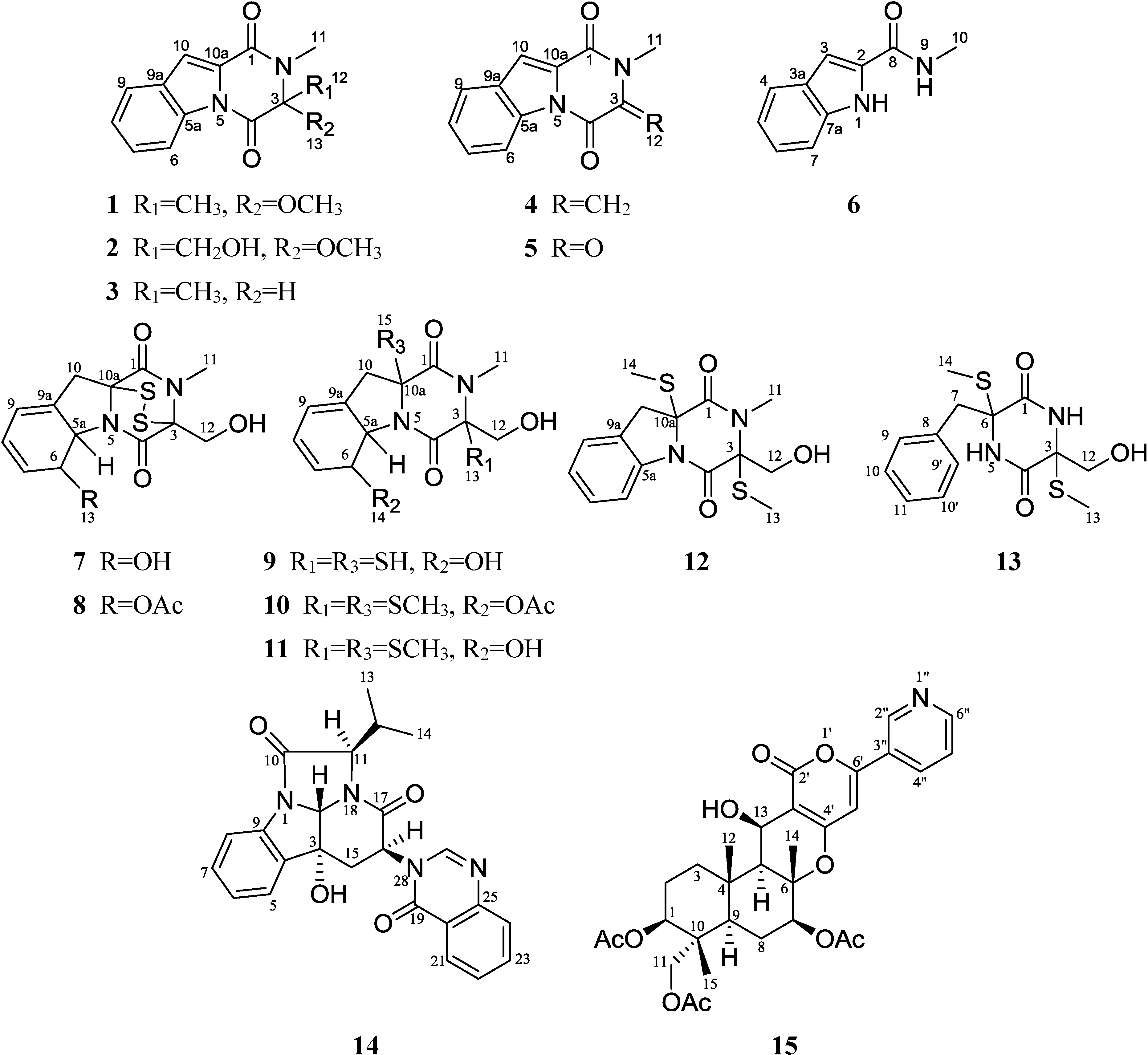
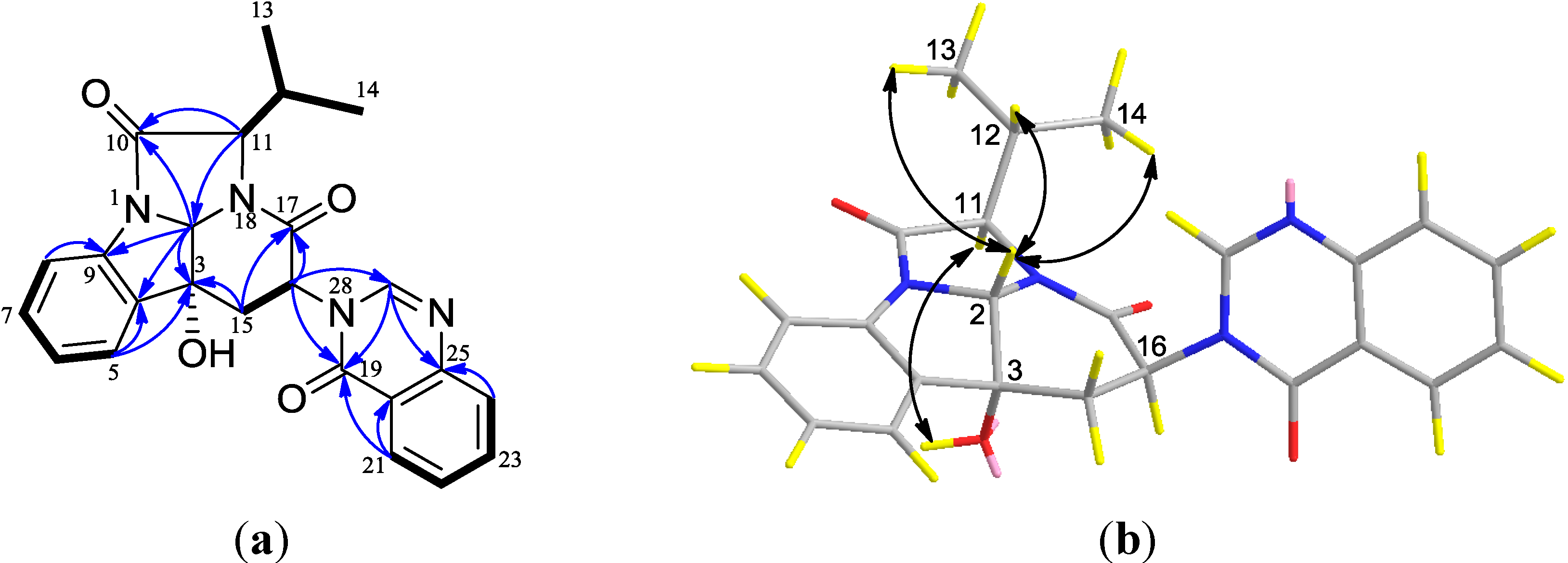
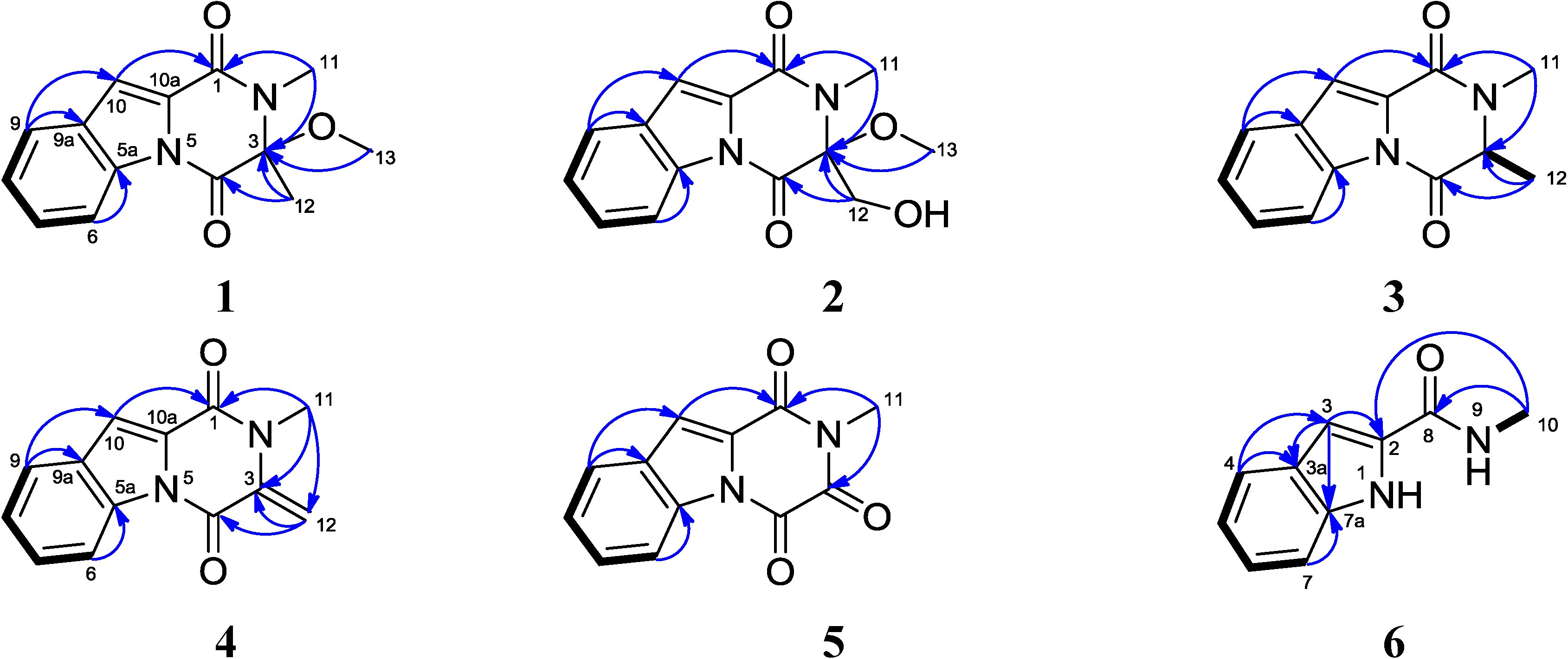
2.1. Structural Elucidation

Compound 1 was obtained as a yellowish solid. The molecular formula was determined as C14H14N2O3 from the HREIMS peak at m/z 258.0998 [M]+ (calcd. 258.0999) (Supplementary Figures S2 and S3), implying nine degrees of unsaturation. The IR spectrum indicated the presence of a carbonyl group (1680 cm−1) and a benzene ring (3073, 1574 and 1507 cm−1). UV maxima at 211, 243 and 295 nm displayed the conjugated system containing a benzene ring. The 13C NMR and DEPT spectra displayed three methyls, five methines and six quaternary carbons (Table 1 and Supplementary Figures S4–S6). Two quaternary carbons at δC 163.7 and 156.7 are amide carbonyls. The amide protons were substituted for the lack of the corresponding signals in IR and 1H NMR spectra in CDCl3. Eight carbon resonance signals appeared in the region of δC 114.9~134.8. Among them, four aromatic methines (δC 116.8, δH 8.50 (d, 8.0, H-6); δC 125.8, δH 7.43 (ddd, 8.0, 8.0, 0.8, H-7); δC 128.2, δH 7.55 (ddd, 8.0, 8.0, 0.8, H-8); and δC 122.7, δH 7.72 (d, 8.0, H-9)) established the partial structure -CH-CH-CH-CH- based on the 1H-1H COSY correlations of H-6/H-7, H-7/H-8 and H-8/H-9 (Figure 2, Supplementary Figure S7). Further analysis on the HMBC correlations of H-6/C-5a and H-9/C-9a suggested a 1,2-disubstutited benzene ring in the molecule. The methine (δC 114.9, δH 7.49, s) and a quaternary carbon C-10a at δC 127.8 constructed an additional trisubstituted double bond. The HMBC correlations of H-9/C-10, H-10/C-9a, H-10/C-10a and H-10/C-1 revealed the trisubstituted double bond connected to a benzene ring and an amide carbonyl. Methyl group C-11 at δH 3.13 (δC 26.6) showed HMBC correlations with amide carbonyl C-1 and quaternary carbon C-3, so it was connected to amide nitrogen N-2. Methyl group C-12 at δH 1.81 (δC 25.3) was connected to C-3 based on the HMBC correlation with the amide carbon C-4 and quaternary carbon C-3. The methoxyl group was also located at C-3, because it showed an HMBC correlation with C-3 (Supplementary Figure S8). Finally, in order to connect the remaining open bonds, C-5a must be linked to the nitrogen atom at the 5-position to form the additional five-membered ring. In the NOESY spectrum, the correlations among the protons of three methyl groups and H-9/H-10, confirmed the connection (Supplementary Figure S9). Therefore, compound 1 was established as 1,2,3,4-tetrahydro-3-methoxyl-2,3-dimethyl-1,4-dioxopyrazino[1,2-a]indole, trivially named neosartin A.
Compound 2 was isolated as a yellowish solid. The molecular formula was established as C14H14N2O4 based on the HREIMS peak at m/z 274.0947 [M]+ and 13C NMR data (Table 1, and Supplementary Figures S11 and S13). The 13C NMR and DEPT spectra displayed two methyls, one methylene, five methines and six quaternary carbons. The NMR data of compound 2 were very similar to those of compound 1 (Figure 2 and Supplementary Figures S12–S17). By comparison of their NMR data, a quick identification was made that the methyl C-12 (δC 25.3, δH 1.81, s) in 1 was replaced by an oxymethylene (δC 64.9, δH 4.18, d, J = 10.7 Hz; 3.99, d, J = 10.7 Hz) in 2. Therefore, the structure of 2 was elucidated as 1,2,3,4-tetrahydro-3-methoxyl-3-hydroxylmethyl-2-methyl-1,4-dioxopyrazino[1,2-a]indole, commonly named neosartin B.
Compounds 1 and 2 did not show optical activity in circular dichroism (CD) spectra; thus, they existed as a racemic mixture of 3R and 3S.
Compound 3 was isolated as a white solid. The molecular formula was established as C13H12N2O2 based on the LREIMS molecular ion at m/z 228 and the NMR data (Table 1 and Supplementary Figure S18). Its NMR spectra data closely resembled those of 1 except for the methoxy group in 1, which was replaced with a hydrogen atom in 3, and that correlated with C-3 in the HMBC spectrum. Due to the vicinal coupling with methyl group C-12, the 1H signal at δ 4.33 appeared as a typical quartet with J = 7.2 Hz. The structure of 3 was confirmed by 1H-1H COSY, HMBC and NOESY data (Supplementary Figures S19–S24). Compound 3 was determined to be 1,2,3,4-tetrahydro-2,3-dimethyl-1,4-dioxopyrazino[1,2-a]indole. It was once synthesized by heating anhydrodethiogliotoxin with acetic anhydride [20]; however, this is the first time that the detailed NMR data have been presented.
Table 1. 1H and 13C NMR data of compounds 13 at 400/100 MHz, respectively, in CDCl3, δ in ppm.
Position123
δCTypeδH, mult., (J in Hz)δCTypeδH, mult., (J in Hz)δCTypeδH, mult., (J in Hz)
1156.7C 158.5C 156.2C
2 N N N
390.7C 93.6C 60.2CH4.33, q (7.2)
4163.7C 162.9C 165.5C
5 N N N
5a129.2C 128.8C 129.2C
6116.9CH8.49, dd (8.0, 0.8)116.6CH8.47, dd (7.6, 0.8)116.5CH8.43, d (8.0)
7128.2CH7.55, ddd (8.0, 8.0, 0.8)128.1CH7.49, ddd (7.6,7.6, 0.8)127.8CH7.51, dd (8.0, 8.0)
8125.8CH7.43, ddd (8.0, 8.0, 0.8)125.7CH7.26, ddd (7.6, 7.6, 0.8)125.4CH7.40, dd (8.0, 8.0)
9122.7CH7.72, dd (8.0, 0.8)122.5CH7.32, dd (7.6, 0.8)122.5CH7.70, d (8.0)
9a134.8C 134.5C 134.8C
10114.9CH7.50, s115.0CH7.19, s114.1CH7.44, s
10a127.8C 127.5C 128.5C
1126.6CH33.13, s26.5CH33.14, s31.8CH33.16, s
1225.3CH31.81, s64.9CH24.17, d (11.6);4.02, d (11.6)19.8CH31.71, d (7.2)
1352.0OCH33.20, s52.0OCH33.24, s
12-OH 2.04, brs
Compound 4 contains a typical terminal C=C double bond (δC 137.7, C-3; δC 106.0, δH 6.15, s; 5.25, s, C-12). Its structure was elucidated as 1,2,3,4-tetrahydro-2-methyl-3-methylene-1,4-dioxopyrazino[1,2-a]indole by analysis of its spectral data (Table 2, Figure 2 and Supplementary Figures S25–S31). Compound 4 was previously obtained as the conversion product of gliotoxin (7) by passing through a column of alkaline alumina at 20 °C [21]. Compound 5 was deduced as 1,2,3,4-tetrahydro-2-methyl-1,3,4-trioxopyrazino[1,2-a]indole by careful analysis of the MS and NMR data (Table 2, Figure 2 and Supplementary Figures S32–S38). Compound 5 was firstly reported in 1945 as a degradation product of gliotoxin by heating with selenium [22]. It was also isolated from the culture of Penicillium terlikowskii [23]. Compound 6 was obtained as a white solid. The molecular formula was established as C10H10N2O on the basis of HREIMS (m/z 174.0789 [M]+, calcd. 174.0788) and NMR data (Table 2 and Supplementary Figures S39–S45). The structure of 6 was elucidated as N-methyl-1H-indole-2-carboxamide by analysis on the 1D and 2D NMR (HMQC, HMBC and 1H-1H COSY) (Table 2 and Figure 2). It is unprecedented that compound 6 was obtained from a natural source.
Figure 2. 1H-1H COSY (bold line) and the main HMBC (arrows) correlations of compounds 16.
Marinedrugs 12 05657 g002
Table 2. 1H and 13C NMR data of compounds 46 at 400/100 MHz, respectively, δ in ppm.
Position4 a5 bPosition6 c
δCTypeδH, mult., (J in Hz)δCTypeδH, mult., (J in Hz)δCTypeδH, mult., (J in Hz)
1154.4C 157.0C 1 NH11.04, brs
2 N N 2132.8C
3137.7C 156.8C 3102.7CH7.07, s
4154.9C 149.9C 3a137.8C
5 N N 4122.5CH7.61, d (8.0)
5a128.9C 128.3C 5124.5CH7.22, dd (8.0, 8.0)
6117.0CH8.50, d (8.0)115.8CH8.32, d (8.0)6120.9CH7.06, dd (8.0, 8.0)
7128.2CH7.53, dd (8.0, 8.0)129.0CH7.63, d (8.0, 8.0)7113.2CH7.58, d (8.0)
8125.5CH7.40, dd (8.0, 8.0)125.7CH7.47, d (8.0, 8.0)7a128.9C
9122.8CH7.72, d (8.0)123.9CH7.88, d (8.0)8163.0C
9a135.6C 135.4C 9 NH7.83, brs
10115.3CH7.51, s116.3CH7.72, s1026.4CH32.97, s
10a127.6C 127.7C
1129.6CH33.41, s26.6CH33.22, s
12106.0CH26.15, s; 5.25, s
a Measured in CDCl3. b Measured in DMSO-d6. c Measured in acetone-d6.
Compound 7 was identified as gliotoxin by comparing the data with the literature values [24,25] (Supplementary Figures S46–S48). The NMR spectra data of compound 8 closely resembled those of 7, except for one additional 1H resonance signal of the acetyl group. Its structure was identified as acetylgliotoxin, which was isolated from fungus strain FO2047 previously, and showed broad activities, including inhibition of fungi, bacteria and viruses [26] (Table 3 and Supplementary Figures S49 and S50). Compound 9, having NMR data similar to those of 7, was identified as reduced gliotoxin, which was the reduced dithiol form of 7 (Table 3 and Supplementary Figures S51 and S52). Daniel et al. found that gliotoxin (7) was generated from the corresponding dithiol (9) by a novel FAD-dependent dithiol oxidase, GliT [27]. Although 46, 8 and 9 are known compounds, their detailed NMR data were never reported previously.
Table 3. 1H and 13C NMR data of compounds 8 and 9 in CDCl3, δ in ppm.
Position8 a9 b
δCTypeδH, mult., (J in Hz)δCTypeδH, mult., (J in Hz)
1168.4C 169.7C
2 N N
378.2C 78.6C
4165.9C 168.1C
5 N N
5a64.9CH5.36, d (14.1)70.6CH4.77, d (13.2)
674.3CH5.82, d (14.1)72.7CH5.04, d (13.2)
7128.6CH5.93, m129.8CH5.93, m
8124.9CH5.93, m123.1CH5.88, m
9120.7CH5.60, d (7.5)120.7CH5.75, m
9a131.7C 130.0C
1041.4CH23.25, d (15.0); CH23.28, d (16.2);
3.08, d (15.0)3.05, d (16.2)
10a77.6C 77.2C
1129.0CH33.10, s28.9CH33.15, s
1262.4CH24.32, d (12.3)62.3CH24.39, d (12.0)
4.00, d (12.3)4.06, d (12.0)
13170.1COCH3
21.4COCH32.17, s
a 1H and 13C NMR data were measured at 300/75 MHz; b 1H and 13C NMR data were measured at 400/100 MHz. CDCl3.
Compounds 1013 were identified as 6-acetylbis(methylthio)gliotoxin (10) [28], bisdethiobis(methylthio)gliotoxin (11) [29], didehydrobisdethiobis(methylthio)gliotoxin (12) [30] and bis-N-norgliovictin (13) [31], respectively, by comparing their spectroscopic data (Table 4 and Supplementary Figures S53–S60) with the literature values. In the literature, only the 1H NMR data of compound 12 were reported; here, we report the detailed 1H and 13C NMR data. The 13C NMR data of compound 13 recorded in DMSO-d6 were shifted about 0.7~2.5 ppm to a higher field compared to the data reported for pyridine-d5.
Compound 14 was obtained as a yellow solid. The molecular formula was deduced as C24H22N4O4 from the HREIMS peak at m/z 430.1635 [M]+ (calcd. 430.1636), implying 16 degrees of unsaturation (Supplementary Figure S62). The 13C NMR and DEPT spectra displayed twenty-four carbons, which were classified into two methyls, one methylene, thirteen methines and eight quaternary carbons (Table 5, Supplementary Figures S63 and S64). The chemical shifts of sixteen carbons were located at δC 115.0–172.0, corresponding to the aromatic or double-bond carbons. The 1H NMR spectrum showed eight proton signals in the downfield region (δH 7.15~8.20) with the coupling constants being about 7.6, suggesting at least two phenyl groups in the molecule. By analysis of the HMQC spectrum, the 1H and 13C NMR data of each carbon were definitely assigned. The 1H-1H COSY correlations of H-15/H-16, H-11/H-12, H-12/H-13 and H-12/H-14 deduced the presence of two partial structures, -CH2CH- and -CHCH(CH3)2, respectively (Figure 3a, Supplementary Figure S66). Furthermore, the COSY correlations of H-5/H-6, H-6/H-7, H-7/H-8, H-21/H-22, H-22/H-23 and H-23/H-24 indicate that there are two -CHCHCHCH- in the molecule. HMBC correlations of H-5/C-4, H-6/C-4, H-7/C-9, H-8/C-9, H-21/C-20, H-21/C-25, H-22/C-20, H-23/C-25 and H-24/C-25 further confirmed the presence of two disubstituted benzene rings. Three quaternary carbons at δC 171.4 (C-10), 163.6 (C-17) and 161.7 (C-19) are amide carbonyl groups. HMBC correlations of H-11/C-10, H-16/C-17, H-21/C-19 revealed that three amide carbonyl groups were connected to -CHCH(CH3)2, -CH2CH- and a disubstituted benzene ring, respectively. The hydroxyl group at δH 4.09 (s) was attached to C-3 (δC 74.8), and C-3 was connected to the methine C-2 (δC 84.8) and C-15 (δC 36.5) based on the HMBC correlations of H-2/C-3 and H-15/C-3. The planar structure of 14 was finally established by the HMBC correlations of H-2/C-10, H-5/C-3, H-15/C-3, H-16/C-19, H-16/C-27 and H-27/C-25 (Supplementary Figure S67).
Table 4. 1H and 13C NMR data of compounds 12 and 13, δ in ppm.
Position12 aPosition13 b
δCTypeδH, mult., (J in Hz)δCTypeδH, mult., (J in Hz)
1166.0C 1165.2C
2 N 2NH8.95, brs
371.7C 365.8C
4161.8C 4165.0C
5 N 5 NH8.40, brs
5a128.9C 665.6C
6127.9CH8.03, d (8.0)743.3CH23.33, d (6.0); 3.31, d (6.0)
7126.2CH7.31, dd (8.0, 8.0)8135.0C
8125.2CH7.19, dd (8.0, 8.0)9/9′130.0CH7.20, m
9118.1CH7.30, d (8.0)10/10′127.6CH7.20, m
9a140.6C 11126.5CH7.20, m
1039.6CH24.50, d (12.0); 3.96, d (12.0)1264. 8CH23.52, d (18.0); 3.00, d (18.0)
10a70.8C 1312.8CH32.11, s
1128.9CH33.20, s1413.5CH32.29, s
1263.9CH23.62, d (16.8); 3.51, d (16.8)
1314.5CH32.32, s
1413.8CH32.24, s
a 1H and 13C NMR data were measured at 400/100 MHz in CDCl3; b 1H and 13C NMR data were measured at 300/75 MHz in DMSO-d6.
The relative stereochemistry of 14 was determined on the basis of NOESY data. The NOESY correlations of H-2 with H-12, H-13 and H-14 and of OH-3 with H-11 suggested that H-2 and H-11 were placed on the opposite face of the ring system and H-11 and the hydroxyl group at C-3 position were placed on the same face (Figure 3b). No NOESY correlation between H-2 and H-16 was observed (Supplementary Figure S68). Therefore, H-2 was assigned a β-orientation, whereas OH-3, H-11 and H-16 were determined to have an α-orientation.
The NMR data of compound 15 recorded in CDCl3 and acetone-d6 showed minor differences (Table 6 and Supplementary Figures S69–S75). By comparison of its spectral data with those reported in the literature, compound 15 was elucidated as pyripyropene A [31]. Pyripyropene A (15), previously isolated from Aspergillus fumigatus FO-1289, showed very potent inhibition of cholesterol acyltransferase (ACAT).
Table 5. 1H and 13C NMR data of compound 14 at 400/100 MHz, respectively, in CDCl3, δ in ppm.
PositionδCTypeδH, mult., (J in Hz)
1 N
284.8CH5.95, s
374.8C
4135.2C
5124.7CH7.43, brd (7.6)
6126.4CH7.15, dd (7.6, 7.6)
7130.9CH7.35, dd (7.6, 7.6)
8115.6CH7.53, d (7.6)
9138.5C
10171.4C
1170.3CH4.43, d (8.4)
1230.1CH2.35, dqq (8.4, 6.4, 6.4)
1320.1CH31.17, d (6.4)
1419.1CH31.21, d (6.4)
1536.5CH22.49, dd (15.2, 5.2); 3.23, dd (15.2, 4.8)
1656.8CH5.11, dd (5.2, 4.8)
17163.6C
18 N
19161.7C
20121.4C
21127.4CH8.20, dd (7.6, 0.8)
22127.8CH7.50, dd (7.6, 7.6)
23134.9CH7.71, ddd (7.6, 7.6, 0.8)
24126.1CH7.64, d (7.6)
25144.4C
26 N
27147.5CH8.61, s
28 N
3-OH 4.09, brs
Figure 3. (a) 1H-1H COSY (bold line), the main HMBC (arrows); and (b) key NOESY correlations of compound 14.
Marinedrugs 12 05657 g003
Table 6. 1H and 13C NMR data of compound 15 at 600/150 MHz, respectively, δ in ppm.
PositionIn CDCl3In Acetone-d6
δCTypeδH, mult., (J in Hz)δCδH, mult., (J in Hz)
173.5CH5.00, dd (10.8, 4.8)74.54.99, dd (10.8, 4.8)
222.7CH21.83, m; 1.90, m23.71.85, m; 1.89, m
336.2CH21.37, m; 2.16, m36.81.49, m; 2.17, ddd (12.6, 4.8, 4.8)
437.9C 38.8
554.7CH1.53, d (4.2)55.11.67, d (4.8)
683.3C 83.9
777.7CH4.78, dd (12.0, 4.8)79.04.81, dd (11.4, 4.8)
825.2CH21.63, ddd (12.0, 12.0, 11.4); 1.78, dd (11.4, 4.8)26.01.74, m; 1.83, ddd (10.8, 5.4, 5.4)
945.4CH1.58, d (12.0)46.21.72, d (12.0)
1040.3C 41.3
1164.8CH23.77, d (11.4); 3.70, d (11.4)65.63.76, d (12.0); 3.72, d (12.0)
1217.4CH31.43, s17.91.53, s
1360.1CH4.99, d (4.2)60.55.00, d (4.8)
1416.2CH31.69, s16.81.76, s
1513.2CH30.83, s13.60.93, s
2′163.6C 163.3
3′103.3C 104.2
4′162.0C 162.7
5′99. 9CH6.48, s100.16.71, s
6′156.4C 158.1
2″145.4CH9.05, s147.79.08, s
3″127.9C 128.5
4″134.3CH8.20, d (8.4)133.88.24, ddd (7.8, 1.8, 1.8)
5″124.2CH7.51, brd ( 8.4)124.77.53, dd (7.8, 4.8)
6″149.8CH8.72, s152.28.69, d (4.8)
1-O-CO-CH3170.5C 170.6
7-O-CO-CH3170.0C 170.3
11-O-CO-CH3170.9C 170.8
1-O-CO-CH321.1CH32.07, s21.12.02, s
7-O-CO-CH321.2CH32.15, s21.22.10, s
11-O-CO-CH320.8CH32.03, s20.72.00, s
13-OH OH3.06, brs 2.90, brs

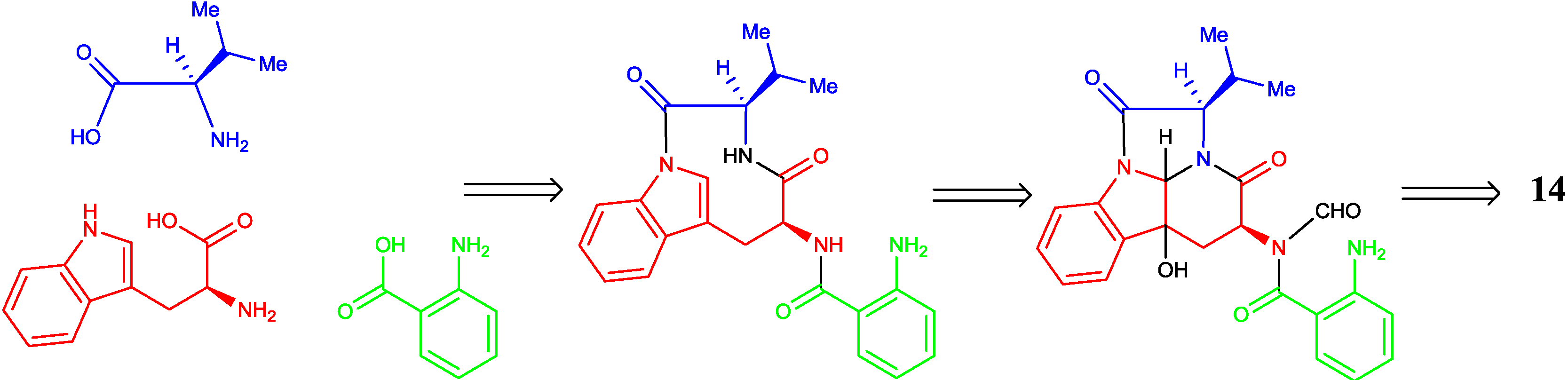
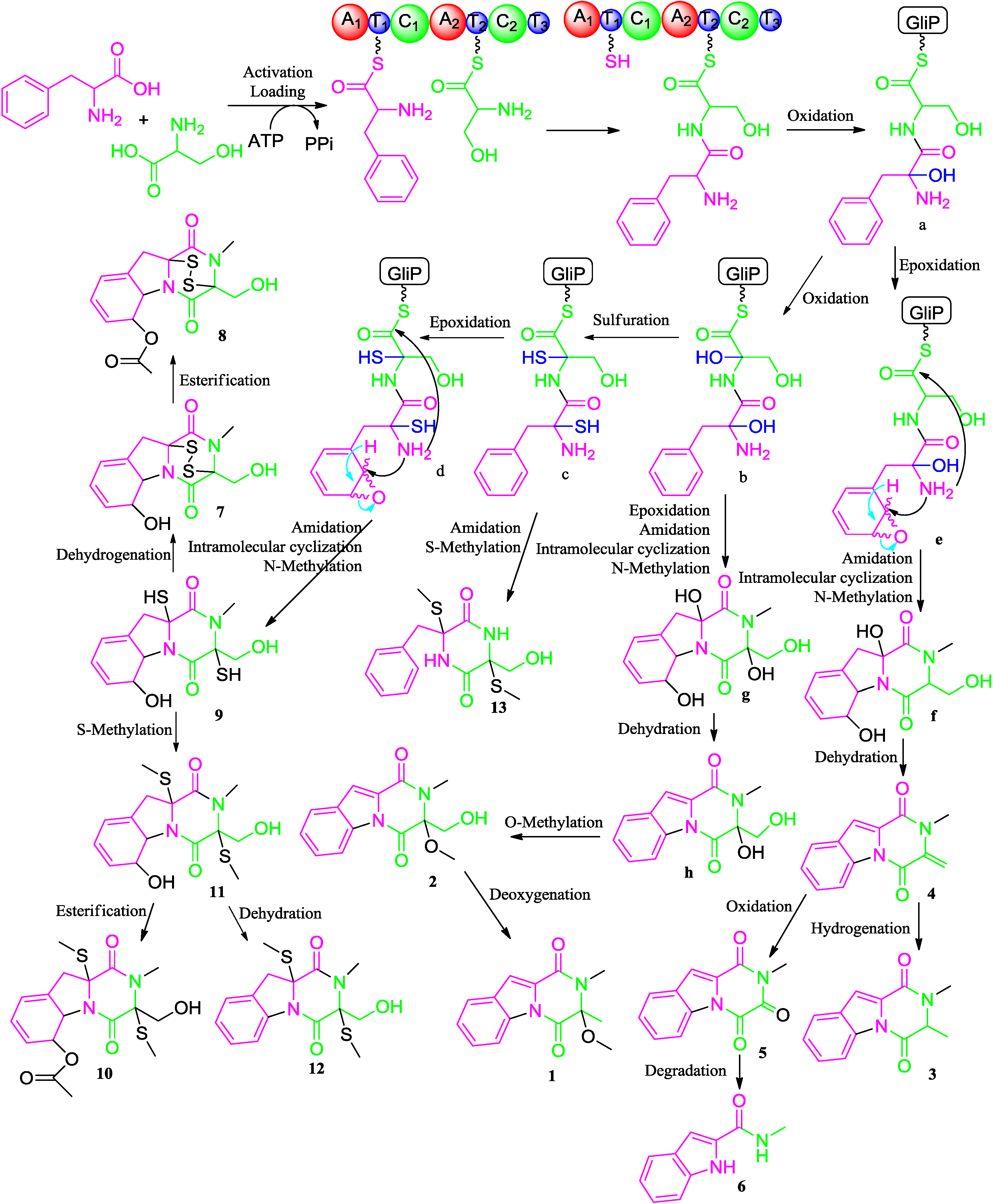
2.2. Proposed Biosynthetic Pathway

Diketopiperazines (DKPs) are typically synthesized via nonribosomal peptide synthetases (NRPS) to incorporate more than one amino acid from fungi [32]. Gliotoxin and its analogues have the diketopiperazine core with a disulfide bridge in an oxidized or reduced form. In the biosynthesis of gliotoxin, two-modular nonribosomal peptide synthetase, GliP, incorporates l-phe and l-ser to form dipeptidyl l-phe-l-ser, and under the action of the enzymes, the latter is converted to the corresponding diketopiperazines [33]. Therefore, the biosynthetic pathways of compounds 113 were proposed in Figure 4. Catalyzed by GliP, the intermolecular condensation between phenylalanine and serine generates an l-phe-l-ser, followed by successive oxidation, sulfuration and epoxidation reactions to obtain the intermediates, ad. Then, a and b undergo epoxidation, amidation, intramolecular nucleophilic cyclization and N-methylation reactions to produce the intermediates, f and g, respectively. Dehydration of f furnishes compound 4. Compound 4 is further hydrogenated and oxidized to afford 3 and 5, respectively. The product, g, is sequentially dehydrated, O-methylated to form 2, followed by the deoxygenation to produce 1. After successive amidation and methylation of the thiol groups, the intermediate c generates compound 13. Amidation, intramolecular nucleophilic cyclization and N-methylation of d afford compound 9. Compound 9 is S-methylated to generate 11 or dehydrogenated to produce 7, which further undergoes esterification to form 10 and 8, separately. Compound 12 is produced by the dehydration of 11, whereas compound 6 is the degradation product of 5.
Figure 4. Proposed biosynthetic pathways of compounds 113.
Marinedrugs 12 05657 g004
Compound 14 possessed a unique tetracyclic-fused skeleton, and it was the diastereomer of pseudofischerine [34]. We postulated that the biosynthetic pathway may involve d-valine, d-tryptophan and anthranilic acid as the precursors (Figure 5). Chaetominine [35] and kapakahines [36] have a similar tetracyclic-fused fragment.
Figure 5. Proposed biosynthetic pathway of compound 14.
Marinedrugs 12 05657 g005

2.3. Biological Activity

Compounds 214 were evaluated for their antibacterial activity against three multidrug-resistant bacteria, i.e., the Gram-positive Staphylococcus aureus (ATCC29213) and Methicillin-resistant staphylococcus aureus (R3708) and the Gram-negative Escherichia coli (ATCC25922), using a broth dilution method (Mueller–Hinton broth) [37]. Vancomycin and ampicillin sodium were used as positive controls. Compounds 7 and 9 displayed significant inhibitory activities against these three bacteria with MIC values ranging from 1.52 to 97.56 μM (Table 7). Compounds 4 and 8 inhibited the growth of Staphylococcus aureus ATCC29213 and R3708 with MIC values of 283.11, 70.70 μM and 86.91, 21.73 μM, respectively. The remaining nine compounds, 2, 3, 5, 6 and 1014, were inactive in this assay (MIC > 256 μg/mL). The results suggested that the bioactive compounds are more active against the Gram-positive bacteria. Especially, compounds 7 and 9 showed potent inhibition against Staphylococcus aureus R3708 with MIC values of 1.53 and 1.52 μM. Preliminary analysis of the structure-activity relationships of these twelve diketopiperazines suggests that the disulfide bridge or reduced disulfide bond is essential for the inhibitory activity. If the thiol groups are substituted, like compounds 1013, the inhibitory effects disappeared. The substitution at the six-membered ring containing two conjugated double bonds influences the intensity of antibacterial activity. The analogues with a hydroxyl group at C-6 enhance the antibacterial activity compared to the analogues with an acetyl group at the same position. Additionally, the α-methylene ketone group is also the pharmacophore for the antibacterial activity.
Table 7. Antibacterial activities of diketopiperazines 4 and 79 (MIC, μM, n = 3).
CompoundStaphylococcus aureus (ATCC29213)Staphylococcus aureus (R3708)Escherichia coli (ATCC25922)
4283.1170.70>1,132
712.201.5324.53
886.9121.73>695.65
948.781.5297.56
Vancomycin0.842.01
Ampicillin sodium8.07129.246.73
Furthermore, compounds 4, 5 and 713 were screened for their cytotoxic activities on the human embryonic kidney (HEK) 293 cell line and human colon cancer cell lines, HCT-116 and RKO (a poorly differentiated colon carcinoma cell line). Compounds 4, 79 and 11 exhibited potent cytotoxicities against these cell lines (Table 8). With a disulfide bridge in the molecule, compounds 7 and 8 showed potent cytotoxic activities. Compound 4 showed stronger inhibitory activities than compound 5; their structural difference is a typical α-methylene ketone group in 4, whereas a diketone in 5. Compared to compounds 9 and 11, compound 10 lacked any activity (IC50 > 50 μM), supposedly since the thiol groups at C-3 and C-10a were methylated and the 6-OH was acetylated. The cytotoxic activities of the other compounds, due to the limited sample amount, were not tested in this assay.
Table 8. Cytotoxicities of compounds 4, 5 and 713 (IC50, μM, n = 5).
Compound Cell line
293HCT-116RKO
430.10 ± 0.9010.34 ± 1.4133.56 ± 1.22
5>50>50>50
71.58 ± 0.031.24 ± 0.380.80 ± 0.20
84.49 ± 0.240.89 ± 0.041.24 ± 0.18
91.26 ± 0.040.43 ± 0.040.41 ± 0.07
10>50>50>50
1116.39 ± 0.388.59 ± 0.9610.32 ± 0.04
12>50>50>50
13>50>50>50
5-Fluorouracil 2.04 ± 0.2245.86 ± 4.58

3. Experimental Section

3.1. General Experimental Procedures

Preparative HPLC was performed using a Shimadzu LC-20AT HPLC pump (Shimadzu Corporation, Nakagyo-ku, Kyoto, Japan) equipped with an SPD-20A dual λ absorbance detector (Shimadzu Corporation, Nakagyo-ku, Kyoto, Japan) and a Shim-pack PRC-ODS HPLC column (250 × 20 mm, Shimadzu Corporation, Nakagyo-ku, Kyoto, Japan). Optical rotations were measured using a Schmidt and Haensch Polartronic HNQW5 optical rotation spectrometer (SCHMIDT + HAENSCH GmbH & Co., Berlin, Germany). CD spectra were measured on a JASCO J-810 circular dichroism spectrometer (JASCO International Co. Ltd., Hachioji, Tokyo, Japan). UV spectra were recorded on a Shimadzu UV-Vis-NIR spectrophotometer (Shimadzu Corporation, Nakagyo-ku, Kyoto, Japan). 1D and 2D NMR spectra were recorded on Bruker Avance IIIT 600HD and Bruker Avance II 400 spectrometers (Bruker BioSpin AG, Industriestrasse 26, Fällanden, Switzerland) and a Varian Mercury-Plus 300 spectrometer (Varian Medical Systems In., Salt Lake City, UT, USA). The chemical shifts are relative to the residual solvent signals (CDCl3: δH 7.26 and δC 77.0; DMSO-d6: δH 2.50 and δC 39.51; acetone-d6: δH 2.05 and δC 29.92). Mass spectra were obtained on Thermo DSQ EI low-resolution and Thermo MAT95XP EI high-resolution mass spectrometers (Thermo Fisher Scientific In., Waltham, MA, USA).

3.2. Fungal Strain and Culture Method

The marine fungus, Neosartorya pseudofischeri (Collection No. 2014F27-1), was isolated from the inner tissue of the sea star, Acanthaster planci, collected from Hainan Sanya National Coral Reef Reserve, China. This fungal strain was maintained in 15% (v/v) glycerol aqueous solution at −80 °C. A voucher specimen was deposited in the School of Pharmaceutical Sciences, Sun Yat-sen University, Guangzhou, China. Analysis of the ITS rDNA (GenBank KF999816) by BLAST database screening provided a 100% match to N. pseudofischeri.
This fungus were cultured in GlyPY medium (glycerol 10 g, peptone 5 g, yeast extract 2 g, CaCO3 1 g, sea water 1 L) and GluPY medium (glucose 10 g, peptone 5 g, yeast extract 2 g, sea water 1 L, pH 7.5), respectively. Fungal mycelia were cut and transferred aseptically to 500 mL Erlenmeyer flasks each containing 200 mL sterilized GlyPY or GluPY liquid media. The flasks were incubated at 28 °C on a rotary shaker (120 rpm) for 25 days.

3.3. Extraction and Isolation

Fifty liters of GlyPY growth culture broth were filtered through cheesecloth. The culture broth was successively extracted with EtOAc (50 L) three times. The EtOAc extract was concentrated by low-temperature rotary evaporation. The extract (9.8 g) was chromatographed on a silica gel column (diameter: 8 cm, length: 70 cm, silica gel, 200 g) using petroleum ether (2 L), EtOAc (2 liters) (100:0–0:100, v/v), followed by EtOAc (2 L) and MeOH (2 L) (100:0–0:100, v/v) as the eluent to afford 10 fractions (code Fraction 1–Fraction 10). Fraction 2 was purified by the recrystallization in the petroleum ether-EtOAc (3:1, v/v) solution to give compounds 4 (13.4 mg) and 5 (52.7 mg). Fraction 3 was separated with a preparative RP HPLC using a gradient elution MeOH-H2O (20:80 up to 100:0, v/v) and then on Sephadex LH-20 using MeOH as the eluent followed by preparative RP HPLC eluted with MeOH–H2O (70:30, v/v) to give compounds 1 (1.3 mg), 2 (2.2 mg), 3 (1.62 mg) and 12 (2.15 mg). Fraction 5 was purified with a preparative RP HPLC (MeOH–H2O, 65:35, v/v) to give compound 6 (2.3 mg). Compound 10 (7.2 mg) was obtained from Fraction 7 with a preparative RP HPLC (MeOH–H2O, 70:30, v/v).
Fifty liters of GluPY growth culture broth were filtered through cheesecloth. The culture broth was successively extracted with EtOAc (50 L) three times to afford 10.2 g of extract. The crude extract was separated as ten fractions (Fraction 1–10) by a silica gel column chromatograph (diameter: 8 cm; length: 70 cm; silica gel, 200 g) employing the gradient elution described above. Fraction 3 displayed interesting signals in the δH 8~9 region and was further purified by RP HPLC using MeCN–H2O (40:60, v/v) as the eluent to yield compounds 14 (3.2 mg) and 15 (4.0 mg). Fraction 4 was separated on a silica gel column chromatograph (diameter: 3 cm; length: 50 cm; silica gel, 50 g) using an elution with petroleum ether (300 mL) and EtOAc (300 mL) (50:50, v/v) to obtain five fractions (Fractions 4–1 to Fractions 4–5). Fractions 4–2 was further purified on preparative RP HPLC eluted with MeOH–H2O (75:25, v/v) to give compound 7 (32 mg), under the same experiment condition. Fraction 4–4 afforded compounds 10 (10.2 mg), 11 (71.3 mg) and 13 (23.1mg). Fraction 7 was purified on RP HPLC (MeOH–H2O, 60:40, v/v) to give compounds 8 (7.5 mg) and 9 (9.3 mg).
Neosartin A (1): Yellowish solid. UV (MeOH) λmax (ε) 211 (25,800), 243 (30,637), 295 (21,930) nm. IR: υmax 3000, 2926, 1716, 1656, 1589, 1576, 1427, 1391, 1357, 1228, 1112, 1043, 844, 750 cm−1. 1H and 13C NMR: Table 1. LREIMS: m/z 256, 243, 227, 215, 199, 187, 170, 156, 143, 129, 115, 103, 92, 89, 78, 72, 63, 56. HREIMS: m/z [M]+ calcd. for C14H14O3N2: 258.0999; found 258.0998.
Neosartin B (2): Yellowish solid. UV (MeOH) λmax (ε) 212 (17,237), 243 (21,941), 295 (16,195) nm. IR: υmax 3301, 3126, 2951, 1706, 1635, 1591, 1577, 1435, 1392, 1360, 1348, 1224, 1151, 1116, 1081, 981, 746, 733 cm−1. 1H and 13C NMR: Table 1. LREIMS: m/z 274, 260, 243, 229, 215, 202, 188, 172, 156, 143, 130, 115, 103, 89, 83, 72, 57. HREIMS: m/z [M]+ calcd. for C14H14O4N2: 274.0948; found 274.0947.
1,2,3,4-Tetrahydro-2,3-dimethyl-1,4-dioxopyrazino[1,2-a]indole (3): White solid. 1H and 13C NMR: Table 1. LREIMS: m/z 228, 213, 200, 185, 172, 143, 131, 115, 100, 89, 71, 62, 56.
1,2,3,4-Tetrahydro-2-methyl-3-methylene-1,4-dioxopyrazino[1,2-a]indole (4): White solid. 1H and 13C NMR: Table 2. LREIMS: m/z 226, 199, 185, 169, 157, 143, 129, 115, 99, 88, 75, 62, 55.
1,2,3,4-Tetrahydro-2-methyl-1,3,4-trioxopyrazino[1,2-a]indole (5): White solid. 1H and 13C NMR: Table 2. LREIMS: m/z 228, 200, 159, 143, 131, 115, 100, 88, 71, 62, 50.
N-methyl-1H-indole-2-carboxamide (6): White solid. 1H and 13C NMR: Table 2. LREIMS: m/z 174, 156, 143, 115, 89, 77, 63, 58.
Gliotoxin (7): Pale yellowish solid. 1H NMR (CDCl3, 400 MHz): 5.98 (1H, d, 4.0, H-7), 5.92 (1H, dd, 9.2, 4.2, H-8), 5.84 (1H, s, OH-13), 5.75 (1H, d, 9.2, H-9), 4.80 (2H, s, H-5a, H-6), 4.43 (1H, d, 17.6, H-12), 4.23 (1H, d, 17.6, H-12), 4.06 (1H, brs, OH-12), 3.73 (1H, d, 12.8, H-10), 3.19 (3H, s, CH3-11), 2.94 (1H, d, 12,8, H-10); 13C (CDCl3, 100 MHz): 166.0 (C-1), 165.1 (C-4), 130.8 (C-9a), 129.8 (C-7), 123.3 (C-8), 120.1 (C-9), 77.3 (C-3), 75.8 (C-10a), 73.1 (C-6), 69.7 (C-5a), 60.4 (C-12), 36.5 (C-10), 27.5 (C-11). LREIMS: m/z 326, 308, 277, 262, 244, 233, 217, 199, 188, 160, 144, 132, 107, 89, 77, 73, 64, 55, 42.
Acetylgliotoxin (8): Yellowish solid. 1H and 13C NMR: Table 3.
Reduced gliotoxin (9): Yellowish solid. 1H and 13C NMR: Table 3.
6-Acetylbis(methylthio)gliotoxin (10): Yellowish oil. 1H NMR (CDCl3, 400 MHz): 6.16 (1H, d, 14.8, H-7), 5.90 (2H, m, H-8, H-9), 5.75 (1H, d, 10.4, H-6), 5.01 (1H, d, 14.4, H-5a), 4.23 (1H, d, 11.2, H-12), 3.76 (1H, d, 11.6, H-12), 3.60 (1H, brs, OH-12), 3.09 (3H, s, CH3), 3.04 (1H, d, 16.0, H-10), 2.90 (1H, d, 16.0, H-10), 2.29 (3H, s, CH3-13), 2.12 (3H, s, CH3-15), 2.05 (3H, s, COCH3-14); 13C (CDCl3, 100 MHz): 170.7 (COCH3-14), 166.4 (C-1), 164.4 (C-4), 133.8 (C-9a), 127.8 (C-7), 123.3 (C-8), 119.8 (C-9), 72.7 (C-3), 72.3 (C-10a), 75.2 (C-6), 65.4 (C-5a), 63.6 (C-12), 40.1 (C-10), 28.7 (C-11), 21.3 (COCH3-14), 15.0 (C-13), 13.0 (CH3-15).
Bisdethiobis(methylthio)gliotoxin (11): Yellowish oil. 1H NMR (CDCl3, 400 MHz): 5.85 (2H, m, H-7, H-8), 5.65 (1H, d, 9.2, H-9), 4.86 (2H, s, H-5a, H-6), 4.30 (1H, d, 11.6, H-12), 3.82 (1H, d, 11.6, H-12), 3.09 (3H, s, CH3-11), 3.01 (1H, d, 16.0, H-10), 2.92 (1H, d, 16.0, H-10), 2.22 (3H, s, CH3-13), 2.19 (3H, s, CH3-15); 13C (CDCl3, 100 MHz): 166.7 (C-1), 165.9 (C-4), 131.7 (C-9a), 129.5 (C-7), 123.2 (C-8), 119.8 (C-9), 74.2 (C-3), 72.1 (C-10a), 71.4 (C-6), 69.4 (C-5a), 63.4 (C-12), 38.7 (C-10), 28.5 (CH3-11), 15.0 (CH3-13), 13.0 (CH3-15) .
Didehydrobisdethiobis(methylthio)gliotoxin (12): Yellowish oil. 1H and 13C NMR: Table 4.
Bis-N-norgliovictin (13): White solid. 1H and 13C NMR: Table 4.
Neosartin C (14): Yellowish solid. [ α ] D 20 : −58.17°(c =0.0822, MeOH). UV (MeOH) λmax (ε) 314 (2910), 301 (3653), 226 (35,975), 212 (29,412) nm. IR: υmax 3348, 3068, 2970, 2934, 2875, 1724, 1667, 1608, 1476, 1465, 1387, 1326, 1292, 1267, 1248, 1172, 1112, 1076, 1007, 976, 915, 891, 844, 764, 742, 700, 593, 560 cm−1. 1H and 13C NMR: Table 5. LREIMS: m/z 430, 412, 341, 329, 314, 298, 286, 266, 238, 224, 184, 147, 130, 102, 83, 76, 55. HREIMS: m/z [M]+ calcd. for C24H22O4N4: 430.1636; found 430.1635.
Pyripyropene A (15): Yellowish solid. UV (MeOH) λmax (ε) 320 (18,029), 231 (29,463), 209 (15,006) nm. IR: υmax 3409, 2975, 2947, 2883, 1723, 1644, 1580, 1481, 1436, 1394, 1370, 1234, 1159, 1109, 1090, 1074, 1040, 1026, 1009, 984, 960, 925, 873, 809, 763, 704, 687, 650, 603, 583 cm−1. 1H and 13C NMR: Table 6. LREIMS: m/z 583, 565, 550, 523, 505, 494, 463, 445, 430, 403, 385, 358, 334, 304, 283, 240, 218, 202, 190, 171, 157, 148, 133, 119, 106, 95, 79, 69, 55.

3.4. Antibacterial Activity Assay

The MIC values were determined using a broth dilution method (Mueller–Hinton broth) based on the National Committee for Clinical Laboratory Standards (NCCLS) standard. The starting concentrations of the tested compounds were 256 µg/mL (from 256 to 0.25). The solution of compound in DMSO (10 µL) was added to 90 µL of bacterial culture (1 × 10⁶ CFU/mL) in the first well of flat-bottomed 96-well tissue culture plates. The solution was then double diluted. The bacterial culture solution containing the appropriate compound (50 µL) was discarded from the last well in order to ensure a 100-µL volume of bacterial culture in every well. A set of tubes containing only inoculated broth and solvent were kept as controls. The plate was incubated at 37 °C overnight in an electro-heating standing-temperature cultivator before the measurement of the absorbance value. The optical density values at 600 nm were measured using a multifunction microplate reader (PowerWaveTM XS2, BioTek® Instruments Inc., Winooski, VT, USA). Vancomycin and ampicillin sodium were used as positive controls.

3.5. Cytotoxicity Assay

Compounds in DMSO at 50 mM were used in the MTS ((3-(4,5-dimethylthiazol-2-yl)-5-(3-carboxymethoxyphenyl)-2-(4-sulfophenyl)-2H-tetrazolium)) assay. 5-Fluorouracil was used as the positive control, and DMSO was used as the negative control. 293, HCT116 and RKO cells were suspended in fresh RPMI-1640 medium containing 10% fetal bovine serum and 100 μg/mL penicillin and streptomycin at a cell density of 1 × 10⁵ cells/mL and seeded into 96-well plates each 100 μL/well. The suspension cells, 293, HCT116 and RKO cells, were incubated at 37 °C for 12 h. Then, compounds were added to the cultures at different concentrations; then, the cells were cultured at 37 °C for 72 h. Twenty microliters of MTS/PMS (phenazine methosulfate) were added into each well, incubated at 37 °C for 4 h in a humidified, 5% CO2 atmosphere. The absorbance at 490 nm was recorded using a Thermo Scientific Varioskan Flash Multimode Reader (Thermo Fisher Scientific Inc., Waltham, MA, USA). The data were analyzed with the GraphPad Prism 6 software package [38].

4. Conclusions

Alternating the cultivation parameters systematically, such as the components of the media, to enhance the diversity of secondary metabolites produced by the fungus is a well-known, simple and efficient technique. Using this strategy, we isolated and identified fifteen compounds, including twelve diketopiperazine derivatives, a meroterpenoid, an alkaloid with a unique tetracyclic-fused skeleton and an imidazole analogue from the two cultures. The discovery of these compounds provided further evidence that the genus of Neosartorya is a rich source of nitrogen-containing natural products. Interestingly, the dominant metabolites from the GluPY medium were the diketopiperazines with disulfide bonds; however, the main compounds from the GlyPY medium were the diketopiperazines without a disulfide bond. The biosynthetic pathways of the unique alkaloids are complex and diverse. Hopefully, further investigation on the secondary metabolites of Neosartorya pseudofischeri in varied culture conditions supplied with amino acid precursors may find more novel alkaloids and improve their production. Most of the metabolites showed significant antibacterial and cytotoxic activities. Based on the structure-activity relationship analysis, the disulfide bridge, the α-methylene ketone group, the hydroxyl group at C-6 and the thiol groups were considered as the pharmacophores.

Supplementary Files

Supplementary File 1

Acknowledgments

This project was financially supported by the National Natural Science Foundation of China (Nos. 30973633 and 81102502), the Special Financial Fund of Innovative Development of the Marine Economic Demonstration Project (GD2012-D01-001), the Guangdong Provincial Science and Technology Research Program (Nos. 2012A031100005, 2013B021100010 and 2013B021100012), the Guangdong Natural Science Foundation (No. S2012010010653), and the Guangzhou Science and Technology Research Program (No. 2014J4100059).

Author Contributions

Conceived of and designed the experiments: Wen-Jian Lan, Hou-Jin Li. Performed the experiments: Wan-Ling Liang, Xiu Le, Xiang-Ling Yang, Jun-Xiong Chen, Huan-Liang Liu, Lai-You Wang, Kun-Teng Wang, Kun-Chao Hu, De-Po Yang. Wrote the paper: Wan-Ling Liang, Wen-Jian Lan, Hou-Jin Li, Jun Xu.

Conflicts of Interest

The authors declare no conflict of interest.

References

  1. Wang, Y.; Zheng, J.K.; Liu, P.P.; Wang, W.; Zhu, W.M. Three new compounds from Aspergillus terreus PT06-2 grown in a high salt medium. Mar. Drugs 2011, 9, 1368–1378. [Google Scholar] [CrossRef] [PubMed]
  2. Wang, W.J.; Li, D.Y.; Hua, H.M.; Ma, E.L.; Li, Z.L. Caryophyllene sesquiterpenes from the marine-derived fungus Ascotricha sp. ZJ-M-5 by the one strain−many compounds strategy. J. Nat. Prod. 2014, 77, 1367–1371. [Google Scholar] [CrossRef] [PubMed]
  3. James, W.; Suzanne, M.S.; David, J.N.; Karina, M.Z. Tetramic acid analogues produced by coculture of Saccharopolyspora erythraea with Fusarium pallidoroseum. J. Nat. Prod. 2014, 77, 173–177. [Google Scholar] [CrossRef] [PubMed]
  4. Samuel, B.; Olivier, S.; Nadine, B.; Michel, M.; Katia, G.; Jean-Luc, W. De novo production of metabolites by fungal co-culture of Trichophyton rubrum and Bionectria ochroleuca. J. Nat. Prod. 2013, 76, 1157–1165. [Google Scholar] [CrossRef] [PubMed]
  5. Wang, F.Z.; Wei, H.J.; Zhu, T.J.; Li, D.H.; Lin, Z.J.; Gu, Q.Q. Three new cytochalasins from the marine-derived fungus Spicaria elegans KLA03 by supplementing the cultures with l- and d-Tryptophan. Chem. Biodivers. 2011, 8, 887–894. [Google Scholar] [CrossRef]
  6. Christopher, T.W.; Stuart, W.H.; Brian, D.A.; Gao, X.; Tang, Y. Short pathways to complexity generation: Fungal peptidyl alkaloid multicyclic scaffolds from anthranilate building blocks. ACS Chem. Biol. 2013, 8, 1366–1382. [Google Scholar] [CrossRef] [PubMed]
  7. Jeremy, B.; Nida, M.; Whittney, N.B.; Lacey, H.; Lindsey, N.S.; Tina, M.; Dennis, E.K.; Betty, B.; Alberto, van O.; Bill, B. Epigenetic tailoring for the production of anti-infective cytosporones from the marine fungus Leucostoma persoonii. Mar. Drugs 2012, 10, 762–774. [Google Scholar] [CrossRef]
  8. Teigo, A.; Takashi, Y.; Yoshiteru, O. Aromatic polyketide productionin cordyceps indigotica, an entomopathogenic fungus, induced by exposure to a histone deacetylase inhibitor. Org. Lett. 2012, 14, 2006–2009. [Google Scholar] [CrossRef] [PubMed]
  9. Li, H.J.; Jiang, W.H.; Liang, W.L.; Huang, J.X.; Mo, Y.F.; Ding, Y.Q.; Lam, C.K.; Qian, X.J.; Zhu, X.F.; Lan, W.J. Induced marine fungus Chondrostereum sp. as a means of producing new sesquiterpenoids chondrosterins I and J by using glycerol as the carbon source. Mar. Drugs 2014, 12, 167–175. [Google Scholar] [CrossRef] [PubMed]
  10. Li, H.J.; Chen, T.; Xie, Y.L.; Chen, W.D.; Zhu, X.F.; Lan, W.J. Isolation and structural elucidation of chondrosterins F-H from the marine fungus Chondrostereum sp. Mar. Drugs 2013, 11, 551–558. [Google Scholar] [CrossRef] [PubMed]
  11. Li, H.J.; Xie, Y.L.; Xie, Z.L.; Chen, Y.; Lam, C.K.; Lan, W.J. Chondrosterins A–E, triquinane-type sesquiterpenoids from soft coral-associated fungus Chondrostereum sp. Mar. Drugs 2012, 10, 627–638. [Google Scholar] [CrossRef] [PubMed]
  12. Li, H.J.; Lan, W.J.; Lam, C.K.; Yang, F.; Zhu, X.F. Hirsutane sesquiterpenoids from the marine- derived fungus Chondrostereum sp. Chem. Biodivers. 2011, 8, 317–324. [Google Scholar] [CrossRef] [PubMed]
  13. Lan, W.J.; Zhao, Y.; Xie, Z.L.; Liang, L.Z.; Shao, W.Y.; Zhu, L.P.; Yang, D.P.; Zhu, X.F.; Li, H.J. Novel sorbicillin analogues from the marine fungus Trichoderma sp. associated with the seastar Acanthaster planci. Nat. Prod. Commun. 2012, 7, 1337–1340. [Google Scholar] [PubMed]
  14. Zhao, Y.; Li, S.Q.; Li, H.J.; Lan, W.J. Lanostane triterpenoids from the fungus Ceriporia lacerate. Chem. Nat. Compd. 2013, 49, 653–656. [Google Scholar] [CrossRef]
  15. Xie, Z.L.; Li, H.J.; Wang, L.Y.; Liang, W.L.; Liu, W.; Lan, W.J. Trichodermaerin, a new lactone from the marine fungus Trichoderma erinaceum associated with the sea star Acanthaster planci. Nat. Prod. Commun. 2013, 8, 67–68. [Google Scholar] [PubMed]
  16. Lan, W.J.; Liu, W.; Liang, W.L.; Xu, Z.; Le, X.; Xu, J.; Lam, C.K.; Yang, D.P.; Li, H.J.; Wang, L.Y. Pseudaboydins A and B: Novel isobenzofuranone derivatives from marine fungus Pseudallescheria boydii associated with starfish Acanthaster planci. Mar. Drugs 2014, 12, 4188–4199. [Google Scholar]
  17. Peterson, S.W. Neosartorya pseudofischeri sp. nov. and its relationship to other species in Aspergillus section Fumigati. Mycol. Res. 1992, 96, 547–554. [Google Scholar]
  18. Yukihiro, A.; Hideaki, K.; Rie, O.; Arika, Y.; Hiroshi, M.; Hiroyuki, O. Azaspirene: A novel angiogenesis inhibitor containing a 1-oxa-7-azaspiro[4.4]non-2-ene-4,6-dione skeleton produced by the fungus Neosartorya sp. Org. Lett. 2002, 4, 2845–2848. [Google Scholar]
  19. Marco, M.; Anna, A.; Veronique, M.; Angela, B.; Alessio, C.; Laetitia, M.Y.B.; Maurizio, V.; Alexander, K.; Robert, K.; Antonio, E. Fischerindoline, a pyrroloindole sesquiterpenoid isolated from Neosartorya pseudofischeri, with in vitro growth inhibitory activity in human cancer cell lines. Tetrahedron 2013, 69, 7466–7470. [Google Scholar]
  20. John, R.J.; James, B.B. Gliotoxin. X. dethiogliotoxin and related compounds. J. Am. Chem. Soc. 1953, 75, 2103–2109. [Google Scholar]
  21. Ajay, K.B.; Das, K.G.; Funke, P.T.; Irene, K.; Shukla, O.P.; Khanchandani, K.S.; Suhadolnik, R.J. Biosynthetic studies on gliotoxin using stable isotopes and mass spectral methods. J. Am. Chem. Soc. 1968, 90, 1038–1041. [Google Scholar] [CrossRef]
  22. Johnson, J.R.; Hasbrouck, R.B.; Dutcher, J.D.; Bruce, W.F. Gliotoxin. V. The Structure of certain indole derivatives related to gliotoxin. J. Am. Chem. Soc. 1945, 67, 423. [Google Scholar] [CrossRef]
  23. Ali, M.S.; Shannon, J.S.; Taylor, A. Isolation and structures of 1,2,3,4-tetrahydro-l,4-dioxopyrazino[l,2-a]-indoles from cultures of Penicillium terlikowskii. J. Chem. Soc. C 1968, 2044–2048. [Google Scholar] [CrossRef]
  24. Mourad, K. Gliotoxin: Uncommon 1H couplings and revised 1H and 13C-NMR assignments. J. Nat. Prod. 1990, 53, 717–719. [Google Scholar] [CrossRef]
  25. Katharine, R.W.; Joseline, R.; Ang, K.H.; Karen, T.; Jennifer, E.C.; James, M.; Phillip, C. Assessing the trypanocidal potential of natural and semi-synthetic diketopiperazines from two deep water marine-derived fungi. Bioorg. Med. Chem. 2010, 18, 2566–2574. [Google Scholar] [CrossRef]
  26. Didier, V.D.; Junji, I.; Kazuro, S.; Hong, Y.; Hideo, T.; Satoshi, O. Inhibition of farnesyl-protein transferase by gliotoxin and acetylgliotoxin. J. Antibiot. 1992, 45, 1802–1805. [Google Scholar] [CrossRef]
  27. Daniel, H.S.; Nicole, R.; Thorsten, H.; Peter, H.; Axel, A.B.; Christian, H. Transannular disulfide formation in gliotoxin biosynthesis and its role in self-resistance of the human pathogen Aspergillus fumigates. J. Am. Chem. Soc. 2010, 132, 10136–10141. [Google Scholar] [CrossRef] [PubMed]
  28. Afiyatullov, S.S.; Kalinovskii, A.I.; Pivkin, M.V.; Dmitrenok, P.S.; Kuznetsova, T.A. Alkaloids from the marine isolate of the fungus Aspergillus fumigatus. Chem. Nat. Compd. 2005, 41, 236–238. [Google Scholar] [CrossRef]
  29. Kirby, G.W.; Robins, D.J.; Sefton, M.A.; Talekar, R.R. Biosynthesis of bisdethiobis(methylthio) gliotoxin, a new metabolite of Gliocladium Deliquescens. J. Chem. Soc. Perkin Trans Ⅰ. 1980, 119–121. [Google Scholar] [CrossRef]
  30. Zhao, W.Y.; Zhu, T.J.; Han, X.X.; Fan, G.T.; Liu, H.B.; Zhu, W.M.; Gu, Q.Q. A new gliotoxin analogue from a marine-derived fungus Aspergillus fumigatus Fres. Nat. Prod. Res. 2009, 23, 203–207. [Google Scholar] [CrossRef] [PubMed]
  31. Atsuki, O.; Kaoru, I.; Masaki, O.; Hiroshi, T.; Satoshi, O.; Tohru, N. Total synthesis of pyripyropene A. Tetrahedron 2011, 67, 8195–8203. [Google Scholar] [CrossRef]
  32. Mootz, H.D.; Marahiel, M.A. Biosynthetic systems for nonribosomal peptide antibiotic assembly. Curr. Opin. Chem. Biol. 1997, 1, 543–551. [Google Scholar] [CrossRef] [PubMed]
  33. Balibar, C.J.; Walsh, C.T. GliP, a multimodular nonribosomal peptide synthetase in Aspergillus fumigatus, makes the diketopiperazine scaffold of gliotoxin. Biochemistry 2006, 45, 15029–15038. [Google Scholar] [CrossRef] [PubMed]
  34. Amnat, E.; Anake, K.; Céline, B.; Véronique, M.; Leka, M.; Florence, L.; Artur, S.; Robert, K.; Werner, H. Secondary metabolites from a culture of the fungus Neosartorya pseudofischeri and their In vitro activity in human cancer cells. Planta Med. 2012, 78, 1767–1776. [Google Scholar] [CrossRef] [PubMed]
  35. Jiao, R.H.; Xu, S.; Liu, J.Y.; Ge, H.M.; Ding, H.; Xu, C.; Zhu, H.L.; Tan, R.X. Chaetominine, a cytotoxic alkaloid produced by endophytic Chaetomium sp. IFB-E015. Org. Lett. 2006, 8, 5709–5712. [Google Scholar] [CrossRef] [PubMed]
  36. Bryan, K.S.; Yeung, Y.N.; Robin, B.K.; John, R.C.; Wesley, Y.Y.; Paul, J.S.; Michelle, K.B. The Kapakahines, cyclic peptides from the marine sponge Cribrochalina olemda. J. Org. Chem. 1996, 61, 7168–7173. [Google Scholar]
  37. Casey, J.T.; O’Cleirigh, C.; Walsh, P.K.; O’Shea, D.G. Development of a robust microtiter plate-based assay method for assessment of bioactivity. J. Microbiol. Methods 2004, 58, 327–334. [Google Scholar] [CrossRef] [PubMed]
  38. GraphPad Prism 6, GraphPad Software, Inc.: La Jolla, CA, USA, 2014.

