
A Rare Case of Facial Artery Branching—A Review of the Literature and a Case Report with Clinical Implications

 , ,
, ,
Abstract
:1. Introduction
2. Materials and Methods
2.1. Narrative Literature Review
2.2. Case Report
3. Results
3.1. Literature Review
3.2. Case Report
4. Discussion
5. Conclusions
Author Contributions
Funding
Institutional Review Board Statement
Informed Consent Statement
Data Availability Statement
Acknowledgments
Conflicts of Interest
References
- Meegalla, N.; Sood, G.; Nessel, T.A.; Downs, B.W. Anatomy, Head and Neck, Facial Arteries. In StatPearls; StatPearls Publishing: Treasure Island, FL, USA, 2021. [Google Scholar]
- Soikkonen, K.; Wolf, J.; Hietanen, J.; Mattila, K. Three Main Arteries of the Face and Their Tortuosity. Br. J. Oral Maxillofac. Surg. 1991, 29, 395–398. [Google Scholar] [CrossRef]
- Von Arx, T.; Tamura, K.; Yukiya, O.; Lozanoff, S. The Face—A Vascular Perspective. A Literature Review. Swiss Dent. J. 2018, 128, 382–392. [Google Scholar]
- Bayram, S.B.; Kalaycioglu, A. Branching Patterns of Facial Artery in Fetuses. New J. Med. 2010, 27, 227–230. [Google Scholar]
- Pretterklieber, M.L.; Krammer, E.B.; Mayr, R. A Bilateral Maxillofacial Trunk in Man: An Extraordinary Anomaly of the Carotid System of Arteries. Cells Tissues Organs 1991, 141, 206–211. [Google Scholar] [CrossRef] [PubMed]
- Stathakios, J.; Carron, M.A. Anatomy, Head and Neck, Neck Triangle. In StatPearls; StatPearls Publishing: Treasure Island, FL, USA, 2021. [Google Scholar]
- Isolan, G.R.; Rowe, R.; Al-Mefty, O. Microanatomy and Surgical Approaches to the Infratemporal Fossa: An Anaglyphic Three-Dimensional Stereoscopic Printing Study. Skull Base 2007, 17, 285–302. [Google Scholar] [CrossRef] [PubMed] [Green Version]
- Loukas, M.; Hullett, J.; Louis, R.G.; Kapos, T.; Knight, J.; Nagy, R.; Marycz, D. A Detailed Observation of Variations of the Facial Artery, with Emphasis on the Superior Labial Artery. Surg. Radiol. Anat. 2006, 28, 316–324. [Google Scholar] [CrossRef] [PubMed]
- Rao, S.B.; Vollala, V.R.; Rao, M.; Samuel, V.P.; Deepthinath, D.; Nayak, S.; Pamidi, N. Unusual Position of External Carotid Artery: A Case Report. Indian J. Plast. Surg. 2005, 38, 170–171. [Google Scholar] [CrossRef] [Green Version]
- Drenckhahn, D.; Zenker, W. Benninghoff Anatomie Makroskopische Anatomie, Embryologie und Histologie des Menschen, 15th ed.; Urban & Schwarzenberg: München-Wien-Baltimore, Germany, 1994; Volume 1, ISBN 3-541-00245-X. [Google Scholar]
- Pilsl, U.; Anderhuber, F.; Neugebauer, S. The Facial Artery-The Main Blood Vessel for the Anterior Face? Dermatol. Surg. 2016, 42, 203–208. [Google Scholar] [CrossRef] [PubMed]
- Lohn, J.W.G.; Penn, J.W.; Norton, J.; Butler, P.E.M. The Course and Variation of the Facial Artery and Vein: Implications for Facial Transplantation and Facial Surgery. Aesthetic Surg. J. 2011, 67, 184–188. [Google Scholar] [CrossRef] [PubMed]
- Koziej, M.; Trybus, M.; Hołda, M.; Polak, J.; Wnuk, J.; Brzegowy, P.; Popiela, T.; Walocha, J.; Chrapusta, A. Anatomical Map of the Facial Artery for Facial Reconstruction and Aesthetic Procedures. Aesthetic Surg. J. 2019, 39, 1151–1162. [Google Scholar] [CrossRef]
- Furukawa, M.; Mathes, D.W.; Anzai, Y. Evaluation of the Facial Artery on Computed Tomographic Angiography Using 64-Slice Multidetector Computed Tomography: Implications for Facial Reconstruction in Plastic Surgery. Plast. Reconstr. Surg. 2013, 131, 526–535. [Google Scholar] [CrossRef] [PubMed]
- Padur, A.A.; Kumar, N. Unusual Branching Pattern and Termination of Facial Artery and Its Clinical Implications for Facial Operations. J. Vasc. Bras. 2019, 18, e20190021. [Google Scholar] [CrossRef] [PubMed] [Green Version]
- Midy, D.; Mauruc, B.; Vergnes, P.; Caliot, P. A Contribution to the Study of the Facial Artery, Its Branches and Anastomoses; Application to the Anatomic Vascular Bases of Facial Flaps. Surg. Radiol. Anat. 1986, 8, 99–107. [Google Scholar] [CrossRef] [PubMed]
- Niemann, K.; Lazarus, L.; Rennie, C. An Anatomical Study of the Facial Artery. Int. J. Morphol. 2019, 37, 1310–1315. [Google Scholar] [CrossRef] [Green Version]
- Lazzeri, D.; Agostini, T.; Figus, M.; Nardi, M.; Pantaloni, M.; Lazzeri, S. Blindness Following Cosmetic Injections of the Face. Plast. Reconstr. Surg. 2012, 129, 995–1012. [Google Scholar] [CrossRef] [PubMed]
- Hammer, N.; Löffler, S.; Bechmann, I.; Steinke, H.; Hädrich, C.; Feja, C. Comparison of Modified Thiel Embalming and Ethanol-Glycerin Fixation in an Anatomy Environment: Potentials and Limitations of Two Complementary Techniques: Modified Thiel Complements Ethanol Fixation. Am. Assoc. Anat. 2015, 8, 74–85. [Google Scholar] [CrossRef]
- Dupoirieux, L.; Plane, L.; Gard, C.; Penneau, M. Anatomical Basis and Results of the Facial Artery Musculomucosal Flap for Oral Reconstruction. Br. J. Oral Maxillofac. Surg. 1999, 37, 25–28. [Google Scholar] [CrossRef]
- Koh, K.S.; Kim, H.J.; Oh, C.S.; Chung, I.H. Branching Patterns and Symmetry of the Course of the Facial Artery in Koreans. Int. J. Oral Maxillofac. Surg. 2003, 32, 414–418. [Google Scholar] [CrossRef]
- Lasjaunias, P.; Berenstein, A.; Doyon, D. Normal Functional Anatomy of the Facial Artery. Radiology 1979, 133, 631–638. [Google Scholar] [CrossRef] [PubMed]
- Lee, H.-J.; Won, S.-Y.; O, J.; Hu, K.-S.; Mun, S.-Y.; Yang, H.-M.; Kim, H.-J. The Facial Artery: A Comprehensive Anatomical Review. Clin. Anat. 2018, 31, 99–108. [Google Scholar] [CrossRef]
- Mağden, O.; Göçmen-Mas, N.; Senan, S.; Edizer, M.; Karaçayli, Ü.; Karabekir, H.S. The Premasseteric Branch of Facial Artery: Its Importance for Craniofacial Surgery. Turk. Neurosurg. 2009, 19, 45–50. [Google Scholar]
- Marx, C.; Kumar, P.; Reddy, S.; Vollala, V.R. Bilateral Variation of Facial Artery: A Case Report. Rom. J. Morphol. Embryol. 2008, 49, 399–401. [Google Scholar]
- Mitz, V.; Ricbourg, B.; Lassau, J.P. Facial branches of the facial artery in adults. Typology, variations and respective cutaneous areas. Ann. Chir. Plast. 1973, 18, 339–350. [Google Scholar]
- Pinar, Y.A.; Bilge, O.; Govsa, F. Anatomic Study of the Blood Supply of Perioral Region. Clin. Anat. 2005, 18, 330–339. [Google Scholar] [CrossRef]
- Vasudha, T.K.; Divya Shanthi, D.; Sadashivana, G. A study on course and variations of facial artery on the face. IJAR 2018, 6, 4928–4932. [Google Scholar] [CrossRef]
- Adachi, B.; Hasebe, K. Das Arteriensystem der Japaner: A. Pulmonalis, Aorta-Arcus Volaris Profundus; Verlag der Kaiserlich-Japanischen Universität zu Kyoto: Kyoto, Japan, 1928; Volume 1. [Google Scholar]
- Bordes, S.J.; Zarrintan, S.; Iwanaga, J.; Loukas, M.; Tubbs, R.S. The Premasseteric Branch of the Facial Artery: A Review and Translation of Adachi’s Work. Cureus 2020, 12, e10538. [Google Scholar] [CrossRef]
- Kato, H.; Oda, T.; Watanabe, S.; Morita, A. Facial Distribution of Squamous Cell Carcinoma in Japanese. Exp. Dermatol. 2019, 28, 72–74. [Google Scholar] [CrossRef] [PubMed] [Green Version]
- Bande, C.R.; Mishra, A.; Gupta, M.K.; Goel, M.; Gawande, M.J. Utility of Superiorly Based Masseter Muscle Flap for Postablative Retromaxillary Reconstruction. J. Oral Maxillofac. Surg. 2017, 75, 224.e1–224.e9. [Google Scholar] [CrossRef] [PubMed]
- Khan, K.; Hinckley, V.; Cassell, O.; Silva, P.; Winter, S.; Potter, M. A Novel Use of the Facial Artery Based Buccinator Musculo-Mucosal Island Flap for Reconstruction of the Oropharynx. J. Plastic Reconstr. Aesthetic Surg. 2013, 66, 1365–1368. [Google Scholar] [CrossRef]
- Ayad, T.; Xie, L. Facial Artery Musculomucosal Flap in Head and Neck Reconstruction: A Systematic Review: Facial Artery Musculomucosal Flap Review. Head Neck 2015, 37, 1375–1386. [Google Scholar] [CrossRef]
- Carruthers, J.D. Discussion: New Anatomical Insights on the Course and Branching Patterns of the Facial Artery. Plast. Reconstr. Surg. 2014, 133, 1083–1084. [Google Scholar] [CrossRef]


| Author | Type of Study | Number of Arteries | Types/Categories | Frequency | Annotations |
|---|---|---|---|---|---|
| Bayram et al. [4] | Anatomical study | n = 25 | 3 types based on termination | Type 1 (angular): 76% Type 2 (superior labial): 12% Type 3 (inferior labial): 12% | Study performed on fetuses |
| Dupoirieux et al. [20] | Anatomical study | n = 20 | 5 classes based on pattern and termination | Type 1 (angular): 20% Type 2 (nasal dorsal): 45% Type 3 (coronal labial): 35% Type 4 (double): 0% Type 5 (weak): 0% | |
| Furukawa et al. [14] | Head and neck CT with contrast agent | n = 187 | 4 types based on termination | Type 1 (superior labial): 34.2% Type 2 (nasolabial): 39.6% Type 3 (lateral nasal): 24% Type 4 (dominant angular): 2.1% | |
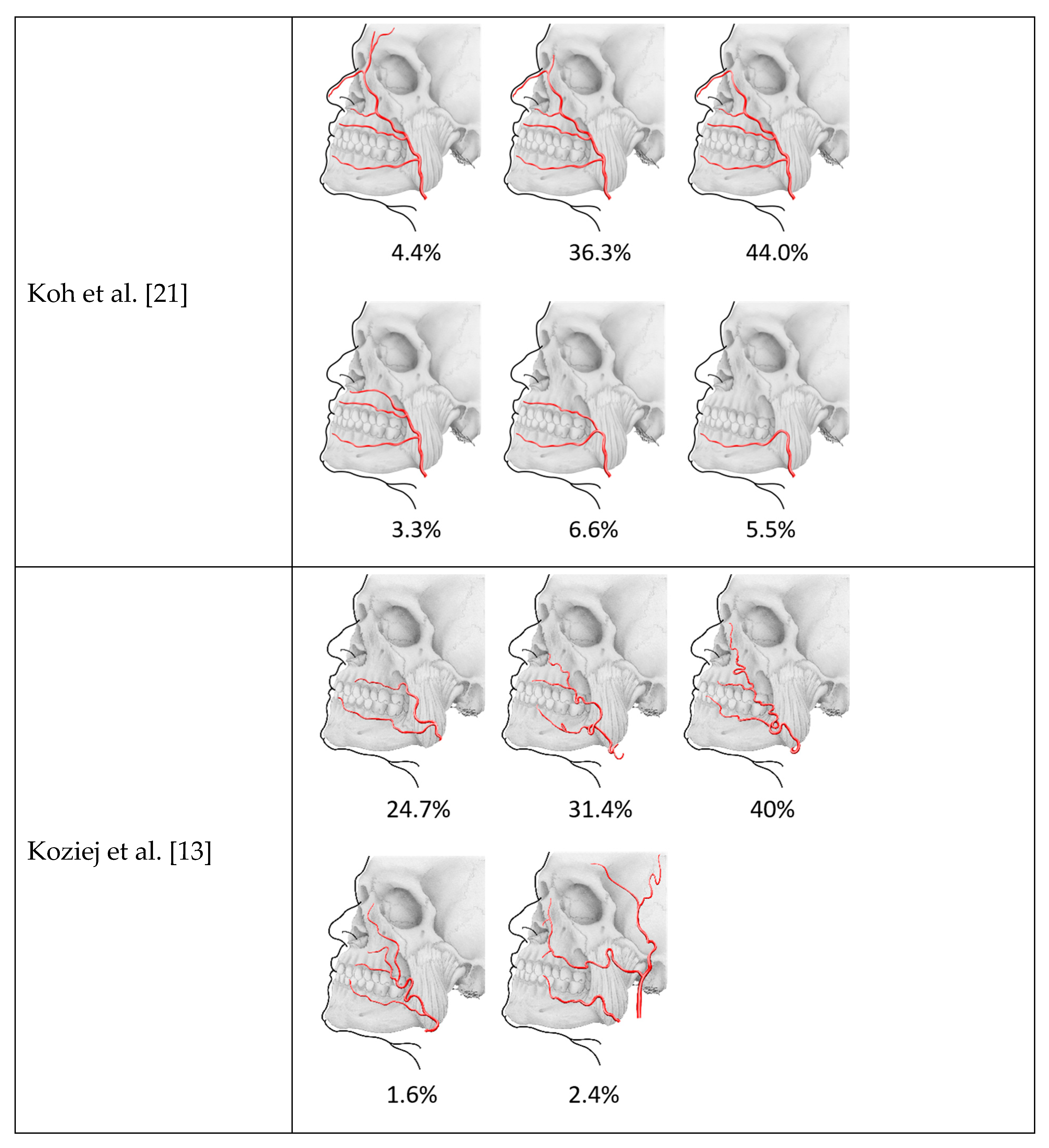
| Koh et al. [21] | Anatomical study | n = 91 | 6 types based on the pattern of final arterial branches | Forehead type: 4.4% Angular type: 36.3% Nasal type: 44% Alar type: 3.3% Superior labial type: 6.6% Inferior labial type: 5.5% | |
| Koziej et al. [13] | Head and neck CT with contrast agent | n = 255 | 5 categories based on termination and branching | Type I (proximal to superior labial): 24.7% Type II (distal to superior labial): 31.4% Type III (textbook course): 40% Type IV (dominant lateral angular): 1.6% Type V (hypoplastic vessel): 2.4% | |
| Lasjaunias et al. [22] | Angiographic findings | Qualitative description of the vascularization of the face and branches of the facial artery and their hemodynamic important anastomoses | Functional arterial monopedicles formed by the following (partly) anastomosing branches: Ascending palatine artery Submaxillary artery Submental artery Inferior masseteric artery Jugal trunk (buccal artery and posterior jugal artery) Middle mental artery Inferior labial artery Superior labial artery Anterior jugal artery Nasal arcade Angular orbital artery | ||
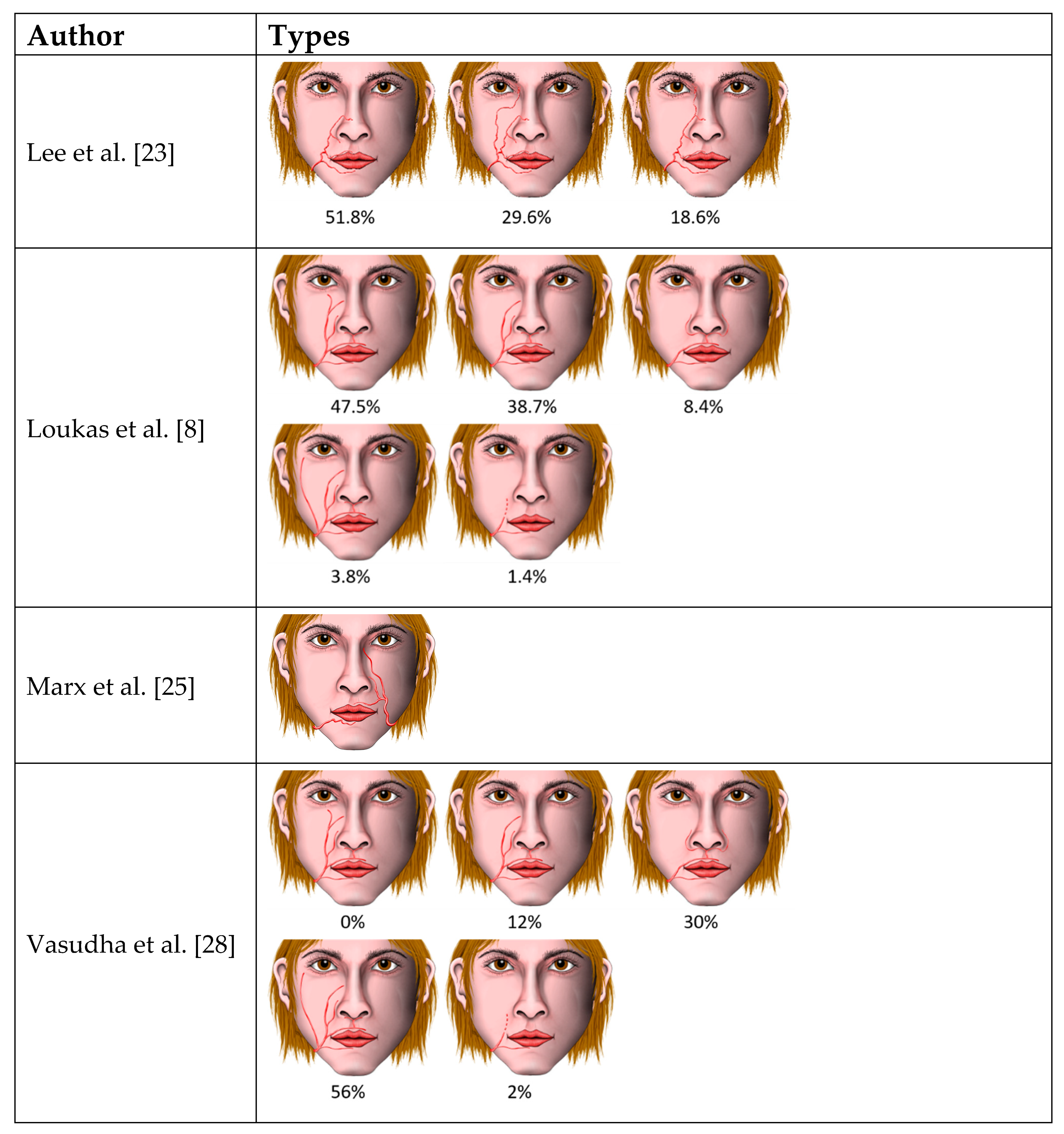
| Lee et al. [23] | Anatomical study | n = 54 | 3 categories based on the branching | Type I (nasolabial type): 51.8% Type II (infraorbital trunk): 29.6% Type III (forehead pattern): 18.6% | |
| Lohn et al. [12] | Anatomical study | n = 201 | 6 categories based on the terminal branch | Type I (angular): 20% Type II (lateral nasal): 48% Type III (inferior alar): 17% Type IV (superior labial): 10% Type V (inferior labial): 3% Type VI (undetected): 2% | |
| Loukas et al. [8] | Anatomical study | n = 200 | 6 types based on the branching and termination with subtypes | Type A (typical arrangement): 47.5% Type B (nasal): 38.7% Type C (superior labial–alar): 8.4% Type D (duplex or long course): 3.8% Type E (rudimentary): 1.4% | Duplex, being an angular artery branching off at the level of the mouth |
| Mağden et al. [24] | Anatomical study | n = 27 | Neurovascular and anatomical features and relations of the premasseteric branch and its branches (origin location, diameter, length, course) | Separate origin of the premasseteric branch from the facial artery in all cases Location determined according to the body of the mandible Diameter 1.12 mm (at level of origin), in 3% of cases larger than the facial artery Observation of the course up to the upper anterior border of the masseter Description of branches and anastomoses | |
| Marx et al. [25] | Case report | Anomalous artery pattern in both sides Termination as inferior labial on the right, missing inferior labial on the left | |||
| Midy et al. [16] | Anatomical study | n = 40 | 4 types based on the termination | Type 1 (angular): 27.5% Type 2 (labial): 40% Type 3 (nasal): 30% Type 4 (abortive): very uncommon, found only once | |
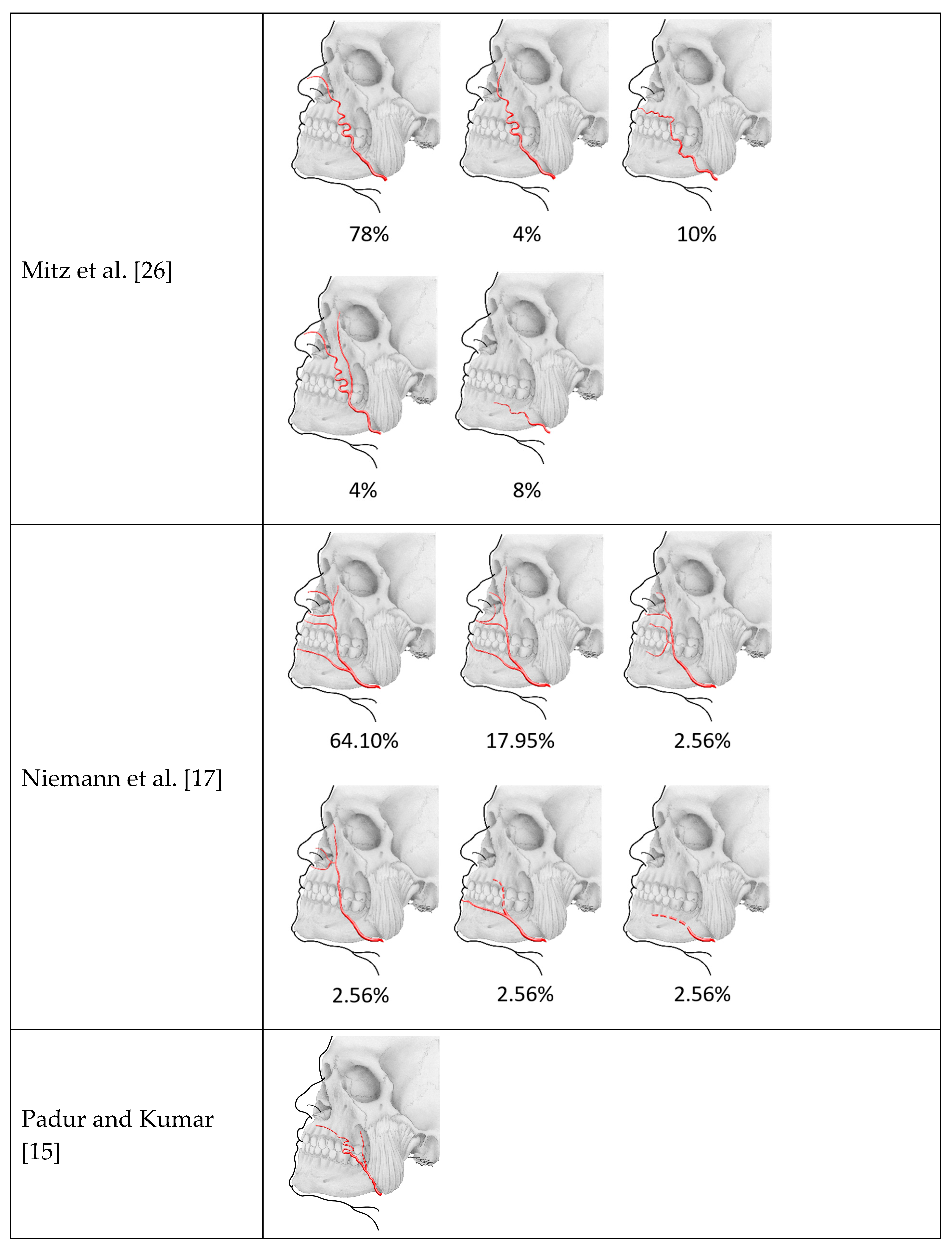
| Mitz et al. [26] | Anatomical study | n = 50 | 5 types based on course, pattern, and termination | Type 1 (nasal): 78% Type 2 (classic): 4% Type 3 (intermediate): 10% Type 4 (duplicate): 4% Type 5 (weak): 8% | |
| Niemann et al. [17] | Anatomical study | n = 39 | 6 types based on the origin of the branches | Type 1, each of the branches arose separately from the facial artery, as in the standard anatomic definition: 64.10% Type 2, the superior labial branch gave off an inferior alar branch: 17.95% Type 3, the superior and inferior labial branches originated from a common trunk and the facial artery had early termination: 2.56% Type 4, the superior and inferior alar branches came from a common trunk and no superior labial and inferior labial branches were present: 2.56% Type 5, the facial artery was rudimentary, which is when the artery terminated after giving off an inferior labial artery, but before reaching the upper lip: 2.56% Type 6, the facial artery was abortive, which is when the facial artery gives off no facial branches: 2.56% | |
| Padur and Kumar [15] | Case report | Bifurcation of the facial artery with posterior branch terminating in the buccal region | |||
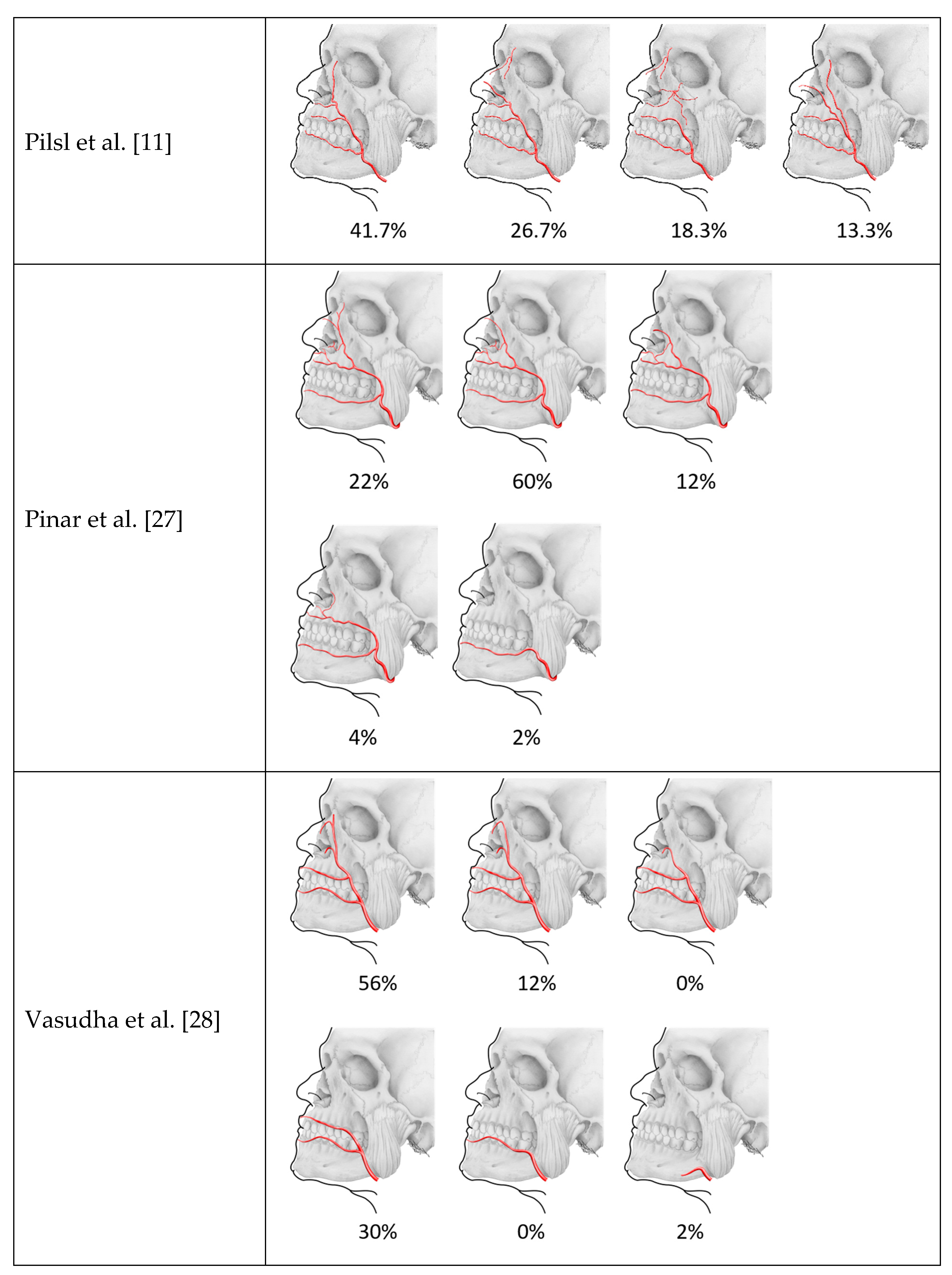
| Pilsl et al. [11] | Anatomical study | n = 60 | 4 types based on course, pattern, and termination | Type 1 (textbook course): 41.7% Type 2 (no angular): 26.7% Type 3 (superior labial): 18.3% Type 4 (two branches): 13.3% | In type 4, the branching was observed at the level of the mouth after release of the inferior labial artery |
| Pinar et al. [27] | Anatomical study | n = 50 | 5 classes based on termination | Type 1 (angular): 22% Type 2 (nasal): 60% Type 3 (alar): 12% Type 4 (superior labial): 4% Type 5 (hypoplastic): 2% | |
| Vasudha et al. [28] | Anatomical study | n = 50 | Classification based on description of Koh et al., Bayram et al. and Loukas et al. | Type 1 (angular): 56% Type 2 (superior labial): 30% Type 3 (inferior labial): 0% Type A (typical arrangement): 0% Type B (nasal): 12% Type C (superior labial—alar): 30% Type D (duplex or long course): 56% Type E (rudimentary): 2% Type I (angular): 56% Type II (lateral nasal): 12% Type III (inferior alar): 0% Type IV (superior labial): 30% Type V (inferior labial): 0% Type VI (undetected): 2% |
Publisher’s Note: MDPI stays neutral with regard to jurisdictional claims in published maps and institutional affiliations. |
© 2021 by the authors. Licensee MDPI, Basel, Switzerland. This article is an open access article distributed under the terms and conditions of the Creative Commons Attribution (CC BY) license (https://creativecommons.org/licenses/by/4.0/).
Share and Cite
Siwetz, M.; Turnowsky, N.; Hammer, N.; Pretterklieber, M.; Wree, A.; Antipova, V. A Rare Case of Facial Artery Branching—A Review of the Literature and a Case Report with Clinical Implications. Medicina 2021, 57, 1172. https://doi.org/10.3390/medicina57111172
Siwetz M, Turnowsky N, Hammer N, Pretterklieber M, Wree A, Antipova V. A Rare Case of Facial Artery Branching—A Review of the Literature and a Case Report with Clinical Implications. Medicina. 2021; 57(11):1172. https://doi.org/10.3390/medicina57111172
Chicago/Turabian StyleSiwetz, Martin, Nicol Turnowsky, Niels Hammer, Michael Pretterklieber, Andreas Wree, and Veronica Antipova. 2021. "A Rare Case of Facial Artery Branching—A Review of the Literature and a Case Report with Clinical Implications" Medicina 57, no. 11: 1172. https://doi.org/10.3390/medicina57111172