Reconstruction for Defects of Total Nail Bed and Germinal Matrix Loss with Acellular Dermal Matrix Coverage and Subsequently Skin Graft


Abstract
:1. Introduction
2. Materials and Methods
2.1. Patients
2.2. Surgical Procedures
2.3. Clinical Results
3. Case Illustrations
3.1. Case 1
3.2. Case 2
4. Discussion
5. Conclusions
Author Contributions
Funding
Conflicts of Interest
References
- Dika, E.; Fanti, P.A.; Patrizi, A.; Misciali, C.; Vaccari, S.; Piraccini, B.M. Mohs Surgery for Squamous Cell Carcinoma of the Nail Unit: 10 Years of Experience. Dermatol. Surg. 2015, 41, 1015–1019. [Google Scholar] [CrossRef] [PubMed]
- Chow, W.T.; Bhat, W.; Magdub, S.; Orlando, A. In situ subungual melanoma: Digit salvaging clearance. J. Plast. Reconstr. Aesthet. Surg. 2013, 66, 274–276. [Google Scholar] [CrossRef] [PubMed]
- Husain, Z.; Allawh, R.M.; Hendi, A. Mohs micrographic surgery for digital melanoma and nonmelanoma skin cancers. Cutis 2018, 101, 346–352. [Google Scholar]
- Cochran, A.M.; Buchanan, P.J.; Bueno, R.A., Jr.; Neumeister, M.W. Subungual melanoma: A review of current treatment. Plast Reconstr. Surg. 2014, 134, 259–273. [Google Scholar] [CrossRef] [PubMed]
- Yun, M.J.; Park, J.U.; Kwon, S.T. Surgical options for malignant skin tumors of the hand. Arch. Plast. Surg. 2013, 40, 238–243. [Google Scholar] [CrossRef] [PubMed] [Green Version]
- Martin-Playa, P.; Foo, A. Approach to Fingertip Injuries. Clin. Plast. Surg. 2019, 46, 275–283. [Google Scholar] [CrossRef] [PubMed] [Green Version]
- Lai, C.S.; Lin, S.D.; Yang, C.C. The reverse digital artery flap for fingertip reconstruction. Ann. Plast. Surg. 1989, 22, 495–500. [Google Scholar] [CrossRef] [PubMed]
- Idone, F.; Sisti, A.; Tassinari, J.; Nisi, G. Fenestrated Adipofascial Reverse Flap: A Modified Technique for the Reconstruction of Fingertip Amputations. J. Investig. Surg. 2017, 30, 353–358. [Google Scholar] [CrossRef] [PubMed]
- Yang, J.; Wang, T.; Yu, C.; Gu, Y.; Jia, X. Reconstruction of large area defect of the nail bed by cross finger fascial flap combined with split-thickness toe nail bed graft: A new surgical method. Medicine (Baltimore) 2017, 96, e6048. [Google Scholar] [CrossRef] [PubMed]
- Tang, J.B.; Elliot, D.; Adani, R.; Saint-Cyr, M.; Stang, F. Repair and reconstruction of thumb and finger tip injuries: A global view. Clin. Plast. Surg. 2014, 41, 325–359. [Google Scholar] [CrossRef] [PubMed]
- Idone, F.; Sisti, A.; Tassinari, J.; Nisi, G. Cooling Composite Graft for Distal Finger Amputation: A Reliable Alternative to Microsurgery Implantation. In Vivo 2016, 30, 501–505. [Google Scholar] [PubMed]
- Laoulakos, D.H.; Tsetsonis, C.H.; Michail, A.A.; Kaxira, O.S.; Papatheodorakis, P.H. The dorsal reverse adipofascial flap for fingertip reconstruction. Plast. Reconstr. Surg. 2003, 112, 121–125. [Google Scholar] [CrossRef] [PubMed]
- Sisti, A.; Oliver, J.D.; Nisi, G. Fenestrated adipofascial reverse flap for the reconstruction of fingertip amputations. Microsurgery 2019, 39, 575. [Google Scholar] [CrossRef] [PubMed]
- Fiedler, D.K.; Barrett, J.E.; Lourie, G.M. Nail Bed Reconstruction Using Single-Layer Bovine Acellular Dermal Matrix. J. Hand. Surg. Am. 2017, 42, e67–e74. [Google Scholar] [CrossRef] [PubMed] [Green Version]
- Hsieh, S.C.; Chen, S.L.; Chen, T.M.; Cheng, T.Y.; Wang, H.J. Thin split-thickness toenail bed grafts for avulsed nail bed defects. Ann. Plast. Surg. 2004, 52, 375–379. [Google Scholar] [CrossRef] [PubMed]
- Delia, G.; Casoli, V.; Sommario, M.; Risitano, G.; D’Alcontres, F.S.; Colonna, M.R. Homodigital dorsal adipofascial reverse flap: Anatomical study of distal perforators and key points for safe dissection. J. Hand Surg. Eur. 2010, 35, 454–458. [Google Scholar] [CrossRef] [PubMed]


| Case | Age | Sex | Past History | Location | Biopsy Report | Pathology With TNM Stage | Stage 1 Surgery | Stage 2 Surgery | Operation Interval | FTSG Take Rate | Follow up Period |
|---|---|---|---|---|---|---|---|---|---|---|---|
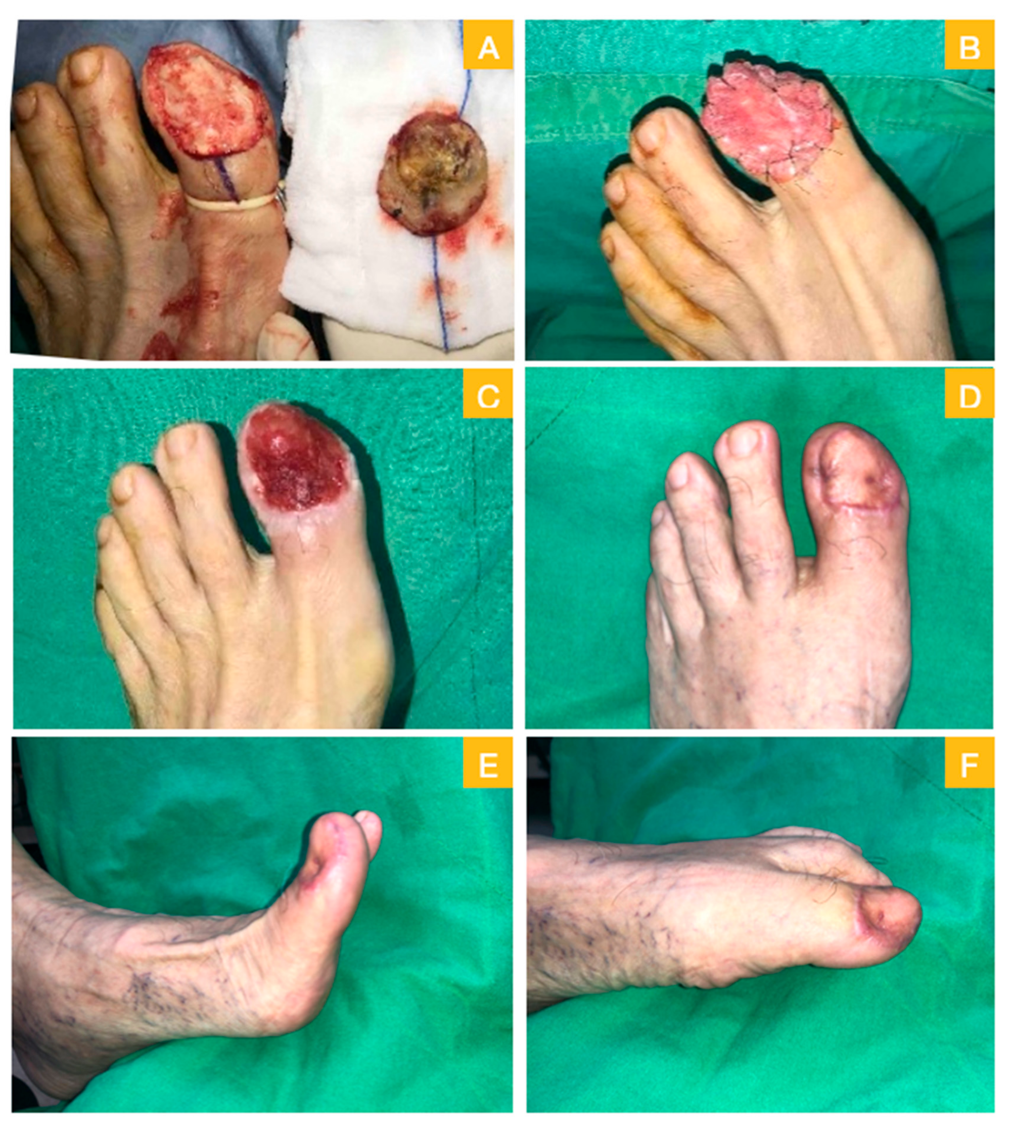
| 1 | 77 | M | HTN, prostate adenocarcinoma | left big toe | melanoma in situ | no residual tumor (pTisN0) | wide excision + PELNAC® | FTSG (3 × 3 cm2) | 21 days | 100% | 13 months |
| 2 | 57 | F | HTN, HBV, breast fibroadenoma | right big toe | malignant melanoma | lentigo maligna (pTisN0) | wide excision + PELNAC® | FTSG (3 × 4.5cm2) | 21 days | 100% | 10 months |
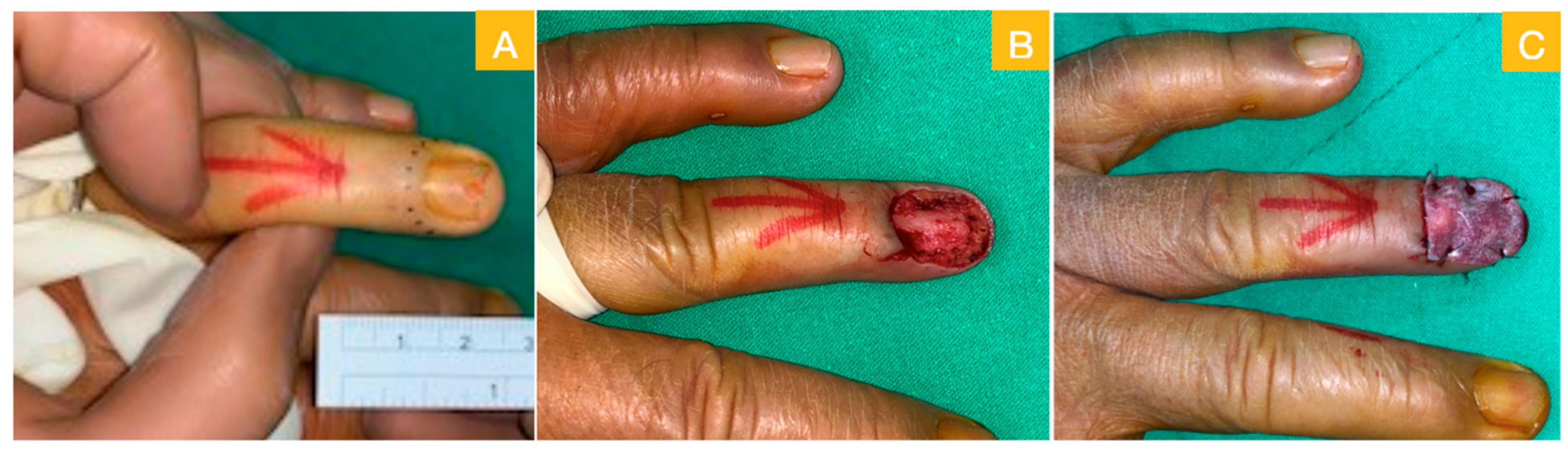
| 3 | 64 | M | HTN, dyslipidemia | left ring finger | SCC | no residual tumor (cT2N0M0 stage 2) | wide excision + PELNAC® | FTSG (2 × 1.5 cm2) | 21 days | 100% | 9 months |
| 4 | 31 | M | nil | right big toe | nil, clinical suspected melanoma | hyperpigmentation in basal layer of epidermis | wide excision + PELNAC® | FTSG (3 × 2 cm2) | 25 days | 98% | 5 months |
© 2020 by the authors. Licensee MDPI, Basel, Switzerland. This article is an open access article distributed under the terms and conditions of the Creative Commons Attribution (CC BY) license (http://creativecommons.org/licenses/by/4.0/).
Share and Cite
Liu, T.-H.; Hsieh, M.-C.; Chou, P.-R.; Huang, S.-H. Reconstruction for Defects of Total Nail Bed and Germinal Matrix Loss with Acellular Dermal Matrix Coverage and Subsequently Skin Graft. Medicina 2020, 56, 17. https://doi.org/10.3390/medicina56010017
Liu T-H, Hsieh M-C, Chou P-R, Huang S-H. Reconstruction for Defects of Total Nail Bed and Germinal Matrix Loss with Acellular Dermal Matrix Coverage and Subsequently Skin Graft. Medicina. 2020; 56(1):17. https://doi.org/10.3390/medicina56010017
Chicago/Turabian StyleLiu, Tsung-Hsien, Meng-Chien Hsieh, Ping-Ruey Chou, and Shu-Hung Huang. 2020. "Reconstruction for Defects of Total Nail Bed and Germinal Matrix Loss with Acellular Dermal Matrix Coverage and Subsequently Skin Graft" Medicina 56, no. 1: 17. https://doi.org/10.3390/medicina56010017