Article

First Survey of Heterobranch Sea Slugs (Mollusca, Gastropoda) from the Island Sangihe, North Sulawesi, Indonesia

1
Centre of Molecular Biodiversity, Zoological Research Museum Alexander Koenig, 53113 Bonn, Germany
2
Faculty of Mathematics and Natural Sciences, Sam Ratulangi University, Manado 95115, Indonesia
3
Politeknik Nusa Utara, Tahuna 95812, Sangihe Islands Regency, Indonesia
4
Faculty of Fisheries and Marine Science, Sam Ratulangi University, Manado 95115, Indonesia
5
Minaesa Institute of Technology, Tomohon 95439, Indonesia
6
Institute for Pharmaceutical Biology, Rheinische Friedrich-Wilhelms-University, 53115 Bonn, Germany
7
Institute for Insect Biotechnology, Justus-Liebig_University Giessen, 35392 Giessen, Germany
8
Fraunhofer Institute for Molecular Biology and Applied Ecology, Department of Bioresources, 35394 Giessen, Germany
*
Author to whom correspondence should be addressed.
Deceased.
Diversity 2019, 11(9), 170; https://doi.org/10.3390/d11090170
Submission received: 23 August 2019 / Revised: 10 September 2019 / Accepted: 12 September 2019 / Published: 17 September 2019
(This article belongs to the Section Marine Diversity)

Abstract

:
Indonesia is famous for its underwater biodiversity, which attracts many tourists, especially divers. This is also true for Sangihe Islands Regency, an area composed of several islands in the northern part of North Sulawesi. However, Sangihe Islands Regency is much less known than, e.g., Bunaken National Park (BNP, North Sulawesi). The main island, Sangihe, has recently experienced an increase in tourism and mining activities with potentially high impact on the environment. Recently, monitoring projects began around BNP using marine Heterobranchia as indicators for coral reef health. No information about this taxon exists from the remote islands in North Sulawesi. The present study represents the first monitoring study ever and focuses on marine Heterobranchia around Sangihe. In total, 250 specimens were collected, which could be assigned to Sacoglossa (3), Anthobranchia (19), and Cladobranchia (1). Despite the low number (23 versus 172 in BNP), at least eight species (35%) are not recorded from BNP, probably indicating differences in habitat, but also influence of a strong El Niño year in 2016. Here we also report for the first time a Chromodoris annae specimen mimicking C. elisabethina, and the discovery of a new Phyllidia species.

1. Introduction

Indonesia is an archipelagic country with a coastline of more than 100,000 km. Coral reefs, sea grasses, and mangrove forests cover approximately 50,875 km2, although this number does not take into consideration the remote areas [1,2]. These tropical ecosystems with a high species and habitat diversity have a tremendous ecological and economic value to nature and humans. They contribute substantially to the community’s income as well as to the national economy. However, many anthropogenic activities form a threat to these natural habitats. Suharsono [3] found that the condition of more than 20% of Indonesian coral reefs were in poor condition and only 6.5% were considered healthy. More recent studies in Indonesia suggest the additional decline of healthy reefs influenced by natural disturbances (e.g., Utama and Hadi, [4]) and up to 50% are severely damaged (e.g., Rudianto and Bintoro [5]).
North Sulawesi is known as a mega-diverse area, and therefore very popular for diving and snorkeling tourists. Thus, the pressure on the reefs has increased dramatically in the last few years. Based on Badan Pusat Statistik Provinsi Sulawesi Utara [6], the number of foreign visitors visiting North Sulawesi Province, via the International Airport of Sam Ratulangi, Manado, approached nearly 11,000 visitors alone in February 2018. This is an increase of 27% compared to January 2018. Comparing foreign visitors in February 2018 with February 2017, the number augmented by more than 100% [6]. Thus, the pressure on the reefs has increased dramatically in the last years. A few local studies conducted in Bunaken National Park (BNP), North Sulawesi, over 10 years clearly indicate a declining state of coral coverage and coral reef fish, and this is related to an increased number of local and foreign visitors, in addition to an increased number of permanent residents [7]. Another study identified diving and snorkeling activities as a major source of the decline in living coral coverage by comparing different sites around Bunaken Island [8]. Undisturbed areas had a live coral coverage of nearly 55% in 3 m depths, while areas with snorkelers and divers showed coverage of only 17% at this depth.
Sangihe Islands in North Sulawesi Province is less known to tourists. It is one of the most northern groups of islands in Indonesia, with Sangihe as the largest island covering an area of approximately 500 km2. The area geographically connects North Sulawesi with Mindanao (Philippine Islands) and forms the eastern boundary of the Celebes Sea. However, biogeographically it is still part of the Wallacea, marked by the Wallace line, which runs between Sangihe Islands and the Philippines. Sangihe has come into focus recently by advertising adventurous diving tourism, including visits to the active underwater volcano Mahengetang in a depth of less than 10 m [9]. Being promoted recently as one of the tourist destinations in the Sangihe Islands Regency, the area is liable to experience a huge pressure on its environment in the near future by many more visitors, both national and international, and a higher demand for hotels, resorts, and diving centers. Higher levels of tourist activities are usually accompanied by threats to ecosystems, such as increased farming, aquaculture, and fisheries due to additional needs of temporary visitors and/or permanent residents. Additionally, Sangihe has come into the focus of mining companies. Since 2007, East Asia Minerals Corporation and local partners were granted exploration permits from the local government within an area of 42,000 ha in the south of Sangihe. The first gold and silver production phase within this Sangihe Gold Project was scheduled for the end of 2018, but did not start yet in 2019 [10]. In terms of minimizing the negative impacts on the environment in the future and helping to build up a sustainable use of the natural resources on and around Sangihe, investigation of the biodiversity in this still rather undisturbed region is paramount. In contrast to BNP, which is already highly affected by diving and snorkeling tourism, monitoring activities in Sangihe Islands Regency with only 12 resorts [11] could provide a good opportunity to study the impact of new infrastructure for tourists and their activities in the marine habitats, as well as other economically important activities on the environment.
Diversity and health of coral reefs is reflected by a diversity of marine organisms, including marine Heterobranchia. These sea slugs use a highly diverse food spectrum, with a high affinity to their specific diet. This spectrum covers nearly all sessile organisms (algae, poriferans, cnidarians, ascidians, bryozoans, tunicates). Thus, this group was already used for monitoring coral reef diversity in North Sulawesi [12,13,14,15]. Because marine Heterobranchs are also very attractive to tourists, additional data are and will be available through citizen science due to documentation in websites or personal information and provision of images on personal bases. This was shown lately by Nimbs et al. [16] and Nimbs and Smith [17] where long-term documentation of scientists and recreational divers led to the identification of new tropical species introduced in Port Stephens, on the central New South Wales coast of Australia, and Tasman Sea. In order to monitor potential damage to the environment around Sangihe, irrespective of its original cause, we have started with a first survey in 2016, focusing on marine Heterobranchia. Here we present the first results from this collecting period and compare our results with former studies in Bunaken National Park [13,14] and other areas in and around Indonesia.

2. Materials and Methods
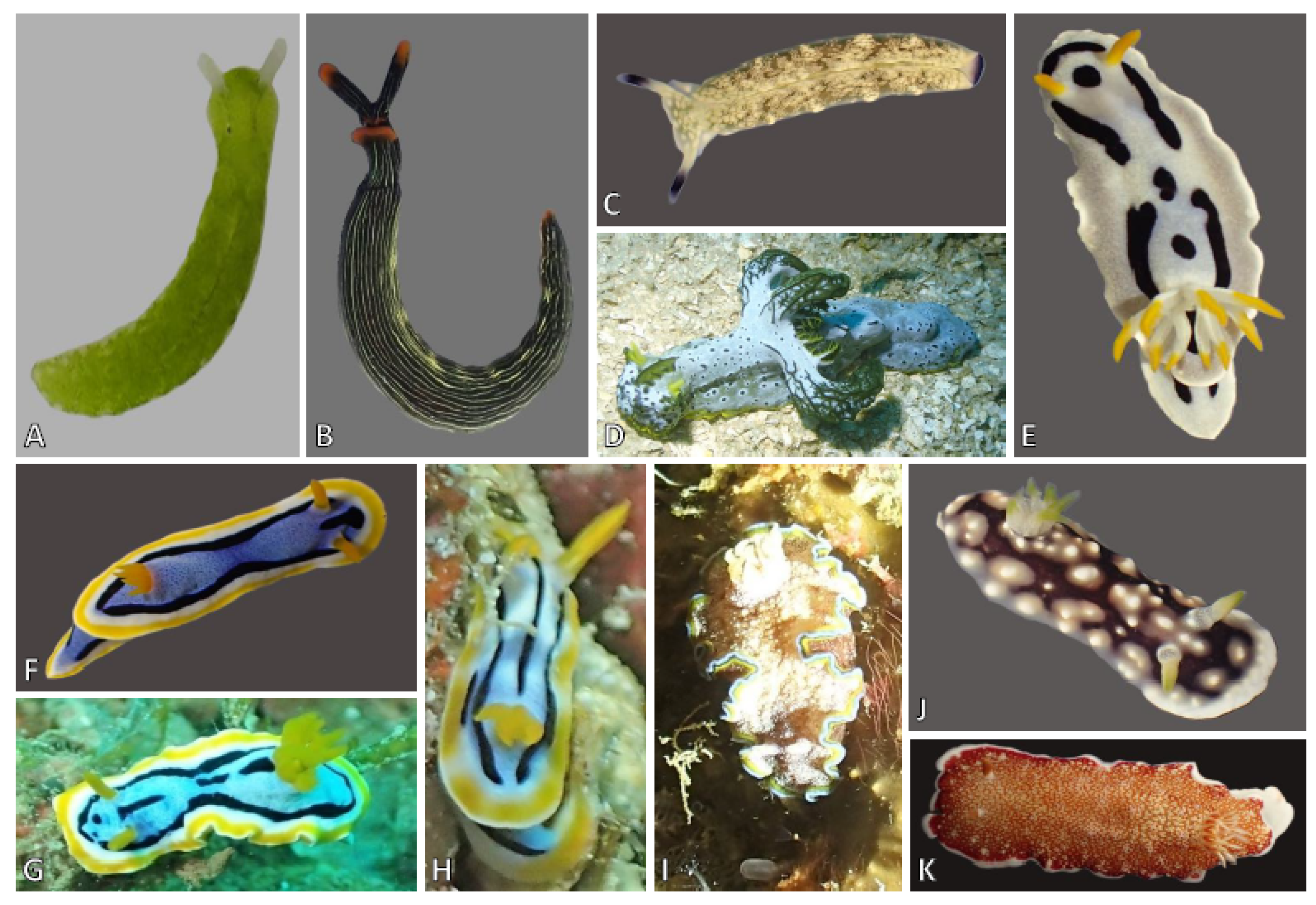
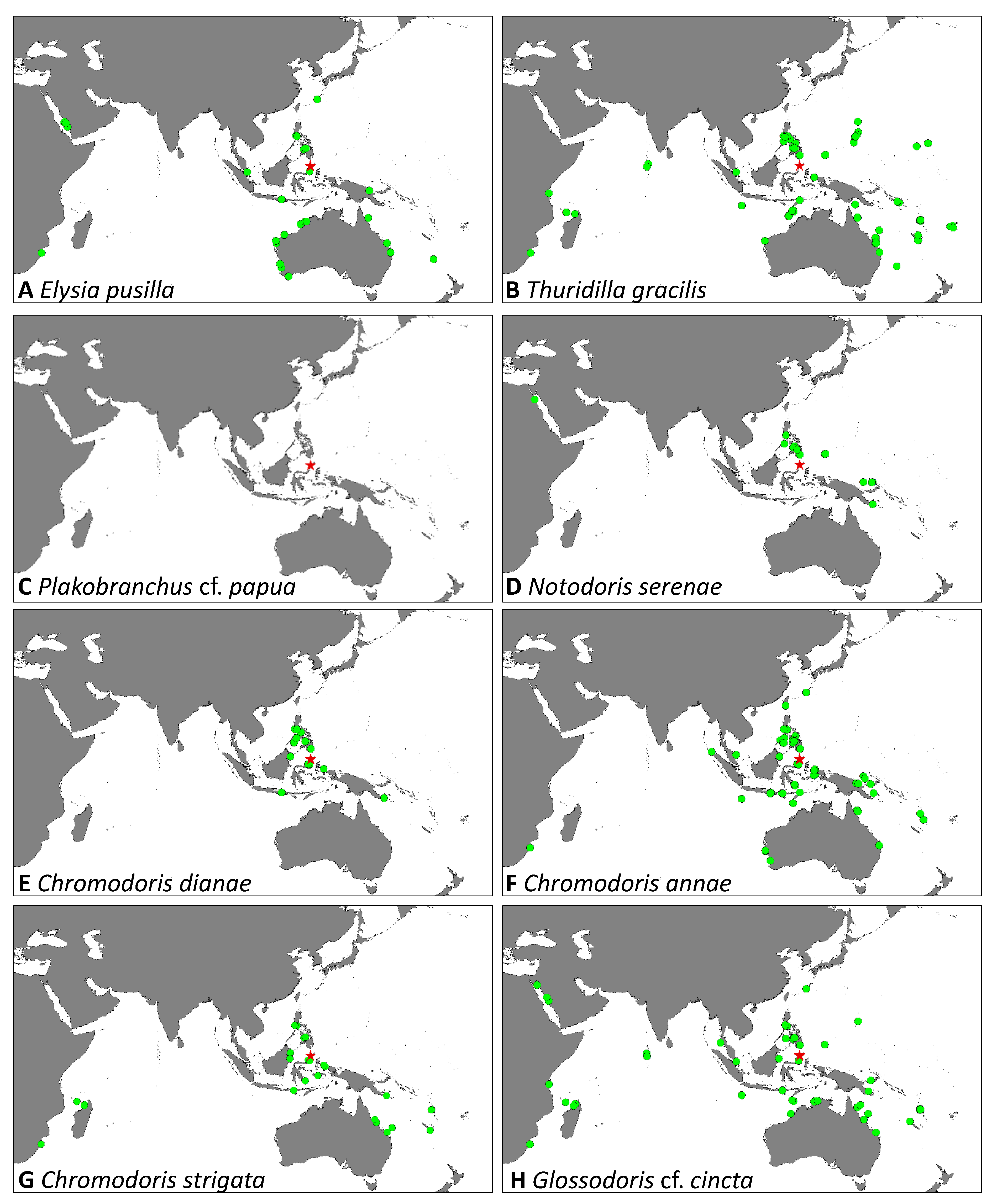
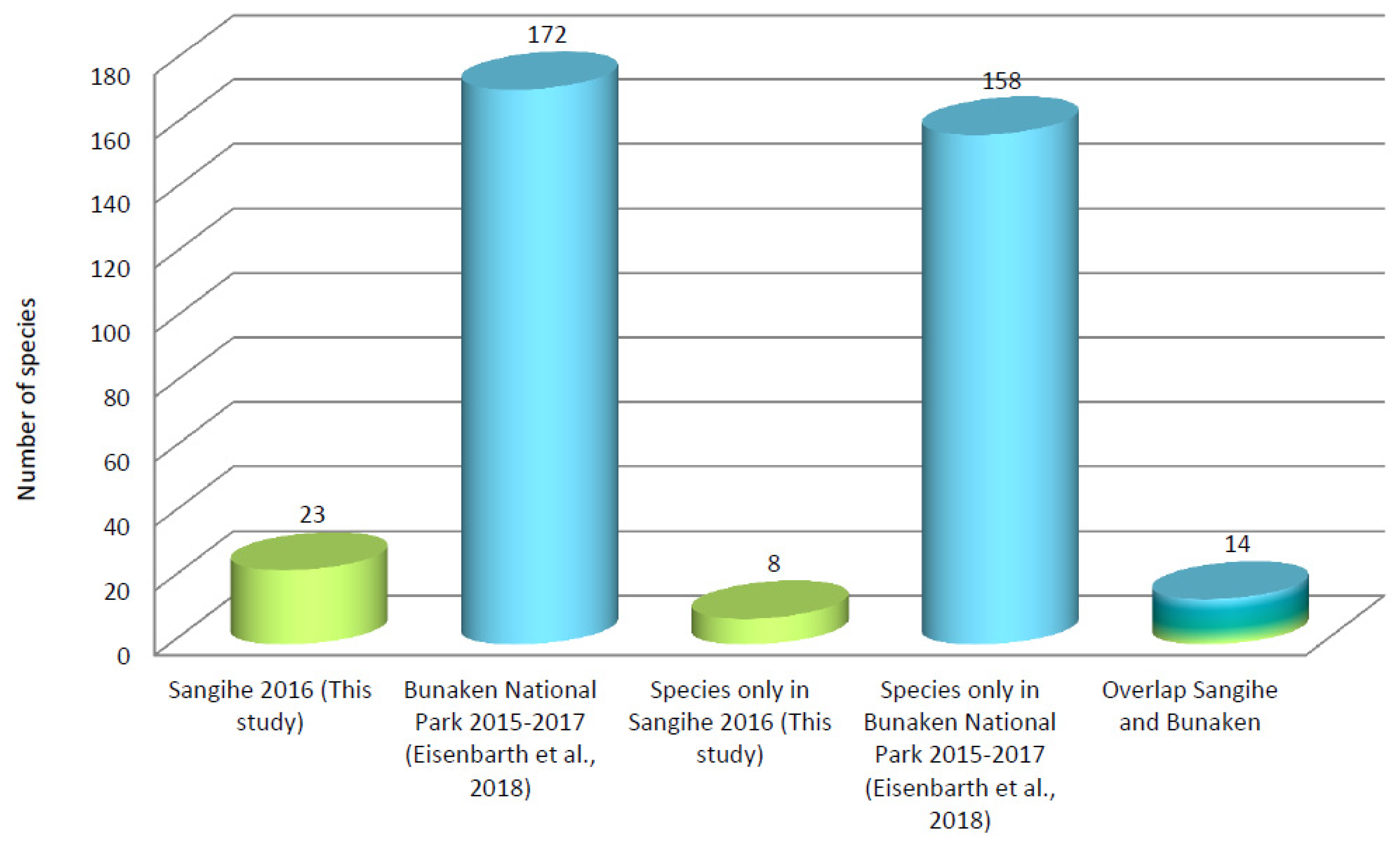
Sampling was carried out during daytime from 3 to 7 August 2016 at seven sites around the island Sangihe (Figure 1, Table 1). Seven scientists and students (three with less and four with good collecting experience from former studies, including the BNP studies) collected in a depth range from the eulittoral to maximum of 28 m. On average, the bottom time for each collecting activity was 60 minutes. In total, underwater searching period correlated to approximately 50 working hours around the island. Additionally, about 10 working hours in total were spent collecting while snorkeling. Specimens were photo-documented in the field on the original substrate before being collected individually. Most specimens were identified before preservation using identification books and original literature [18,19,20,21,22,23,24,25,26,27,28,29,30], as well as websites (e.g., The Sea Slug Forum [31]). Regarding species validity, the World Register of Marine Species [32] was used. The sea slugs were usually preserved in 96% alcohol for further study (including barcoding). All animals were recorded with metadata that are available in the database Diversity Collection (Part of Diversity Workbench) using the data brokerage service of the German Federation for Biological Data (GFBio) [33]. Geographic names were not available for all collection sites. We then used the name of the village close to the respective study area. This is the case for the villages of Palahanaeng and Talengen. Available data on the distribution of respective sea slugs are downloaded from the Global Biodiversity Information Facility (GBIF). For visualization in maps, the geographic information system ArcGIS, release 10.0, was used. The material is registered in the Sam Ratulangi University (UNSRAT, Manado) reference collection under the number SRU2016/01.
Traditional barcoding genes (partial CO1 and partial 16 S) were analyzed for most specimens to verify identification. DNA-Isolation has been carried out by means of QIAgen® DNeasy Blood and Tissue-Kit (QIagen, Hilden, Germany), following the manufacturer’s instructions. Partial sequences of mitochondrial CO1 (about 680 bp) and ribosomal 16 S (about 450 bp) were amplified by polymerase chain reaction using the primers LCO1490-JJ (5′–CHACWAAYCATAAAGATATYGG-3′) and HCO2198-JJ (5′-AWACTTCVGGRTGVCCAAARAATCA-3′) [34] for CO1 and 16 Sar-L (5′-CGCCTGTTTATCAAAAACAT-3′) and 16Sbr-H (5′-CCGGTCTGAACTCAGATCACGT-3’) [35] for 16 S. Amplification of CO1 was performed by an initial step (95 °C for 15 min) followed by 40 touch-down cycles of denaturation (94 °C for 35 s), annealing (55 °C for 90 s) and extension (72 °C for 90 s), with a final extension step 72 °C for 10 min. For 16 S rRNA, the PCR started with an initial step (95 °C for 15 min), denaturation (94 °C for 45 s), followed by 34 touch-down cycles, annealing (56 °C for 45 s), extension (72 °C for 90 s), and final extension step at 72 °C for 10 min. PCR products were sequenced by Macrogen Europe Laboratory (Amsterdam, Netherlands). The software GENEIOUS Pro 7.1.9 (Biomatters Ltd., Auckland, New Zealand) was used to extract the consensus sequence between the primer regions, to construct the final alignments, including sequences from the National Center for Biotechnology Information (NCBI, Bethesda, Maryland, USA), in order to analyze species assignment.

3. Results

250 specimens were collected comprising 23 species (Table 2, Figure 2 and Figure 3). These can be assigned to the Sacoglossa (3) and within the Nudibranchia to Anthobranchia (19) and Cladobranchia (1) (Figure 2 and Figure 3). Out of the 250 specimens, identification was verified by barcoding for 236 specimens (partial CO1 and 16 S genes, see NCBI accession numbers in Table 3). Distribution of the species based on data in GBIF, including the new results from the island Sangihe, are depicted in Figure 4, Figure 5 and Figure 6 with a restriction to the Indian and Western Pacific Ocean.
Systematics
HETEROBRANCHIA
SACOGLOSSA
PLAKOBRANCHOIDEA
Family: Plakobranchidae Gray, 1840
Elysia, Risso, 1818
Elysia pusilla, Bergh, 1871 (Figure 2A and Figure 4A, Table 2)
Description
Three specimens of Elysia pusilla with lengths of 2–6 mm were collected from Mendaku and Tahuna Bay South (Figure 2A). All three specimens had the typical green coloration with whitish rhinophores.
Remarks
One specimen from Mendaku was crawling out of a patch of the chlorophyte Caulerpa racemosa; another one at the same locality was sitting on a Halimeda species with small thalli. The third specimen from Tahuna Bay South was associated with Halimeda cf. macroloba. The species is widely distributed in the Indo-Pacific Ocean (Figure 4A).
Thuridilla, Bergh, 1872
Thuridilla gracilis, Risbec, 1928 (Figure 2B and Figure 4B, Table 2)
Description
Six specimens with lengths of 20–30 mm were collected in front of Palahanaeng village (3 specimens), Talengen village (1 specimen), and in Sapaeng (2 specimens). All specimens show the typical black background and whitish to light green colored fine longitudinal lines. The anterior part of the foot, the tips of the rhinophores, and the tip of the tail show the typical orange color, but the orange rim of the parapodia is only very narrow. No distinct blue spots, which are described from some specimens of the form bayeri, are visible.
Remarks
One specimen from Sapaeng was observed on an algae looking very similar to Dictyota, the other from the same locality and one animal from Palahanaeng village were sitting close to the same algal species. The specimen from Talengen village was crawling on the base of small Halimeda thalli. The remaining two specimens were crawling on unspecified sediment. The species is widely distributed in the Indian and Western Pacific Ocean (Figure 4B).
Plakobranchus, van Hasselt, 1824
Plakobranchus cf. papua, Meyers-Muñoz and van der Velde, 2016 (Figure 2C and Figure 4C, Table 2)
Description
Two specimens of Plakobranchus cf. papua with lengths of 25 and 30 mm were collected in Tahuna Bay South and Mendaku at depths of 5 and 15 m. Our animals show yellowish to white spots of various sizes arranged in a distinct pattern on a darker olive to green background. Our animals differ from the animals described and depicted by Meyers-Muñoz et al. [56] in so far as that they exhibit more spots and thus appeared lighter in color than the animals described from West Papua, Indonesia. However, our animals match with regard to the rhinophores, which are nearly completely violet in color.
Remarks
Recently, Yonow and Jensen [57] reviewed and discussed the complicated situation within the genus Plakobranchus with at least 14 species described from the Pacific Ocean. Many species have never been found again; descriptions were poor, rendering assignment of new material very difficult. The authors depict two specimens, one from Ambon, one from Malaysia, assigned tentatively to P. cf. papua. They look very similar to our specimens, especially in the number of spots and the arrangement of these. Eisenbarth et al. [14] assigned their specimens from Bunaken Island to P. ocellatus. In contrast to our specimens which were collected in depths of 5 and 15 m, the animals collected in BNP lived in the eulittoral. Both animals from our collection were crawling on sediment surrounded by various species of algae. No further distribution records are listed in GBIF (Figure 4C).
NUDIBRANCHIA
DORIDINA
Family: Aegiridae P. Fischer, 1883
Notodoris Bergh, 1875
Notodoris serenae Gosliner and Behrens, 1997 (Figure 2D and Figure 4D, Table 2)
Description
Two specimens of Notodoris serenae with lengths of 60 and 90 mm were collected in Manalu at depths of 24 and 27 m. They show the same typical coloration as depicted in Kaligis et al. [13].
Remarks
Only Notodoris serenae from the family Aegiridae, which usually feeds on hexactinellid sponges, was collected during the present survey. Both animals were crawling on sediment. The species is mainly known from the Coral Triangle (Figure 4D).
Family: Chromodorididae Bergh, 1891
Chromodoris Alder and Hancock, 1855
Chromodoris dianae Gosliner and Behrens, 1998 (Figure 2E and Figure 4E, Table 2)
Description
Seven specimens of Chromodoris dianae with lengths of 5–45 mm were collected in Manalu (1 specimen) and Mendaku (6 specimens) at depths of 15–27 m. The body is elongate and the color of this species is white with a tinge of blue and a pattern of distinct interrupted black lines and spots. The rhinophores are yellowish to orange, whereas the gills are white with yellow tips.
Remarks
Gosliner and Behrens [58] mentioned in their first description the similarity in color with Chromodoris quadricolor (Rüppell and Leuckart, 1830), another pale blue chromodorid. However, C. quadricolor has an orange marginal band. Our Chromodoris dianae specimens are very similar to those depicted in Yonow [27] and Kaligis et al. [13]. Our specimens were also mainly collected from sponges. The mantle glands of C. dianae, which can be seen clearly in the live animal, are well separated from each other and are highly ramified with digitate branches. Species records are mainly confined to the Coral Triangle (Figure 4E).
Chromodoris annae Bergh 1877 (Figure 2F,G and Figure 4F, Table 2)
Description
Thirteen specimens of Chromodoris annae with lengths of 8–41 mm were collected in Manalu, Palahanaeng village, Mendaku, Sapaeng, and Talengen village at depths of 5–23 m (Table 2). Our specimens show the typical blue color with darker miniature spots. They are lacking a mid-dorsal longitudinal line and any small black dots within the blue areas. The rhinophores exhibit the typical yellow color. However, one specimen shows differences in coloration by exhibiting a lighter blue and an interrupted black line in the middle.
Remarks
Some Chromodoris species are difficult to distinguish by color only [13]. Chromodoris elisabethina Bergh, 1877 looks similar to Chromodoris annae, but C. annae usually does not have a median black line and the blue areas of the mantle are not uniform blue as is the case of C. elisabethina (Rudman, 1982). However, we collected one animal in front of Talengen village (Figure 2G) which is quite similar to C. elisabethina: the specimen shows the usual elongate bluish body with the mantle margin encircled by a black, a white, and finally a yellow band. However, the animal mimicking C. elisabethina had additionally a medially lying black line, which was interrupted several times. Barcoding and comparison with our unpublished sequences, and the few available from NCBI, clearly indicate its correct assignment to C. annae, and therefore provides here the first example of mimicry involving C. annae and C. elisabethina. Mimicry forms between members of the Phyllidiidae and Chromodorididae are depicted in several identification books [19,20], and described in Cheney et al. [59] and Padula et al. [60]. However, mimicry between closely related Chromodoris species was described in a broader context for the first time only recently [61]. This is the first example of Chromodoris annae mimicking C. elisabethina. The species is widely distributed in the Indo-Pacific Ocean, including subtropical areas (Figure 4F).
Chromodoris strigata Rudman, 1982 (Figure 2H and Figure 4G, Table 2)
Description
Only one specimen of Chromodoris strigata with a length of 10 mm was collected in Mendaku at a depth of 15 m. The mantle of this specimen shows a white background with bluish tinges. The gills and rhinophores are the same yellow to orange as the mantle border. The yellow band along the mantle rim is interrupted.
Remarks
Although having similarities to many bluish to white Chromodoris species, C. strigata is easily recognised in this color group by the fading blue on white background as well as the areas of light yellow to white in the yellow mantle rim. This renders the species paler than other species [21]. Its distribution is recorded from the Indo-Pacific Ocean (Figure 4G).
Glossodoris Ehrenberg, 1831
Glossodoris cf. cincta (Bergh, 1888) (Figure 2I and Figure 4H, Table 2)
Description
Two specimens of Glossodoris cf. cincta with lengths of 21 and 48 mm were collected in Manalu and Mendaku at depths of 8 and 13 m. The animals show an elongate to oval shape with mottled reddish brown and white on the notum. The gills and rhinophores are brown.
Remarks
Nudibranchs of the genus Glossodoris are moderately large and easily spotted. They are widely distributed in tropical and temperate reef environments around the world [23,62]. Most recently several new species with similar color patterns to G. cincta were described [63,64]. Doriprismatica kyanomarginata Yonow, 2018 differs from our specimen by having a diffuse inner yellow ribbon at the mantle margin, which is characteristic for this new species. Our animal is very close in coloration to Glossodoris acosti Matsuda and Gosliner, 2018. Especially the coloration of the mantle margin with a light blue outermost ring, followed by a dark green and then a lighter yellow-green ring is very similar in both species. However, the rings are wider in G. acosti and furthermore, the gills are mentioned to be larger, forming an arch opening to the posterior and with two distinct spirals. Our animal had all gill branches on one level and the arrangement was forming a complete circle. It thus resembles the animal depicted as Glossodoris cf. cincta in Matsuda and Gosliner [63]. Bergh [65] in his original description of G. cincta mentioned dark brown rhinophores with white dots and the gills with six larger branches in the anterior part, followed by eight smaller ones on each side in the posterior part of the circle. Thus, our specimen also differs from the original description. We therefore only tentatively assign our animal to Glossodoris cincta. The specimen was collected from a brownish sponge. According to GBIF data, Glossodoris cincta shows a broad distribution from the Red Sea until Fiji Islands (Figure 4H). However, difficulties in correct identification probably blur the correct distribution area.
Goniobranchus Pease, 1866
Goniobranchus geometricus (Risbec, 1928) (Figure 2J and Figure 5A, Table 2)
Description
Four specimens of Goniobranchus geometricus with lengths of 10–15 mm were collected in Tahuna Bay South (1 specimen), Palahanaeng village (1 specimen), and Sapaeng (2 specimens) at depths of 6–19 m. Our specimens are rose colored with opaque white tubercles and a network of thick black lines in between the tubercles. The mantle rim is whitish. The translucent white gills and rhinophores have bright green to yellow tips.
Remarks
The color pattern of Goniobranchus geometricus from Sangihe is very similar to that depicted in various identification books, and is also shown by Yonow [27], Kaligis et al. [13], and Eisenbarth et al. [14]. The slug usually can be found under stones or coral rubble [20], where we also found our animals. The species is widely distributed in the Indo-Pacific Ocean (Figure 5A).
Goniobranchus reticulatus (Quoy and Gaymard, 1832) (Figure 2K and Figure 5B, Table 2)
Description
Two specimens of Goniobranchus reticulatus with lengths of 25 and 55 mm were collected in Sapaeng at depths of 6 and 9 m. The specimens show an elongate body with a reticulated network of red lines over the surface mantle. The mantle rim exhibits a narrow white area. The rhinophores are white with red tips. The gills are reddish with the inner rachis opaque white.
Remarks
Our specimen is very similar to the animals depicted by Kaligis et al. [13] and Eisenbarth et al. [14], which were also identified as G. reticulatus. Barcoding and comparison with our unpublished sequences, and the few available from NCBI, indicate its correct assignment to G. reticulatus. However, Yonow [27] discussed Chromodoris inopinata Bergh, 1905 as a very common form in the Indo-Pacific and probably often misidentified as G. reticulatus. C. inopinata shows very similar color patterns as G. reticulatus. No CO1 sequences assigned to C. inopinata are available at NCBI GenBank yet. The records in GBIF show a more limited distribution than is known from G. geometricus, with findings mainly from the Coral Triangle (Figure 5B).
Hypselodoris Stimpson, 1855
Hypselodoris tryoni (Garrett, 1873) (Figure 3A and Figure 5C, Table 2)
Description
Three specimens of Hypselodoris tryoni with lengths of 25–60 mm were collected in Sapaeng at depths of 10–16 m. The specimens show a cream to dirty brown mantle with bluish to dark violet spots. These spots are surrounded by a ring of white pigment and then a paler area. The rim of the mantle is purple. The gill and rhinophores are translucent white with the rachis of the gills brownish and the rachis of the rhinophores purple.
Remarks
Hypselodoris tryoni, Goniobranchus leopardus (Rudman, 1987), and Goniobranchus cavae (Eliot, 1904) are very similar in external appearance, exhibiting a dark cream background color with dark violet round patches surrounded by a light colored area. Additionally, G. cavae can be highly variable in color [30,66]. However, in G. cavae the gills and rhinophores are white with usually purple tips, whereas in H. tryoni, the rachis of the gills and rhinophores shows a purple coloration throughout the full length and the tips of the rhinophores are not distinctively purple. The species has a wide distribution in the Indo-Pacific Ocean with many records also from the subtropics (Figure 5C).
Family: Phyllidiidae Rafinesque, 1814
Phyllidia, Cuvier, 1797
Phyllidia ocellata Cuvier, 1804 (Figure 3B and Figure 5D, Table 2)
Description
Seven specimens of Phyllidia ocellata with lengths of 18–35 mm were collected in Tahuna Bay South (2 specimens), Manalu (2 specimens), Palahanaeng village (1 specimen), and Sapaeng (2 specimens) at depths of 4–18 m. All our animals exhibit the typical yellow coloration with white tubercles, some of which are surrounded by black circles, followed by a thin white line. All other white tubercles are sticking out of the orange background color.
Remarks
Phyllidia ocellata with the yellow to orange background and the tubercles surrounded by black rings is unique in its coloration and therefore cannot be confused with any other Phyllidia species. Gosliner et al. [20] depicted color morphs that lack white tubercles, which were not found during the present study. The species is very common in the Indo-Pacific with a range into subtropics of Australia (Figure 5E).
Phyllidia picta Pruvot-Fol, 1957 (Figure 3C and Figure 5E, Table 2)
Description
Twenty-one specimens of Phyllidia picta with lengths between 13 and 30 mm were collected in Tahuna Bay South (6 specimens), Manalu (3 specimens), Palahanaeng village (2 specimens), Mendaku (2 specimens), Sapaeng (6 specimens), and Talengen village (2 specimens) at depths of 1–15 m. All of our animals show an oval shape, with black reticulate pattern and single yellow tubercles on a blue background. The rhinophores are yellow and the foot sole has no black stripe.
Remarks
Brunckhorst [67] considered Phyllidia picta to be a junior synonym of P. coelestis, but Yonow [26] and Stoffels et al. [68] confirmed its validity. The species is not recorded from Bunaken National Park [13,14] but was reported from Ambon [29] and is also recorded in GBIF from few other places in Indonesia down to Australia (Figure 5D).
Phyllidia spec. (Figure 3D, Table 2)
Description
Figure 3D exhibits an unidentified Phyllidia specimen with a length of 28 mm. It was found only once in Talengen village. This specimen has an elongate to oval shape with greenish to greyish background and black lines between tubercles arranged in ridges. Tubercles are single rather than compound. The rhinophores are yellow. The foot sole shows a black line as is typical for Phyllidia elegans Bergh, 1869, to which it is very similar.
Remarks
The specimen cannot be assigned to any described species. Genetic information indicates no relationship to P. elegans, but to Phyllidia picta; however, therefore its assignment to the genus Phyllidia is confirmed. The specimen of the undescribed Phyllidia species depicted by Eisenbarth et al. [14] looks very different from ours.
Phyllidia madangensis Brunckhorst, 1993 (Figure 3E and Figure 5F, Table 2)
Description
Phyllidia madangensis was collected in front of Talengen village with one specimen with a length of 28 mm. Our animal shows the typical features, the lack of the dark stripe on the foot sole and its overall blackish color. Few white tubercles capped in bright yellow are scattered over the notum. The rhinophores are dark yellow.
Remarks
Phyllidia madangensis is very similar to P. carlsonhoffi Brunckhorst, 1993; however, our animal has smaller tubercles and is more blackish than P. carlsonhoffi, as is depicted by e.g., Gosliner et al. [20]. Rudman [69] illustrated a specimen of P. madangensis with whitish tubercles, similar to our specimen, whereas the tubercles of some P. carlsonhoffi can be more bluish. Phyllidia carlsonhoffi also has tubercles more evenly distributed over the notum, whereas P. madangensis has sparsely scattered tubercles. Brunckhorst [67] described rhinophoral tubercles to occur in all Phyllidia species, but the presence of a small tubercle directly in front of each rhinophoral pocket appears to be unique to P. madangensis. Our specimen did not really show this tubercle. However, the overall appearance and the coloration allow the assignment to P. madangensis, which is a rather rare species (Figure 5F).
Phyllidia coelestis Bergh, 1905 (Figure 3F and Figure 5G, Table 2)
Description
Nine specimens of Phyllidia coelestis with lengths of 7–32 mm were collected in Tahuna Bay South (3 specimens), Manalu (1 specimen), Palahanaeng village (1 specimen), Sapaeng (1 specimen), and Talengen village (3 specimens). The specimens display the typical background blue color with three black lines. The line in the middle is interrupted by few single yellow tubercles, whereas the outer two lines run lateral to the smaller yellow tubercles. The rhinophores are yellow.
Remarks
Phyllidia coelestis is a smaller and widely distributed species (Figure 5G), which has neither a foot stripe nor a median tuberculate ridge. The species can be distinguished from other similar looking phyllidiids, such as P. varicosa, by the central black stripe on the notum, interrupted by large yellow tubercles. Additionally, it has a characteristic black Y-shaped pattern between and in front of the rhinophores. Brunckhorst [70] and Yonow [29] mentioned a dark form that has a central oval region where the ground color is black and only a marginal band around it depicts the bluish-white color.
Phyllidia varicosa, Lamarck, 1801 (Figure 3G and Figure 5H, Table 2)
Description
Fifty-eight specimens with lengths of 7–87 mm were collected at all sampling sites, except ship wreck, at depths of 3–15 m. All specimens show a light blue background with yellow tubercles in rows and blackish lines between these tubercle ridges. The rhinophores are yellowish.
Remarks
Phyllidia varicosa is a large species that can be distinguished by its black stripe at the foot sole, which is absent in most Phyllidia species. It has three to six longitudinal, tuberculate notal ridges [67]. Our animals are quite similar to this description, with an elongate to oval shape, the yellow rhinophores, and the black stripe along the foot sole. The species is very common in the Indo-Pacific Ocean and also occurs in the subtropics of Australia (Figure 5H).
Phyllidiella, Bergh 1869
Phyllidiella lizae Brunckhorst, 1993 (Figure 3H and Figure 6A, Table 2)
Description
Twelve specimens with lengths of 6–68 mm were assigned preliminarily to Phyllidiella lizae. They were collected in Tahuna Bay South (3 specimens), Manalu (3 specimens), Palahanaeng village (1 specimen), Mendaku (3 specimens), and Talengen village (2 specimens). All specimens show the pale pink background, pale pink tubercles and irregular, narrow black lines on the dorsum like a pale ‘x’. The rhinophores are black with pink at the base.
Remarks
Brunckhorst [67] stated that Phyllidiella lizae is recognizable by its pale pink notum with simple, rounded, pale pink tubercles and narrow black lines crossing the dorsum. The rhinophores are black at the tip, pink in the central area, and white at the base. Other distinguishing characters are the pale, pinkish white oral tentacles and foot sole. Our animals match this description, except that the rhinophores are more pinkish at the base, instead of white. However, molecular data indicate cryptic speciation (A.P. unpublished data). Records in GBIF are confined to the coral triangle and the northern parts of Australia (Figure 6A).
Phyllidiella pustulosa (Cuvier, 1804) (Figure 3I–K and Figure 6B, Table 2)
Description
In this study, 77 specimens of Phyllidiella pustulosa with lengths of 12–68 mm were found at all sampling sites in Sangihe in depths of 1–23 m. Our animals have elongate bodies, and diverse color variations; from reddish to pink or even green tubercles surrounded by black lines.
Remarks
Stoffels et al. [68] described Phyllidiella pustulosa with a high intraspecific variation and cryptic speciation, based on molecular analyses. Already Brunckhorst [67] stated that ontogenetic variation also might have contributed to the confusion in the literature. Burghardt et al. [12] assigned one specimen to Phyllidiella nigra, which actually looks very similar to P. pustulosa. In our collection, P. pustulosa is the species with the highest number of color-morphs and our own unpublished molecular data confirm cryptic speciation. Thus, the broad distribution data in GBIF in the tropic and subtropic Indo-Pacific Ocean probably reflect the distribution of several cryptic species (Figure 6B).
Phyllidiella nigra (van Hasselt, 1824) (Figure 3L and Figure 6C, Table 2)
Description
One specimen of Phyllidiella nigra with a length of 29 mm was collected in Sapaeng. This specimen has an elongate body, and its overall color appears blackish with pinkish to brownish tubercles. The tubercles are evenly scattered and not arranged in rows, however they cluster together, as typical for Phyllidiella pustulosa. The rhinophores are black.
Remarks
Brunckhorst [67] distinguished Phyllidiella nigra from conspecifics by its tall, rounded, dark pink to red tubercles, which are evenly distributed (not clustered) over the dorsum (e.g., specimens from Ambon in Yonow [29]). Stoffels et al. [68] already depicted several specimens with tubercles clustering and surrounded by black patterns. In our study, P. nigra appears blackish with darker pinkish tubercles, but the overall appearance is quite similar to the P. nigra specimens depicted in Stoffels et al. [68]. Our genetic analyses group this specimen with published sequences also assigned to P. nigra; however, the quality of our sequence is poor and needs repetition. The species is mainly recorded from the Coral Triangle and Northern Australia (Figure 6C).
Phyllidiopsis Bergh, 1876
Phyllidiopsis krempfi Pruvot-Fol, 1957 (Figure 3M and Figure 6D, Table 2)
Description
Fifteen specimens of Phyllidiopsis krempfi with lengths of 14–50 mm were collected in just one locality, Palahanaeng village, at depths of 13–16 m. The oral tentacles are fused, as is typical for the genus. The animals are elongate to oval and have two black lines on the dorsum extending around the rhinophores, meeting in front of the rhinophores. Additionally, black lines run from these longitudinal lines perpendicularly toward the notum margin, similar to the patterns depicted by Stoffels et al. [68]. The color of our animals varies from reddish (Figure 3L) to pale pink. The rhinophores are black.
Remarks
Phyllidiopsis krempfi is characterised by a predominantly pink coloration and wide shape [67]. Phyllidiopsis gemmata (Pruvot-Fol, 1957) is very similar to P. krempfi, but Tibiriçá et al. [66] described characteristic differences. Phyllidiopsis krempfi has pink rhinophores with only the apical part in black, while P. gemmata has mainly black rhinophores with only the base pinkish [67]. Our animals therefore more resemble P. gemmata. However, the tubercles are simple in P. gemmata, while they are compound in P. krempfi [67], as this is the case in our animals. P. gemmata is also mentioned to be more elongate than P. krempfi. This character is difficult to distinguish, when no other material is available for comparison. Molecular data confirm the assignment to P. krempfi and indicate a higher color variation as previously described. Records in GBIF are rare (Figure 6D) but reach from the Red Sea to Fiji Islands.
Phyllidiopsis shireenae Brunckhorst, 1990 (Figure 3N and Figure 6E, Table 2)
Description
Two specimens of Phyllidiopsis shireenae with a length of 77 and 81 mm were collected in Manalu and Sapaeng at depth of 8 and 15 m. The body is elongate to oval, with a typical longitudinal mid-dorsal ridge, which is covered with large whitish tubercles. The body color is white with opaque white spots and a typical black lining. The foot is also white. The rhinophores are salmon pink.
Remarks
The specimens are very similar to the one depicted by Stoffels et al. [68] from the northern Moluccas and from Ambon [29]. Ours have the two black transversal lines connecting the longitudinal stripes in common with them. One of our specimens shows a black dot in the middle of the white ridge, similar to the animal depicted by Gosliner et al. [20]. Brunckhorst [67] considered the mid-dorsal crest as the characteristic feature of Phyllidiopsis shireenae, which is lacking in most other Phyllidiopsis species. Another characteristic is the salmon pink rhinophores. Phyllidiopsis pipeki Brunckhorst, 1993, Phyllidiopsis burni Brunckhorst, 1993, and Phyllidiopsis fissuratus Brunckhorst, 1993 differ from P. shireenae in having large compound tubercles, black or pale pink rhinophores, and pink to grey ventral coloration (white in P. shireenae) [67]. The species is mainly distributed from the Coral Triangle to Northern Australia (Figure 6E).
Aeolidioidea spec. (Figure 3O, Table 2)
Description
A tiny aeolidid species, probably a juvenile, with a length of 1 mm was collected in Mendaku at 1 m depth. The animal (Figure 3O) is whitish with orange rhinophores and with orange to opaque white cerata. The rhinophores showed irregular swellings or rings. Oral tentacles are short.
Remarks
There are many members of the Aeolidioidea with similar rhinophores, and similar cerata shape and arrangement, but overall habitus resembles probably most a Flabellina species. Proper identification will need barcoding methods, resulting in the complete loss of this specimen for further investigation.

4. Discussion

This is the first study describing the diversity of marine Heterobranchia around the island of Sangihe, Sangihe Islands Regency, North Sulawesi Province. Collecting at different locations (Tahuna Bay South, Ship Wreck, Mendaku, and Manalu, on the eastern coastline; in front of the villages of Palahanaeng and Talengen, and Sapaeng, on the western coastline) ensured the cover of differing habitats and degrees of exposure. Strong currents did not allow extensive sampling in many exposed areas, especially at the outer reef areas and drop offs. This first record is based on a high number of specimens (250), which can be assigned to 23 species (Table 2). The species number cannot compare with the higher numbers of other recent studies at North Sulawesi (Figure 7) [13,14], which might be due to several factors. Collecting time was lower than in BNP, but the differences in habitats were more pronounced. We observed a high sedimentation rate in the water column, resulting in many organisms (sponges, corals, and algae) being covered by a thin layer of silt or mud. This is probably caused by unusually heavy rainfall in this particular season and/or the higher impact of many small river systems close to the collection areas.
Figure 8 and Table 2 provide detailed information about numbers of species/specimens found at the various collection localities around Sangihe. Collection time and effort were similar for all localities. The highest number of sea slug species was found at Sapaeng (13 species, 60 specimens), followed by Tahuna Bay South (11 species, 58 specimens), Palahanaeng village (11 species, 42 specimens), Manalu (11 species, 29 specimens), Mendaku (10 species, 22 specimens), and Talengen village (9 species, 35 specimens). The lowest overall species number was recorded on the Ship Wreck (1 species, 4 specimens), a locality highly influenced by Tahuna harbor and the city of Tahuna. Members of the Anthobranchia (with 238 specimens assigned to 19 species) were present in all seven localities, followed by sacoglossans (11 specimens assigned to 3 species), present in five localities. The Cladobranchia was represented by only one specimen, an unidentified member of the Aeolidioidea.
Recent studies have shown that several species represent cryptic species complexes, while species treated earlier as different taxa are simply color-variants of the same species [60,61,66,71,72,73,74,75]. With regard to our listed taxa, cryptic speciation has been recorded for the genus Plakobranchus [57]. We did not barcode our specimens, but the color patterns allow the tentative assignment to P. papua. We can confirm that Phyllidiella pustulosa is a species complex with similarly colored species or subspecies [68]. Therefore, our animals are tentatively assigned to this species, although they group within different clades (unpublished data; see also Stoffels et al. [68]). Color variation and mimicry appear quite common in Chromodorididae (e.g., Cheney et al. [59], Padula et al. [60], Layton et al. [61], Johnson and Gosliner [62], Epstein et al. [76]). Thus, identification only by color might lead to errors, and therefore we barcoded these taxa to verify identification by including sequences from our specimens into a preliminary phylogenetic analysis of this family (unpublished data). We could therefore identify the first mimicry forms within the species Chromodoris annae exhibiting the color of C. elisabethina.
Phyllidiidae show the highest dominance (three genera represented by 11 species, with 205 specimens) in our study. Of the five valid phyllidiid genera, Reticulidia and Ceratophyllidia were not present in our study. These genera are also not recorded from BNP, but Reticulidia halgerda Brunckhorst and Burn in Brunckhorst [50] was recorded from Ambon [29]. The second most commonly recorded group is the family Chromodorididae. Seventeen chromodoridid genera are recorded by WoRMS. In our study three genera are represented by nine species with 35 specimens; therefore, this family is not well represented in our collection. Only one further anthobranch family besides Chromodorididae was found, the hexactinellid sponge-feeding Aegiridae with Notodoris serenae. Thus, in total 19 anthobranch species are now recorded from Sangihe, in contrast to the 69 anthobranch species mentioned by Eisenbarth et al. [14] from BNP.
Interestingly, the number of cladobranchs with only one very tiny unidentified aeolidid species was extremely low, compared to other study areas close by, e.g., Ambon, Bali, Vietnam, Papua New Guinea, Taiwan, and Hong Kong (Table 4). According to these studies, usually one-quarter to one-third of collected nudibranchs comprise members of Cladobranchia (Table 4). A similar proportion of Anthobranchia to Cladobranchia as seen around Sangihe (20:1) was only recorded from Mauritius [77]. Eisenbarth et al. [14], covering the Bunaken National Park, mentioned 28 species of Aeolidioidea and in total 47 cladobranch species, compared to 69 anthobranch species (Table 4). The low cladobranch number around Sangihe might be explained by the more sheltered sampling localities with a dominance of algae and sponges, and no hydro-dynamically exposed areas so typical of outer reefs and necessary for their hydrozoan prey. The number of sacoglossan species (3) with 11 collected specimens is also rather low. However, our overall numbers are in line with other studies from Indonesia, which show the general dominance of Nudibranchia and particularly the Anthobranchia, versus all other marine heterobranch groups (Table 4). This is also consistent with the overall diversity in these different groups [78,79].
Comparing results from the collecting sites, a few species clearly dominate the various habitats: Phyllidiella pustulosa species complex (77 recorded specimens) was collected from all localities. The species has a high number of records (Figure 6B) which also indicates a very common distribution with high specimens’ numbers; however, it has to be emphasized here that the map depicts actually a species complex with several cryptic species looking all very similar to P. pustulosa. The second most common species around Sangihe was Phyllidia varicosa (58), which is also very common in the Indo-Pacific (Figure 5H). Phyllidia picta (21) was also collected from all sites around Sangihe except Ship Wreck. Phyllidiopsis krempfi was found only at four sampling sites. With 15 specimens, it was quite common around Sangihe, but this species probably is not so commonly distributed in the Indo-Pacific (Figure 6C). It is also not recorded from BNP. Chromodoris annae, Phyllidia coelestis, and Phyllidiella lizae (13, 9, and 8 specimens respectively) were also found at only four sampling sites. Phyllidia madangensis seems to be very rare and our specimen probably represents the only record from Indonesia at the moment (Figure 5F).
In comparison to the study by Eisenbarth et al. [14] (2018) covering the Bunaken National Park (BNP) and including several collection periods between 2015 and 2017, the number of species is much lower (23 versus 172 species) (Figure 7). When including a former collection period in 2003 [12], the total species number increases to 215 in BNP. Interestingly, we collected seven species that are not yet recorded from BNP (Figure 7), despite the extensive studies around this area. Three of them were very common around Sangihe: Phyllidia picta (21 specimens), Phyllidiella lizae (12 specimens), and Phyllidiopsis krempfi (15 specimens). The other four were less common: Phyllidia madangensis (1 specimen), Phyllidiella nigra (1 specimen), Phyllidiopsis shireenae (2 specimens), and Plakobranchus cf. papua (2 specimens). An undescribed Phyllidia species was also collected, which is not recorded from BNP or any other locality. Since nothing can be said about the affiliation of the small aeolidid, the number might even be nine. Overlap of species when comparing these two areas in North Sulawesi was therefore less than 70%, despite the rather short distance of approximately 200 km.
By comparing our preliminary results on the largest island of the Sangihe Islands Regency, not only with the studies from North Sulawesi, but also with other studies from Indonesia and nearby countries, the overlap of species lies mainly in the most common phyllidiid species, including the Phyllidiella pustulosa complex and Phyllidia varicosa, as well as the chromodorid Chromodoris annae. Sangihe is still heavily under-sampled and more collecting events are necessary to better understand the marine Heterobranch fauna from this highly remote area. However, differences outlined here between species composition clearly show the distinctiveness of this region from other areas close by. With this first sampling period, we have created the first baseline for future biodiversity studies and monitoring projects, especially with regard to human activities.

Author Contributions

All authors except N.U. and A.P. were involved in collecting the animals. N.U., A.P., and H.W. analyzed the material; N.U. and H.W. wrote the manuscript and designed the figures; all other authors contributed with comments and corrections to the manuscript. All authors read and approved the final manuscript.

Funding

The study was funded by the Federal Ministry of Education and Research (BMBF) in the frame of the “Biodiversity and Health - from biodiversity to biomedical innovation” program (Indobio, grants 16GW0117K and 16GW0118) to H. Wägele, G.M. König, and T.F. Schäberle. Nani Undap and Adelfia Papu have grants provided by the German Academic Exchange System (DAAD).

Acknowledgments

The authors thank the Republic of Indonesia for making this research possible. We are very grateful to staff members of the Politeknik Nusa Utara, Sangihe, Indonesia who supported and helped in the fieldwork. All material was collected in accordance to the Nagoya Protocol, with all necessary permits and ABS papers provided by the director of the Politeknik Nusa Utara, Tahuna, Sangihe Islands Regency, North Sulawesi, Indonesia. No material collected during this study is listed in IUCN or CITES. Special thanks go to Katja Fisch (Bonn) helping in preparing the specimens after collection and Claudia Etzbauer (Bonn) for her support in the molecular laboratory. We thank Morris Flecks (Bonn) for assistance in using the ArcGIS program and Birgit Klasen for her support in data management in the program Diversity Workbench. We also want to thank two anonymous reviewers, as well as Nathalie Yonow (Swansea), who provided many valuable comments, and improved the English.

Conflicts of Interest

The authors declare that they have no conflict of interests.

Availability of Data and Materials

The material was made available by the late Fontje Kaligis. Some material is used for further studies within the project funded by the Federal Ministry of Education and Research, Germany. Metadata of each individual is documented in the database Diversity Collection (Part of Diversity Workbench) using the data brokerage service of the German Federation for Biological Data (GFBio) [33]. Data are publicly available at www.gfbio.org for browsing and the archived data can be downloaded at https://doi.org/10.20363/heterobranchia-sangihe-prj-1.1. Photographs are available from Heike Wägele with copyright from Zoological Research Museum Alexander Koenig, Bonn. Material not used for further studies will be stored in the Sam Ratulangi University collection under the number SRU2016/01. Sequences are uploaded to NCBI.

References

  1. Suharsono. Jenis-Jenis Karang di Indonesia (Corals of Indonesia). In COREMAP Program; Indonesian Institute of Sciences (LIPI) Pusat Penelitian Oseanografi: Jakarta, Indonesia, 2008; p. 382. [Google Scholar]
  2. Wilkinson, C. Status of Coral Reefs of the World; Global Coral Reef Monitoring Network and Reef and Rainforest Research Centre: Townsville, Australia, 2008; p. 152. [Google Scholar]
  3. Suharsono. Condition of coral reef resources in Indonesia. J. Pesisir Lautan 1998, 1, 44–52. [Google Scholar]
  4. Utama, R.S.; Hadi, T.A. Recent coral reef conditions in Weh Island, Aceh Province, Indonesia. Ocean. Life 2018, 2, 47–53. [Google Scholar]
  5. Bintoro, G. Building resilience for communities in the face of damaged coastal ecosystems: A case study in Gerokgak Village, Buleleng Regency, Bali, Indonesia. IOP Conf. Ser. Earth Environ. Sci. 2018, 239, 012042. [Google Scholar]
  6. Perkembangan Pariwisata Sulawesi Utara Bulan Februari 2018. Available online: Sulut.bps.go.id/pressrelease/2018/04/02/393/perkembangan-pariwisata-sulawesi-utara-bulan-februari-2018.html (accessed on 1 May 2019).
  7. Setiawan, F.; Kusen, J.A.; Kaligis, G.J.F. Community change of coral reef fishes in Bunaken National Park, North Sulawesi, Indonesia (Perubahan struktur ikan karang di Taman Nasional Bunaken, Sulawesi Utara). Aquat. Sci. Manag. 2013, 1, 117–123. [Google Scholar]
  8. Towoliu, R. Kondisi terumbu karang pada beberapa pusat penyelamatan di Bunaken, Sulawesi Utara (Coral reef condition in several dive points around coral Bunaken island, North Sulawesi). Aquat. Sci. Manag. 2014, 2, 44–48. [Google Scholar]
  9. Indonesia Tourism. Available online: http://www.indonesia-tourism.com/northsulawesi/sangihe_archipelago.html (accessed on 3 May 2019).
  10. East Asia Minerals Corporation. Available online: https://eastasiaminerals.com/projects-indonesia/sangihe-gold-project/ (accessed on 10 May 2019).
  11. Kabupaten Kepulauan Sangihe Dalam Angka 2017. Available online: https://sangihekab.bps.go.id/publication/2017/08/13/18b493674a849a55b6a0b5ee/kabupaten-kepulauan-sangihe-dalam-angka-2017.html (accessed on 13 August 2017).
  12. Burghardt, I.; Carvalho, R.; Eheberg, D.; Gerung, G.; Kaligis, F.; Mamangkey, G.; Schroedl, M.; Schwabe, E.; Vonnemann, V.; Waegele, H. Molluscan diversity at Bunaken National Park, Sulawesi. J. Zool. Soc. Wallacea 2006, 2, 29–43. [Google Scholar]
  13. Kaligis, F.; Eisenbarth, J.H.; Schillo, D.; Dialao, J.; Schäberle, T.F.; Böhringer, N.; Bara, R.; Reumschüssel, S.; König, G.M.; Wägele, H. Second survey of heterobranch sea slugs (Mollusca, Gastropoda, Heterobranchia) from Bunaken national Park, North Sulawesi, Indonesia—How much do we know after 12 years. Mar. Biodiver. Rec. 2018, 11, 2. [Google Scholar] [CrossRef]
  14. Eisenbarth, J.H.; Undap, N.; Papu, A.; Schillo, D.; Dialao, J.; Reumschüssel, S.; Kaligis, F.; Bara, R.; Schäberle, T.F.; König, G.M.; et al. Marine Heterobranchia (Gastropoda, Mollusca) in Bunaken National Park, North Sulawesi, Indonesia—A follow-up diversity study. Diversity 2018, 10, 127. [Google Scholar] [CrossRef]
  15. Ompi, M.; Lumoindong, F.; Undap, N.; Papu, A.; Wägele, H. Monitoring marine Heterobranchia in Lembeh Strait, North Sulawesi (Indonesia), in a changing environment. AACL Bioflux 2019, 12, 664–667. [Google Scholar]
  16. Nimbs, M.J.; Larkin, M.; Davis, T.R.; Harasti, D.; Willan, R.C.; Smith, S.D.A. Southern range extensions for twelve heterobranch sea slugs (Gastropoda: Heterobranchia) on the eastern coast of Australia. Mar. Biodiv. Rec. 2016, 9, 27. [Google Scholar] [CrossRef]
  17. Nimbs, M.J.; Smith, D.A. Beyond Capricornia: Tropical sea slugs (Gastropoda, Heterobranchia) extend their distributions into the Tasman Sea. Diversity 2018, 10, 99. [Google Scholar] [CrossRef]
  18. Debelius, H.; Kuiter, R.H. Nacktschnecken der Weltmeere; IKAN Unterwasserarchiv: Frankfurt, Germany, 2007; p. 359. [Google Scholar]
  19. Gosliner, T.M.; Behrens, D.W.; Valdès, A. Indo-Pacific Nudibranchs and Sea Slugs: A Field Guide to the World’s Most Diverse Fauna; Sea Challengers: San Francisco, CA, USA, 2008; p. 425. [Google Scholar]
  20. Gosliner, T.M.; Valdès, A.; Behrens, D.W. Nudibranch and Sea Slug Identification Indo-Pacific; New World Publications: Jacksonville, FL, USA, 2015; p. 408. [Google Scholar]
  21. Rudman, W.B. The Chromodorididae (Opisthobranchia, Mollusca) of the Indo-West Pacific: Chromodoris quadricolor, C. lineolata and Hypselodoris nigrolineata color groups. Zool. J. Linn. Soc. 1982, 76, 183–241. [Google Scholar] [CrossRef]
  22. Rudman, W.B. The Chromodorididae (Opisthobranchia, Mollusca) of the Indo-West Pacific: A review of the genera. Zool. J. Linn. Soc. 1984, 81. [Google Scholar] [CrossRef]
  23. Rudman, W.B. The Chromodorididae (Opisthobranchia: Mollusca) of the Indo-West Pacific: The genus Glossodoris Ehrenberg (Casella, H. & A. Adams). Zool. J. Linn. Soc. 1986, 86, 101–184. [Google Scholar]
  24. Rudman, W.B. The Chromodorididae (Opisthobranchia, Mollusca) of the Indo-West Pacific: Chromodoris epicuria, C. aureopurpurea, C. annulata, C. coi and Risbecia tryoni color groups. Zool. J. Linn. Soc. 1987, 90, 305–407. [Google Scholar] [CrossRef]
  25. Yonow, N. Opisthobranchs from the Maldive Islands, including descriptions of seven new species (Mollusca: Gastropoda). Rev. Franç. Aquar. Herpet 1994, 20, 97–130. [Google Scholar]
  26. Yonow, N. Systematic Revision of the Family Phyllidiidae in the Indian Ocean Province: Part 1 (Opisthobranchia: Nudibranchia: Doridoidea). J. Conch. 1996, 35, 483–516. [Google Scholar]
  27. Yonow, N. Results from the Rumphius Biohistorical Expedition to Ambon (1990). Part 11. Doridacea of the families Chromodorididae and Hexabranchidae (Mollusca, Gastropoda, Opisthobranchia, NudibranchiaI). Zool. Meded. 2001, 75, 1–50. [Google Scholar]
  28. Yonow, N. Sea Slugs of the Red Sea; Pensoft Publishers: Sofia, Bulgaria, 2008; p. 304. [Google Scholar]
  29. Yonow, N. Results of the Rumphius Biohistorical Expedition to Ambon (1990). Part 15. The suborder Doridina (Mollusca, Gastropoda, Opisthobranchia, Nudibranchia). Zool. Meded. 2011, 85, 905–956. [Google Scholar]
  30. Yonow, N. Opisthobranchs from the western Indian Ocean, with descriptions of two new species and ten new records (Mollusca, Gastropoda). ZooKeys 2012, 197, 1–129. [Google Scholar] [CrossRef] [Green Version]
  31. The Sea Slug Forum. Available online: www.seaslugforum.net (accessed on 05 June 2019).
  32. WoRMS Editorial Board. World Register of Marine Species. Available online: http://www.marinespecies.org (accessed on 10 June 2019).
  33. Diepenbroek, M.; Glöckner, F.; Grobe, P.; Güntsch, A.; Huber, R.; König-Ries, B.; Kostadinov, I.; Nieschulze, J.; Seeger, B.; Tolksdorf, R.; et al. Towards an Integrated Biodiversity and Ecological Research Data Management and Archiving Platform: The German Federation for the Curation of Biological Data (GFBio). Informatik 2014, 232, 1711–1724. [Google Scholar]
  34. Astrin, J.J.; Stüben, P.E. Phylogeny in cryptic weevils: Molecules, morphology and genera of western Palaearctic Cryptorhynchinae (Coleoptera: Curculionidae); Invertebrate Systematics; CSIRO Publishing: Clayton, Australia, 2008; pp. 503–522. [Google Scholar]
  35. Palumbi, S.R.; Martin, A.; Romano, S.; MacMillan, W.O.; Stice, L.; Grabowski, G. The Simple Fool’s Guide to PCR; Version 2.0; Department of Zoology and Kewalo Marine Laboratory, University of Hawaii: Honolulu, HI, USA, 2002; p. 45. [Google Scholar]
  36. GBIF.org. Available online: https://doi.org/10.15468/dl.mhkexk (accessed on 19 August 2019).
  37. GBIF.org. Available online: https://doi.org/10.15468/dl.a8jpj7 (accessed on 19 August 2019).
  38. GBIF.org. Available online: https://doi.org/10.15468/dl.3vlodn (accessed on 19 August 2019).
  39. GBIF.org. Available online: https://doi.org/10.15468/dl.u1dqtv (accessed on 19 August 2019).
  40. GBIF.org. Available online: https://doi.org/10.15468/dl.2jdtpp (accessed on 19 August 2019).
  41. GBIF.org. Available online: https://doi.org/10.15468/dl.8sm78 (accessed on 19 August 2019).
  42. GBIF.org. Available online: https://doi.org/10.15468/dl.lycuc9 (accessed on 19 August 2019).
  43. GBIF.org. Available online: https://doi.org/10.15468/dl.flgtfy (accessed on 19 August 2019).
  44. GBIF.org. Available online: https://doi.org/10.15468/dl.2xa8go (accessed on 19 August 2019).
  45. GBIF.org. Available online: https://doi.org/10.15468/dl.3fmoxu (accessed on 19 August 2019).
  46. GBIF.org. Available online: https://doi.org/10.15468/dl.xplg0z (accessed on 19 August 2019).
  47. GBIF.org. Available online: https://doi.org/10.15468/dl.uzsfrc (accessed on 19 August 2019).
  48. GBIF.org. Available online: https://doi.org/10.15468/dl.hhqftx (accessed on 19 August 2019).
  49. GBIF.org. Available online: https://doi.org/10.15468/dl.4lqgzh (accessed on 19 August 2019).
  50. GBIF.org. Available online: https://doi.org/10.15468/dl.nhnbso (accessed on 19 August 2019).
  51. GBIF.org. Available online: https://doi.org/10.15468/dl.y94qqg (accessed on 19 August 2019).
  52. GBIF.org. Available online: https://doi.org/10.15468/dl.108oy5 (accessed on 19 August 2019).
  53. GBIF.org. Available online: https://doi.org/10.15468/dl.rgidut (accessed on 19 August 2019).
  54. GBIF.org. Available online: https://doi.org/10.15468/dl.unlgk3 (accessed on 19 August 2019).
  55. GBIF.org. Available online: https://doi.org/10.15468/dl.mimhpp (accessed on 19 August 2019).
  56. Meyers-Muñoz, M.A.; van der Velde, G.; van der Meij, S.E.T.; Stoffels, B.E.M.W.; van Alen, T.; Tuti, Y.; Hoeksema, B.W. The phylogenetic position of a new species of Plakobranchus from West Papua, Indonesia (Mollusca, Opisthobranchia, Sacoglossa). ZooKeys 2016, 594, 73–98. [Google Scholar]
  57. Yonow, N.; Jensen, K.R. Result of the Rumphius Biohistorical Expedition to Ambon (1990). Part 17. The Cephalaspidea, Anaspidea, Pleurobranchida, and Sacoglossa (Mollusca: Gastropoda: Heterobranchia). Arch. Molluskenkd. 2018, 147, 1–48. [Google Scholar] [CrossRef]
  58. Gosliner, T.M.; Behrens, D.W. Five new species of Chromodoris (Mollusca: Nudibranchia; Chromodorididae) from the tropical Indo-West Pacific. Proc. Calif. Acad. Sci. 1998, 50, 139–165. [Google Scholar]
  59. Cheney, K.L.; White, A.; Mudianta, I.W.; Winters, A.E.; Quezada, M.; Capon, R.J.; Mollo, E.; Garson, M.J. Choose your weaponry: Selective storage of a single toxic compound, Latrunculin A, by closely related nudibranch molluscs. PLoS ONE 2016, 11, e0145134. [Google Scholar] [CrossRef] [PubMed]
  60. Padula, V.; Bahia, J.; Stöger, I.; Camacho-Garcia, Y.; Malaquias, M.A.E.; Cervera, J.L.; Schrödl, M. A test of color-base taxonomy in nudibranchs: Molecular Phylogeny and species delimitation of the Felimida clenchi (Mollusca: Chromodorididae) species complex. Mol. Phys. Evol. 2016, 103, 215–229. [Google Scholar] [CrossRef] [PubMed]
  61. Layton, K.K.S.; Gosliner, T.M.; Wilson, N.G. Flexible color patterns obscure identification and mimicry in Indo–Pacific Chromodoris nudibranchs (Gastropoda: Chromodorididae). Mol. Phyl. Evol. 2018, 124, 27–36. [Google Scholar] [CrossRef]
  62. Johnson, R.F.; Gosliner, T.M. Traditional taxonomic groupings mask evolutionary history: A molecular phylogeny and new classification of the chromodorid nudibranchs. PLoS ONE 2012, 7, e33479. [Google Scholar] [CrossRef]
  63. Matsuda, S.B.; Gosliner, T.M. Glossing over cryptic species: Descriptions of four new species of Glossodoris and three new species of Doripsimatica (Nudibranchia: Chromodorididae). Zootaxa 2018, 4444, 501–529. [Google Scholar] [CrossRef]
  64. Yonow, N. Red Sea Opisthobranchia 5: New species and new records of chromodorids from the Red Sea (Heterobranchia, Nudibranchia, Chromodorididae). ZooKeys 2018, 770, 9–42. [Google Scholar] [CrossRef]
  65. Bergh, L.S.R. Malacologische Untersuchungen. In Reisen imArchipel der Philippinen. Zweiter Theil; Semper, C., Ed.; Wissenschaftliche Resultate: Berlin, Germany, 1888; pp. 815–872. [Google Scholar]
  66. Tibiriçá, Y.; Pola, M.; Cervera, J.L. Astonishing diversity revealed: An annotated and illustrated inventory of Nudipleura (Gastropoda: Heterobranchia) from Mozambique. Zootaxa 2017, 1, 1–133. [Google Scholar] [CrossRef]
  67. Brunckhorst, D.J. The systematics and phylogeny of Phyllidiid Nudibranch (Doridoidea). Rec. Austr. Mus. Suppl. 1993, 16, 1–107. [Google Scholar] [CrossRef]
  68. Stoffels, B.E.M.W.; van der Meij, S.E.T.; Hoeksema, B.W.; van Alphen, J.; van Alen, T.; Meyers-Munoz, M.A.; de Voogd, N.J.; Tuti, Y.; van der Velde, G. Phylogenetic relationships within the Phyllidiidae (Opisthobranchia, Nudibranchia). ZooKeys 2016, 605, 1–35. [Google Scholar]
  69. Rudman, W.B. Comment on Phyllidia madangensis from New Caledonia by Jean-François Hervé. Available online: http://www.seaslugforum.net (accessed on 16 June 2019).
  70. Brunckhorst, D.J. Redescription of Phyllidia coelestis Bergh, 1905 (Ophisthobranchia: Nudibranchia: Doridoidea). J. Malac. Soc. Aust. 1989, 10, 35–45. [Google Scholar]
  71. Carmona, L.; Malaquias, M.A.E.; Gosliner, T.M.; Pola, M.; Cervera, J.L. Amphi-Atlantic Geographic distributions and cryptic species in Sacoglossan sea slugs. J. Moll. Stud. 2011, 77, 401–412. [Google Scholar] [CrossRef]
  72. Ornelas-Gatdula, E.; Dupont, A.; Valdès, A. The tail tells the tale; taxonomy and biogeography of some Atlantic Chelidonura (Gastropoda; Cephalaspidea; Aglajidae) inferred from nuclear and mitochondrial gen data. Zool. J. Linn. Soc. 2011, 163, 1077–1095. [Google Scholar] [CrossRef]
  73. Pola, M.; Camacho-García, Y.E.; Gosliner, T.M. Molecular data illuminate cryptic nudibranch species: The evolution of the Scyllaeidae (Nudibranchia; Dendrotina) with a revision of Notobryon. Zool. J. Linn. Soc. 2012, 165, 311–336. [Google Scholar] [CrossRef]
  74. Pola, M.; Roldan, P.; Padilla, S. Molecular data on the genus Okenia (Nudibranchia; Goniodorididae) reveal a new cryptic species from New South Wales (Australia). J. Mar. Biol. Assoc. UK 2014, 94, 587–598. [Google Scholar] [CrossRef]
  75. Matsuda, S.B.; Gosliner, T.M. Molecular phylogeny of Glossodoris (Ehrenberg, 1831) nudibranchs and related genera reveals cryptic and pseudocryptic species complexes. Cladistics. 2017, 34, 41–56. [Google Scholar] [CrossRef]
  76. Epstein, H.E.; Hallas, J.M.; Johnson, R.F.; Lopez, A.; Gosliner, T.M. Reading between the lines: Revealing cryptic species diversity and color patterns in Hypselodoris nudibranchs (Mollusca: Heterobranchia: Chromodorididae). Zool. J. Linnean Soc. 2018, 20, 116–189. [Google Scholar] [CrossRef]
  77. Yonow, N.; Hayward, P.J. Opisthobranches de I’lle Maurice, avec la description de deux espèces nouvelles (Mollusca: Opithobranchia). Rev. Franç Aquar. Herpet 1991, 18, 1–30. [Google Scholar]
  78. Wägele, H. Potential key characters in Opisthobranchia (Gastropoda, Mollusca) enhancing adaptive radiation. Org. Divers. Evol. 2004, 4, 175–188. [Google Scholar] [Green Version]
  79. Goodheart, J.A.; Ellingson, R.A.; Vital, X.G.; Filho, H.C.; McCarthy, J.B.; Medrano, S.M.; Bhave, V.J.; Mendez, K.G.; Jimenez, L.M.; Lopez, G.; et al. Identification guide to the heterobranch sea slugs (Mollusca: Gastropoda) from Bocas del Toro, Panama. Mar. Biodiv. Rec. 2016, 9, 56. [Google Scholar] [CrossRef]
  80. Yonow, N. Results from the Rumphius Biohistorical Expedition to Ambon (1990). Part 16. Nudibranchia- Dendronotina, Arminina, Aeolidina, and Doridina (Mollusca, Gastropoda, Heterobranchia). Arch. Molluskenkd. 2017, 146, 135–172. [Google Scholar]
  81. Tonozuka, T. Opisthobranchs of Bali and Indonesia; Hankyu Communications Co. Ltd.: Tokyo, Japan, 2003. [Google Scholar]
  82. Martynov, A.V.; Korshunova, T.A. Opisthobranch molluscs of Vietnam (Gastropoda: Opisthobranchia). In Benthic Fauna of the Bay of Nhatrang, Southern Vietnam; Britayev, T.A., Pavlov, D.S., Eds.; KMK Scientific Press: Moscow, Russia, 2012; Volume 2, pp. 142–257. [Google Scholar]
  83. Gosliner, T.M. Biodiversity of tropical opisthobranch gastropod faunas. Proc. Int. Coral Reef Symp. Guam 1992, 2, 702–709. [Google Scholar]
  84. Huang, H.-D.; Tsai, Y.-C.; Chen, C.-K.; Hung, H.-T.; Chen, J.-C.; Lin, H.; Lee, K.-S. Diverse opisthobranchs and polyclad flatworms in Houwan, Kenting National Park, Southern Taiwan. Coll. Res. 2015, 28, 55–65. [Google Scholar]
  85. Orr, J. Hong Kong Nudibranchs; Urban Council: Hong Kong, China, 1981. [Google Scholar]
  86. Yonow, N.; Anderson, R.A.; Buttress, S.G. Opisthobranch Molluscs from the Chagos Archipelago, Central Indian Ocean. J. Nat. Hist. 2002, 36, 831–882. [Google Scholar] [CrossRef]
  87. Johnson, S.; Boucher, L.M. Notes on some Opisthobranchia (Mollusca: Gastropoda) from the Marshall Islands, including 57 new records. Pac. Sci. 1983, 37, 251–291. [Google Scholar]
  88. Wägele, H.; Burghardt, I.; Anthes, N.; Evertsen, J.; Klussmann-Kolb, A.; Brodie, G. Species diversity of opisthobranch molluscs on Lizard Island, Great Barrier Reef, Australia. Rec. West. Austr. Mus. 2006, 69, 33–59. [Google Scholar] [CrossRef]
  89. Wells, F.E.; Bryce, C.W. Sea Slugs and Their Relatives of Western Australia; Western Australian Museum: Perth, Australia, 1993; p. 184. [Google Scholar]
  90. Brodie, G.D.; Brodie, J.E. A checklist of the opisthobranchs molluscs of Fiji. J. Malacol. Soc. Austr. 1990, 11, 53–63. [Google Scholar] [CrossRef]
  91. Bouchet, P.; Lozouet, P.; Maestrati, P.; Heros, V. Assessing the magnitude of species richness in tropical marine environments: Exceptionally high numbers of molluscs at a New Caledonia site. Biol J. Linn. Soc. 2002, 75, 421–436. [Google Scholar] [CrossRef]
  92. Marshall, J.G.; Willan, R.C. Nudibranchs of Heron Island, Great Barrier Reef; Backhuys Publishers: Leiden, The Netherlands, 1999; p. 268. [Google Scholar]
  93. Apte, D. Opisthobranch fauna of Lakshadweep Islands, India, with 52 new records to Lakshadweep and 40 new records to India: Part 1. J. Bombay Nat. Hist. Soc. 2009, 106, 162–175. [Google Scholar]
  94. Hervé, J.-F. Guide des Nudibranches de Nouvelle-Calédonie et Autres Opisthobranches; Editions Catherine Ledru: Nouméa, France, 2010; p. 404. [Google Scholar]
  95. Nimbs, M.J.; Smith, D.A. An illustrated inventory of the sea slugs of New South Wales, Australia (Gastropoda: Heterobranchia). R. Soc. Vic. 2016, 128, 44–113. [Google Scholar] [CrossRef]
  96. Bertsch, H. Biogeography of northeast Pacific opisthobranchs: Comparative faunal province studies between point conception, California, USA, and Punta Aguja, Piura, Perú. Persp. Malacol. Mexicana 2010, 13, 219–259. [Google Scholar]
Figure 1. Details on North Sulawesi with collection sites in Sangihe (upper insert, and see also Table 1) and the collection area around Bunaken Island [13,14] (lower insert) for comparison.
Diversity 11 00170 g001
Figure 2. Sacoglossa and Anthobranchia: (A) Elysia pusilla, Elpu16Sa-3; (B) Thuridilla gracilis, Thgr 16Sa-2; (C) Plakobranchus cf. papua, Ploc16Sa-2; (D) Notodoris serenae, Aese16Sa-2; (E) Chromodoris dianae, Chdi16Sa-2; (F) Chromodoris annae, Chan16Sa-2; (G) Chromodoris annae mimicking C. elisabethina, Chel16Sa-1; (H) Chromodoris strigata, Chst16Sa-1; (I) Glossodoris cf. cincta, Glci16Sa-1; (J) Goniobranchus geometricus, Goge16Sa-2; (K) Goniobranchus reticulatus, Gore16Sa-1.
Diversity 11 00170 g002
Figure 3. Anthobranchia and Cladobranchia/Aeolidioidea: (A) Hypselodoris tryoni, Goku16Sa-1; (B) Phyllidia ocellata, Phoc16Sa-3; (C) Phyllidia picta, Phpic_16Sa-13; (D) Phyllidia spec., Phsp3_16Sa-1; (E) Phyllidia madangensis, Phma16Sa-1; (F). Phyllidia coelestis, Phco16Sa-1; (G) Phyllidia varicosa, Phva16Sa-6; (H) Phyllidiella lizae, Phli16Sa-4; (I) Phyllidiella pustulosa complex, Phpu16Sa-29; (J) Phyllidiella pustulosa complex, Phpu16Sa-91; (K) Phyllidiella pustulosa complex, Phpu16Sa-95; (L) Phyllidiella nigra, Phpu 16Sa-64; (M) Phyllidiopsis krempfi Phpu16Sa-58; (N) Phyllidiopsis shireenae, Phsh16Sa-2; (O) Aeolidioidea Flsp16Sa-1.
Diversity 11 00170 g003
Figure 4. Distribution data of respective species in the Indo-Pacific Ocean. Data from this study (Sangihe) and downloaded from GBIF. (A) Elysia pusilla [36]; (B) Thuridilla gracilis [37]; (C) Plakobranchus cf. papua (no data in GBIF available yet); (D) Notodoris serenae [38]; (E) Chromodoris dianae [39]; (F) Chromodoris annae [40]; (G) Chromodoris strigata [41]; (H) Glossodoris cf. cincta [42].
Diversity 11 00170 g004
Figure 5. Distribution data of respective species in the Indo-Pacific Ocean. Data from this study (Sangihe) and downloaded from GBIF. (A) Goniobranchus geometricus [43]; (B) Goniobranchus reticulatus [44]; (C) Hypselodoris tryoni [45]; (D) Phyllidia ocellata [46]; (E) Phyllidia picta [47]; (F) Phyllidia madangensis [48]; (G) Phyllidia coelestis [49]; (H) Phyllidia varicosa [50].
Diversity 11 00170 g005
Figure 6. Distribution data of respective species in the Indo-Pacific Ocean. Data from this study (Sangihe) and downloaded from GBIF. (A) Phyllidiella lizae [51]; (B) Phyllidiella pustulosa [52]; (C) Phyllidiella nigra [53]; (D) Phyllidiopsis krempfi [54]; (N) Phyllidiopsis shireenae [55].
Diversity 11 00170 g006
Figure 7. Comparison of species diversity in this study (Sangihe) with (Bunaken National Park) [14]. Note that one-third of the species collected in Sangihe were not found during surveys in Bunaken National Park.
Diversity 11 00170 g007
Figure 8. Comparison of marine Heterobranch species collected around the island Sangihe. The numbers in front of the species names indicate the number of collected specimens. The numbers after the locality names indicate the number of species collected, followed by the number of specimens.
Diversity 11 00170 g008
Table 1. Details on collection sites (Figure 1). When sites do not have a geographic name, we used the name of the village nearby. Abbreviations of localities are used in Table 2.
NameAbbreviationArea and Geographic DataDate of Collection
Ship WreckShW3°36′28.00″ N 125°29′38.00″ E04.08.2016
Tahuna Bay SouthTBS3°35′59.40″ N 125°29′23.40″ E04.08.2016
Mendaku Men3°22′01.94″ N 125°34′26.67″ E03.08.2016
Palahanaeng (village)Pal3°35′18.92″ N 125°34′26.67″ E07.08.2016
Talengen (village)Tal3°34′49.92″ N 125°34′34.93″ E05.08.2016
Manalu Man3°32′08.87″ N 125°37′25.46″ E06.08.2016
SapaengSap3°34′55.81″ N 125°34′49.04″ E06.08.2016
Table 2. Species records around Sangihe with details about specimens and locality, as well as first authorities. Species recorded in Eisenbarth et al. [14] around BNP are indicated in the last column.
TaxonSpecies NameLocalitiesDepths (m)Number of SpecimensSize (mm)Eisenbarth et al. [14]
TBSShWManPalMenSapTal
SacoglossaElysia pusilla (Bergh, 1871)1---2--232–6x
Thuridilla gracilis (Risbec, 1928)---3-214–10620–30x
Plakobranchus cf. papua (Meyers-Muñoz & van der Velde, 2016)1---1--5–15225,30-
AnthobranchiaNotodoris serenae (Gosliner & Behrens, 1997)--2----24–27260,90x
Chromodoris dianae (Gosliner & Behrens, 1998)--1-6--15–2775–45x
Chromodoris annae (Bergh, 1877)--112545–23138–41x
Chromodoris strigata (Rudman, 1982)----1--15110x
Glossodoris cf. cincta (Bergh, 1888)---1--18, 13221, 48x
Goniobranchus geometricus (Risbec, 1928)1--1-2-6–19410–15x
Goniobranchus reticulatus (Quoy & Gaimard, 1832)2------6, 9225, 55x
Hypselodoris tryoni (Garret, 1873)-----3-10, 16325–60x
Phyllidia ocellata (Cuvier, 1804)2-21-2-4–18716–35x
Phyllidia picta (Pruvot-Fol, 1957)6-322621–152113–30-
Phyllidia spec. (Phsp3_16Sa-1)-----1-1125-
Phyllidia madangensis (Brunckhorst, 1993)------18128-
Phyllidia coelestis (Bergh, 1905)3-11-133–1297–32x
Phyllidia varicose (Lamarck, 1801)19-210215103–15587–87x
Phyllidiella lizae (Brunckhorst, 1993)3-313-25–23126–68-
Phyllidiella pustulosa (Cuvier, 1804)1941115215111–237712–47x
Phyllidiella nigra (van Hasselt, 1824)-----1-8129x
Phyllidiopsis krempfi (Pruvot-Fol, 1957)1-26-6-6–281514–50-
Phyllidiopsis shireenae (Brunckhorst, 1990)--1--1-8, 15277, 81-
CladobranchiaAeolidioidea (Flsp16Sa-1)----1--211-
Table 3. Species used in this study, identification number, and Genbank accession numbers as also mentioned in Diversity Workbench.
FamilySpecies NameIDGenBank Accession Numbers
16 SCO1
Chromodorididae (Bergh, 1891)Chromodoris dianae (Gosliner & Behrens, 1998)Chdi16Sa-1MN104702MN320502
Chdi16Sa-2MN104703MN320503
Chdi16Sa-3MN104704MN320504
Chdi16Sa-4MN104705MN320505
Chdi16Sa-5MN104706MN320506
Chdi16Sa-6MN104707MN320507
Chdi16Sa-7MN104708MN320508
Chromodoris annae (Bergh, 1877)Chan16Sa-1MN104690MN124751
Chan16Sa-2MN104691MN124752
Chan16Sa-3MN104692MN124753
Chan16Sa-4MN104693MN124754
Chan16Sa-5MN104694MN124755
Chan16Sa-6MN104695MN124756
Chan16Sa-7MN104696MN124757
Chan16Sa-8MN104698MN124758
Chan16Sa-9MN104699MN124759
Chan16Sa-10MN104700MN124760
Chan16Sa-11MN104701MN124761
Chan16Sa-12MN104702MN124762
Chel16Sa-1MN104709MN124763
Chromodoris strigata (Rudman, 1982)Chst16Sa-1MN104710MN365022
Glossodoris cf. cincta (Bergh, 1888)Glci16Sa-1MN104711MN339440
Glci16Sa-2MN104712MN339441
Goniobranchus geometricus (Risbec, 1928)Goge16S-1MN104715MN339442
Goge16S-2MN104716MN339443
Goge16S-3MN104717MN339444
Goge16S-4-MN339445
Goniobranchus reticulatus (Quoy & Gaimard, 1832)Gore16Sa-1MN104719MN339446
Gore16Sa-2MN104720MN339447
Hypselodoris tryoni (Garret, 1873)Goca16S-1MN104713MN339448
Goca16S-2MN104714MN339450
Goku16Sa1MN104718MN339449
Phyllidiidae (Rafinesque, 1814)Phyllidia picta (Pruvot-Fol, 1957)Phpic16Sa-1MN217674MN248545
Phpic16Sa-5MN217680MN248543
Phpic16Sa-6MN217675MN248546
Phpic16Sa-8MN217671MN248540
Phpic16Sa-9MN217669MN248539
Phpic16Sa-10MN217672MN248542
Phpic16Sa-11MN217679MN248549
Phpic16Sa-12MN217678MN248547
Phpic16Sa-13MN217676MN248548
Phpic16Sa-14MN217681MN248544
Phsp616Sa-3MN217677MN248550
Phspec116Sa-2MN217670MN248541
Phyllidia spec.Phsp316Sa-1MN217673MN265389
Phyllidia ocellata (Cuvier, 1804)Phoc16S-1MN173896MN173896
Phoc16S-2MN173895MN173895
Phoc16S-4MN173894MN173894
Phoc16S-5MN173893MN173893
Phoc16S-6-MN173892
Phoc16S-7MN173891MN173891
Phyllidia coelestis (Bergh, 1905)Phco16Sa-1MN172238MN234119
Phco16Sa-2MN172237MN234113
Phco16Sa-3MN172236MN234115
Phco16Sa-4MN172235MN234118
Phco16Sa-5MN172234MN234112
Phco16Sa-7MN172233MN234116
Phco16Sa-9MN172232MN234114
Phco16Sa-10MN172231MN234112
Phyllidia varicosa (Lamarck, 1801)Phva16Sa-2MN243776-
Phva16Sa-3MN243779-
Phva16Sa-4MN243778MN248554
Phva16Sa-5MN243774-
Phva16Sa-7MN243747-
Phva16Sa-8MN243735-
Phva16Sa-9MN243783MN248572
Phva16Sa-10MN243750-
Phva16Sa-11MN243761-
Phva16Sa-12MN243781-
Phva16Sa-13MN243760MN248571
Phva16Sa-15MN243782MN248555
Phva16Sa-16MN243775-
Phva16Sa-17MN243759-
Phva16Sa-18MN243780-
Phva16Sa-20MN243758MN248556
Phva16Sa-21MN243734MN248563
Phva16Sa-22MN243777-
Phva16Sa-23MN243773-
Phva16Sa-24MN243757MN248568
Phva16Sa-25MN243746-
Phva16Sa-26MN243733-
Phva16Sa-27MN243771MN248573
Phva16Sa-28MN243748-
Phva16Sa-29MN243745-
Phva16Sa-30MN243740-
Phva16Sa-31MN243770MN248567
Phva16Sa-32MN243768-
Phva16Sa-33MN243767MN248574
Phva16Sa-34MN243756-
Phva16Sa-36MN243772-
Phva16Sa-37MN243755MN248569
Phva16Sa-38MN243763-
Phva16Sa-39MN243744-
Phva16Sa-40MN243754MN248557
Phva16Sa-41MN243739-
Phva16Sa-42MN243749MN248562
Phva16Sa-43MN243764MN248565
Phva16Sa-44MN243766MN248561
Phva16Sa-45MN243741MN248564
Phva16Sa-46MN243738-
Phva16Sa-47MN243737MN248558
Phva16Sa-48MN243753-
Phva16Sa-49MN243743-
Phva16Sa-50MN243742MN248559
Phva16Sa-52MN243765MN248560
Phva16Sa-53MN243762MN248570
Phva16Sa-54MN243752-
Phva16Sa-55MN243751-
Phva16Sa-56MN243769MN248566
Phva16Sa-57MN243736-
Phva16Sa-58MN243732-
Phyllidiella lizae (Brunckhorst, 1993)Phli16Sa-1MN243971MN248575
Phli16Sa-2MN243973MN248577
Phli16Sa-5MN243972MN248576
Phli16Sa-6MN243974MN248578
Phyllidiella pustulosa (Cuvier, 1804)Phpu16Sa-1MN243977MN248624
Phpu16Sa-2MN244015MN248636
Phpu16Sa-3MN243991MN248601
Phpu16Sa-4MN243992MN248606
Phpu16Sa-5MN243996MN248608
Phpu16Sa-6MN244006MN248602
Phpu16Sa-7MN244007MN248594
Phpu16Sa-8MN243999-
Phpu16Sa-9MN243980MN248627
Phpu16Sa-13MN243969MN248581
Phpu16Sa-14-MN248580
Phpu16Sa-15MN243960MN248585
Phpu16Sa-18MN243983MN248632
Phpu16Sa-20-MN248590
Phpu16Sa-23MN243962MN248586
Phpu16Sa-24MN243978MN248625
Phpu16Sa-25MN244008MN248595
Phpu16Sa-26MN243979MN248626
Phpu16Sa-27MN244009MN248596
Phpu16Sa-28MN243970MN248591
Phpu16Sa-29MN243955MN248639
Phpu16Sa-30MN244000MN248620
Phpu16Sa-31MN243963MN248587
Phpu16Sa-33MN243985MN248614
Phpu16Sa-34MN244017MN248637
Phpu16Sa-35MN243957MN248640
Phpu16Sa-36MN244011MN248597
Phpu16Sa-38MN243997MN248609
Phpu16Sa-39MN244010MN248598
Phpu16Sa-40MN243981MN248628
Phpu16Sa-46MN244001MN248620
Phpu16Sa-48MN243975MN248590
Phpu16Sa-50MN243958MN248641
Phpu16Sa-52MN244002MN248621
Phpu16Sa-53MN243968MN248584
Phpu16Sa-55MN244081-
Phpu16Sa-56MN243994MN248605
Phpu16Sa-60MN244014MN248634
Phpu16Sa-61MN244006MN248613
Phpu16Sa-62MN243995MN248607
Phpu16Sa-68-MN248610
Phpu16Sa-69MN244016MN248635
Phpu16Sa-70MN244018MN248638
Phpu16Sa-71MN243986MN248616
Phpu16Sa-73MN243998-
Phpu16Sa-74MN243956MN248642
Phpu16Sa-75MN243987MN248617
Phpu16Sa-76MN243989MN248615
Phpu16Sa-77MN243993MN248604
Phpu16Sa-79MN243988MN248619
Phpu16Sa-80MN244019MN248600
Phpu16Sa-84MN244012MN248599
Phpu16Sa-85MN243990MN248618
Phpu16Sa-86MN244003MN248611
Phpu16Sa-87MN243982MN248629
Phpu16Sa-90-MN248630
Phpu16Sa-91MN243984MN248631
Phpu16Sa-92MN243967MN248592
Phpu16Sa-94-MN248603
Phpu16Sa-95MN244004MN248612
Phli16Sa-4MN243976MN248623
Phli16Sa-7MN244013MN248633
Phyllidiella nigra (van Hasselt, 1824)Phpu16Sa-64--
Phyllidiopsis krempfi (Pruvot-Fol, 1993)Phfi16Sa-1MN244067MN248643
Phfi16Sa-2MN244068MN248644
Phpu16Sa-19-MN248652
Phpu16Sa-47MN244076MN248654
Phpu16Sa-54MN244077MN248653
Phpu16Sa-57MN244071MN248651
Phpu16Sa-58MN244074MN248650
Phpu16Sa-65MN244072MN248649
Phpu16Sa-66MN244073MN248647
Phpu16Sa-67MN244069MN248645
Phpu16Sa-72MN244070MN248646
Phpu16Sa-82-MN248658
Phpu16Sa-83MN244080MN248657
Phpu16Sa-88MN244075MN248646
Phpu16Sa-93MN244078MN248655
Phyllidiopsis shireenae (Brunckhorst, 1990)Phsh16Sa-2MN244082MN248659
Table 4. Marine Heterobranch species records of several studies from the Indo-Pacific split into main taxa
ActeonoideaCephalaspidea + RuncinaceaAnaspideaSacoglossaUmbraculidaPleurobranchomorphaAnthobranchiaCladobranchiaTotal Species NumberReferences
Sangihe 201600030019123This Study
BNP 2015–2017024426026947172[14]
Ambon011612049015138[27,29,80]
Bali and Indonesia 3127110912835205[81]
Vietnam01176169525151[82]
Papua New Guinea07196108257132538[83]
Taiwan020401531070[84]
Hong Kong000000401454[85]
Chagos Archipelago02120030641[86]
Maldives04220221435[25]
Marshall Islands513510015314101[87]
Lizard Island428621046629158[88]
Mauritius05500222135[77]
Western Australia72212212611531215[89]
Fiji Islands10306261612745251[90]
New Caledonia16821017149830258[91]
Heron Island0205310715147261[92]
Red Sea74117160814065294[28]
Great Barrier Reef06412420921077414[92]
Lakshadweep Islands16590427860[93]
New Caledonia419122501123765373[94]
New South Wales035172721220980378[95]
Tropical East Pacific0891330011131125399[96]

