Most naturally occurring 7-deazapurine ribonucleosides [
1,2] – both of the adenosine as well as of the guanosine type – carry substituents at the 7-position, and many of them which are found in tRNA represent Mannich bases such as the nucleoside “Q” {2-amino-5-(4,5-cis-dihydroxy-1-cyclopenten-3-yl-trans-aminomethyl-(7-ß-D-ribofuranosyl)-3,7-dihydro-4H-pyrrolo[2,3-d]pyrimidin-4-one, Queuosine, 1}. Many attempts have been made to direct a Mannich side chain regioselectively into position 7. In contrast to 7-deazaadenosine derivatives [
3,4] this is not an easy task for 7-deazaguanosine precursors [
5] and requires chemical detours [
6]. For example, regioselective C(7) Mannich alkylation (morpholine, HCOH/HOAc) can be performed at 3’,5’-bis-O-toluoylated 6-methoxy-2-(methylthio)-7-deazapurine 2’-deoxy-ß-D-ribonucleoside, followed by a three-step aglycone conversion to 7-deazaguanine and deprotection of the glycone [
7].
It has been reported for 7-deazaguanines that the position of electrophilic substitution strongly depends on the particular substituent pattern of the base as well as of the reaction conditions [
8,9,10]. Of decisive importance is the observation that a free 2-amino group directs the electrophilic attack into the undesired 8-position (position 6 using systematic numbering) of the 7-deazaguanine moiety. This is the result of mesomeric stabilization of the σ-complex formed during electrophilic attack at the 8-position. On the other hand, 2-acylamino-7-deazaguanine derivatives form the desired 7-substituted compounds [
8]
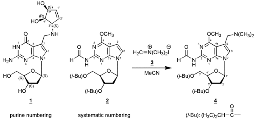
In this communication we disclose that reaction of the fully protected 7-deaza-2’-deoxy-7-deazaguanosine derivative
2 [
8] with N,N-dimethyl-methyleniminium iodide (Eschenmoser’s salt) afforded the C(7) alkylated Mannich compound
4 in moderate yield. This reaction presents a new and alternative route to 7-deazapurine nucleosides with a Mannich side chain in position 7. The structure of
4 was unequivocally assigned by
1H- and
13C-NMR spectroscopy as well as by
1H-NMR NOE difference spectroscopy (see Experimental Procedure).
13C-NMR resonances were assigned applying DEPT-135 and [C,H]HETCOR spectra.
A further transformation of the tertiary amino group of 4 into other Mannich compounds can be accomplished after quarternization. Furthermore, the synthetic or enzymatic incorporation of Mannich bases derived from compounds such as compound 4 allows the introduction of reporter groups in a favourable position of a DNA molecule because the Mannich side chain protrudes into the major groove of a B-DNA double helix.
Experimental Procedure
7-[2-Deoxy-3,5-bis-O-(2-methylpropanoyl)-ß-D-erythro-pentofuranosyl]-5-(dimethylaminomethyl)-2-(formylamino)-4-methoxy-7H-pyrrolo[2,3-d]pyrimidine (4).
Compound 2 (305 mg, 0.7 mmol) was dissolved in acetonitrile (3 mL) and N,N-dimethyl-methyleniminium iodide (3, Eschenmoser’s Salz, 260 mg, 1.4 mmol) was added. After heating to 80°C for 24h, the reaction mixture was evaporated to a small volume, and compound 4 (67 mg, 20 %) was isolated as an amorphous solid by thick-layer chromatography (chloroform/methanol, 9:1, two developments, Rf, 0.4), elution from the silica gel with methanol and centrifugation. 1H-NMR ((D6)DMSO, 500.1 MHz): δ 10.73 (1H, d, HC=O, J = 9.9 Hz); 9.44 (1H, d, NH, J = 9.3 Hz); 7.31 (1H, s, H-C(6)); 6.49 (1H, dd, H-C(1’), J = 6.4, 8.0 Hz); 5.36 (1H, dd, H-C(3’), J = 2.8, 3.6 Hz); 4.28 - 4.17 (3H, m, H-C(4’), H2-C(5’)); 4.03 (3H, s, OCH3); 3.67 (2H, s, CH2); 2.86 (1H, m, iBu-CH); 2.63 – 2.57 (2H, m, H2-C(2’)); 2.47 (1H, m, iBu-CH); 2.28 (6H, s, N(CH3)2); 1.15 and 1.09 (12H, 4 iBu-CH3). 1H-NMR NOE Data ((D6)DMSO): proton irradiated: H-C(1’). NOE observed: H-C(6), 1.5 %; Hα-C(2’), 2.5 %; H-C(4’), 1.5 %. 13C-NMR ((D6)DMSO, 125.8 MHz): δ 175.6, 175.8 (2 C=O, iBu); 163.4 (C-4); 163.3 (C=O, formyl); 152.4 (C-7a); 152.0 (C-2); 121.6 (C-6); 111.4 (C-5); 101.5 (C-4a); 82.9 (C-1’); 81.1 (C-4’); 74.2 (C-3’); 63.6 (C-5’); 53.8 (CH2); 53.7 (OCH3); 44.1 and 33.1 (4 Me, iBu); 35.9 (C-2’); 18.6 (2 Me, N(CH3)2).