Intramolecular C−H···π Interactions in Metal-Porphyrin Complexes

1
"Vinča" Institute of Nuclear Sciences, Laboratory of Theoretical Physics and Condensed Matter Physics, 11001, Belgrade, P. O. Box 522, Serbia and Montenegro
2
Department of Chemistry, Studentski trg 16, 11000 Belgrade, Serbia and Montenegro
*
Author to whom correspondence should be addressed.
Int. J. Mol. Sci. 2004, 5(4), 174-185; https://doi.org/10.3390/i5040174
Submission received: 15 December 2003
/
Accepted: 11 March 2004
/
Published: 1 April 2004
(This article belongs to the Special Issue Proceedings of the Workshop on Modeling Interaction in Biomolecules)
Abstract
:Cambridge Structural Database (CSD) was screened in order to find intramolecular C−H···π interactions with a chelate ring of coordinated porphyrin. It was found 154 crystal structures with 244 intramolecular C−H···π interactions in transition metal complexes with derivatives of porphyrin. Comparison of interacting distances indicates that interactions of hydrogen atoms in positions 2 and 6 of axially coordinated pyridine are more favorable with ruffled than with planar porphyrin.
Introduction
Noncovalent interactions with aromatic rings have been studied very intensively. It has been documented that these interactions are important in different molecular systems, from molecular biology to material science [1]. Noncovalent interactions in metal complexes between π-systems and ligands with X − H (X = N, O, C) have been noticed and investigated by searching data bases of crystal structures, by quantum chemical calculations and by spectroscopic methods [2]. Noncovalent interactions of chelate rings with delocalized π-bonds were observed and investigated in a few studies [3,4,5]. In our previous work we observed that chelate ring, as π-system, can be involved in C−H···π interactions. By searching Cambridge Structural Database (CSD) we found out a number of crystal structures of metal complexes where six membered chelate ring with delocalized π-bonds is proton acceptor in C−H···π interactions [3]. The calculated energy of the interaction is about 1 kcal/mol [3]. The calculated energy and geometry observed in crystal structures are comparable with C−H···π interactions where proton acceptor is organic aromatic ring [6].
Metalloproteins that contain derivatives of porphyrin coordinated to a metal center are involved in many different processes in living organisms. Studies of metal center in heme proteins and model systems showed that many factors, including noncovalent interactions can play important role in properties of these metalloproteins. The orientations of histidines, axially ligated to the heme, are considered to have a strong influence on the redox potential and can control the coordination of substrates to heme-proteins [7]. By analyzing crystal structures of heme-proteins [8] it was shown that there are two main factors that determine the orientations of imidazole ligated to heme. Both of them are noncovalent interactions. These are the interactions of imidazole with the propionic side groups on porphyrin in heme and interactions with the histidine backbone. Generally the NH group of imidazole is oriented towards the propionic groups of the heme [8].
Iron porphyrinato complexes with axially coordinated imidazoles and pyridines are model systems of cytochromes. Quantum chemical calculations show that there is difference in behavior of complexes with axially coordinated imidazoles and pyridines [9,10]. Axially coordinated ligands (imidazol or pyridine) can be in mutually parallel or orthogonal orientation; in complexes with parallel orientation of axial ligands porphyrin ring is planar, in complexes with orthogonal orientation pophyrin ring is ruffled. Iron(III) complex with mutual orthogonal orientation of pyridines has more ruffled porphyrin ring than complex with imidazoles. Also, for complex with pyridines orthogonal orientation of pyridines is by 16 kcal/mol more stable than parallel orientation [10], while in complex with imidazoles there is no difference in stability for orthogonal and parallel orientation [9]. There is open question why there are differences in stability for complexes with imidazoles and pyridines. The assumption was made that these differences are consequence of steric interactions of α-hydrogen atoms of pyridine with equatorial porphyrin [10]. Namely, imidazole is five membered ring, while pyridine is six membered ring, hence, α-hydrogen atoms (atoms in positions 2 and 6) of pyridine are closer to the equatorial porphyrin. In complexes with parallel orientation of pyridines and ruffled porphyrin there is less steric interaction of α-hydrogen atoms with porphirin ring.
Porphyrin molecule and derivatives of porphyrin are delocalized π-systems. When pophyrin is coordinated to a metal as a tetradentate ligand there are four six membered chelate rings with delocalized π -bonds, hence, there are additional rings with delocalized π -systems. The chelate rings of the coordinated porphyrin could be involved in the C−H···π interactions and it has prompted us to analyse C−H···π interactions in crystal structures of metal-porphyrin complexes from Cambridge Structural Database (CSD). To the best of our knowledge this is the first report of intramolecular C−H···π interactions with the chelate ring of porphyrinato ligand.
Data Screening and Computational Methods
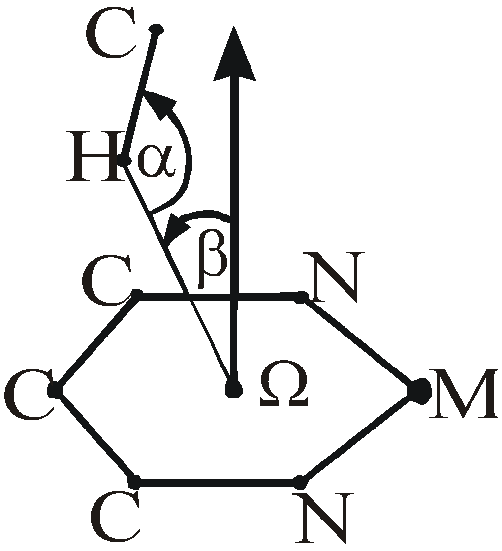
Cambridge Structural Database (CSD) [11] was screened in order to find intramolecular C−H···π interactions with a chelate ring of coordinated porphyrin. We searched for the crystal structures of metal complexes with coordinated porphyrin or derivatives of porphyrin. In these structures we demanded that there is a hydrogen atom at a distance shorter than 3.0 Å from the center of the six membered chelate ring, and presenting a X−H···Ω axis a narrow cone perpendicular to the ring (α > 110°, β < 16°) (Figure 1). These screening criteria are more restrictive than criteria used before for similar screening [12,13].
Model system was built from geometry of crystal structure VATXIX by substituting side groups of porphyrin with hydrogen atoms. The single point calculations were done for two different conformers. In the conformers there are two different orientations of axial ligand, in one conformer there is the C−H···π interaction with chelate ring, in the other conformer there is no interaction. The energy of the C−H···π interaction is evaluated as the difference in the energy of the two conformers.
The single point calculations on model systems have been done using DFT, specifically Becke three-parameter exchange functional (B3) [14] and the Lee-Yang-Parr correlation functional (LYP) [15]. These B3LYP calculations have been carried out with GAUSSIAN98 program [16]. The LANL2DZ basis set was chosen for zinc atom and 6-31G** basis sets were chosen for all other atoms.
Figure 1.
Geometrical parameters for C−H···π interaction with chelate ring of porphyrinato ligand.
Results and discussion
Searching crystal structures of transition metal complexes from the Cambridge Structural Database (CSD) shows that specific C−H···π interactions, interactions between C-H groups and the π-system of porphyrinato chelate ring, can be observed in many crystal structures. By using described criteria 154 crystal structures with 244 intramolecular C−H···π interactions in transition metal complexes with derivatives of porphyrin were found. Most of these intramolecular C−H···π interactions (116 interactions) are interactions in iron complexes. Geometrical data for some of these interactions are given in Table 1 and Table 2. In Table 1 there are data for different complexes, while data for iron-porphyrin complexes with axially coordinated pyridines are given in Table 2.
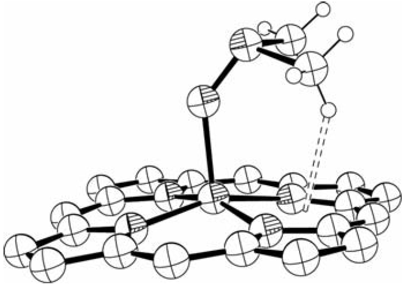
The C-H group involved in the intramolecular C−H···π interaction with the chelate ring of porphyrinato ligand is in most cases part of axial ligand. In Figure 2 crystal structure KACGIE is shown.

Table 1.
Some of geometrical dataa for intramolecular C−H···π interaction in metal-porphyrin complexes
| Refcode | Metal | β (o) | H… Ω (Å) | α(o) | Ref. No. | |
|---|---|---|---|---|---|---|
| 1 | DIQWOP | Ru4+ | 12.2 | 2.260 | 123.7 | 17 |
| 2 | HAMDII | Zn2+ | 5.8 | 2.396 | 118.9 | 18 |
| 3 | MAZVAKb | 2 Zn2+ | 9.8 | 2.397 | 125.5 | 19 |
| 4 | POZQIE | Co3+ | 7.0 | 2.344 | 139.0 | 20 |
| 11.2 | 2.240 | 135.1 | ||||
| 5 | VATXIX | Zn2+ | 14.6 | 2.324 | 144.8 | 21 |
| 6 | YIYPOLb | 2 Zn2+ | 3.9 | 2.145 | 153.1 | 22 |
| 7 | BAFKEZ | Ru2+ | 15.6 | 2.425 | 120.6 | 23 |
| 8 | LODQEA | Zn2+ | 1.9 | 2.466 | 125.6 | 24 |
| 9 | NACTUGb | 2 Ni2+ | 11.8 | 2.445 | 135.3 | 25 |
| 10 | HUHLEB | Ru 2+ | 9.6 | 2.454 | 144.9 | 26 |
| 11 | BMPRCU | Cu2+ | 9.8 | 2.561 | 161.1 | 27 |
| 12 | GITLEA | Ni2+ | 2.4 | 2.501 | 112.0 | 28 |
| 13 | KACGIE | Fe2+ | 9.3 | 2.513 | 116.6 | 29 |
| 8.9 | 2.722 | 110.2 | ||||
| 14 | ZAWBAA01 | Fe3+ | 10.8 | 2.576 | 163.9 | 30 |
| 15 | BONREB | Zr4+ | 6.3 | 2.615 | 117.2 | 31 |
| 16 | CAFBANb | 2 Zn2+ | 12.9 | 2.647 | 139.9 | 32 |
| 17 | HAMMIR | Zn2+ | 15.2 | 2.824 | 117.2 | 33 |
| 18 | PAFTUL | Fe3+ | 13.0 | 2.734 | 161.6 | 34 |
| 19 | QOHWEP | Ti2+ | 15.6 | 2.601 | 129.7 | 35 |
| 20 | TAKVOQb | Cu2+ Fe3+ | 12.2 | 2.858 | 147.2 | 36 |
| 21 | VUPKUM | Ru2+ | 15.7 | 2.655 | 161.3 | 37 |
| 22 | WAVDOM10b | Cu2+ Fe3+ | 12.8 | 2.735 | 127.3 | 38 |
aExplanation of geometrical parameters is given on Figure 1bBinuclear complex
Figure 2.
Crystal structure of KACGIE. Dashed lines represent C−H···π interactions. Some of atoms have been omitted for clarity. Coordinates are taken from CSD.
Figure 2.
Crystal structure of KACGIE. Dashed lines represent C−H···π interactions. Some of atoms have been omitted for clarity. Coordinates are taken from CSD.
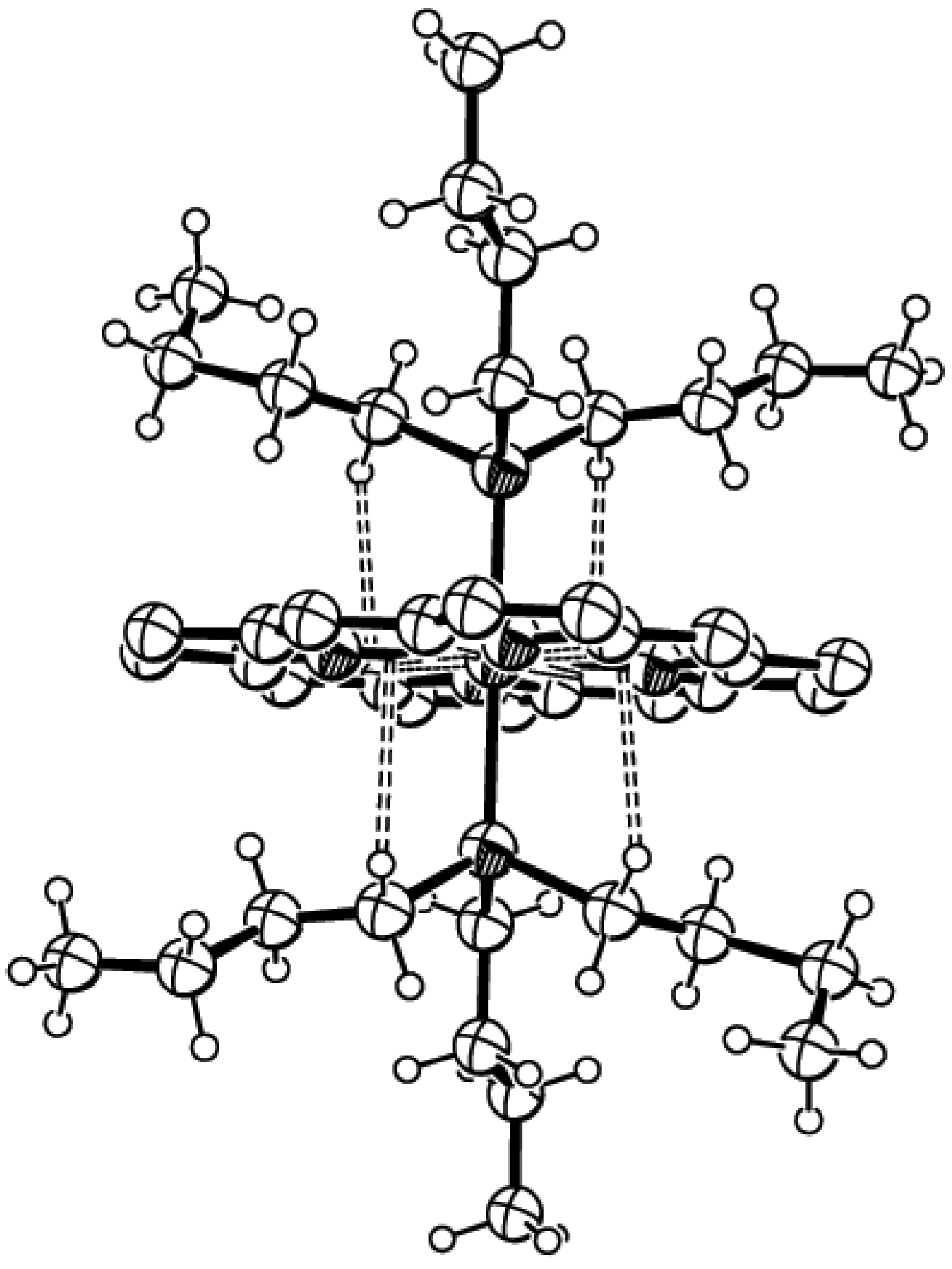
Usually there is no geometrical flexibility and the hydrogen atom of the axial ligand is forced to be close to the chelate ring. In some cases there are very short H···Ω distances caused by constrains in geometry of complex. The examples are structure YIYPOL and complexes with axially coordinated pyridines (Table 2). However there are some structures with geometrical flexibility in axial ligand. One example, crystal structure VATXIX is shown in Figure 3.
Figure 3.
Crystal structure of VATXIX. Dashed line represent C−H···π interaction. Some of atoms have been omitted for clarity. Coordinates are taken from CSD.
Figure 3.
Crystal structure of VATXIX. Dashed line represent C−H···π interaction. Some of atoms have been omitted for clarity. Coordinates are taken from CSD.

In this complex of zinc there is dimethylsulfoxide as axial ligand. Dimethylsulfoxide ligand can be oriented with methyl groups toward the porphyrin ring (Figure 3) or in opposite direction.
In the first conformation there is C−H···π interaction (Figure 3), in the second conformation interaction does not exist. For this complex calculations have been done in order to evaluate the energy of the interaction. Evaluated energy obtained as a difference in the energy of the conformer shown on the Figure 3 and the energy of the conformer without C−H···π interaction is 0.7 kcal/mol. The evaluated energy is somewhat smaller than previously calculated energy for intermolecular C−H···π interactions that was above 1 kcal/mol. The reason could be some repulsion that exist in case of intramolecular interaction in VATXIX structure.
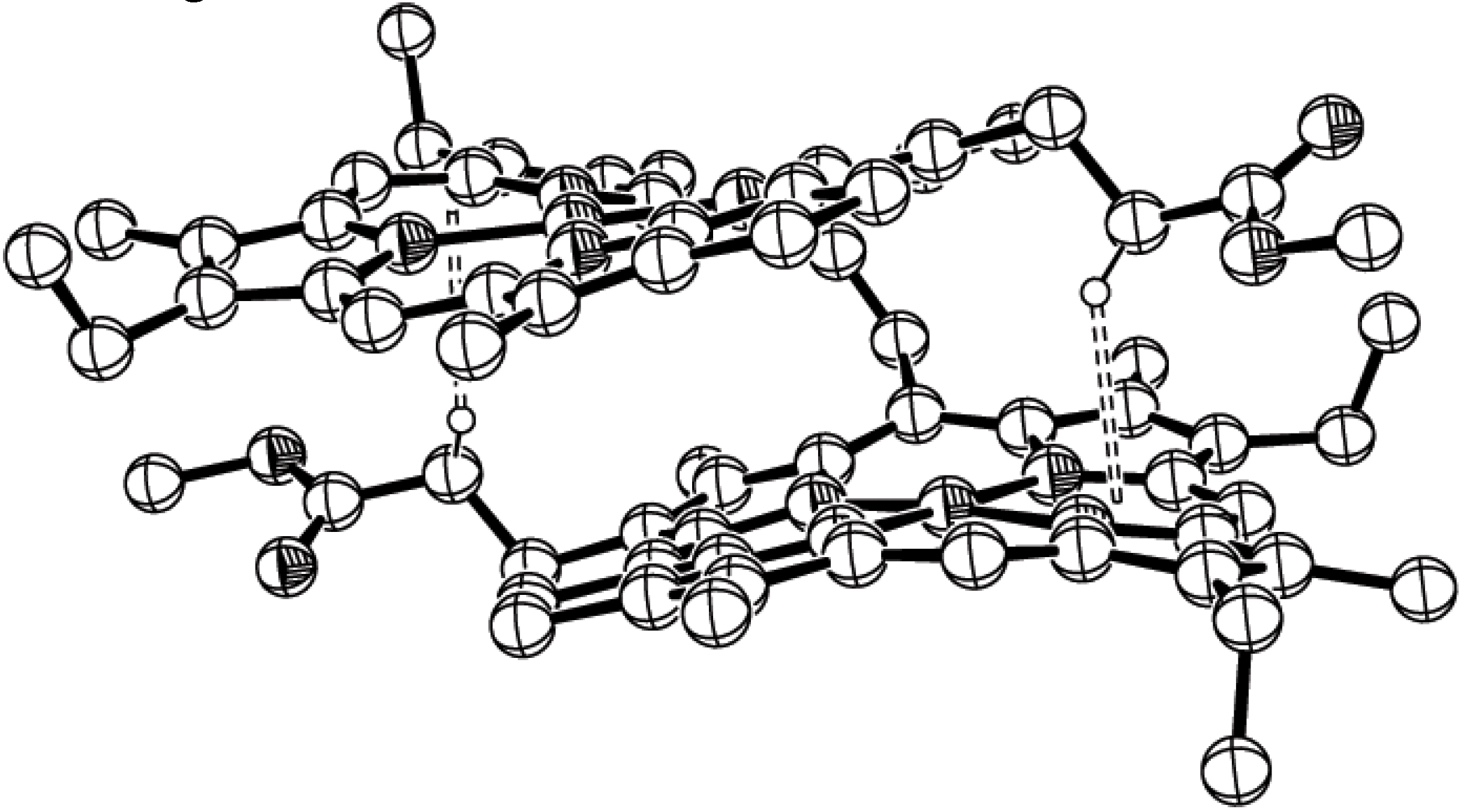
There are crystal structures with C−H···π interaction where C-H group is not part of axial ligand. In Figure 4 there is crystal structure of binuclear complex NACTUG with two nickel atoms with porphyrinato rings. The C-H of side group from one porphyrinato ligand interacts with chelate ring from the second porpyirinato ring.
Figure 4.
Crystal structure of NACTUG. Dashed lines represent C−H···π interactions. Some of atoms have been omitted for clarity. Coordinates are taken from CSD.
Figure 4.
Crystal structure of NACTUG. Dashed lines represent C−H···π interactions. Some of atoms have been omitted for clarity. Coordinates are taken from CSD.

Table 2.
Some of geometrical dataa for intramolecular C−H···π interaction in Fe complexes with porphyrin derivate and two axially coordinated pyridines.
| Refcode | Metal | β (o) | H…Ω (Å) | α(o) | Fe…Ω (Å) | P/Nb | Ref. No. | |
|---|---|---|---|---|---|---|---|---|
| 1 | CPOEFE10c | Fe3+ | 3.7 | 2.40 | 119.3 | 1.98 | P | 39 |
| 6.2 | 2.21 | 123.0 | ||||||
| 2 | DAJJAZc | Fe2+ | 10.3 | 2.22 | 117.2 | 1.97 | P | 40 |
| 9.5 | 2.22 | 118.7 | ||||||
| 3 | FUXTUNc | Fe2+ | 6.6 | 2.16 | 120.2 | 1.91 | P | 41 |
| 3.3 | 2.17 | 119.2 | ||||||
| 4 | NIWLOUc | Fe2+ | 4.3 | 2.16 | 117.0 | 1.98 | P | 42 |
| 6.1 | 2.19 | 115.9 | ||||||
| 5 | NIWLUAc | Fe2+ | 5.6 | 2.15 | 117.3 | 1.95 | P | 42 |
| 2.7 | 2.22 | 116.5 | ||||||
| 6 | NIWMAHc | Fe2+ | 2.9 | 2.24 | 115.7 | 1.96 | P | 42 |
| 5.7 | 2.17 | 117.3 | ||||||
| 7 | VOFLORc | Fe3+ | 8.4 | 2.18 | 116.1 | 1.98 | P | 43 |
| 8.1 | 2.23 | 116.5 | ||||||
| 8 | GEWKOI | Fe3+ | 9.9 | 2.30 | 115.8 | 1.96 | N | 44 |
| 5.6 | 2.32 | 115.2 | ||||||
| 3.7 | 2.22 | 116.9 | ||||||
| 6.6 | 2.26 | 116.3 | ||||||
| 9 | HERZIN | Fe3+ | 9.8 | 2.38 | 115.6 | 1.93 | N | 45 |
| 10.1 | 2.42 | 114.6 | ||||||
| 8.1 | 2.32 | 116.6 | ||||||
| 8.9 | 2.35 | 115.4 | ||||||
| 10 | KEFFOQ | Fe2+ | 7.6 | 2.40 | 116.4 | 1.93 | N | 46 |
| 6.3 | 2.37 | 116.3 | ||||||
| 6.8 | 2.38 | 116.2 | ||||||
| 7.2 | 2.39 | 116.9 | ||||||
| 11 | PALVED | Fe3+ | 4.5 | 2.30 | 116.6 | 1.94 | N | 47 |
| 2.5 | 2.25 | 116.0 | ||||||
| 2.0 | 2.23 | 117.5 | ||||||
| 7.1 | 2.42 | 115.4 | ||||||
| 12 | PALVIH | Fe3+ | 5.0 | 2.33 | 116.1 | 1.95 | N | 47 |
| 4.1 | 2.33 | 116.2 | ||||||
| 10.5 | 2.27 | 114.8 | ||||||
| 9.8 | 2.29 | 113.4 | ||||||
| 13 | PALVON | Fe3+ | 1.2 | 2.15 | 118.9 | 1.94 | N | 47 |
| 5.8 | 2.42 | 115.2 | ||||||
| 2.6 | 2.19 | 115.3 | ||||||
| 9.0 | 2.47 | 115.1 | ||||||
| 14 | TUBJAB | Fe2+ | 11.6 | 2.45 | 116.7 | 1.93 | N | 48 |
| 12.1 | 2.38 | 117.9 | ||||||
| 7.2 | 2.37 | 118.5 | ||||||
| 7.5 | 2.38 | 117.8 | ||||||
| 15 | VOFLUX | Fe3+ | 9.2 | 2.32 | 116.3 | 1.94 | N | 43 |
| 10.5 | 2.41 | 115.2 | ||||||
| 5.2 | 2.41 | 114.3 | ||||||
| 6.0 | 2.32 | 117.1 |
aExplanation of geometrical parameters is given on Figure 1bP - planar conformation of chelate ring and mutual parallel orientation of pyridines, N - ruffled conformation of chelate ring and mutual orthogonal orientation of pyridines cOnly two interactions are presented, the other two interactions are equivalent because of symmetry of complexes with parallel orientation of pyridines.
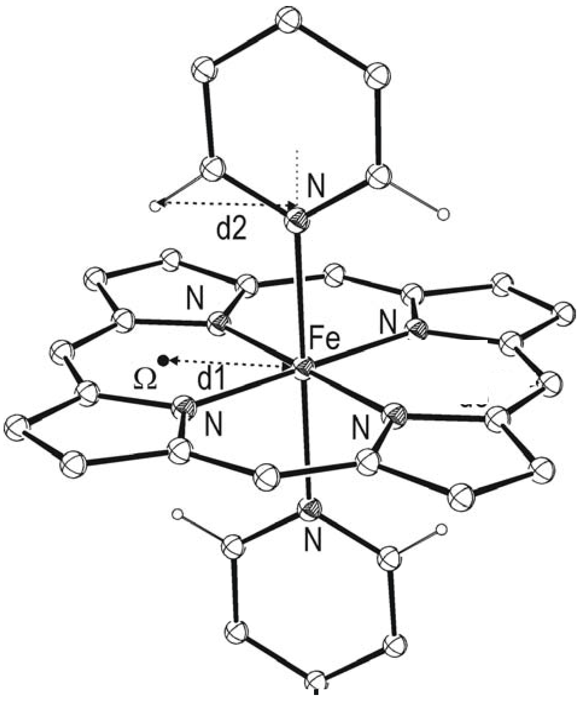
The best examples of structures with geometrical constrains are structures with axially coordinated pyridines, where the α-hydrogen atoms of pyridine are involved in C−H···π interactions with chelate rings (Figure 5,Figure 6,Figure 7). It is interesting that most of found intramolecular C−H···π interactions in iron-porphyrin complexes are in complexes with axially coordinated pyridine. In these structures H···Ω distances are very short, mainly below 2.4 Å. Some geometrical data about C−H···π interactions in iron-porphyrin complexes with two axially coordinated pyridines are shown in Table 2.
The observation that H···Ω distances in pyridine complexes are very short is in accord with previously made assumption that steric interactions of α-hydrogen atoms from pyridine with porphyrin can contribute to the relative stability of conformers with parallel and orthogonal orientation of pyridines [10]. Because of importance, as well as large number of complexes, we analyzed interactions of α-hydrogen atoms of pyridine with chelate rings of porphyrinato ligand.
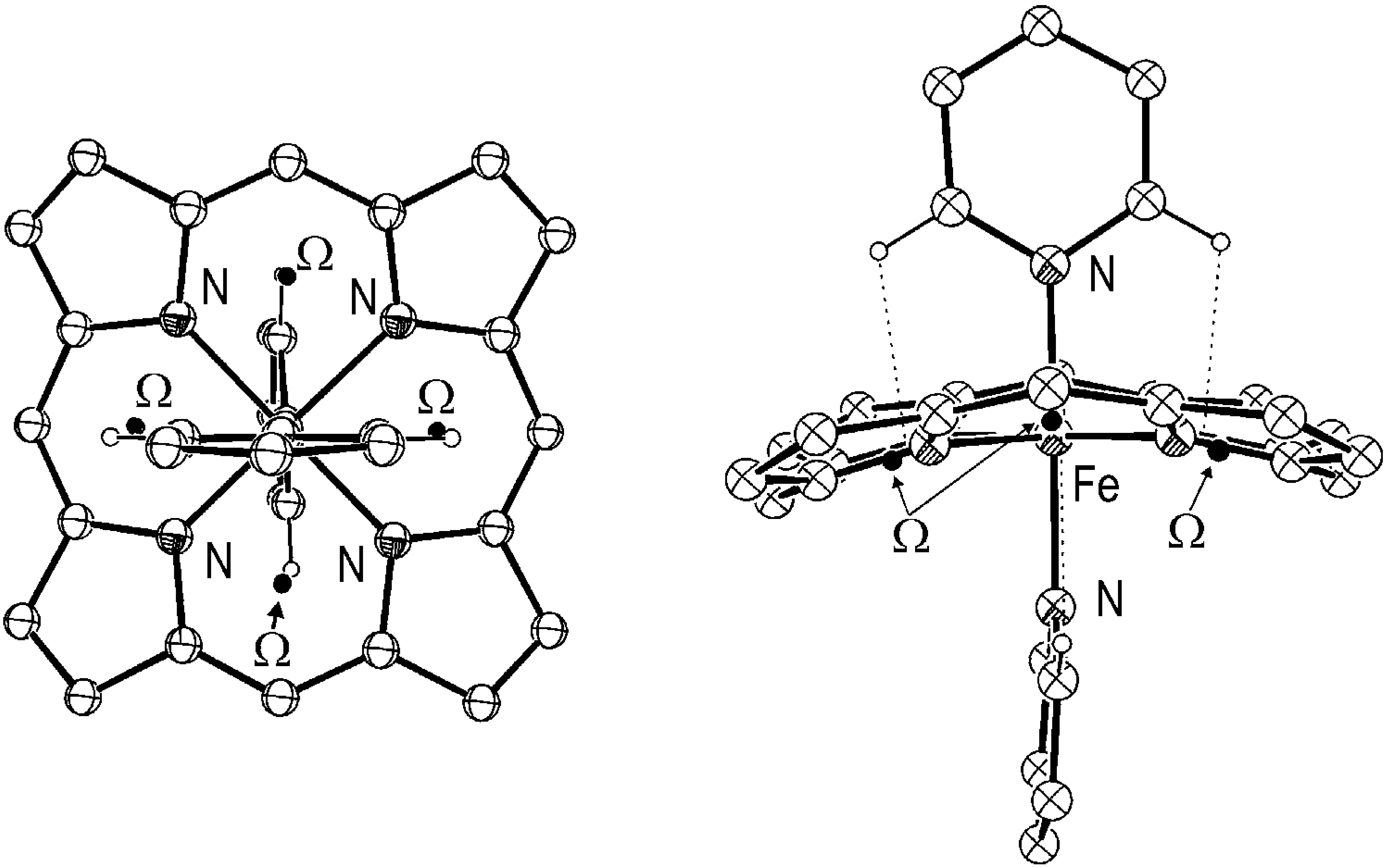
Figure 5.
Crystal structure of CPOEFE10 representing some of significant distances. Some of atoms have been omitted for clarity. Coordinates are taken from CSD.
Figure 5.
Crystal structure of CPOEFE10 representing some of significant distances. Some of atoms have been omitted for clarity. Coordinates are taken from CSD.

In the complexes with two axially coordinated pyridines there are four intramolecular C−H···π interactions with one porphyrin ring (Figure 5,Figure 6,Figure 7). Analyzing geometrical data for pyridine and porphyrin ring shows that α-hydrogen atoms of pyridine is in suitable position (with small value of angle в) for intramolecular C−H···π interactions with chelate ring of porphyirin. Figure 5 represents CSD structure CPOEFE01 with C−H···π interaction between axial pyridines and chelate rings of porphyrin. Center of the six membered chelate ring (Ω) is on Fe-C line and Fe-Ω distance (d1) is 1.96 Å. Distance between hydrogen in position 2 or 6 and normal to the porphyrin, which goes through the Fe atom (d2), is around 2.03 Å. In the iron complexes with ruffled porphyrin, d1 distance is somewhat smaller, 1.94 Å (Table 2). Similar values for the distances d1 and d2 show that hydrogens in position 2 and 6 of pyridine are in the favorable position in respect to the center of the six membered chelate ring. Projection of hydrogens in 2 and 6 position of pyridine on the six membered chelate ring plane is only 0.07 Å away from the center of the ring in case of planar porphyrin ring. Position of hydrogen in regard to the center of the chelate ring can be described with angle β, Figure 1. If angle β is smaller hydrogen atom is orientated closer to the center of the ring. Data for angle β given in Table 2 show that angle β has small values, in most of cases below 10o.
Figure 6.
Crystal structure of CPOEFE10 with parallel orientation of pyridine ligands. Two orthogonal projections of molecule are shown. Dashed lines represent C−H···π interactions between four hydrogen atoms and two chelate rings. Some of atoms have been omitted for clarity. Coordinates are taken from CSD.
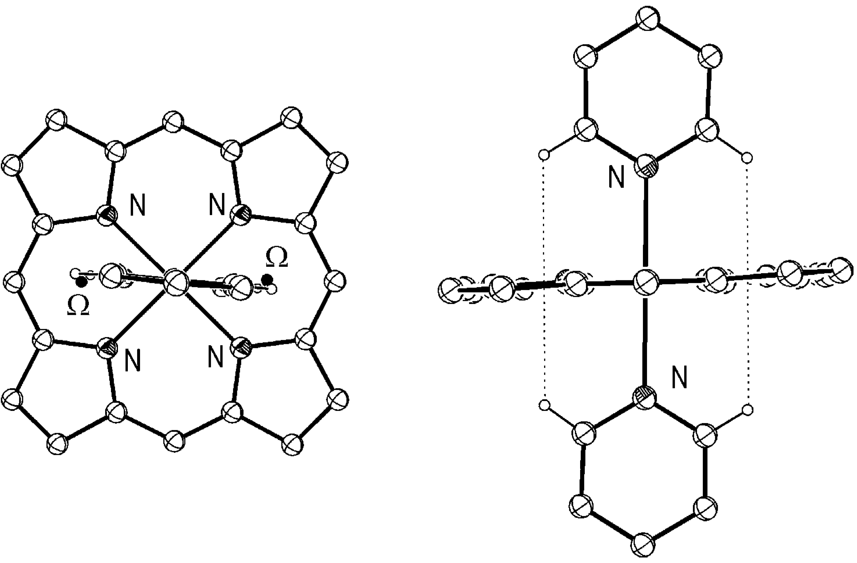
Figure 7.
Crystal structure of KEFFOG with orthogonal orientation of pyridine ligands. Two orthogonal projections of molecule are shown. Dashed lines represent C−H···π interactions between four hydrogen atoms and four chelate rings. Some of atoms have been omitted for clarity. Coordinates are taken from CSD.
As it was mentioned there is correlation between porphyrin conformation and mutual orientation of two pyridines. In the complexes with planar porphyrin mutual orientation of pyridines is parallel and in the complexes with ruffled porphyrin pyridines are orthogonal each to other. In Figure 6 and Figure 7 there are crystal structures of complexes with parallel and orthogonal orientation of axially coordinated pyridines.
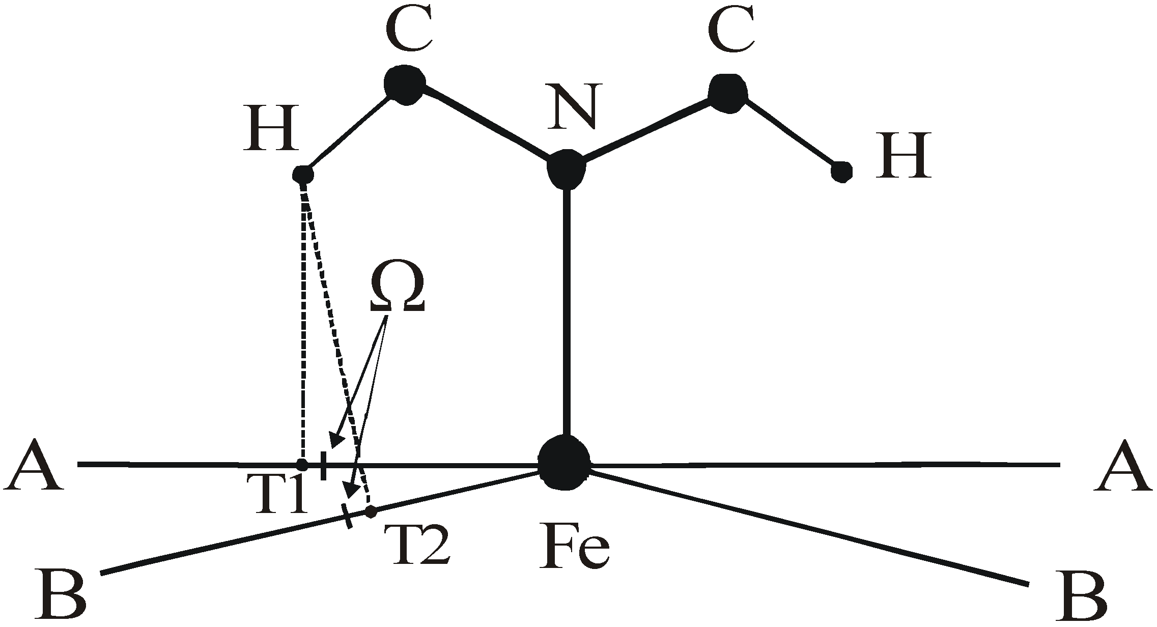
Possible positions of hydrogen atom of pyridine in respect to the centre of six membered chelate ring in the cases of planar and ruffled porphyrin are shown in Figure 8. In both cases there is possibility for C−H···π interactions, because projections of the hydrogen atom to the plane of chelate ring (T1 and T2) are close to the center of the ring (Ω). In case of the ruffled porphyrin H···Ω distances are somewhat longer.
Figure 8.
Schematic representation of C−H···π interactions with chelate ring of porphyrin in case of planar (A) and ruffled porphyrin (B). Only part of pyridine is presented. Porphyrin is presented as a line. In both cases projection of the hydrogen atom to the plane of chelate ring (T1 and T2) is close to the center of the ring (Ω). In case of ruffled porphyrin H···Ω distance is longer.

Data in Table 2 show that distances between hydrogen atom and center of the chelate ring (H···Ω) are in range of 2.15 to 2.47 Å and that values of angle β are between 1.2 and 11.6º. There is no important difference in values of angle β for structures with planar and ruffled porphyrins, while H···Ω distances are shorter for structures with planar porphyrin. In structures with planar porphyrin the distances are between 2.15 and 2.24 Å, with one exception, while in structures with ruffled porphyrin the distances are above 2.22 Å, with two exceptions.
The H···Ω distances in structures with parallel orientations of pyridines (Table 2) are shorter than previously reported distances for C−H···π interactions with chelate rings [3], and shorter that distances in Table 1. In previously reported data [3] there were just a few structures with H···Ω distances below 2.35 Å and large number of interactions with H···Ω distances above 2.40 Å [3]. These data were for intermolecular C−H···π interactions, hence there were not geometrical constrains. We assume that H···Ω distances for these intermolecular interactions respond to the most stable interactions.
Based on that, H···Ω distances in pyridine complexes are shorter than the most favorable distances. By ruffling porphyrin ring in pyridine complexes with orthogonal orientation of pyridine ligands H···Ω distances are getting longer and energetically more favorable. That contribute to the stability of orthogonal orientations.
Conclusion
By using geometrical criteria 244 intramolecular C−H···π interactions with chelate rings of coordinated porphyrins were found in 154 crystal structures from CSD. In most of the structures a hydrogen atom of the axial ligand is involved in the interaction, although there are structures where C-H group is not part of the axial ligand. In some cases where C-H group is part of the axial ligand there are very short H···Ω distances caused by a constrains in the geometry of the complex. Among these structures there are structures of iron porphyrin complexes with axially coordinated pyridines.
In iron porphyrin complexes with pyridines distances between hydrogen and the center of the ring are shorter than the most favorable distance. By ruffling porphyrin H···Ω distances are getting longer and energetically more favorable. That contribute to the stability of conformation with orthogonal orientations of pyridine ligands.
References
- Ma, J. C.; Daugherty, D. A. Chem. Rev. 1997, 97, 1303. Daugherty, D. A. Science 1996, 271, 163. Steiner, T. Angew. Chem. Int. Ed. 2002, 41, 48. Yamauchi, O. O.; Takani, M. J. Chem. Soc.-Dalton Trans. 2002, 3411. Meyer, E. A.; Castellano, R. K.; Diederich, F. Angew. Chem.-Int. Edit. 2003, 42, 1210. Schmitt, W.; Anson, C. E.; Hill, J. P.; Powell, A. K. J. Am. Chem. Soc. 2003, 125, 11142. Amunugama, R.; Rodgers, M. T. Int. J. Mass Spectrom. 2003, 227, 339. Zhu, W. L.; Tan, X. J.; Shen, J. H.; Luo, X. M.; Cheng, F.; Mok, P. C.; Ji, R. Y.; Chen, K. X.; Jiang, H. L. J. Phys. Chem. A 2003, 107, 229. Pletneva, E. V.; Laederach, A. T.; Fulton, D. B.; Kostić, N. M. J. Am. Chem. Soc. 2001, 123, 623. Burghardt, T. P.; Juranić, N.; Macura, S.; Ajtai, K. Biopolimers 2002, 63, 261. Kim, D.; Hu, S.; Tarakeshwar, P.; Kim, K.S.; Lisy, J.M. J. Phys. Chem. A 2003, 107, 1228.
- Zarić, S. D. Eur. J. Inorg. Chem. 2003, 2197. Zarić, S. D. Chem. Phys. Lett. 1999, 311, 77. Zarić, S. D. Chem. Phys. 2000, 256, 213. Zarić, S. D.; Popović, D. M.; Knapp, E. W. Chemistry 2000, 6, 3935. Milčić, M.; Zarić, S. D. Eur. J. Inorg. Chem. 2001, 2143. Žmirić, A.; Milčić, M.; Zarić, S. D. Int. J. Quantum Chem. 2002, 87, 354. McFail-Isom, L.; Shui, X.; Williams, L. D. Biochemistry 1998, 37, 17105. Kumita, H.; Kato, T.; Jitsukawa, K.; Einaga, H.; Masuda, H. Inorg. Chem. 2001, 40, 393. Suezawa, H.; Yoshida, T.; Umezawa, Y.; Tsuboyama, S.; Nishio, M. Eur. J. Inorg. Chem. 2002, 3148. Sénèque, O.; Giorgi, M.; Reinaud, O. Chem. Commun. 2001, 984. [poner, J.; [poner, J. E.; Leszczynski, J. J. Biomol. Struct. Dyn. 2000, 17, 1087.
- Bogdanović, G. A.; Bire, A. S.; Zarić, S. D. Eur. J. Inorg. Chem. 2002, 599.
- Tomić, Z. D.; Leovac, V. M.; Pokorni, S. V.; Zobel, D.; Zarić, S. D. Eur. J. Inorg. Chem. 2003, 1222.
- Castineiras, A.; Sicilia-Zafra, A. G.; Gonzalez-Perez, J. M.; Choquesillo-Lazarte, D.; Niclos-Gutierrez, J. Inorg. Chem. 2002, 41, 6956.
- Nishio, M.; Hirota, M.; Umezawa, Y. The CH/р Interaction, Evidence, Nature and Consequences; John Wiley & Sons, Inc.: New York, 1998. [Google Scholar]
- Walker, F. A.; Huynh, B.H.; Schedt, W.R.; Osvath, S.R. J. Am. Chem. Soc. 1986, 108, 5288. Walker, F. A. Chem. Rev. 2004, 104, 589. Menyhard, D.K.; Keseru, G.M. J. Am. Chem. Soc. 1998, 120, 7991.
- Zarić, S. D.; Popović, D. M.; Knapp, E. W. Biochemistry 2000, 40, 7914. [CrossRef]
- Medaković, V. B.; Zarić, S. D. Inorg. Chim. Acta 2003, 349, 1.
- Ghosh, A.; Gonzales, E.; Vangberg, T. J. Phys. Chem. 1999, 103, 1363.
- Allen, F. H.; Bellard, S.; Brice, M. D.; Cartwright, B. A.; Doubleday, A.; Higgs, H.; Hummelink, T.; Hummelink-Peters, B. G.; Kennard, O.; Motherwell, W. D. S.; Rodgers, J. R.; Watson, D. G. Acta Cryst. 1979, B35, 2331.
- Ciunik, Z.; Desiraju, G. R. Chem. Commun. 2001, 703.
- Steiner, T.; Desiraju, G. R. Chem. Commun. 1998, 891.
- Becke, A. D. J. Chem. Phys. 1993, 98, 5648.
- Lee, C.; Yang, W.; Parr, R. G. Phys. Rev. B 1988, 37, 785. [CrossRef]
- Frisch, M. J.; Trucks, G. W.; Schlegel, H. B.; Scuseria, G. E.; Robb, M.A.; Cheeseman, J. R.; Zakrzewski, V. G.; Montgomery, J. A., Jr.; Stratmann, J. A.; Burant, J. C.; Dapprich, S.; Millam, J. M.; Daniels, A. D.; Kudin, K. N.; Strain, M. C.; Farkas, O.; Tomasi, J.; Barone, V.; Cossi, M.; Cammi, R.; Mennucci, B.; Pomelli, C.; Adamo, C.; Clifford, S.; Ochterski, J.; Petersson, G. A.; Ayala, P.Y.; Cui, Q.; Morokuma, K.; Malick, D. K.; Rabuck, A. D.; Raghavachari, K.; Foresman, J. B.; Cioslowski, J.; Ortiz, J. V.; Stefanov, B.B.; Liu, G.; Liashenko, A.; Piskorz, P.; Komaromi, I.; Gomperts, R.; Martin, R. L.; Fox, D. J.; Keith, T.; Al-Laham, M. A.; Peng, C. Y.; Nanayakkara, A.; Gonzalez, C.; Challacombe, M.; Gill, P. M. W.; Johnson, B.; Chen, W.; Wong, M. W.; Andres, J. L.; Gonzalez, C.; Head-Gordon, M.; Replogle, E. S.; Pople, J. A. Gaussian 98, Revision A.6, Gaussian, Inc., Pittsburgh PA 1998.
- Huang, J.S.; Sun, X.R.; Leung, S.K.Y.; Cheung, K.K.; Che, C.M. Chem. Eur. J. 2000, 6, 334.
- Byrn, M.P.; Curtis, C.J.; Hsiou, Y.; Khan, S.I.; Sawin, P.A.; Tendick, S.K.; Terzis, A.; Strouse, C.E. J.Am.Chem.Soc. 1993, 115, 9480. [CrossRef]
- Nakash, M.; Clyde-Watson, Z.; Feeder, N.; Teat, S.J.; Sanders, J.K.M. Chem.-Eur.J. 2000, 6, 2112.
- Simonato, J.P.; Pecaut, J.; Marchon, J.C. J.Am.Chem.Soc. 1998, 120, 7363. [CrossRef]
- Jagessar, R.C.; Shang, M.; Scheidt, W.R.; Burns, D.H. J.Am.Chem.Soc. 1998, 120, 11684. [CrossRef]
- Bag, N.; Chern, S.S.; Peng, S.M.; Chang, C.K. Inorg.Chem. 1995, 34, 753.
- Lee, J.; Twamley, B.; Richter-Addo, G.B. Chem.Commun. 2002, 380.
- Senge, M.O.; Speck, M.; Wiehe, A.; Dieks, H.; Aguirre, S.; Kurreck, H. Photochem.Photobiol. 1999, 70, 206. [CrossRef]
- Jaquinod, L.; Nurco, D.J.; Medforth, C.J.; Pandey, R.K.; Forsyth, T.P.; Olmstead, M.M.; Smith, K.M. Angew.Chem.,Int.Ed.Engl. 1996, 35, 1013. [CrossRef]
- Stulz, E.; Maue, M.; Feeder, N.; Teat, S.J.; Ng, Y. F.; Bond, A.D.; Darling, S.L.; Sanders, J.K.M. Inorg.Chem. 2002, 41, 5255.
- Collmann, J.P.; Chong, A.O.; Jameson, G.B.; Oakley, R.T.; Rose, E.; Schmittou, E.R.; Ibers, J.A. J.Am.Chem.Soc. 1981, 103, 516. [CrossRef]
- Jia, S.L.; Jentzen, W.; Shang, M.; Song, X.Z.; Ma, J.G.; Scheidt, W.R.; Shelnutt, J.A. Inorg.Chem. 1998, 37, 4402.
- Belani, R.M.; James, B.R.; Dolphin, D.; Rettig, S.J. Can.J.Chem. 1988, 66, 2072.
- Chen, L.; Yi, G.B.; Wang, L.S.; Dharmawardana, U.R.; Dart, A.C.; Khan, M.A.; Richter-Addo, G.B. Inorg.Chem. 1998, 37, 4677.
- Kim, H.J.; Kim, K. Acta Crystallogr.,Sect.C:Cryst.Struct.Commun. 1999, 55, 1814.
- Fletcher, J.T.; Therien, M.J. Inorg.Chem. 2002, 41, 331.
- Byrn, M.P.; Curtis, C.J.; Hsiou, Y.; Khan, S.I.; Sawin, P.A.; Tendick, S.K.; Terzis, A.; Strouse, C.E. J.Am.Chem.Soc. 1993, 115, 9480. [CrossRef]
- Schulz, L.D.; Fallon, G.D.; Moubaraki, B.; Murray, K.S.; West, B.O. Chem.Commun. 1992, 971.
- Thorman, J.L.; Young Junior, V.G.; Boyd, P.D.W.; Guzei, I.A.; Woo, L.K. Inorg.Chem. 2001, 40, 499.
- Scott, M.J.; Goddard, C.A.; Holm, R.H. Inorg.Chem. 1996, 35, 2558.
- Seyler, J.W.; Fanwick, P.E.; Leidner, C.R. Inorg.Chem. 1992, 31, 3699.
- Lee, S.C.; Holm, R.H. Inorg.Chem. 1993, 32, 4745.
- Scheidt, W.R.; Geiger, D.K.; Haller, K.J. J.Am.Chem.Soc. 1982, 104, 495.
- Naiyin, L.; Petricek, V.; Coppens, P.; Landrum, J. Acta Crystallogr., Sect.C (Cr.Str.Comm.) 1985, 41, 902. [CrossRef] [Green Version]
- Naiyin, L.; Coppens, P.; Landrum, J. Inorg.Chem. 1988, 27, 482.
- Safo, M.K.; Nesset, M.J.M.; Walker, F.A.; Debrunner, P.G.; Scheidt, W.R. J.Am.Chem.Soc. 1997, 119, 9438. [CrossRef]
- Safo, M.K.; Gupta, G.P.; Walker, F.A.; Scheidt, W.R. J.Am.Chem.Soc. 1991, 113, 5497. [CrossRef]
- Inniss, D.; Soltis, S.M.; Strouse, C.E. J.Am.Chem.Soc. 1988, 110, 5644. [CrossRef]
- Safo, M.K.; Walker, F.A.; Raitsimring, A.M.; Walters, W.P.; Dolata, D.P.; Debrunner, P.G.; Scheidt, W.R. J.Am.Chem.Soc. 1994, 116, 7760. [CrossRef]
- Moore, K.T.; Fletcher, J.T.; Therien, M.J. J.Am.Chem.Soc. 1999, 121, 5196. [CrossRef]
- Safo, M.K.; Gupta, G.P.; Watson, C.T.; Simonis, U.; Walker, F.A.; Scheidt, W.R. J.Am.Chem.Soc. 1992, 114, 7066.
- Balch, A.L.; Noll, B.C.; Olmstead, M.M.; Phillips, S.L. Inorg.Chem. 1996, 35, 6495.
© 2004 by MDPI (http://www.mdpi.org).
Share and Cite
MDPI and ACS Style
Bogdanović, G.A.; Medaković, V.; Milčić, M.K.; Zarić, S.D. Intramolecular C−H···π Interactions in Metal-Porphyrin Complexes. Int. J. Mol. Sci. 2004, 5, 174-185. https://doi.org/10.3390/i5040174
AMA Style
Bogdanović GA, Medaković V, Milčić MK, Zarić SD. Intramolecular C−H···π Interactions in Metal-Porphyrin Complexes. International Journal of Molecular Sciences. 2004; 5(4):174-185. https://doi.org/10.3390/i5040174
Chicago/Turabian StyleBogdanović, Goran A., Vesna Medaković, Miloš K. Milčić, and Snežana D. Zarić. 2004. "Intramolecular C−H···π Interactions in Metal-Porphyrin Complexes" International Journal of Molecular Sciences 5, no. 4: 174-185. https://doi.org/10.3390/i5040174